printer-friendly version (PDF) - Reflex Sympathetic Dystrophy ...
printer-friendly version (PDF) - Reflex Sympathetic Dystrophy ...
printer-friendly version (PDF) - Reflex Sympathetic Dystrophy ...

Create successful ePaper yourself
Turn your PDF publications into a flip-book with our unique Google optimized e-Paper software.
OPINION | GETSON<br />
The Use of Thermography in the<br />
Diagnosis of CRPS: A Physician’s Opinion<br />
BY PHILIP GETSON, DO<br />
EXPERTS WHO EVALUATE PATIENTS WITH CRPS make the diagnosis<br />
based upon history and physical examination. However,<br />
because of the wide variation in symptom complexes, not every<br />
individual presents with the “classic” symptoms that are frequently<br />
associated with CRPS (e.g., temperature change, color<br />
change, and hair growth change).<br />
In the past, attempts have been made to diagnosis CRPS<br />
with triple phase bone scans. Some literature suggests that these<br />
are about 40% accurate, but I believe that in reality the number<br />
is closer to 15%. This test is frequently non-specific in its representation,<br />
and rarely do radiologists offer a diagnosis of CRPS<br />
when they have not been provided with that historical information.<br />
Electrodiagnostic testing (EMGs), CAT Scans, MRIs, etc.,<br />
have no appreciable value in assisting in the diagnosis of CRPS.<br />
Thermography has been utilized in medical application<br />
since the 1950s. Prior to that it had, and still does have, industrial<br />
applications. The use of infrared imaging for neuromuscular<br />
purposes dates back to the 1960’s and has continued despite<br />
lack of widespread acceptance. Numerous articles have been<br />
written regarding the value of thermography in the diagnosis<br />
of sympathetically mediated pain syndromes and work in this<br />
area continues. The July 2002 United States Department of<br />
Health and Human Services document on reflex sympathetic<br />
dystrophy, suggests thermography as the diagnostic tool for the<br />
evaluation of CRPS.<br />
In the 24 years since I began using neuromuscular thermography<br />
in my practice, we have examined thousands of patients<br />
with neuromuscular disorders. Using electronic thermographic<br />
apparatus, the cameras (which were initially driven by liquid<br />
nitrogen) are now hi-tech computer-generated images that<br />
allow us to view the nervous system by measuring changes in<br />
skin temperature. These changes are controlled by the sympathetic<br />
nervous system and alterations in the sympathetics cause<br />
alterations in thermal (infrared) imaging which do not conform<br />
to dermatomal patterns.<br />
While electrodiagnostic testing may show a radiculopathic<br />
pattern, such testing often errs because EMGs measure motor<br />
function as opposed to sensory function, which is the fundamental<br />
basis for CRPS. The mechanism of thermal imaging<br />
allows for perception of altered skin temperature to one-tenth<br />
of one degree centigrade. The lack of symmetry which is out of<br />
conformation to dermatomal distribution patterns goes a long<br />
way to confirming the clinical diagnosis of CRPS.<br />
Measurements taken on an individual within approximately<br />
the first six months of the onset of the pathology will show the<br />
affected side to be warmer than the contra lateral side by temperature<br />
gradient in excess of 0.9 degrees centigrade (considered<br />
by this observer to be the standard for sympathetically mediated<br />
thermal asymmetry). Frequently this asymmetry exceeds 1.5 or<br />
2 degrees and is clearly not the result of vascular pathology per<br />
se. After approximately six months the pattern changes with the<br />
affected side being the “cold side.” It is therefore imperative<br />
that a history of the traumatic event which precipitated CRPS<br />
be afforded the thermographic expert.<br />
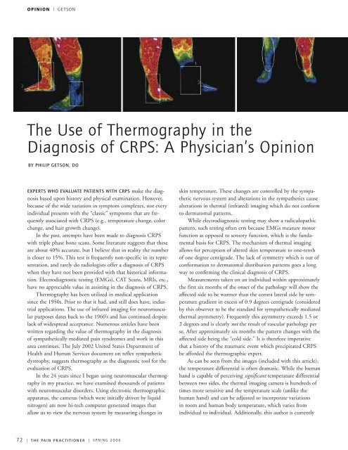
As can be seen from the images (included with this article),<br />
the temperature differential is often dramatic. While the human<br />
hand is capable of perceiving significant temperature differential<br />
between two sides, the thermal imaging camera is hundreds of<br />
times more sensitive and the temperature scale (unlike the<br />
human hand) and can be adjusted to incorporate variations<br />
in room and human body temperature, which varies from<br />
individual to individual. Additionally, this author is currently<br />
72 | T H E PA I N P R A C T I T I O N E R | S P R I N G 2 0 0 6