PHYSICS OF TRANSDUCERS FOR IMAGING AND DOPPLER ...
PHYSICS OF TRANSDUCERS FOR IMAGING AND DOPPLER ...
PHYSICS OF TRANSDUCERS FOR IMAGING AND DOPPLER ...

Create successful ePaper yourself
Turn your PDF publications into a flip-book with our unique Google optimized e-Paper software.
DLM 14 - <strong>PHYSICS</strong> <strong>OF</strong> <strong>TRANSDUCERS</strong> <strong>FOR</strong> <strong>IMAGING</strong> <strong>AND</strong> <strong>DOPPLER</strong><br />
Benjamin Adeyemi MB M.Sc<br />
North Middlesex University Hospital<br />
London<br />
Objectives<br />
• To understand the properties of the basic ultrasound pulse as applied to 2D and Doppler imaging<br />
• To understand the design and development of the modern day phased array transducer, the<br />
piezo-electric materials and physical construction of a phased array transducer as applied to<br />
clinical echocardiography, and how it functions effectively to improve image quality<br />
• To understand the principles behind the development of the modern day transducer, its clinical<br />
applications, and improvement in technology<br />
• To understand broadband imaging, why image texture is different in harmonic imaging, and how<br />
this knowledge can be used effectively to optimise imaging techniques<br />
• To understand recent technological advances in the design of transducers.<br />
1. Introduction<br />
The development of ultrasound transducers has been driven by efforts to improve image quality. Image<br />
quality is largely determined by beam width, frame rates, the length of the ultrasound pulse (l/bandwidth),<br />
and sensitivity.<br />
Reducing beam width and increasing frame rates have benefited from previous developments that have<br />
have largely concentrated on electronic beam forming techniques such as focussing to reduce beam<br />
width and improve lateral resolution, faster signal processing to improve display of the ultrasound image,<br />
and increasing frame rates using faster digital processing. Most modern machines now have greatly<br />
increased frame rates.<br />
In recent years, manufacturers have concentrated on other aspects of transducer design, and have<br />
made substantial improvements which include:<br />
1. Using acoustics and vibration properties such as electromechanical properties of PZT and other<br />
ceramics, and impedance matching to improve sensitivity and axial resolution.<br />
2. Developments in transducer materials and impedance matching to improve sensitivity and<br />
bandwidth, with greater penetration and increased dynamic range, and improved axial, spatial<br />
and contrast resolution.<br />
3. Focussing in the elevation plane which controls slice thickness and produces higher resolution.<br />
4. Focussing in both elevation and lateral planes with improved image quality in 3D applications.<br />
5. Further improvements in digital signal processing which affects all aspects of image quality.<br />
6. Wider imaging field distribution for 3D imaging
2<br />
2. The Basic Ultrasound pulse and Pulse generation<br />
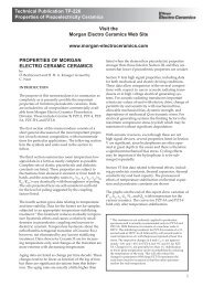
A typical ultrasound pulse consists of cycles of oscillating amplitudes (figure 1a), and contains a<br />
spectrum of frequencies (bandwidth) dominated by a centre frequency.<br />
A high frequency transducer generates a short pulse (1-3 cycles), producing a wide bandwidth<br />
(broadband), this gives accurate distance information with good axial resolution, and is ideal for<br />
diagnostic 2D imaging (figure 1a).<br />
Figure 1a. Short Pulse<br />
1 – 3 cycles<br />
Wide bandwidth<br />
A low frequency transducer generates a long pulse about 5 – 30 cycles (figure 1b ), producing a narrow<br />
bandwidth, this gives accurate frequency information but poor axial resolution, therefore used typically<br />
for Doppler imaging (figure 1b).<br />
Figure 1b. Long Pulse<br />
5 – 30 cycles<br />
Narrow bandwidth<br />
The ultrasound pulse occurs in microseconds therefore difficult to measure. The pulse generated by the<br />
transducer is sent to tissues, and the reflected echo is received by the same transducer within a time<br />
interval determined by the depth or range of the reflector (Figure 1c). A brief pulse generates a single<br />
echo allowing the delay to be measured, with continuous transmission individual echoes can’t be<br />
identified.<br />
Figure 1c.
3<br />
3. Structure of Phased Array Transducers<br />
All transducers have a thin piezoelectric ceramic plate often made of lead zirconate titanate (PZT), a<br />
matching layer, and a backing layer with a single lens across the transducer arrays. (figure 2a). The<br />
piezoelectric plate generates and detects ultrasound waves. Some piezoelectric materials (e.g quartz)<br />
occurs naturally, but (PZT) is a synthetic ceramic material commonly used. The curved cylindrical lens<br />
after the matching layer is used to focus the beam in the elevation plane and improve electronic<br />
focussing, producing the greatest transmitted amplitude and receive sensitivity at the focal zone.<br />
Figure 2a: Cross-section of a typical phased array transducer<br />
Figure 2b: Cross-section of a typical phased array transducer<br />
4. PZT and generation of ultrasound pulse<br />
The properties of piezoelectric materials are that they deform in response to an electrical voltage, and<br />
generate electrical voltages when stretched or compressed by an external force. Newer versions of PZT<br />
have been developed to improve sensitivity and produce larger acoustic power, and other piezoelectric<br />
materials are being developed to generate stronger electrical signals when receiving echo pulses with<br />
greater sensitivity and penetration and wider bandwidth, producing better axial resolution.<br />
PZT generates and transmits ultrasound imaging pulses when fired by rapidly changing voltages. In<br />
order to transmit an ultrasound pulse, the oscillating voltage of the required frequency is applied across
4<br />
the PZT plates making it vibrate at this frequency sending the ultrasonic pulse into tissues. During<br />
reception, the pressure variation of returning echoes cause the PZT plate to contract and expand<br />
generating voltage variations across the plate that form an electronic version of the received echo<br />
signal.<br />
For the PZT plate to vibrate most strongly, its thickness must be exactly half the wavelength of the<br />
signal produced by the vibrating frequency produced ie “half-wave resonance”. Resonance occurs<br />
because the ultrasound wave propagating across the thickness of the PZT plate reverberates within the<br />
plate. If the thickness of the PZT is equal to half the wavelength of the vibrating frequency, it will travel a<br />
full wavelength within the PZT before arriving at its starting point, this means that it will be in phase with<br />
the original wave and add constructively to produce a greater output. A PZT plate with thickness that is<br />
equal to half the wavelength at the required centre frequency will resonate and produce a larger output<br />
at this frequency.<br />
PZT crystals transmit impulses 1% of the time, and receive impulses 99% of the time. The electrical<br />
pulse applied across the PZT plate typically consists of 1-3 cycles of oscillating voltages of peak to peak<br />
amplitude of as much as 200 to 300 Volts determined by the output power control. The frequency and<br />
duration of the oscillating pulse is determined by the transducer centre frequency and pulse length.<br />
Excess “internal ringing”<br />
The main disadvantage of PZT is that it has a high density therefore high characteristic acoustic<br />
impedance up to 20 times higher than that of soft tissue. This results in up to 80% reflection of the<br />
ultrasound energy at the PZT-tissue interface, multiple prolonged internal reverberation (ringing), and<br />
very long pulses with ultimately poor axial resolution, poor sensitivity and reverberation artifarct.<br />
Backing layer<br />
A backing (damping) layer with a high characteristic acoustic impedance and ability to absorb ultrasound<br />
is used behind the PZT plate to reduce unwanted ringing (figure 2a). Modern transducers have a<br />
backing layer with impedance somewhat lower to give a useful reduction in ringing without lowering<br />
sensitivity too much, but prevent total absorption of ultrasound. The remaining ringing is removed by<br />
adding a matching layer (figure 2a).<br />
Apart from the ringing problem, poor sensitivity is made worse because only 20% of the ultrasound<br />
wave’s power would be transmitted through the front PZT-tissue interface. To correct this problem a<br />
single “impedance matching layer” is bonded to the front face of the PZT plate to increase transmission<br />
across the front face of the PZT by nearly 100%, and increase the efficiency of the transmitted wave as<br />
it exits the transducer surface (figure 2a). This is done effectively by ensuring that the matching layer<br />
has a thickness equal to a quarter of a wavelength, and an impedance equal to :<br />
Impedance of PZT x Impedance of Tissue<br />
This is called the geometric mean of the impedances.<br />
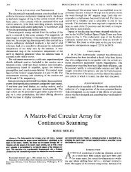
Figure 2c shows reverberation within PZT plate that produces multiple transmission into tissues<br />
resulting in reinforced signals and a larger amplitude pulse. Multiple reflections back into PZT cancel<br />
out the original (top) reflection into backing layer. The most efficient (near 100%) transmission through<br />
the matching layer only occurs when its thickness is exactly one-quarter of a wavelength of its vibrating<br />
frequency.
5<br />
Figure 2c: Quarter-wave length matching layer<br />
Matching layer and bandwidth<br />
Although the matching layer improves sensitivity, it gives high sensitivity at a single frequency, and good<br />
performance only over a narrow range of frequencies. It also acts as a filter therefore reducing<br />
bandwidth. Figure 3 below shows the bandwidth or range of frequencies for a transducer. A -3db<br />
bandwidth is the range of frequencies over which the transducer is most efficient. A transducer with a<br />
matching layer will have a -3db bandwidth of about 60% of the centre frequency, therefore a 3MHz<br />
transducer with a matching layer will have a -3dB lower frequency of 1.8MHz. and an upper frequency of<br />
4.2MHz.<br />
Figure 3: Transducer bandwidth<br />
5. Improvements in efficiency of Transducers<br />
A large transducer bandwidth is produced by a short pulse, and is crucial for good axial resolution.<br />
Transducer bandwidth can be increased by using two or more matching layers of different thickness.<br />
Improvements in backing layers and introduction of multiple matching layer technology have led to<br />
transducers with wider bandwidths. Up to -3dB bandwidth with greater than 100% centre frequency is<br />
now available.<br />
Larger transducer bandwidths are now a common feature in modern day transducers. This allows a<br />
choice of operating frequencies according to penetration or resolution required. Modern day transducers<br />
can now operate at three frequencies for example a 1, 2 and 3MHz transducer would need to have a<br />
centre frequency of 2MHz and a bandwidth of 2MHz which is 100% of the centre frequency. Axial
6<br />
resolution of bandwidths generated at the upper and lower frequencies is however less than if the whole<br />
transducer bandwidth were used.<br />
Figure 4: Broadband Transducer bandwidth<br />
6. Improvements in PZT and transducer design<br />
The Piezoelectric (PZT) material is the most effective determinant of beam penetration and image<br />
quality. For many years, there has been slow progress in the development of PZT crystals with superior<br />
electromechanical properties. Recently transducer design has been focussed on development of new<br />
types of PZT materials (e.g. PureWave crystals, as developed by Philips Medical Ltd)) with improved<br />
electromechanical properties compared to old PZT ceramics or PZT composites. These are being been<br />
used with specially designed matching layers and backing materials producing dramatic improvements<br />
in efficiency, sensitivity and bandwidth.<br />
“Composite PZT” is made by cutting closely spaced narrow channels through a solid plate of PZT<br />
ceramic and filling them with an inert polymer, this has a lower characteristic acoustic impedance than<br />
PZT itself, therefore produces transducers with greater sensitivity and wider bandwidth, and alleviates<br />
the problems associated with matching.<br />
PureWave crystals are more efficient than PZT ceramic or PZT composites with electro-mechanical<br />
properties improved by up to 68 – 85%, and ten times the ability to deform in the presence of an<br />
electrical field. When combined with multiple matching layers and backing material, they greatly<br />
increase bandwidth (1 - 5MHz), and sensitivity at transmission and reception, with improved dynamic<br />
range, greater penetration, greater clarity of images, and greater uniformity throughout the entire image<br />
field. They provide better endocardial border delineation in difficult-to-image patients, with significant<br />
benefits in both tissue and contrast harmonics applications. Improved sensitivity means higher<br />
frequencies can be used with better image resolution, and provides the main benefit for contrast<br />
harmonics allowing the detection of bubbles more easily. There is also better sensitivity at the lower<br />
frequency spectrum for colour and spectral Doppler frequencies.
7<br />
Figure 5a: Broadband versus Pure-wave crystal technology<br />
Figure 5b: PureWave crystal transducer with large bandwidth
8<br />
7. Broadband Transducers and imaging frequencies<br />
These are now used on modern ultrasound scanners. They have a large bandwidth, so they can<br />
transmit and receive pulses with several different center frequencies (Pulse spectra shown as dashed in<br />
figure 3). A large transducer bandwidth is crucial for good axial resolution and harmonic imaging. It<br />
allows the operator a choice of transmitting and receiving frequencies, and to toggle between them<br />
according to the penetration or resolution required.<br />
Typical imaging frequencies in adult echocardiography can be anything from 1.0MHz to 5MHz.<br />
Compared to a lower frequency band, a higher frequency band will have shorter near zone, more<br />
divergent beam, greater side-lobes, poor penetration, but better axial & near field resolution. Attenuation<br />
or energy loss is greatly increased at higher frequencies, and explains the poor beam penetration.<br />
Beam intensity at a particular depth is lower in the higher frequency transducer due early beam<br />
divergence.<br />
A lower frequency band will have a longer near zone, less divergent beam, better penetration, and<br />
poorer axial & near field resolution therefore ideal for Doppler imaging. Narrower beam width results<br />
from a less divergent beam, therefore better lateral resolution. The beam characteristics are also ideal<br />
for harmonic imaging which requires transmitting at low frequencies, and receiving at higher<br />
frequencies. The short wavelengths associated with higher frequencies lead to improved resolution of<br />
images. The aim is to choose the optimum frequency band for each particular application. This is a<br />
compromise that ensures that the best resolution is obtained while allowing echoes to be received from<br />
the required depth.<br />
8. Phased Array Transducers (Beam steering and focusing)<br />
Phased array transducers have typically 128 rectangular elements or arrays arranged with individual<br />
connections. All arrays are used to transmit and receive beams for every scan line, and each scan line<br />
represents the axis of the transmission-receive beam. The larger the arrays on a phased array<br />
transducer, the lower the resonation, and the lower the transmitting frequency.<br />
Beam steering<br />
These transducers allow the beam to be moved electronically with the benefit of being able to change<br />
the shape and size of the beam to suit imaging needs. The beam former is the part of the electronics of<br />
the scanner that determines the shape size and position of the interrogating beams by controlling the<br />
delay period of signals to and from the transducer array elements. During transmission it generates the<br />
electronic signals that pulse each array and during reception combines the individual echo sequences<br />
from all the elements into a single echo sequence.<br />
The beam is angled and then swept as a sector electronically by firing all the arrays as a complete<br />
group, with a small delay (< 1 microsecond) called “Phasing”, the time delay is also changed with each<br />
successive transmitted echo signal, so that the wavefronts are inclined, and beam direction is<br />
continuously changed and perpendicular to transducer face (figure 6a). However angling the wave-front<br />
gives rise to side lobe artefacts affecting near field resolution (figure 6b).
9<br />
Figure 6a.<br />
Electronic Steering of a Phased Array Sector Transducer<br />
Sector scan<br />
format of a<br />
phased array<br />
probe<br />
Each waveform merges to form a compound wave,<br />
generating a sector beam.<br />
Constructive<br />
interference<br />
from wavelets<br />
generating<br />
sector beam<br />
Huygen’s principle<br />
This states that every point on a wavefront can be considered as a new point source emitting a<br />
spherical wave of the same frequency and phase.<br />
The width of the source producing the sound wave from a phased array probe is greater than the<br />
wavelength of the wave, producing a wave that propagates at 90 0 to the source ie in form a beam. Each<br />
small source generates a sound wave with same frequency and amplitude, and are in phase with each<br />
other The spherical waves from each source propagate outwards with parts of the wave parallel to the<br />
surface of the source. Some interfere destructively and cancel out, while others interfere constructively<br />
and align to form a plane wavefront generating the ultrasound beam.<br />
Figure 6b.<br />
Typical Ultrasound Beam From a Phased Arrray Transducer<br />
Note!! side lobe artefacts.<br />
These grow stronger at<br />
larger steering angles
10<br />
Beam focusing<br />
The ultrasound beam is focussed during transmission and reception. Like beam steering, transmit<br />
focussing is achieved by using an electronic delay in firing the arrays resulting in a wavefront curvature<br />
that directs the beam to a focal zone, which is the zone of highest beam intensity.<br />
Figure 6c.<br />
FOCAL<br />
ZONE<br />
The same electronic time delay in receiving early echoes is then used during reception allowing echoes<br />
from different depths to arrive at the same time. This is achieved by adjusting the delay periods during<br />
the receive phase allowing late arriving echoes also to be in focus. This is called Dynamic Receive<br />
Focussing.This reduces beam width and improves lateral resolution, but has no effect on focussing in<br />
the elevation plane. This is achieved by using a curved cylindrical lens producing higher resolution.<br />
Multi-zone Focusing is achieved by dividing each individual scan line into two or more sections, and<br />
interrogating each section with a separate transmission pulse producing transmission foci at different<br />
depths. This allows the operator to select two or more or multiple focal zones. This improves lateral<br />
resolution, however temporal resolution and frame rate is affected due to the need to remain longer on<br />
each scan line while interrogating several zones. Parameters such as centre frequency and pulse length<br />
are not affected, but high frequencies within the pulse spectrum will be affected by attenuation because<br />
several focal zones are selected at greater depths.<br />
Phased array transducers have been established for many years and used in all modern echo<br />
transducers. An effective phased array transducer will need to have the right balance between number<br />
of elements and crystal size. More advanced phased array transducers for three-dimensional imaging<br />
have now up to 3000 much smaller elements compared to 128 elements of standard two-dimensional<br />
phased arrays.
11<br />
9. Transducer design and development<br />
Figure 7.<br />
LARGE<br />
PROBE<br />
2D Phased Array<br />
Up to 128 elements<br />
Field of view<br />
(Widely used for 2D echo)<br />
A1.5D Array enables<br />
dynamic<br />
focusing in the<br />
elevation plane with better<br />
resolution & dynamic range<br />
(Potentially useful for 3D<br />
volume reconstruction<br />
imaging)<br />
3D Matrix Array<br />
3000 Elements<br />
Real-time<br />
(Live 3D echo)<br />
10. The Three Dimensional Matrix Array<br />
This transducer represents a breakthrough in the design of ultrasound transducers. Reduction in PZT<br />
crystal size has made it possible for up to 3000 arrays to form a three dimensional (3D) image, that is<br />
also ideal for two-dimensional bi-plane imaging. Using a pyramidal burst of ultrasound (Raster scanning),<br />
a real-time dynamic 3D image is formed. The main limitation is due to variable distances between<br />
individual scan planes with less structural information available, and less resolution. The transducer is<br />
bulky, with images limited to a pyramid or cone shape, and acquisition restricted by small acoustic<br />
windows and chest wall movement. However improvements in digital signal processing, and high speed<br />
acquisition with rapid data processing, resolution and image quality has been improved. Improvements in<br />
beam forming techniques have made it possible for electronic steering and beam focusing in both the<br />
lateral and elevation planes, and 3D volume quantification. Beam focusing results in a narrow slice<br />
thickness, with superior resolution.<br />
Benjamin Adeyemi
12<br />
References<br />
1 Apfel RE, Holland K : (1991) Gauging the likely hood of cavitation from short pulse, low duty<br />
cycle diagnostic ultrasound. Ultrasound Med Biol Vol 17 p 175 185.<br />
2 Desser TS, Jedrzejewicz T, Bradley C : (2000) Ultrasound Quart. : Native tissue harmonic<br />
imaging : Basic principles and clinical applications. C 16 p 40 – 48.<br />
3 Feigenbaum H, Armstrong WF, Ryan T : (2005) Feigenbaum’s Echocardiography physics and<br />
instrumentation p 11 – 19 Lippincott Williams & Wilkins, Philadelphia Baltimore New York<br />
London Buenos Aires Hong Kong Sydney Tokyo.<br />
4 Fish P : (1990) Physics And Instrumentation of Diagnostic Medical Ultrasound. Real time<br />
scanners. p 83 – 96 John Wiley and Sons, New York Chichester Brisbane Toronto<br />
Singapore.<br />
5 Hill CR : (1986) Physical Principles of Medical Ultrasonics. Beam formation by transducer arrays.<br />
p 83 – 89 John Wiley and Sons, New York Chichester Brisbane Toronto Singapore.<br />
6 Hoskins P, Thrush A, Martin K, Whittingham TA : (2003) Diagnostic Ultrasound Physics<br />
and equipment. Transducers and beam forming p 23 – 29 1 st edition Greenwich Medical<br />
Media London San Francisco.<br />
7 Kremkau FW : (1993) Diagnostic Ultrasound Principles and Instruments. Transducers 4 th Edition<br />
p 69 – 128 W B Saunders and Co, Philadelphia London Toronto Montreal Sydney Tokyo.<br />
8 McDicken WN : (1991) Diagnostic Ultrasonics Principles And Use Of Instruments. Introduction to<br />
basic ultrasonics and basic ultrasonic instruments 3 rd Edition p1 – 28 Churchill Livingstone<br />
Edinburgh London Melbourne New York.<br />
9 Merrit CRB, In Rumack CM, Wilson SR, Charboneau JW : Diagnostic Ultrasound. Physics of<br />
Ultrasound. Vol 1 2 nd Editiion p 10 – 33 St Louis Mosby.<br />
10 Monaghan MJ : (190) Practical Echocardiography and Doppler. Basic physics and technology of<br />
echocardiography p1 – 10 John Wiley and Sons New York Chichester Brisbane Toronto<br />
Singapore.<br />
11 Salustri A, Roelandt JRTC (1995) Ultrasonic three-dimensional reconstruction of the heart.<br />
Ultrasound Med Biol Vol 21 p 281 - 293.<br />
12 Wells PNT (1977) Biomedical Ultrasonics. Academic Press London.<br />
13 Whittingham TA (1995). Modern developments in diagnostic ultrasound. Radiography I<br />
p 61 – 73.<br />
14 Wilde P Oakley CM : (1993) Clinical Ultrasound A Comprehensive Text. Introduction to cardiac<br />
ultrasound p1 – 40 Churchill Livingstone Edinburgh London Madrid Melbourne New York and<br />
Tokyo.<br />
15 Hylon B Ferrant M Ferrant P : (1995) Basic Ultrasound : John Wiley and Sons New York<br />
Highlighted references indicate compulsory reading.