- Page 1 and 2: UNIVERSITÀ DEGLI STUDI DI MILANO B
- Page 3 and 4: CONTENTS 3 3.2 Dynamic Light scatte
- Page 5 and 6: CONTENTS 5 5.15.2 Spectral characte
- Page 7 and 8: Introduction In the last two decade
- Page 9 and 10: Chapter 9 then we have applied the
- Page 11 and 12: Chapter 1 Metal nanoparticles 1.1 N
- Page 13 and 14: Chapter 1 13 carry out reactions in
- Page 15 and 16: Chapter 1 15 donor and the metal at
- Page 17 and 18: Chapter 1 17 Figure 1.4: Surface pl
- Page 19 and 20: Chapter 1 19 where the scattering a
- Page 21 and 22: Chapter 1 21 screens the surface ch
- Page 23 and 24: Chapter 1 23 easily be seen from eq
- Page 25 and 26: Chapter 1 25 The first is equal to
- Page 27 and 28: Chapter 1 27 dipole approximation a
- Page 29 and 30: Chapter 1 29 used for disease or ca
- Page 31 and 32: Bibliography [1] Nanotoxicology: na
- Page 33 and 34: Chapter 1 33 [30] Bohren CF; Huffma
- Page 35 and 36: Chapter 2 Principles 2.1 Basics of
- Page 37 and 38: Chapter 2 37 A direct consequence o
- Page 39 and 40: Chapter 2 39 be considerably more c
- Page 41 and 42: Chapter 2 41 Figure 2.5: Effect of
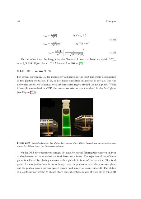
- Page 43 and 44: Chapter 2 43 2.4 Two-photon excited
- Page 45 and 46: Chapter 2 45 m = λV MI hc 2 N A (2
- Page 47: Chapter 2 47 P SF OP E (u, v) = |h(
- Page 51 and 52: Bibliography [1] Lakowicz J.R. In p
- Page 53 and 54: Chapter 2 53 [29] Born M. and Wolf
- Page 55 and 56: Chapter 3 55 3.2 Dynamic Light scat
- Page 57 and 58: Chapter 3 57 where n is the refract
- Page 59 and 60: Chapter 3 59 with g V H (t) ∝ β
- Page 61 and 62: Chapter 3 61 κ m (Γ) = dm K(−τ
- Page 63 and 64: Chapter 3 63 that plagues conventio
- Page 65 and 66: Chapter 3 65 set equal to the new p
- Page 67 and 68: Chapter 3 67 the molecules. In orde
- Page 69 and 70: Chapter 3 69 number of molecules in
- Page 71 and 72: Chapter 3 71 γ 1 G(τ) = V ex 〈C
- Page 73 and 74: Chapter 3 73 Figure 3.2: Auto-corre
- Page 75 and 76: Chapter 3 75 electronic distortions
- Page 77 and 78: Chapter 3 77 Figure 3.5: left panel
- Page 79 and 80: Chapter 3 79 p 1 (k, V 0 , ɛ) = 1
- Page 81 and 82: Chapter 3 81 3.9.3 Photon Moment An
- Page 83 and 84: Chapter 3 83 Figure 3.6: Schematic
- Page 85 and 86: Bibliography [1] Chu B. In Laser Li
- Page 87 and 88: Chapter 3 87 [27] Eigen S.M. and Ri
- Page 89 and 90: Chapter 3 89 [53] Chen Y., Tekmen M
- Page 91 and 92: Chapter 4 91 • L.Sironi, S.Freddi
- Page 93 and 94: Chapter 4 93 die. Mutations in the
- Page 95 and 96: Chapter 4 95 arm of chromosome 17.
- Page 97 and 98: Chapter 4 97 shown that one of the
- Page 99 and 100:
Chapter 4 99 DNA replication while
- Page 101 and 102:
Chapter 4 101 4.2). The ratio R = [
- Page 103 and 104:
Chapter 4 103 The polydispersity of
- Page 105 and 106:
Chapter 4 105 The fitting of the FC
- Page 107 and 108:
Chapter 4 107 the streptavidin (4.5
- Page 109 and 110:
Chapter 4 109 29 nm for the 10 nm s
- Page 111 and 112:
Chapter 4 111 A 1 Γ 1 (nm) λ 1 (n
- Page 113 and 114:
Chapter 4 113 Figure 4.8: (Left: Fl
- Page 115 and 116:
Chapter 4 115 been systematically i
- Page 117 and 118:
Chapter 4 117 Figure 4.12: Left: Me
- Page 119 and 120:
Chapter 4 119 at the basis of the o
- Page 121 and 122:
Chapter 4 121 presence of p53, comp
- Page 123 and 124:
Chapter 4 123 Figure 4.17: The figu
- Page 125 and 126:
Chapter 4 125 from probably aspecif
- Page 127 and 128:
Chapter 4 127 Figure 4.21: Panel A:
- Page 129 and 130:
Bibliography [1] Anker J. N.; Hall
- Page 131 and 132:
Chapter 4 131 [47] Das P.; Metiu H.
- Page 133 and 134:
Chapter 5 133 In this chapter the c
- Page 135 and 136:
Chapter 5 135 Figure 5.1: Left:Symm
- Page 137 and 138:
Chapter 5 137 5.3 Experimental deta
- Page 139 and 140:
Chapter 5 139 back aperture of the
- Page 141 and 142:
Chapter 5 141 5.4.2 Transmission El
- Page 143 and 144:
Chapter 5 143 For anisotropic branc
- Page 145 and 146:
Chapter 5 145 coefficient D is then
- Page 147 and 148:
Chapter 5 147 Figure 5.9: ACF of th
- Page 149 and 150:
Chapter 5 149 measured through a ve
- Page 151 and 152:
Chapter 5 151 objective in epifluor
- Page 153 and 154:
Chapter 5 153 The fitting of FCS cu
- Page 155 and 156:
Chapter 5 155 Sample Population (%)
- Page 157 and 158:
Chapter 5 157 5.8 Concentration eva
- Page 159 and 160:
Chapter 5 159 Figure 5.17: Emission
- Page 161 and 162:
Chapter 5 161 5.12 Dependence of TP
- Page 163 and 164:
Chapter 5 163 Figure 5.22: Histogra
- Page 165 and 166:
Chapter 5 165 5.13 Cellular uptake
- Page 167 and 168:
Chapter 5 167 Figure 5.29: NPs obta
- Page 169 and 170:
Chapter 5 169 Figure 5.33: Autofluo
- Page 171 and 172:
Chapter 5 171 5.14 Photothermal eff
- Page 173 and 174:
Chapter 5 173 Figure 5.37: ζ-poten
- Page 175 and 176:
Chapter 5 175 Temperature [ ◦ C]
- Page 177 and 178:
Chapter 5 177 In figure 5.39 the fl
- Page 179 and 180:
Chapter 5 179 P [mW] < τ > [ns] 10
- Page 181 and 182:
Chapter 5 181 The dependence of the
- Page 183 and 184:
Chapter 6 183 Currently, only alumi
- Page 185 and 186:
Chapter 6 185 tact organ studies, a
- Page 187 and 188:
Chapter 6 187 with liquid flow chan
- Page 189 and 190:
Chapter 6 189 Figure 6.1: DC in clo
- Page 191 and 192:
Chapter 6 191 • New contrast agen
- Page 193 and 194:
Chapter 6 193 experiments by flowin
- Page 195 and 196:
Chapter 6 195 it has discontinuitie
- Page 197 and 198:
Chapter 6 197 P t. rection. If all
- Page 199 and 200:
Chapter 6 199 Figure 6.6: Commonly
- Page 201 and 202:
Chapter 6 201 Figure 6.9: 3D and 2D
- Page 203 and 204:
Chapter 6 203 Figure 6.11: (A) NK c
- Page 205 and 206:
Chapter 6 205 are somehow confined,
- Page 207 and 208:
Chapter 6 207 Figure 6.15: Distance
- Page 209 and 210:
Chapter 6 209 Figure 6.18: (A)The s
- Page 211 and 212:
Chapter 6 211 Figure 6.21: The figu
- Page 213 and 214:
Chapter 6 213 Figure 6.24: Paramete
- Page 215 and 216:
Chapter 6 215 of the natural killer
- Page 217 and 218:
Chapter 6 217 and CD8 + T cells, is
- Page 219 and 220:
Chapter 6 219 6.10.1 TLRs TLRs are
- Page 221 and 222:
Chapter 6 221 arise from myeloid pr
- Page 223 and 224:
Chapter 6 223 of infection, primari
- Page 225 and 226:
Chapter 6 225 Figure 6.30: Lymph-no
- Page 227 and 228:
Bibliography [1] A. Diaspro, G. Chi
- Page 229 and 230:
Chapter 6 229 [25] E. O. Long. Regu
- Page 231 and 232:
Chapter 6 231 [47] S. H. Wei, M. J.
- Page 233 and 234:
Chapter 6 233 [72] S. Fossum and W.
- Page 235 and 236:
Chapter 235 mode-locked cavity. Thi
- Page 237 and 238:
Chapter 237 Figure 6.34: Energy lev
- Page 239 and 240:
Chapter 239 Figure 6.37: Prisms seq
- Page 241 and 242:
Chapter 241 part of the TE300 and t
- Page 243 and 244:
Chapter 243 Figure 6.42: The Olympu
- Page 245 and 246:
Chapter 245 a f=100 mm bispherical
- Page 247 and 248:
Chapter 247 are just events’ dete
- Page 249 and 250:
Chapter 249 smaller quartz cell (He
- Page 251 and 252:
Chapter 251 needed to a linear corr
- Page 253 and 254:
Chapter 253 Figure 6.48: Channel ar
- Page 255 and 256:
Chapter 255 ming for burst height a
- Page 257:
Chapter 257 • M. Caccia, T. Gorle