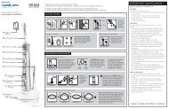
Philips Sonicare FlexCare - Sonicare.com - Sonicare
Philips Sonicare FlexCare - Sonicare.com - Sonicare
Philips Sonicare FlexCare - Sonicare.com - Sonicare

Create successful ePaper yourself
Turn your PDF publications into a flip-book with our unique Google optimized e-Paper software.
2% sucrose in MatTek culture plates b at 37°C and 5%<br />
CO 2<br />
. The culture medium was replaced daily with<br />
fresh 37°C medium over a 3-day culture period. The<br />
biofilms were stained to show the bacterial cells in<br />
green and the slime matrix in blue. The images were<br />
constructed using Imaris software. c<br />
b<br />
MatTek Corp; Ashland, MA.<br />
c<br />
Bitplane, AG; Zurich, Switzerland.<br />
Diffusion Chamber<br />
A previously developed method 22-24 was used to<br />
assess the effect of power brushing on the diffusion<br />
of fluoride through biofilms. A specially designed 2-<br />
chamber diffusion chamber was fabricated from<br />
polycarbonate (Figure 2).<br />
The 2 chambers were separated by a cellulose<br />
ester membrane. By growing biofilms on the membrane<br />
and measuring how quickly fluoride passed<br />
from one chamber to the other, we could measure<br />
the effect of power brushing on delivering fluoride<br />
through the biofilm-colonized membrane. The membrane<br />
filter holder was easily inserted into the diffusion<br />
chamber allowing rapid replacement with new<br />
biofilm-colonized filters for repeated measurements.<br />
First, the biofilm-side chamber (right-hand chamber)<br />
was gently filled with 1100 ppm sodium fluoride<br />
solution in phosphate buffer salts (PBS). The<br />
measurement-side chamber contained PBS with<br />
approximately 0.5 ppm fluoride. By starting with a<br />
small concentration of fluoride in the measurement<br />
chamber, the stabilization time of the electrode was<br />
decreased. Thus, at time zero, the measurement-side<br />
chamber had 0.5 ppm fluoride, whereas the biofilmside<br />
chamber had 1100 ppm. The filling process took<br />
approximately 5 seconds. The appearance of fluoride<br />
in the measurement chamber, after traveling through<br />
the biofilm and the membrane, was measured by an<br />
ion-selective fluoride electrode. The measurementside<br />
chamber was mixed vigorously with a magnetic<br />
stir bar. Over the 4-minute monitoring period, the<br />
concentration in the biofilm-side chamber never fell<br />
to less than 1050 ppm, so it was assumed that the<br />
concentration gradient driving the fluoride flux was<br />
constant.<br />
Biofilm Growth for Diffusion Measurements<br />
Biofilms were grown on the cellulose ester<br />
membranes from S mutans UA159 by immersing a<br />
filter holder, with an attached filter, in each well of a<br />
6-well plate with 5 mL of BHI and 2% sucrose.<br />
Because the filter assemblies were slightly buoyant,<br />
they floated, so that the filter surface was level with<br />
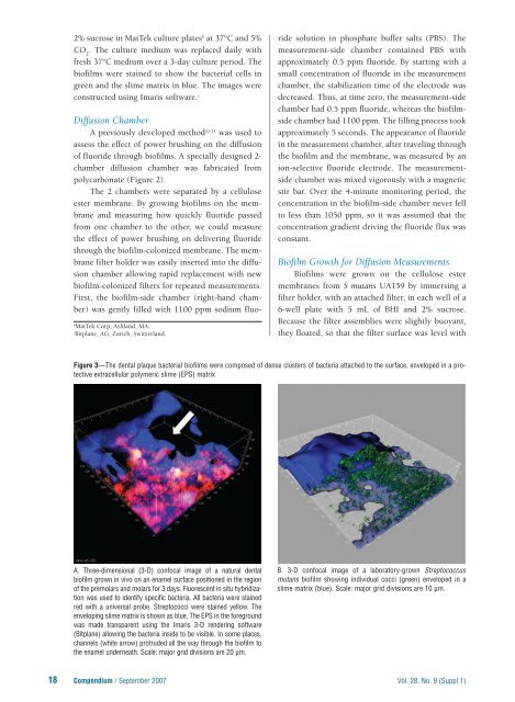
Figure 3—The dental plaque bacterial biofilms were <strong>com</strong>posed of dense clusters of bacteria attached to the surface, enveloped in a protective<br />
extracellular polymeric slime (EPS) matrix<br />
A. Three-dimensional (3-D) confocal image of a natural dental<br />
biofilm grown in vivo on an enamel surface positioned in the region<br />
of the premolars and molars for 3 days. Fluorescent in situ hybridization<br />
was used to identify specific bacteria. All bacteria were stained<br />
red with a universal probe. Streptococci were stained yellow. The<br />
enveloping slime matrix is shown as blue. The EPS in the foreground<br />
was made transparent using the Imaris 3-D rendering software<br />
(Bitplane) allowing the bacteria inside to be visible. In some places,<br />
channels (white arrow) protruded all the way through the biofilm to<br />
the enamel underneath. Scale: major grid divisions are 20 µm.<br />
B. 3-D confocal image of a laboratory-grown Streptococcus<br />
mutans biofilm showing individual cocci (green) enveloped in a<br />
slime matrix (blue). Scale: major grid divisions are 10 µm.<br />
18 Compendium / September 2007 Vol. 28, No. 9 (Suppl 1)