Wound Closure Manual (PDF) - Penn Medicine
Wound Closure Manual (PDF) - Penn Medicine
Wound Closure Manual (PDF) - Penn Medicine

You also want an ePaper? Increase the reach of your titles
YUMPU automatically turns print PDFs into web optimized ePapers that Google loves.
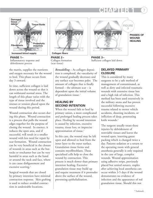
CHAPTER 1 7<br />
Damaged<br />
tissue<br />
debris<br />
Fibroblasts<br />
Tissue fluids<br />
Proteolytic<br />
enzymes<br />
FIGURE<br />
3<br />
PHASES OF<br />
WOUND<br />
HEALING<br />
Increased blood supply<br />
PHASE 1–<br />
Inflammatory response and<br />
debridement process<br />
Collagen fibers<br />
PHASE 2–<br />
Collagen formation<br />
(scar tissue)<br />
PHASE 3–<br />
Sufficient collagen laid down<br />
the matrix, supplies the nutrients<br />
and oxygen necessary for the wound<br />
to heal. This phase occurs from<br />
day 3 onward.<br />
In time, sufficient collagen is laid<br />
down across the wound so that it<br />
can withstand normal stress. The<br />
length of this phase varies with the<br />
type of tissue involved and the<br />
stresses or tension placed upon the<br />
wound during this period.<br />
<strong>Wound</strong> contraction also occurs during<br />
this phase. <strong>Wound</strong> contraction<br />
is a process that pulls the wound<br />
edges together for the purpose of<br />
closing the wound. In essence, it<br />
reduces the open area, and if<br />
successful, will result in a smaller<br />
wound with less need for repair by<br />
scar formation. <strong>Wound</strong> contraction<br />
can be very beneficial in the closure<br />
of wounds in areas such as the buttocks<br />
or trochanter but can be very<br />
harmful in areas such as the hand<br />
or around the neck and face, where<br />
it can cause disfigurement and<br />
excessive scarring. 3<br />
Surgical wounds that are closed<br />
by primary intention have minimal<br />
contraction response. Skin grafting<br />
is used to reduce avoided contraction<br />
in undesirable locations.<br />
Remodelling – As collagen deposition<br />
is completed, the vascularity of<br />
the wound gradually decreases and<br />
any surface scar becomes paler. The<br />
amount of collagen that is finally<br />
formed – the ultimate scar – is<br />
dependent upon the initial volume<br />
of granulation tissue. 2<br />
HEALING BY<br />
SECOND INTENTION<br />
When the wound fails to heal by<br />
primary union, a more complicated<br />
and prolonged healing process takes<br />
place. Healing by second intention<br />
is caused by infection, excessive<br />
trauma, tissue loss, or imprecise<br />
approximation of tissue. 3<br />
In this case, the wound may be left<br />
open and allowed to heal from the<br />
inner layer to the outer surface.<br />
Granulation tissue forms and<br />
contains myofibroblasts. These<br />
specialized cells help to close the<br />
wound by contraction. This<br />
process is much slower than primary<br />
intention healing. Excessive<br />
granulation tissue may build up<br />
and require treatment if it protrudes<br />
above the surface of the wound,<br />
preventing epithelialization.<br />
DELAYED PRIMARY<br />
CLOSURE<br />
This is considered by many<br />
surgeons to be a safe method of<br />
management of contaminated, as<br />
well as dirty and infected traumatic<br />
wounds with extensive tissue loss<br />
and a high risk of infection. This<br />
method has been used extensively in<br />
the military arena and has proven<br />
successful following excessive<br />
trauma related to motor vehicle<br />
accidents, shooting incidents, or<br />
infliction of deep, penetrating<br />
knife wounds. 3<br />
The surgeon usually treats these<br />
injuries by debridement of<br />
nonviable tissues and leaves the<br />
wound open, inserting gauze<br />
packing which is changed twice a<br />
day. Patients sedation or a return to<br />
the operating room with general<br />
anesthesia generally is only required<br />
in the case of large, complex<br />
wounds. <strong>Wound</strong> approximation<br />
using adhesive strips, previously<br />
placed but untied sutures, staples<br />
after achieving local anesthesia can<br />
occur within 3-5 days if the wound<br />
demonstrates no evidence of<br />
infection and the appearance of red<br />
granulation tissue. Should this not<br />
* Trademark