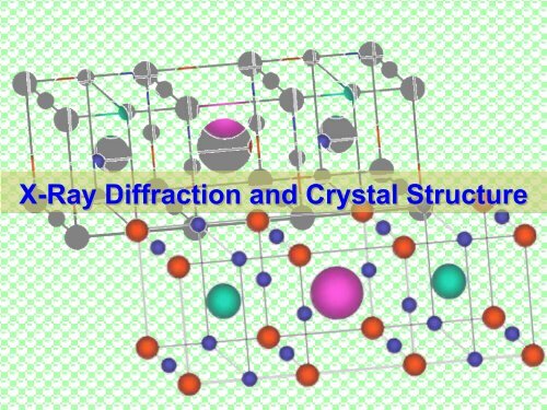
X-Ray Diffraction and Crystal Structure
X-Ray Diffraction and Crystal Structure
X-Ray Diffraction and Crystal Structure
Create successful ePaper yourself
Turn your PDF publications into a flip-book with our unique Google optimized e-Paper software.
X-<strong>Ray</strong> <strong>Diffraction</strong> <strong>and</strong> <strong>Crystal</strong> <strong>Structure</strong>
X-<strong>Ray</strong> <strong>Diffraction</strong> <strong>and</strong> <strong>Crystal</strong> <strong>Structure</strong> (XRD)<br />
X-ray diffraction (XRD) is one of the most important non-destructive tools to<br />
analyse all kinds of matter - ranging from fluids, to powders <strong>and</strong> crystals. From<br />
research to production <strong>and</strong> engineering, XRD is an indispensible method for<br />
structural materials characterization <strong>and</strong> quality control which makes use of the<br />
Debye-Scherrer method. This technique uses X-ray (or neutron) diffraction on<br />
powder or microcrystalline samples, where ideally every possible crystalline<br />
orientation is represented equally. The resulting orientational averaging causes<br />
the three dimensional reciprocal space that is studied in single crystal diffraction<br />
to be projected onto a single dimension. One describes the three dimensional<br />
space with reciprocal axes x*,y* <strong>and</strong> z* or alternatively in spherical coordinates q,<br />
φ*, χ*. The Debye-Scherrer method averages over φ* <strong>and</strong> χ* <strong>and</strong> only q remains as<br />
an important measurable quantity. To eliminate effects of texturing <strong>and</strong> to achieve<br />
true r<strong>and</strong>omness one rotates the sample orientation. In the socalled diffractogram<br />
the diffracted intensity is shown as function either of the scattering angle 2θ or as<br />
a function of the scattering vector q which makes it independent of the used X-ray<br />
wavelength. The diffractogram is like a unique “fingerprint” of materials.<br />
This method gives laboratories the ability to quickly analyze unknown materials<br />
<strong>and</strong> characterize them in such fields as metallurgy, mineralogy, forensic science,<br />
archeology <strong>and</strong> the biological <strong>and</strong> pharmazeutical sciences. Identification is<br />
performed by comparison of the diffractogram to known st<strong>and</strong>ards or to<br />
international databases.
Water Pump <strong>and</strong> Supply<br />
Detector<br />
HV<br />
Data<br />
Output<br />
X-<strong>Ray</strong> Source<br />
Sample<br />
Electronic Circuit Panel<br />
HV Power Supply<br />
Angle Control<br />
Computer
Utility<br />
Binary ASCII<br />
Conversion<br />
Measurement<br />
Server<br />
Control<br />
Wavelength<br />
Table<br />
Display<br />
Rigaku Control<br />
Panel<br />
Miniflex II<br />
Right System<br />
Rigaku<br />
Right<br />
Measurement<br />
Manual<br />
Measurement<br />
St<strong>and</strong>ard<br />
Measurement<br />
Present<br />
Position<br />
Peak Search<br />
St<strong>and</strong>ard Data<br />
Processing<br />
Integral<br />
Intensity<br />
Multi Record<br />
Application Data<br />
Processing
X-<strong>Ray</strong> <strong>Diffraction</strong> (XRD) <strong>and</strong> <strong>Crystal</strong> <strong>Structure</strong> : Required Knowledge<br />
‣ Bragg’s relation for X-ray scattering<br />
‣ Max von Laue <strong>and</strong> Bragg method<br />
‣ Debye-Scherrer technique<br />
‣ <strong>Crystal</strong> lattice <strong>and</strong> reciprocal space<br />
‣ Lattice parameters<br />
‣ Identification of compounds<br />
‣ Powder <strong>Diffraction</strong> File (PDF)<br />
‣ Cambridge Structural Database<br />
(CSD)<br />
‣ Electron density determination<br />
‣ Principles of a Bragg spectrometer<br />
‣ X-ray tube <strong>and</strong> X-ray production<br />
‣ X-ray optics, slits, Soller slits,<br />
X-ray reflexion<br />
‣ Wavelength discrimination<br />
‣ X-ray detectors<br />
‣ Sample preparation<br />
‣ Compare with Neutron <strong>Diffraction</strong><br />
‣ Application of the XRD method to<br />
different other fields
X-<strong>Ray</strong> <strong>Diffraction</strong> <strong>and</strong> <strong>Crystal</strong> <strong>Structure</strong> : Tasks <strong>and</strong> Goals<br />
‣ Set-up<br />
‣ Produce<br />
‣ Set-up<br />
‣ Determine<br />
‣ Determine<br />
‣ Determine energy<br />
‣ Determine the<br />
‣ Measure energy<br />
‣ Determine<br />
‣ Compare energy<br />
WARNINGS<br />
‣ Be careful.<br />
‣ Shut down<br />
‣ Never touch<br />
‣ Remove source after measurement
The RIGAKU MINIFLEX II<br />
X-ray diffractometer
Vitamins are another group of materials commonly tested with XRD. The wrong<br />
polymorph of a vitamin is usually inert <strong>and</strong> provides no benefit to the consumer. For<br />
example, the B vitamin pantothenic acid has two polymorphs, one of which is inert. The<br />
data in Figure 2 is from a B-complex vitamin supplement. Using XRD it is possible to<br />
identify each component, <strong>and</strong> whether the correct polymorph is present to ensure proper<br />
potency.<br />
Measured XRD pattern of a B-complex vitamin supplement
Simulated XRD patterns of cocaine-maltose mixtures<br />
Simulated diffraction patterns of cocaine <strong>and</strong> maltose are displayed along with the peak<br />
positions of the International Centre for <strong>Diffraction</strong> Data (ICDD) st<strong>and</strong>ard. Two mixtures, 50%<br />
cocaine / 50% maltose <strong>and</strong> 10% cocaine / 90% maltose, show the qualitative differences in<br />
the XRD pattern as the components are varied. Qualitative identification is based on the<br />
presence of the unique diffraction lines for each substance, the "X-ray fingerprint." In<br />
addition, a quantitative determination can be made for each component by measuring its<br />
peak intensity <strong>and</strong> comparing it to the intensities measured from one or more samples of<br />
known concentration.
General XRD phase/<br />
composition identification<br />
analysis<br />
General X-ray diffraction<br />
phase/composition identification<br />
will distinguish the<br />
major, minor, <strong>and</strong> trace<br />
compounds present in a<br />
sample. The data usually<br />
includes mineral (common)<br />
name of the substance,<br />
chemical formula, crystalline<br />
system, <strong>and</strong> reference<br />
pattern number from the<br />
ICDD International database.<br />
A summary table of<br />
analysis results <strong>and</strong> diffraction<br />
plot with reference<br />
pattern markers for visual<br />
comparison is shown below.<br />
Minor phases Minor phases Trace phases<br />
Anhydrite,<br />
CaSO4 - orthorhombic<br />
ICDD # 72-0916<br />
Gypsum,<br />
syn CaSO4.2H2O - monoclinic<br />
ICDD # 33-031<br />
Calcite,<br />
syn CaCO3 - rhombohedral<br />
ICDD # 05-0586<br />
Brucite,<br />
Mg(OH)2 - hexagonal<br />
ICDD # 74-2220<br />
Portl<strong>and</strong>ite,<br />
syn, Ca(OH)2 - hexagonal<br />
ICDD # 44-1181<br />
Quartz,<br />
SiO2 - hexagonal<br />
ICDD # 64-0312


