Rapid radiographic film calibration for IMRT verification using ...
Rapid radiographic film calibration for IMRT verification using ...
Rapid radiographic film calibration for IMRT verification using ...

Create successful ePaper yourself
Turn your PDF publications into a flip-book with our unique Google optimized e-Paper software.
<strong>Rapid</strong> <strong>radiographic</strong> <strong>film</strong> <strong>calibration</strong> <strong>for</strong> <strong>IMRT</strong> <strong>verification</strong> <strong>using</strong> automated<br />
MLC fields<br />
Nathan L. Childress, a) Lei Dong, and Isaac I. Rosen<br />
Department of Radiation Physics, U.T. M.D. Anderson Cancer Center, 1515 Holcombe Blvd., Box 94,<br />
Houston, Texas 77030<br />
Received 28 March 2002; accepted <strong>for</strong> publication 1 August 2002; published 30 September 2002<br />
A method <strong>for</strong> measuring a <strong>film</strong> sensitometric curve <strong>using</strong> a single sheet of <strong>film</strong> exposed with a two<br />
field step-and-shoot MLC treatment was developed and tested with Kodak XV2 and EDR2 <strong>film</strong>s.<br />
With this technique a <strong>film</strong> sensitometric curve can be completed in only 10 minutes, making it<br />
practical to generate new <strong>film</strong> <strong>calibration</strong>s daily. This method is applicable to <strong>film</strong> <strong>calibration</strong>s <strong>for</strong><br />
all purposes, but is particularly useful in <strong>IMRT</strong> treatment <strong>verification</strong> due to the method’s use of<br />
small fields. This method agrees with the traditional large-field multi<strong>film</strong> <strong>calibration</strong> within 0.5%<br />
and will produce sensitometric curves with errors less than 1% throughout the dose range, including<br />
uncertainties in dose delivery, <strong>film</strong> response, and optical density measurements. OD values <strong>for</strong> XV2<br />
and EDR2 <strong>film</strong>s were consistent in the middle of exposure areas at high depths, but the XV2 <strong>film</strong><br />
penumbra regions showed large amounts of over-response as the <strong>calibration</strong> depth increased. If<br />
XV2 <strong>film</strong> is used <strong>for</strong> <strong>IMRT</strong> treatment <strong>verification</strong>, it is necessary to reduce the fluence of low<br />
energy photons in areas around the <strong>film</strong> by <strong>using</strong> thin lead shields. EDR2 <strong>film</strong> was shown to have<br />
minimal energy dependence, as it accurately represented penumbra areas and yielded identical<br />
sensitometric curves generated with 6 and 18 MV photons. However, its darker tint may make it<br />
more sensitive to scanning laser <strong>film</strong> digitizers’ horizontal nonuni<strong>for</strong>mities. This single <strong>film</strong> method<br />
proved to be superior to the traditional <strong>calibration</strong> method and allows fast daily <strong>calibration</strong>s of <strong>film</strong>s<br />
<strong>for</strong> highly accurate <strong>IMRT</strong> delivery <strong>verification</strong>s. © 2002 American Association of Physicists in<br />
Medicine. DOI: 10.1118/1.1509441<br />
Key words: <strong>film</strong> dosimetry, <strong>IMRT</strong> treatment plan <strong>verification</strong>, <strong>film</strong> digitization<br />
I. INTRODUCTION<br />
Film dosimetry can be used to quickly obtain a two dimensional<br />
dose distribution of a radiation field. The purpose of<br />
this research was to develop a simple method to quickly<br />
produce a <strong>calibration</strong> curve of <strong>film</strong> optical density OD versus<br />
dose exposure, commonly called a sensitometric curve.<br />
This was accomplished by <strong>using</strong> step-and-shoot multileaf<br />
collimator sMLC files to generate regions of varying doses<br />
on a single sheet of <strong>film</strong>. This method was tested with Kodak<br />
XV2 and EDR2 <strong>film</strong>s, and compared with the standard<br />
method of delivering different doses to 1010 cm fields in<br />
the centers of individual XV2 <strong>film</strong>s. It is now in use at U.T.<br />
M.D. Anderson Cancer Center to produce daily <strong>film</strong> <strong>calibration</strong>s<br />
<strong>for</strong> quantitative dose <strong>verification</strong>s of intensity modulated<br />
radiotherapy <strong>IMRT</strong> treatments.<br />
In a developed <strong>film</strong>, the light transmission through any<br />
point is inversely related to the amount of metallic silver,<br />
which is a direct but nonlinear function of dose deposited<br />
at the point. For radiation dosimetry, the <strong>film</strong> is scanned with<br />
a densitometer. The OD is measured at each point in the <strong>film</strong><br />
and converted to dose <strong>using</strong> a sensitometric curve.<br />
While the process of <strong>film</strong> dosimetry appears to be simple,<br />
it has many sources of error. Table I summarizes the sources<br />
of uncertainty in <strong>film</strong> dosimetry. Their magnitudes can vary<br />
greatly among different machines and institutions. Analytical<br />
methods to combine these errors are unclear. Because of error<br />
and measurement uncertainties in <strong>film</strong>, it is common to<br />
find conflicting results in <strong>film</strong> research publications. For example,<br />
there has been a study that examined the wide variations<br />
of published research containing <strong>film</strong> OD measurements<br />
at different depths. 3<br />
The primary source of error in <strong>film</strong> dosimetry is the large<br />
amount of high-Z silver halides in <strong>film</strong>s, resulting in overresponses<br />
to low energy photons primarily due to their increased<br />
photoelectric attenuation values when compared to<br />
biological tissue. Because small <strong>IMRT</strong> fields create many<br />
penumbra regions with low energy photons, <strong>IMRT</strong> <strong>film</strong> dosimetry<br />
can have many areas of <strong>film</strong> over-response. Traditional<br />
methods of dealing with <strong>film</strong> over-response, such as<br />
mathematical modeling, 4 are not applicable to multi-field<br />
treatments. For this reason, lateral scatter filtering 5 has been<br />
recently <strong>for</strong>mulated. The purpose of this filtering is to remove<br />
low-energy scattered photons. The thin lead sheets<br />
preferentially attenuate low-energy photons, while allowing<br />
most higher-energy photons to be transmitted. It has been<br />
found that 0.15 mm thick sheets of lead placed parallel to the<br />
<strong>film</strong> at distances of 6–12 mm results in optimal filtering. 6<br />
While lateral scatter filtering was originally intended <strong>for</strong><br />
<strong>film</strong>s placed parallel to the photon beam axis, it may also be<br />
useful <strong>for</strong> use in nonparallel orientations. Even though the<br />
lead shields attenuate the primary beam, their low-energy<br />
photon absorption can result in an overall improvement in<br />
<strong>film</strong> response. 7 Most photon interactions are Compton<br />
events, which result in average electron energies of around<br />
2384 Med. Phys. 29 „10…, October 2002 0094-2405Õ2002Õ29„10…Õ2384Õ7Õ$19.00 © 2002 Am. Assoc. Phys. Med. 2384
2385 Childress, Dong, and Rosen: <strong>Rapid</strong> <strong>radiographic</strong> <strong>film</strong> <strong>calibration</strong> 2385<br />
TABLE I. Possible sources of error in <strong>IMRT</strong> absolute <strong>film</strong> dosimetry Refs.<br />
1–3.<br />
Error source<br />
Air gaps in either side of <strong>film</strong> may exist<br />
Spectral variations due to different phantom depths may<br />
exist<br />
The beam orientation may be changed throughout<br />
treatment <strong>verification</strong>, thus changing both<br />
depths and photon spectrum along the <strong>film</strong><br />
Low-energy photons originating from penumbra region or<br />
edge of MLC treatment fields may<br />
cause <strong>film</strong> to overrespond<br />
Daily linac output variations exist<br />
Film processor temperature, solvent, and miscellaneous<br />
variations may exist<br />
The <strong>film</strong> response may be changed by storage and<br />
irradiation environmental conditions<br />
Differing radiation responses from different <strong>film</strong> batches<br />
may exist<br />
There are variations in <strong>film</strong> densitometer OD<br />
measurements<br />
The <strong>calibration</strong> field will measure a slightly different<br />
spectrum than the spectra that are<br />
present in larger or smaller treatment fields<br />
Avoidable?<br />
Yes<br />
No<br />
half the incident photon’s energy. With ranges of approximately<br />
5 mm per MeV, some of the secondary electrons created<br />
by high-energy photon interactions in lead can still expose<br />
the <strong>film</strong>. Lateral scatter filtering can cause <strong>film</strong> to<br />
slightly under-respond when compared to biological tissue.<br />
However, it improves results and can yield agreement to<br />
within 3% of ion chamber measurements in the penumbra<br />
region. 7 Results are similar <strong>for</strong> parallel and perpendicular<br />
<strong>film</strong> orientations, though the primary beam attenuation is a<br />
function of the angle of incidence.<br />
Film over-response can also be minimized by the use of<br />
high dose <strong>film</strong>s. Kodak’s EDR2 <strong>film</strong> contains only 50% as<br />
many silver halide particles as their XV2 <strong>film</strong>, thus expanding<br />
its upper dose limit by a factor of four. It is also less<br />
sensitive to low doses in general. EDR2 <strong>film</strong> can handle<br />
doses large enough to simulate full patient treatments up to<br />
400 cGy, rather than requiring the halving of the patient<br />
plan’s monitor units MUs as with XV2 <strong>film</strong>. However, <strong>film</strong><br />
sensitivity is sacrificed due to the more sparse disbursement<br />
of crystals. With fewer high-Z silver halides, the effective Z<br />
of EDR2 <strong>film</strong> as a whole is lower. The reduced effective Z<br />
lowers the photoelectric attenuation coefficient of the <strong>film</strong><br />
<strong>for</strong> low photon energies, and the <strong>film</strong> responds to photons in<br />
a manner more similar to tissue.<br />
Due to the <strong>film</strong>’s high sensitivity to its environment during<br />
developing and scanning, it is essential that routine<br />
planned maintenance be per<strong>for</strong>med on all <strong>film</strong> processors<br />
and densitometers. 8 During the simultaneous development of<br />
multiple <strong>film</strong>s with low-duty processors, the temperature of<br />
solutions can change by a few degrees if not properly filled<br />
and maintained. Appropriate phantom selection is required<br />
when measuring the high photon energies found in linear<br />
accelerators, as most photon interactions that lead to <strong>film</strong><br />
exposure will occur in the material surrounding the <strong>film</strong>. The<br />
No<br />
Yes<br />
No<br />
Yes<br />
Yes<br />
Yes<br />
No<br />
No<br />
proper compression in phantoms is also a necessity to minimize<br />
air packet <strong>for</strong>mation between the <strong>film</strong> and its outer<br />
light–tight packaging. Reasonable error estimates due to<br />
variations in the <strong>film</strong> uni<strong>for</strong>mity and response, processor, and<br />
densitometer would be 6–15% if no method was used to<br />
compensate <strong>for</strong> the standard <strong>film</strong> overresponse, or 3–8% otherwise.<br />
One solution to reduce many <strong>film</strong> errors is to create a<br />
<strong>calibration</strong> curve each day, rather than relying on old <strong>calibration</strong>s<br />
or attempting to scale a standard curve. This method<br />
minimizes errors due to <strong>film</strong> storage, exposure conditions,<br />
<strong>film</strong> developer, and scanner variations assuming all <strong>film</strong>s are<br />
simultaneously processed. The daily production of a sensitometric<br />
curve requires a quick way to accurately generate<br />
different known exposures on <strong>film</strong> so that labor costs can be<br />
minimized and dosimetrists’ tasks can be focused on treatment<br />
<strong>verification</strong>s rather than <strong>film</strong> <strong>calibration</strong>s.<br />
The sMLC <strong>calibration</strong> method can be used to calibrate<br />
<strong>film</strong>s <strong>for</strong> any purpose. However, it is especially accurate <strong>for</strong><br />
<strong>IMRT</strong> treatment <strong>verification</strong>s due to the small field sizes used<br />
in both techniques. The beam’s energy spectrum varies with<br />
field size, and <strong>film</strong>’s OD response is dependent on the energy<br />
spectrum. Thus, it is beneficial to calibrate <strong>film</strong> <strong>using</strong> field<br />
sizes that are similar to the application in which <strong>film</strong> dosimetry<br />
will be used. This allows <strong>for</strong> a more accurate <strong>calibration</strong>,<br />
which is particularly important when <strong>using</strong> <strong>film</strong> as the sole<br />
method to verify complex <strong>IMRT</strong> treatment deliveries. While<br />
this <strong>calibration</strong> method could easily be used <strong>for</strong> non-<strong>IMRT</strong><br />
treatment <strong>verification</strong>s, 2D confirmations of conventional<br />
treatments are not a standard practice.<br />
II. METHODS AND MATERIALS<br />
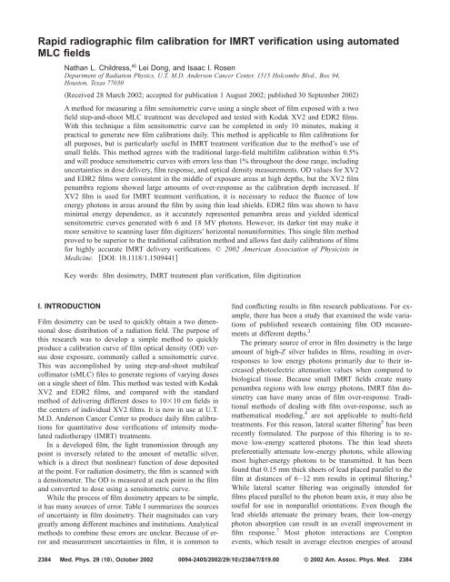
As shown in Fig. 1, step-and-shoot MLC leaf motion files<br />
were created to automatically deliver eight 33 cm squares<br />
of different doses spaced 4 cm apart vertically and 7 cm<br />
apart laterally to one sheet of <strong>film</strong> as a two-field treatment.<br />
Each MLC file corresponds to one side of the <strong>film</strong> exposure<br />
and requires asymmetric jaw settings. The y-axis jaws were<br />
set to have a symmetric 24.4 cm opening, while the x-axis<br />
inner jaw was placed 2 cm across the beam centerline and<br />
the outer jaw was pulled 7 cm from the beam centerline. This<br />
utilized two sMLC files that controlled leaf motions as functions<br />
of dose fractions. The dose is initially delivered to four<br />
open areas, then the MLC closes to deliver doses to fewer<br />
regions until the four areas are irradiated to different levels.<br />
The MLC leaves are parked 1 cm away from the jaw openings<br />
to ensure that minimal leakage from the MLC closing<br />
gaps is allowed to penetrate to the <strong>film</strong>.<br />
Ion chamber measurements were taken of the individual<br />
doses delivered to the eight regions of the <strong>film</strong> <strong>using</strong> EDR2<br />
MUs. The ion chamber used, a Wellhofer CC04 with a4mm<br />
diameter and 4 mm length, was calibrated on Machine 1<br />
Varian Clinac 2100EX with a Varian Millenium 120 leaf<br />
MLC to obtain a dose per charge value. A Wellhofer electrometer<br />
was used with frequent redefinition of its background<br />
compensation. Calibration <strong>film</strong>s were placed at 95<br />
cm SSD, with 5 cm solid water attenuation and 3 cm solid<br />
Medical Physics, Vol. 29, No. 10, October 2002
2386 Childress, Dong, and Rosen: <strong>Rapid</strong> <strong>radiographic</strong> <strong>film</strong> <strong>calibration</strong> 2386<br />
depths of 2, 5, 11, and 19 cm while changing the MU delivered<br />
to closely match the doses obtained at 5 cm. These<br />
additional depths were generated by adding acrylic blocks to<br />
the attenuation layer while keeping the source to <strong>film</strong> distance<br />
the same. Ion chamber and <strong>film</strong> measurements of 6<br />
MV photons were taken on four occasions <strong>using</strong> Machine 1.<br />
One set of ion chamber measurements and a <strong>film</strong> <strong>calibration</strong><br />
of 18 MV photons was acquired. Four sequential sMLC <strong>film</strong><br />
<strong>calibration</strong>s were per<strong>for</strong>med <strong>for</strong> repeatability testing. The<br />
above runs were per<strong>for</strong>med <strong>using</strong> both Kodak XV2 and<br />
EDR2 <strong>film</strong>s taken from the same batches. Daily machine<br />
output fluctuation was recorded by measuring the middle of a<br />
1010 cm field with an ion chamber. Traditional static <strong>film</strong><br />
<strong>calibration</strong>s were per<strong>for</strong>med by irradiating 1010 cm sections<br />
on individual XV2 <strong>film</strong>s at a depth of 5 cm. Ion chamber<br />
measurements were taken once on Machines 2–4 <strong>for</strong><br />
comparison purposes. Machine 2 was identical to Machine 1,<br />
while Machines 3 and 4 were Varian 2100 accelerators with<br />
Varian 80 leaf MLCs. Interleaf leakage was explored by determining<br />
the OD in the regions of the sMLC <strong>calibration</strong><br />
<strong>film</strong>s between exposed boxes.<br />
A Kodak <strong>film</strong> processor that is maintained by physicists<br />
developed all <strong>film</strong>s, and a laser <strong>film</strong> digitizer Kodak LS75<br />
was used to measure ODs with Wellhofer Dosimetrie software.<br />
The horizontal uni<strong>for</strong>mity was previously calibrated<br />
<strong>for</strong> use with XV2 <strong>film</strong>s, but was not altered in this study to<br />
allow <strong>for</strong> the darker tint of EDR2 <strong>film</strong>s. In the weeks following<br />
the initial measurements, daily <strong>film</strong> <strong>calibration</strong>s were<br />
per<strong>for</strong>med and recorded that used different batches of EDR2<br />
<strong>film</strong>. The slopes of these <strong>calibration</strong> curves were estimated<br />
by <strong>using</strong> numerical three point derivatives. Errors were calculated<br />
by finding the standard error in the mean estimation<br />
<strong>using</strong> 95% t-distribution confidence levels. To estimate the<br />
total <strong>calibration</strong> error, standard errors <strong>for</strong> daily dose variations<br />
and single date OD repeatability measurements were<br />
added in quadrature. Standard deviations <strong>for</strong> same day measurements<br />
were compared to each other to assess dose variations.<br />
These deviations were not used in computing the total<br />
error, as the <strong>film</strong> repeatability study includes these fluctuations.<br />
FIG. 1. Visual representation of the sMLC <strong>calibration</strong> method. a The exposed<br />
<strong>film</strong> pattern and its collimator settings dashed lines. Isocenter is<br />
represented by the crosshairs. b The MLC movements and their exposures<br />
in MU used <strong>for</strong> file 2 and MLCs with 1 cm leaves.<br />
water backscatter layers. The ion chamber was inserted in the<br />
middle of a custom drilled 2 cm thick acrylic slab and placed<br />
at 95 SSD with 4 cm solid water attenuation and 2 cm solid<br />
water backscatter layers. Ion chamber readings were corrected<br />
<strong>for</strong> temperature and pressure. Doses delivered to XV2<br />
<strong>film</strong>s ranged from 15–120 MU in 15 MU increments, while<br />
EDR2 <strong>film</strong>s received 30–240 MU doses in 30 MU increments.<br />
Film <strong>calibration</strong>s <strong>using</strong> the new method were done at<br />
III. RESULTS<br />
Table II shows ion chamber measurement data <strong>for</strong> Machine<br />
1 on the four dates on which data were collected.<br />
These numbers can be halved exclusive of error <strong>for</strong> XV2<br />
<strong>film</strong> runs. The standard error in the table represents the standard<br />
error in the four averages measured on four separate<br />
dates, and is mainly due to machine output fluctuation and<br />
ion chamber inaccuracies. While the dose imparted to the<br />
<strong>film</strong> is fairly constant <strong>for</strong> regions not blocked by a collimator,<br />
sequential same day measurements showed the dose to have<br />
slightly higher standard deviations <strong>for</strong> areas that receive high<br />
amounts of MLC and main collimator scatter. The MLC remains<br />
open the entire time the 240 and 120 MU doses are<br />
delivered, but the 30 MU box is blocked by the MLC 75% of<br />
the time the beam delivers the dose to its side. In addition, all<br />
areas receive small doses while protected by the low trans-<br />
Medical Physics, Vol. 29, No. 10, October 2002
2387 Childress, Dong, and Rosen: <strong>Rapid</strong> <strong>radiographic</strong> <strong>film</strong> <strong>calibration</strong> 2387<br />
TABLE II. Data from the four daily sets of ion chamber measurements collected<br />
on Machine 1 <strong>for</strong> the sMLC <strong>calibration</strong> method with EDR2 <strong>film</strong><br />
doses.<br />
MU<br />
Interdate<br />
average<br />
dose cGy<br />
95% confidence<br />
level in interdate<br />
average dose<br />
Intradate daily<br />
dose standard<br />
deviations<br />
240 219.7 0.29% 0.05%<br />
210 192.2 0.32% 0.04%<br />
180 164.1 0.42% 0.10%<br />
150 137.9 0.61% 0.06%<br />
120 110.8 0.40% 0.06%<br />
90 84.18 0.65% 0.17%<br />
60 56.69 0.92% 0.18%<br />
30 29.83 0.78% 0.54%<br />
mission jaws during exposure of the opposite side. Ion chamber<br />
measurements found that this dose was approximately<br />
0.57 cGy <strong>for</strong> the low column and 0.28 cGy <strong>for</strong> the high<br />
column at EDR2 doses. Table III shows comparative data <strong>for</strong><br />
other Varian machines that were collected on only one date.<br />
These data show the amounts of machine variations that<br />
could be expected <strong>for</strong> this method of <strong>calibration</strong>.<br />
When comparing initial <strong>film</strong> <strong>calibration</strong>s produced by the<br />
various machines, it was noted that both machines with<br />
Varian 120 leaf MLCs had a more even interleaf leakage<br />
pattern than the 80 leaf MLC machines. Figure 2 shows<br />
scans of XV2 <strong>film</strong>s that visually compare the leakages. The<br />
ODs ranged from 0.27 to 0.35 <strong>for</strong> the 80 leaf MLC, while the<br />
120 leaf MLC <strong>film</strong> scan produced a fairly constant OD near<br />
0.31. Though it may seem that the 120 leaf MLCs exhibit<br />
less interleaf leakage, the average OD in the leakage areas is<br />
nearly the same <strong>for</strong> both MLCs, indicating that the leakage is<br />
merely more evenly dispersed in Varian 120 leaf MLCs. The<br />
leakage is much less prominent on EDR2 <strong>film</strong>, which does<br />
not exhibit as high of sensitivity to the low-energy photons<br />
that escape between the MLC leaves.<br />
Figure 3 shows sensitometric curves produced on four<br />
separate dates by Machine 1. The final EDR2 run had ODs<br />
6% higher than the other <strong>calibration</strong>s at high doses. This<br />
demonstrates the need <strong>for</strong> a daily <strong>calibration</strong>, regardless of<br />
previous per<strong>for</strong>mance. All <strong>film</strong>s used to prepare this graph<br />
were taken from the same batch. A two dimensional polynomial<br />
provided excellent fits to one set of daily data <strong>for</strong> each<br />
FIG. 2. Scans of interleaf leakage areas in XV2 <strong>film</strong>s <strong>for</strong> two sizes of Varian<br />
MLCs. While the 80 leaf scans produced darker stripes up to an OD of<br />
0.35, the average OD of the entire interleaf area was 0.31 <strong>for</strong> both <strong>film</strong>s.<br />
<strong>film</strong>. High agreement less than 0.5% difference between<br />
traditional static <strong>calibration</strong> and the single sheet <strong>IMRT</strong><br />
method was found. While data from only one date are<br />
shown, they are representative of data collected on all four<br />
dates. The largest discrepancy between the two methods was<br />
1.0%. Calibrations with doses ranging up to 360 MU 330<br />
cGy <strong>for</strong> EDR2 <strong>film</strong> showed a continued linear OD response.<br />
Repeatability errors were low <strong>for</strong> both <strong>film</strong>s. Figure 4<br />
shows the variation of four sequential <strong>calibration</strong>s. In this<br />
study we give an estimate of the maximum precision that can<br />
be obtained with this method of absolute <strong>film</strong> dosimetry. The<br />
deviation from the average value of all points was less than<br />
1%. The average difference between high and low values <strong>for</strong><br />
all eight dose regions was 1.44% <strong>for</strong> XV2 and 1.22% <strong>for</strong><br />
EDR2 <strong>film</strong>. The maximum difference <strong>for</strong> these points was<br />
the high dose region <strong>for</strong> both <strong>film</strong>s, 2.15% <strong>for</strong> XV2 and<br />
1.58% <strong>for</strong> EDR2.<br />
The behavior of the OD of the <strong>film</strong> at different depths was<br />
explored to predict deviations from calibrated behavior when<br />
<strong>film</strong> is used in phantoms. Figure 5 shows results <strong>for</strong> sensitometric<br />
curve generation at different depths <strong>using</strong> the sMLC<br />
TABLE III. EDR2 single data set ion chamber measurements <strong>for</strong> other Varian accelerators <strong>using</strong> the sMLC <strong>calibration</strong> method.<br />
Machine 2 Machine 3 Machine 4<br />
MU<br />
Dose<br />
cGy<br />
% diff. from<br />
Machine 1<br />
Dose<br />
cGy<br />
% diff. from<br />
Machine 1<br />
Dose<br />
cGy<br />
% diff. from<br />
Machine 1<br />
240 219.4 0.15% 220.2 0.22% 220.4 0.29%<br />
210 191.9 0.16% 191.7 0.26% 193.9 0.87%<br />
180 163.9 0.10% 165.6 0.92% 168.5 2.71%<br />
150 137.5 0.25% 139.8 1.41% 141.2 2.41%<br />
120 111.2 0.41% 110.7 0.05% 110.7 0.10%<br />
90 84.63 0.53% 83.61 0.68% 84.64 0.54%<br />
60 56.96 0.47% 56.95 0.45% 57.96 2.24%<br />
30 29.97 0.46% 30.17 1.13% 30.73 3.01%<br />
Medical Physics, Vol. 29, No. 10, October 2002
2388 Childress, Dong, and Rosen: <strong>Rapid</strong> <strong>radiographic</strong> <strong>film</strong> <strong>calibration</strong> 2388<br />
FIG. 3. Sensitometric curves generated on four dates, <strong>using</strong> a XV2 <strong>film</strong> and<br />
b EDR2 <strong>film</strong>. The lines are second order polynomial fits of Day 1 data<br />
whose equations are printed in the graphs.<br />
FIG. 4. Single date repeatability sensitometric curves <strong>for</strong> a XV2 <strong>film</strong> and<br />
b EDR2 <strong>film</strong>. These data were taken sequentially on the same machine.<br />
<strong>calibration</strong> method. The middle 11 cm portion of the <strong>film</strong><br />
exposure areas were measured, and varied by less than 2%<br />
<strong>for</strong> either <strong>film</strong> at any depth. While the middle portions of the<br />
dose boxes gave desired results <strong>for</strong> these small field sizes,<br />
Fig. 6 shows that the penumbra areas are overrepresented by<br />
XV2 <strong>film</strong>, since they contain progressively more low-energy<br />
photons as the depth is increased. It should also be noted that<br />
the visibly exposed portions of the EDR2 <strong>film</strong> at a depth of 5<br />
cm measure nearly 33 cm, while at any depth XV2 <strong>film</strong><br />
shows them to be much larger.<br />
When the difference in the horizontal OD uni<strong>for</strong>mity in<br />
the laser <strong>film</strong> scanner was explored, it was found that EDR2<br />
<strong>film</strong> is more affected by the brightness difference from the<br />
lighter center to the darker edges than XV2 <strong>film</strong>. Figure 7<br />
demonstrates that the ODs determined <strong>for</strong> low doses will<br />
have more error than high doses.<br />
Figure 8 compares sensitometric curves between 6 and 18<br />
MV photons <strong>for</strong> both types of <strong>film</strong>. EDR2 <strong>film</strong> again demonstrates<br />
that it has less energy dependence than XV2 <strong>film</strong>,<br />
as the <strong>calibration</strong> curves are nearly identical <strong>for</strong> both energies.<br />
When viewing the <strong>film</strong> scans, it was noted that 18 MV<br />
photons exhibited a significantly more varied dose distribution<br />
in the small 33 cm field than 6 MV photons. The<br />
EDR2 <strong>film</strong> again displayed a more uni<strong>for</strong>m and compact<br />
dose delivery than XV2 <strong>film</strong> due to its lower-energy sensitivity.<br />
The total <strong>calibration</strong> error can be thought of as the minimum<br />
amount of error in dose values that are determined<br />
from the derived sensitometric curve. The average error<br />
across the <strong>calibration</strong> dose range was 0.68% <strong>for</strong> EDR2 <strong>film</strong><br />
and 0.75% <strong>for</strong> XV2 <strong>film</strong>. Errors <strong>for</strong> XV2 <strong>film</strong> are slightly<br />
higher due to its higher variations in the OD repeatability<br />
study. Machine 1’s daily output never fluctuated more than<br />
0.3% from its calibrated value. Other errors will be present in<br />
the clinical implementation of <strong>film</strong> dosimetry, as previously<br />
discussed in the Introduction section. Potential errors generated<br />
by the horizontal inconsistency of the <strong>film</strong> scanner were<br />
not incorporated into these error estimates.<br />
In the initial weeks of <strong>using</strong> the sMLC method to generate<br />
<strong>film</strong> <strong>calibration</strong> curves, daily <strong>calibration</strong> data was saved and<br />
compared. Figure 9 shows 21 <strong>calibration</strong> curves taken <strong>for</strong><br />
clinical usage. The two curves with significantly greater OD<br />
than the others were created after the <strong>film</strong> processor underwent<br />
extensive repairs. The standard deviations <strong>for</strong> all points<br />
were consistently around 7.5% of the OD. This collection of<br />
curves emphasizes the day-to-day variations that occur in<br />
<strong>film</strong> dosimetry. Since the EDR2 <strong>film</strong> sensitometric curve is<br />
linear, a 7.5% OD daily standard deviation translates into a<br />
7.5% uncertainty in dose calculations. The difference in <strong>film</strong><br />
OD response to dose can be even larger if changes were<br />
made to the processor or its chemicals. The large magnitude<br />
of this variation highlights the need to complete daily <strong>film</strong><br />
<strong>calibration</strong>s <strong>for</strong> accurate <strong>IMRT</strong> <strong>film</strong>-based quality assurance,<br />
rather than the relying on the average response curves.<br />
Medical Physics, Vol. 29, No. 10, October 2002
2389 Childress, Dong, and Rosen: <strong>Rapid</strong> <strong>radiographic</strong> <strong>film</strong> <strong>calibration</strong> 2389<br />
FIG. 7. Percent difference between OD measurements taken from the edge<br />
and middle of the scanning laser <strong>film</strong> densitometer. The differences in the<br />
measurements are due to the Vignetting effect, as the laser must pass<br />
through a greater distance of <strong>film</strong> along the edges than the central area<br />
measurements.<br />
FIG. 5. Generation of sensitometric curves <strong>using</strong> the sMLC method at different<br />
<strong>film</strong> depths, measuring only the center of exposed portions of <strong>film</strong>.<br />
Curves show responses of a XV2 <strong>film</strong> and b EDR2 <strong>film</strong>.<br />
IV. DISCUSSION AND CONCLUSIONS<br />
The more even distribution of interleaf leakage from<br />
Varian 120 leaf MLCs as compared to 80 leaf MLCs produced<br />
better <strong>film</strong> <strong>calibration</strong>s. While 120 leaf MLCs do not<br />
reduce the amount of interleaf leakage, ion chamber measurements<br />
will account <strong>for</strong> the added dose and the dose measurements<br />
will be more independent of the ion chamber’s<br />
placement within the fields. Homogeneous dose distributions<br />
within the exposed regions also reduce the repeatability error<br />
in measuring average ODs.<br />
EDR2 <strong>film</strong>’s darker base tint causes it to have a greater<br />
response to the <strong>film</strong> scanner’s horizontal nonuni<strong>for</strong>mity<br />
FIG. 6. Scans of high dose box of EDR2 <strong>film</strong> left and XV2 <strong>film</strong> right <strong>for</strong><br />
different depths. The increased area of the XV2 boxes at high depths demonstrates<br />
its low-energy photon overresponse.<br />
FIG. 8. A comparison of 6 MV and 18 MV sensitometric curves <strong>for</strong> a XV2<br />
<strong>film</strong> and b EDR2 <strong>film</strong>. It can be seen that EDR2 <strong>film</strong> is not dependent on<br />
beam energy <strong>for</strong> <strong>calibration</strong>s.<br />
Medical Physics, Vol. 29, No. 10, October 2002
2390 Childress, Dong, and Rosen: <strong>Rapid</strong> <strong>radiographic</strong> <strong>film</strong> <strong>calibration</strong> 2390<br />
FIG. 9. 21 <strong>calibration</strong> curves taken over a period of 6 weeks that show<br />
variations in <strong>film</strong> batches and the effects of processor maintenance and<br />
repair. The line represents the average of all data.<br />
when compared to XV2 <strong>film</strong>. The densitometer has a single<br />
laser directed towards a mirror that pivots to scan the laser<br />
beam in a horizontal path across the <strong>film</strong>, leading to a range<br />
of laser pathlengths through the <strong>film</strong>. The laser light will be<br />
more attenuated at outer regions due to its increase in pathlength<br />
known as the vignetting effect. Film densitometers<br />
are usually calibrated to adjust laser intensity as a function of<br />
pivot angle to maintain a uni<strong>for</strong>m image <strong>for</strong> a range of OD<br />
values, but EDR2 <strong>film</strong>’s dark bluish tint exhibits increased<br />
attenuation of the red HeNe laser. Film densitometers that<br />
use a pivoting laser and were previously calibrated to maintain<br />
horizontal uni<strong>for</strong>mity with XV2 <strong>film</strong> may need to be<br />
recalibrated <strong>for</strong> use with EDR2 <strong>film</strong>. This can introduce<br />
problems when the same densitometer is used <strong>for</strong> both types<br />
of <strong>film</strong>. The <strong>calibration</strong> is further complicated by the fact that<br />
the non-uni<strong>for</strong>m attenuation is a function of OD; thus software<br />
corrections may need to be applied.<br />
The error due to the vignetting effect will affect clinical<br />
results <strong>for</strong> large <strong>film</strong> measurements that exhibit nonuni<strong>for</strong>mity<br />
across their surface or <strong>for</strong> small <strong>film</strong>s that are measured<br />
in the center of the scanner. Small fields are affected due to<br />
the <strong>calibration</strong> areas being scanned in a different location<br />
than the <strong>film</strong> measurements. The vignetting effect essentially<br />
makes the <strong>calibration</strong> curve specific to the horizontally<br />
scanned region of the pivoting laser densitometer. While this<br />
error is small enough to ignore with lightly tinted XV2 <strong>film</strong>,<br />
it cannot be assumed that EDR2 <strong>film</strong> will exhibit similar<br />
per<strong>for</strong>mance. At clinically relevant doses above 100 cGy,<br />
EDR2 <strong>film</strong> exhibits a 3% horizontal uni<strong>for</strong>mity uncertainty<br />
error. When this error is combined with the uncertainties in<br />
the <strong>calibration</strong> method and other <strong>film</strong> errors, it is possible to<br />
have total <strong>verification</strong> errors exceeding 4%. As EDR2’s OD<br />
is linear with dose, this becomes a 4% dose uncertainty.<br />
When making a clinical transition from XV2 to EDR2 <strong>film</strong><br />
measured on a pivoting laser densitometer, it is necessary to<br />
analyze the horizontal OD uni<strong>for</strong>mity in order to obtain accurate<br />
<strong>IMRT</strong> <strong>verification</strong>s.<br />
The two field treatment method described earlier yielded<br />
minimal XV2 heterogeneities and virtually no detectable<br />
leakage with EDR2 <strong>film</strong>s. The use of a single sMLC file to<br />
deliver the full pattern in one field was also studied. Although<br />
no quantitative measurements were taken to compare<br />
the increased dose fluctuations, interleaf leakage <strong>using</strong> one<br />
field was visible with XV2 <strong>film</strong> and noticeable when EDR2<br />
<strong>film</strong> was read with a densitometer. Additionally, the use of<br />
two fields only adds around a minute to the delivery time and<br />
allows the <strong>calibration</strong> to be scaled to different doses simply<br />
by inputting different MU settings. The use of the secondary<br />
collimator jaws to create this exposure pattern Fig. 1a was<br />
found to be too slow, difficult to scale <strong>for</strong> different dose<br />
ranges, and yielded minimal scatter improvements over the<br />
two field sMLC method.<br />
The high reproducibility, low error, quick delivery time,<br />
ease of use, and agreement with the traditional static <strong>film</strong><br />
<strong>calibration</strong> method shows that the two field <strong>IMRT</strong> <strong>calibration</strong><br />
method is superior to previous procedures. It is further recommended<br />
that EDR2 <strong>film</strong> be used clinically due to its near<br />
tissue equivalent response to low-energy photons, its linear<br />
response <strong>for</strong> large doses, and its better repeatability <strong>for</strong> highenergy<br />
doses compared to XV2 <strong>film</strong>. However, scanning laser<br />
<strong>film</strong> densitometers may have to have their horizontal uni<strong>for</strong>mity<br />
recalibrated <strong>for</strong> use with EDR2 <strong>film</strong>. The depth study<br />
revealed inconsistencies in XV2’s response at central and<br />
penumbra areas, perhaps partially explaining the inconsistent<br />
reporting of XV2’s OD depth dependence in previous research.<br />
The use of thin lead shields and proper phantom<br />
placement should improve the accuracy of XV2 <strong>film</strong> dosimetry<br />
<strong>for</strong> <strong>IMRT</strong> <strong>verification</strong> but additional research is needed.<br />
Accurate average doses <strong>for</strong> each exposure region were found<br />
by taking only four sets of ion chamber measurements on<br />
different days. Large variations in both the amplitude and<br />
slopes of daily sensitometric curve generation were found<br />
and emphasized the need <strong>for</strong> full dose range daily <strong>calibration</strong>s<br />
which can be completed easily and accurately with the<br />
proper use of MLCs. The entire two field sMLC <strong>calibration</strong><br />
process will require around ten minutes, and its success<br />
would negate the need to per<strong>for</strong>m an ion chamber measurement<br />
on <strong>IMRT</strong> plans in addition to <strong>film</strong> <strong>verification</strong>s.<br />
a Corresponding author. Electronic mail: nchildre@mdanderson.org<br />
1 I. J. Das and C. W. Cheng, Basic Film Dosimetry, AAPM Annual Meeting<br />
Presentation Salt Lake City, Utah, 2001.<br />
2 F. H. Attix et al., Radiation Dosimetry Academic, New York, 1966, Vol.<br />
2, pp. 344–357.<br />
3 C. Danciu, B. S. Proimos, J. C. Rosenwald, and B. J. Mijnheer, ‘‘Variation<br />
of sensitometric curves of <strong>radiographic</strong> <strong>film</strong>s in high energy photon<br />
beams,’’ Med. Phys. 28, 966–974 2001.<br />
4 J. F. Williamson, F. M. Khan, and S. C. Sharma, ‘‘Film dosimetry of<br />
megavoltage photon beams: A practical method of isodensity-to-isodose<br />
curve conversion,’’ Med. Phys. 8, 94–981981.<br />
5 S. E. Burch et al., ‘‘A new approach to <strong>film</strong> dosimetry <strong>for</strong> high energy<br />
photon beams: Lateral scatter filtering,’’ Med. Phys. 24, 775–783 1997.<br />
6 I. J. Yeo, C. K. C. Wang, and S. E. Burch, ‘‘A filtration method <strong>for</strong><br />
improving <strong>film</strong> dosimetry in photon radiation therapy,’’ Med. Phys. 24,<br />
1943–1953 1997.<br />
7 S. G. Ju, Y. C. Ahn, S. J. Huh, and I. J. Yeo, ‘‘Film dosimetry <strong>for</strong> intensity<br />
modulated radiation therapy: Dosimetric evaluation,’’ Med. Phys. 29,<br />
351–355 2002.<br />
8 T. W. Holmes and E. C. McCullough, ‘‘Acceptance testing and quality<br />
assurance of automated scanning <strong>film</strong> densitometers used in the dosimetry<br />
of electron and photon therapy beams,’’ Med. Phys. 10, 698–700<br />
1983.<br />
Medical Physics, Vol. 29, No. 10, October 2002