trs430 commissioning and quality assurance of computerized ...
trs430 commissioning and quality assurance of computerized ...
trs430 commissioning and quality assurance of computerized ...

Create successful ePaper yourself
Turn your PDF publications into a flip-book with our unique Google optimized e-Paper software.
Technical Reports SeriEs No.430<br />
Commissioning <strong>and</strong><br />
Quality Assurance <strong>of</strong><br />
Computerized Planning<br />
Systems for Radiation<br />
Treatment <strong>of</strong> Cancer
COMMISSIONING AND<br />
QUALITY ASSURANCE OF<br />
COMPUTERIZED PLANNING<br />
SYSTEMS FOR RADIATION<br />
TREATMENT OF CANCER
The following States are Members <strong>of</strong> the International Atomic Energy Agency:<br />
AFGHANISTAN<br />
ALBANIA<br />
ALGERIA<br />
ANGOLA<br />
ARGENTINA<br />
ARMENIA<br />
AUSTRALIA<br />
AUSTRIA<br />
AZERBAIJAN<br />
BANGLADESH<br />
BELARUS<br />
BELGIUM<br />
BENIN<br />
BOLIVIA<br />
BOSNIA AND HERZEGOVINA<br />
BOTSWANA<br />
BRAZIL<br />
BULGARIA<br />
BURKINA FASO<br />
CAMEROON<br />
CANADA<br />
CENTRAL AFRICAN<br />
REPUBLIC<br />
CHILE<br />
CHINA<br />
COLOMBIA<br />
COSTA RICA<br />
CÔTE D’IVOIRE<br />
CROATIA<br />
CUBA<br />
CYPRUS<br />
CZECH REPUBLIC<br />
DEMOCRATIC REPUBLIC<br />
OF THE CONGO<br />
DENMARK<br />
DOMINICAN REPUBLIC<br />
ECUADOR<br />
EGYPT<br />
EL SALVADOR<br />
ERITREA<br />
ESTONIA<br />
ETHIOPIA<br />
FINLAND<br />
FRANCE<br />
GABON<br />
GEORGIA<br />
GERMANY<br />
GHANA<br />
GREECE<br />
GUATEMALA<br />
HAITI<br />
HOLY SEE<br />
HONDURAS<br />
HUNGARY<br />
ICELAND<br />
INDIA<br />
INDONESIA<br />
IRAN, ISLAMIC REPUBLIC OF<br />
IRAQ<br />
IRELAND<br />
ISRAEL<br />
ITALY<br />
JAMAICA<br />
JAPAN<br />
JORDAN<br />
KAZAKHSTAN<br />
KENYA<br />
KOREA, REPUBLIC OF<br />
KUWAIT<br />
KYRGYZSTAN<br />
LATVIA<br />
LEBANON<br />
LIBERIA<br />
LIBYAN ARAB JAMAHIRIYA<br />
LIECHTENSTEIN<br />
LITHUANIA<br />
LUXEMBOURG<br />
MADAGASCAR<br />
MALAYSIA<br />
MALI<br />
MALTA<br />
MARSHALL ISLANDS<br />
MAURITIUS<br />
MEXICO<br />
MONACO<br />
MONGOLIA<br />
MOROCCO<br />
MYANMAR<br />
NAMIBIA<br />
NETHERLANDS<br />
NEW ZEALAND<br />
NICARAGUA<br />
NIGER<br />
NIGERIA<br />
NORWAY<br />
PAKISTAN<br />
PANAMA<br />
PARAGUAY<br />
The Agency’s Statute was approved on 23 October 1956 by the Conference on the Statute <strong>of</strong><br />
the IAEA held at United Nations Headquarters, New York; it entered into force on 29 July 1957.<br />
The Headquarters <strong>of</strong> the Agency are situated in Vienna. Its principal objective is “to accelerate <strong>and</strong><br />
enlarge the contribution <strong>of</strong> atomic energy to peace, health <strong>and</strong> prosperity throughout the world’’.<br />
© IAEA, 2004<br />
Permission to reproduce or translate the information contained in this publication may be<br />
obtained by writing to the International Atomic Energy Agency, Wagramer Strasse 5, P.O. Box 100,<br />
A-1400 Vienna, Austria.<br />
Printed by the IAEA in Austria<br />
October 2004<br />
STI/DOC/010/430<br />
PERU<br />
PHILIPPINES<br />
POLAND<br />
PORTUGAL<br />
QATAR<br />
REPUBLIC OF MOLDOVA<br />
ROMANIA<br />
RUSSIAN FEDERATION<br />
SAUDI ARABIA<br />
SENEGAL<br />
SERBIA AND MONTENEGRO<br />
SEYCHELLES<br />
SIERRA LEONE<br />
SINGAPORE<br />
SLOVAKIA<br />
SLOVENIA<br />
SOUTH AFRICA<br />
SPAIN<br />
SRI LANKA<br />
SUDAN<br />
SWEDEN<br />
SWITZERLAND<br />
SYRIAN ARAB REPUBLIC<br />
TAJIKISTAN<br />
THAILAND<br />
THE FORMER YUGOSLAV<br />
REPUBLIC OF MACEDONIA<br />
TUNISIA<br />
TURKEY<br />
UGANDA<br />
UKRAINE<br />
UNITED ARAB EMIRATES<br />
UNITED KINGDOM OF<br />
GREAT BRITAIN AND<br />
NORTHERN IRELAND<br />
UNITED REPUBLIC<br />
OF TANZANIA<br />
UNITED STATES OF AMERICA<br />
URUGUAY<br />
UZBEKISTAN<br />
VENEZUELA<br />
VIETNAM<br />
YEMEN<br />
ZAMBIA<br />
ZIMBABWE
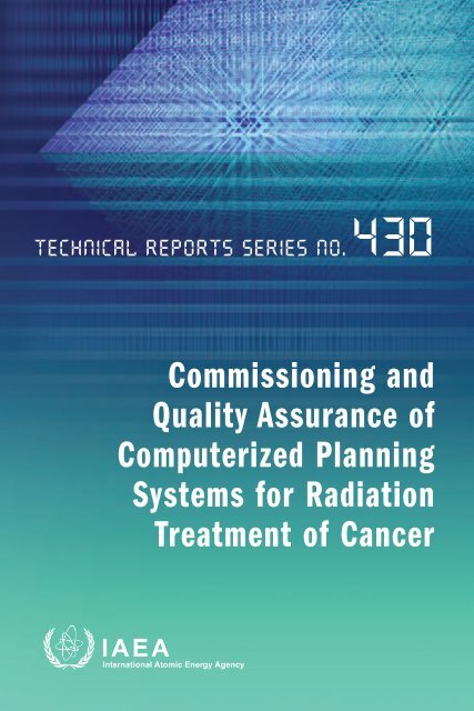
TECHNICAL REPORTS SERIES No. 430<br />
COMMISSIONING AND<br />
QUALITY ASSURANCE OF<br />
COMPUTERIZED PLANNING<br />
SYSTEMS FOR RADIATION<br />
TREATMENT OF CANCER<br />
INTERNATIONAL ATOMIC ENERGY AGENCY<br />
VIENNA, 2004
IAEA Library Cataloguing in Publication Data<br />
Commissioning <strong>and</strong> <strong>quality</strong> <strong>assurance</strong> <strong>of</strong> <strong>computerized</strong> planning systems<br />
for radiation treatment <strong>of</strong> cancer. — Vienna : International Atomic<br />
Energy Agency, 2004.<br />
p. ; 24 cm. — (Technical reports series, ISSN 0074–1914 ; no. 430)<br />
STI/DOC/010/430<br />
ISBN 92–0–105304–5<br />
Includes bibliographical references.<br />
1. Cancer — Treatment — Quality control. 2. Cancer —<br />
Radiotherapy — Quality control. I. International Atomic Energy<br />
Agency. II. Technical reports series (International Atomic Energy<br />
Agency) ; 430.<br />
IAEAL 04–00363
FOREWORD<br />
The radiation therapy treatment planning process is complicated, has<br />
many steps <strong>and</strong> is potentially a high risk procedure, as it involves the h<strong>and</strong>ling<br />
<strong>of</strong> multiple sources <strong>of</strong> information <strong>and</strong> the interaction <strong>of</strong> different pr<strong>of</strong>essional<br />
groups all dedicated to treating cancer patients with radiation. The IAEA has<br />
analysed a series <strong>of</strong> accidental exposures in radiotherapy to learn about<br />
methods <strong>of</strong> preventing future occurrences. This analysis included a review <strong>of</strong><br />
accidents that occurred owing to improper, or lack <strong>of</strong>, <strong>commissioning</strong> <strong>and</strong><br />
appropriate <strong>quality</strong> control procedures for <strong>computerized</strong> treatment planning<br />
systems (TPSs) at purchase, <strong>commissioning</strong> or during the use <strong>of</strong> the equipment.<br />
The IAEA report Investigation <strong>of</strong> an Accidental Exposure <strong>of</strong> Radiotherapy<br />
Patients in Panama, published in 2001, presented a further example <strong>of</strong> very<br />
significant errors related to the improper use <strong>of</strong> TPSs that affected cancer<br />
patients in Panama.<br />
Quality <strong>assurance</strong> (QA) in the radiation therapy treatment planning<br />
process is essential for minimizing the possibility <strong>of</strong> accidental exposure. It is <strong>of</strong><br />
special importance to support hospitals in Member States in developing<br />
procedures for the <strong>commissioning</strong> <strong>and</strong> QA <strong>of</strong> <strong>computerized</strong> TPSs. The<br />
relatively low cost <strong>of</strong> today’s equipment has made TPSs widely available in<br />
industrialized <strong>and</strong> developing countries, but with the exception <strong>of</strong> a few<br />
national recommendations for QA in North America <strong>and</strong> western Europe, no<br />
publications are available for pr<strong>of</strong>essionals to follow to check their TPSs.<br />
Responding to the need to develop an IAEA publication with such<br />
recommendations, a group <strong>of</strong> experts (J. Van Dyk (Canada), J.-C. Rosenwald<br />
(France), B. Fraass (United States <strong>of</strong> America), J. Cramb (Australia) <strong>and</strong><br />
F. Ionescu-Farca (Switzerl<strong>and</strong>)) was appointed in 1999 <strong>and</strong> prepared such a<br />
document during 2000–2002. The main issues that deserve attention in QA<br />
protocols for TPSs were discussed at length during two Consultants Meetings<br />
held in 1999 <strong>and</strong> 2000 in Vienna. These meetings covered the range <strong>of</strong> ancillary<br />
equipment from that available in poorly equipped hospitals to that required for<br />
the sophisticated <strong>and</strong> modern treatment techniques available in better equiped<br />
facilities. A detailed outline for a publication with sections that deal with both<br />
external beam radiotherapy <strong>and</strong> brachytherapy, describing tolerances <strong>and</strong><br />
errors, resource requirements for QA, issues to be considered at purchase,<br />
acceptance tests, <strong>commissioning</strong> <strong>and</strong> the continuing QA process <strong>and</strong> its<br />
management, was developed, <strong>and</strong> the final report was prepared for publication<br />
as this technical report.
Owing to the complexity <strong>of</strong> the treatment planning process, this report<br />
does not provide a simple protocol that can be followed step by step by the user<br />
at a radiotherapy centre for the <strong>commissioning</strong> <strong>and</strong> QA <strong>of</strong> a specific TPS.<br />
Instead, this report provides guidance on the tests <strong>and</strong> procedures that should<br />
be considered. Specific examples <strong>of</strong> tests <strong>and</strong> procedures are given, <strong>and</strong> the<br />
medical physicist may have to modify these depending on his or her TPS, on the<br />
irradiation facilities available or on the specific treatment techniques to be<br />
employed. It must be emphasized that the rationale for the multiple tests<br />
described in this report is related to the four major issues <strong>of</strong> a well structured<br />
QA programme in <strong>computerized</strong> treatment planning, namely education,<br />
verification, documentation <strong>and</strong> communication. The implementation <strong>of</strong> such a<br />
programme will ensure confidence that each patient will receive the radiation<br />
treatment as planned <strong>and</strong> that no errors will occur in the process <strong>of</strong> using the<br />
TPS. This report is addressed to all those individuals who participate in any<br />
aspect <strong>of</strong> TPS <strong>commissioning</strong> <strong>and</strong> its QA programme.<br />
The IAEA wishes to express its thanks to all authors <strong>and</strong> reviewers <strong>of</strong> this<br />
report as listed in the Contributors to Drafting <strong>and</strong> Review section at the end<br />
<strong>of</strong> this report. The editorial contribution <strong>of</strong> J. Van Dyk is especially<br />
acknowledged.<br />
The IAEA staff members responsible for the preparation <strong>of</strong> this report<br />
were P. Andreo, J. Izewska, K. Shortt <strong>and</strong> S. Vatnitsky <strong>of</strong> the Division <strong>of</strong><br />
Human Health.<br />
EDITORIAL NOTE<br />
The mention <strong>of</strong> names <strong>of</strong> specific companies or products (whether or not indicated<br />
as registered) does not imply any intention to infringe proprietary rights, nor should it be<br />
construed as an endorsement or recommendation on the part <strong>of</strong> the IAEA.<br />
The authors are responsible for having obtained the necessary permission for the<br />
IAEA to reproduce, translate or use material from sources already protected by<br />
copyrights.
CONTENTS<br />
1. INTRODUCTION . . . . . . . . . . . . . . . . . . . . . . . . . . . . . . . . . . . . . . . . . 1<br />
1.1. Background . . . . . . . . . . . . . . . . . . . . . . . . . . . . . . . . . . . . . . . . . . 1<br />
1.2. Target audience . . . . . . . . . . . . . . . . . . . . . . . . . . . . . . . . . . . . . . . 2<br />
1.3. Clinical use <strong>of</strong> treatment planning systems . . . . . . . . . . . . . . . . 2<br />
1.4. Common errors . . . . . . . . . . . . . . . . . . . . . . . . . . . . . . . . . . . . . . . 3<br />
1.4.1. Education . . . . . . . . . . . . . . . . . . . . . . . . . . . . . . . . . . . . . . 7<br />
1.4.2. Verification . . . . . . . . . . . . . . . . . . . . . . . . . . . . . . . . . . . . . 8<br />
1.4.3. Documentation . . . . . . . . . . . . . . . . . . . . . . . . . . . . . . . . . 8<br />
1.4.4. Communication . . . . . . . . . . . . . . . . . . . . . . . . . . . . . . . . . 8<br />
1.5. Why is <strong>quality</strong> <strong>assurance</strong> required? . . . . . . . . . . . . . . . . . . . . . . . 8<br />
1.5.1. Accuracy in the radiation treatment . . . . . . . . . . . . . . . . 8<br />
1.5.2. Avoidance <strong>of</strong> treatment errors . . . . . . . . . . . . . . . . . . . . 9<br />
1.6. Brief overview <strong>of</strong> the clinical implementation <strong>of</strong><br />
a treatment planning system . . . . . . . . . . . . . . . . . . . . . . . . . . . . . 9<br />
1.7. Total <strong>quality</strong> management . . . . . . . . . . . . . . . . . . . . . . . . . . . . . . 10<br />
1.8. Previous reports . . . . . . . . . . . . . . . . . . . . . . . . . . . . . . . . . . . . . . . 10<br />
1.9. How to use this report . . . . . . . . . . . . . . . . . . . . . . . . . . . . . . . . . . 11<br />
2. CLINICAL TREATMENT PLANNING PROCESS . . . . . . . . . . . . 12<br />
2.1. Radiation treatment planning process . . . . . . . . . . . . . . . . . . . . 12<br />
2.2. Clinical implementation <strong>of</strong> a treatment planning system . . . . 15<br />
3. DESCRIPTION OF RADIATION TREATMENT<br />
PLANNING SYSTEMS . . . . . . . . . . . . . . . . . . . . . . . . . . . . . . . . . . . . 16<br />
3.1. Hardware . . . . . . . . . . . . . . . . . . . . . . . . . . . . . . . . . . . . . . . . . . . . 16<br />
3.2. S<strong>of</strong>tware . . . . . . . . . . . . . . . . . . . . . . . . . . . . . . . . . . . . . . . . . . . . . 16<br />
3.2.1. Three dimensional <strong>and</strong> two dimensional<br />
treatment planning systems . . . . . . . . . . . . . . . . . . . . . . . 17<br />
3.2.2. Computed tomography simulation <strong>and</strong><br />
three dimensional treatment planning systems . . . . . . . 17<br />
3.2.3. Input–output . . . . . . . . . . . . . . . . . . . . . . . . . . . . . . . . . . . 18<br />
3.2.4. Contouring <strong>and</strong> image display . . . . . . . . . . . . . . . . . . . . . 18<br />
3.2.5. Beam input <strong>and</strong> calculation . . . . . . . . . . . . . . . . . . . . . . . 19<br />
3.2.6. Dose display . . . . . . . . . . . . . . . . . . . . . . . . . . . . . . . . . . . . 19<br />
3.2.7. Plan evaluation tools . . . . . . . . . . . . . . . . . . . . . . . . . . . . . 19
3.2.8. Other features . . . . . . . . . . . . . . . . . . . . . . . . . . . . . . . . . . 20<br />
3.3. Single or multistation systems . . . . . . . . . . . . . . . . . . . . . . . . . . . 20<br />
3.4. Ancillary components . . . . . . . . . . . . . . . . . . . . . . . . . . . . . . . . . . 20<br />
3.5. Third party s<strong>of</strong>tware . . . . . . . . . . . . . . . . . . . . . . . . . . . . . . . . . . . 21<br />
4. ALGORITHMS USED IN<br />
RADIATION TREATMENT PLANNING . . . . . . . . . . . . . . . . . . . 21<br />
4.1. Introduction . . . . . . . . . . . . . . . . . . . . . . . . . . . . . . . . . . . . . . . . . . 21<br />
4.2. Processing <strong>and</strong> display <strong>of</strong> anatomical data . . . . . . . . . . . . . . . . . 22<br />
4.2.1. Contours <strong>and</strong> automated segmentation . . . . . . . . . . . . . 22<br />
4.2.2. Building three dimensional objects (structures)<br />
from a series <strong>of</strong> contours . . . . . . . . . . . . . . . . . . . . . . . . . 22<br />
4.2.3. Multiplanar reconstruction <strong>and</strong> three<br />
dimensional display . . . . . . . . . . . . . . . . . . . . . . . . . . . . . . 25<br />
4.2.4. Exp<strong>and</strong>ing three dimensional objects (structures) . . . . 25<br />
4.2.5. Creating digital reconstructed radiographs . . . . . . . . . . 27<br />
4.2.6. Registration <strong>of</strong> multiple anatomical data sets . . . . . . . . 28<br />
4.3. Beam or source related tools . . . . . . . . . . . . . . . . . . . . . . . . . . . . 29<br />
4.3.1. Automatic design <strong>of</strong> beam apertures . . . . . . . . . . . . . . . 30<br />
4.3.2. Geometrical reconstruction <strong>of</strong> sources<br />
in brachytherapy . . . . . . . . . . . . . . . . . . . . . . . . . . . . . . . . 30<br />
4.4. Dose calculation in external beam radiotherapy . . . . . . . . . . . . 31<br />
4.4.1. Dose calculation problem in external<br />
beam radiotherapy . . . . . . . . . . . . . . . . . . . . . . . . . . . . . . 31<br />
4.4.2. Relative dose distribution . . . . . . . . . . . . . . . . . . . . . . . . 34<br />
4.4.3. Monitor unit/time calculations <strong>and</strong><br />
plan normalization . . . . . . . . . . . . . . . . . . . . . . . . . . . . . . 35<br />
4.4.4. Other issues in dose calculation for<br />
external beam radiotherapy . . . . . . . . . . . . . . . . . . . . . . . 36<br />
4.5. Dose calculation in brachytherapy . . . . . . . . . . . . . . . . . . . . . . . 40<br />
4.5.1. Dose calculation problem in brachytherapy . . . . . . . . . 40<br />
4.5.2. Dose from point sources . . . . . . . . . . . . . . . . . . . . . . . . . 43<br />
4.5.2.1. Activity based method. . . . . . . . . . . . . . . . . . . . 43<br />
4.5.2.2. TG 43 formalism. . . . . . . . . . . . . . . . . . . . . . . . . 44<br />
4.5.3. Dose from tubes or wires . . . . . . . . . . . . . . . . . . . . . . . . . 46<br />
4.5.4. Dose for stepping sources <strong>and</strong> optimization . . . . . . . . . 46<br />
5. QUALITY ASSESSMENT . . . . . . . . . . . . . . . . . . . . . . . . . . . . . . . . . . 47<br />
5.1. Introduction . . . . . . . . . . . . . . . . . . . . . . . . . . . . . . . . . . . . . . . . . . 47
5.2. Uncertainties, deviations, tolerances <strong>and</strong> errors . . . . . . . . . . . . 47<br />
5.2.1. Uncertainty . . . . . . . . . . . . . . . . . . . . . . . . . . . . . . . . . . . . 47<br />
5.2.2. Deviation . . . . . . . . . . . . . . . . . . . . . . . . . . . . . . . . . . . . . . 48<br />
5.2.3. Tolerance . . . . . . . . . . . . . . . . . . . . . . . . . . . . . . . . . . . . . . 49<br />
5.2.4. Error . . . . . . . . . . . . . . . . . . . . . . . . . . . . . . . . . . . . . . . . . . 49<br />
5.3. Quality st<strong>and</strong>ards, reference data, tolerances <strong>and</strong><br />
methods <strong>of</strong> assessment for a treatment planning system . . . . . 50<br />
5.3.1. Quality st<strong>and</strong>ards . . . . . . . . . . . . . . . . . . . . . . . . . . . . . . . 50<br />
5.3.2. Reference data . . . . . . . . . . . . . . . . . . . . . . . . . . . . . . . . . 50<br />
5.3.3. Tolerances for dose calculations . . . . . . . . . . . . . . . . . . . 51<br />
5.3.4. Confidence limits . . . . . . . . . . . . . . . . . . . . . . . . . . . . . . . 53<br />
5.4. Sources <strong>of</strong> uncertainties <strong>and</strong> limitations for a given plan . . . . . 56<br />
6. QUALITY ASSURANCE MANAGEMENT . . . . . . . . . . . . . . . . . 58<br />
6.1. Quality management process . . . . . . . . . . . . . . . . . . . . . . . . . . . . 58<br />
6.2. Treatment planning <strong>quality</strong> <strong>assurance</strong> plan . . . . . . . . . . . . . . . . 58<br />
6.3. Physicist responsible for the treatment planning system . . . . . 58<br />
6.4. Personnel . . . . . . . . . . . . . . . . . . . . . . . . . . . . . . . . . . . . . . . . . . . . 61<br />
6.5. Communication . . . . . . . . . . . . . . . . . . . . . . . . . . . . . . . . . . . . . . . 62<br />
6.6. Equipment . . . . . . . . . . . . . . . . . . . . . . . . . . . . . . . . . . . . . . . . . . . 62<br />
6.7. Staff training <strong>and</strong> education . . . . . . . . . . . . . . . . . . . . . . . . . . . . . 64<br />
6.8. Computer systems management <strong>and</strong> security . . . . . . . . . . . . . . 65<br />
6.9. Policies, procedures <strong>and</strong> documentation . . . . . . . . . . . . . . . . . . . 66<br />
6.10. Common errors . . . . . . . . . . . . . . . . . . . . . . . . . . . . . . . . . . . . . . . 67<br />
6.10.1. S<strong>of</strong>tware misinterpretation . . . . . . . . . . . . . . . . . . . . . . . 67<br />
6.10.2. Normalization . . . . . . . . . . . . . . . . . . . . . . . . . . . . . . . . . . 68<br />
6.10.3. Beam parameterization . . . . . . . . . . . . . . . . . . . . . . . . . . 68<br />
6.10.4. Monitor unit/time calculations . . . . . . . . . . . . . . . . . . . . 68<br />
6.10.5. Inhomogeneity corrections . . . . . . . . . . . . . . . . . . . . . . . 69<br />
6.10.6. Underst<strong>and</strong>ing s<strong>of</strong>tware capabilities <strong>and</strong> limitations . . 69<br />
6.10.7. Error management . . . . . . . . . . . . . . . . . . . . . . . . . . . . . . 70<br />
7. PURCHASE PROCESS . . . . . . . . . . . . . . . . . . . . . . . . . . . . . . . . . . . . 70<br />
7.1. Assessment <strong>of</strong> need . . . . . . . . . . . . . . . . . . . . . . . . . . . . . . . . . . . . 71<br />
7.2. Request for information . . . . . . . . . . . . . . . . . . . . . . . . . . . . . . . . 73<br />
7.3. Vendor demonstrations, presentations <strong>and</strong> site visits . . . . . . . . 73<br />
7.4. Tender process: Definitions <strong>of</strong> system specifications . . . . . . . . 74<br />
7.5. Selection criteria . . . . . . . . . . . . . . . . . . . . . . . . . . . . . . . . . . . . . . 76<br />
7.6. Purchase . . . . . . . . . . . . . . . . . . . . . . . . . . . . . . . . . . . . . . . . . . . . . 76
7.7. Vendor <strong>and</strong> user responsibilities . . . . . . . . . . . . . . . . . . . . . . . . . 77<br />
7.7.1. Vendor responsibilities . . . . . . . . . . . . . . . . . . . . . . . . . . . 77<br />
7.7.2. User responsibilities . . . . . . . . . . . . . . . . . . . . . . . . . . . . . 78<br />
8. ACCEPTANCE TESTING . . . . . . . . . . . . . . . . . . . . . . . . . . . . . . . . . 79<br />
8.1. Introduction . . . . . . . . . . . . . . . . . . . . . . . . . . . . . . . . . . . . . . . . . . 79<br />
8.2. Hardware . . . . . . . . . . . . . . . . . . . . . . . . . . . . . . . . . . . . . . . . . . . . 80<br />
8.3. Network integration . . . . . . . . . . . . . . . . . . . . . . . . . . . . . . . . . . . 80<br />
8.4. Data transfer . . . . . . . . . . . . . . . . . . . . . . . . . . . . . . . . . . . . . . . . . 81<br />
8.5. S<strong>of</strong>tware . . . . . . . . . . . . . . . . . . . . . . . . . . . . . . . . . . . . . . . . . . . . . 82<br />
8.5.1. Verifying system capabilities . . . . . . . . . . . . . . . . . . . . . . 82<br />
8.5.2. Verifying calculation capabilities . . . . . . . . . . . . . . . . . . . 83<br />
8.5.2.1. Calculation on benchmark data . . . . . . . . . . . . 83<br />
8.5.2.2. Calculation on generic data . . . . . . . . . . . . . . . 84<br />
8.5.2.3. Calculation on the institution’s data . . . . . . . . 84<br />
8.5.3. Utility s<strong>of</strong>tware checks . . . . . . . . . . . . . . . . . . . . . . . . . . . 84<br />
8.6. Documentation . . . . . . . . . . . . . . . . . . . . . . . . . . . . . . . . . . . . . . . 85<br />
9. COMMISSIONING . . . . . . . . . . . . . . . . . . . . . . . . . . . . . . . . . . . . . . . . 86<br />
9.1. Introduction . . . . . . . . . . . . . . . . . . . . . . . . . . . . . . . . . . . . . . . . . . 86<br />
9.1.1. Purpose . . . . . . . . . . . . . . . . . . . . . . . . . . . . . . . . . . . . . . . . 86<br />
9.1.2. General documentation guidelines . . . . . . . . . . . . . . . . . 86<br />
9.1.3. General organization . . . . . . . . . . . . . . . . . . . . . . . . . . . . 87<br />
9.1.4. How to use Section 9 . . . . . . . . . . . . . . . . . . . . . . . . . . . . 87<br />
9.2. System set-up <strong>and</strong> machine–source configuration . . . . . . . . . . . 88<br />
9.2.1. General comments . . . . . . . . . . . . . . . . . . . . . . . . . . . . . . 89<br />
9.2.2. Computer hardware . . . . . . . . . . . . . . . . . . . . . . . . . . . . . 90<br />
9.2.3. Computer s<strong>of</strong>tware . . . . . . . . . . . . . . . . . . . . . . . . . . . . . . 91<br />
9.2.4. Treatment planning system configuration . . . . . . . . . . . 91<br />
9.2.5. Patient database . . . . . . . . . . . . . . . . . . . . . . . . . . . . . . . . 92<br />
9.2.6. Treatment planning system data exchange . . . . . . . . . . 92<br />
9.2.7. Display configuration . . . . . . . . . . . . . . . . . . . . . . . . . . . . 92<br />
9.2.8. Planning protocols . . . . . . . . . . . . . . . . . . . . . . . . . . . . . . 93<br />
9.2.9. Computed tomography conversions . . . . . . . . . . . . . . . . 93<br />
9.2.10. Machine database . . . . . . . . . . . . . . . . . . . . . . . . . . . . . . . 94<br />
9.2.11. Source data for brachytherapy . . . . . . . . . . . . . . . . . . . . 94<br />
9.2.12. Dose calculation algorithms . . . . . . . . . . . . . . . . . . . . . . . 95<br />
9.3. Patient anatomical representation . . . . . . . . . . . . . . . . . . . . . . . . 96<br />
9.3.1. Acquisition <strong>of</strong> patient information . . . . . . . . . . . . . . . . 96
9.3.1.1. Acquisition test 1: Manual contour<br />
acquisition . . . . . . . . . . . . . . . . . . . . . . . . . . . . . . 97<br />
9.3.1.2. Acquisition test 2: Computed tomography<br />
data acquisition. . . . . . . . . . . . . . . . . . . . . . . . . . 97<br />
9.3.2. Entry or transfer <strong>of</strong> input anatomical data . . . . . . . . . . 97<br />
9.3.2.1. Input test 1: Digitizer calibration . . . . . . . . . . 97<br />
9.3.2.2. Input test 2: Manual contour entry . . . . . . . . . 99<br />
9.3.2.3. Input test 3: Computed tomography data<br />
acquisition . . . . . . . . . . . . . . . . . . . . . . . . . . . . . . 99<br />
9.3.2.4. Input test 4: Computed tomography tools<br />
in the treatment planning system . . . . . . . . . . . 100<br />
9.3.2.5. Input issue 1: Other imaging modalities . . . . . 102<br />
9.3.2.6. Input issue 2: Patient database . . . . . . . . . . . . . 103<br />
9.3.3. Anatomical model . . . . . . . . . . . . . . . . . . . . . . . . . . . . . . 103<br />
9.3.3.1. Anatomy test 1: Representation <strong>of</strong> contours<br />
without imaging . . . . . . . . . . . . . . . . . . . . . . . . . 104<br />
9.3.3.2. Anatomy test 2: Manual contouring from<br />
computed tomography. . . . . . . . . . . . . . . . . . . . 105<br />
9.3.3.3. Anatomy test 3: Automatic contouring . . . . . . 107<br />
9.3.3.4. Anatomy test 4: Editing <strong>of</strong> contours . . . . . . . . 107<br />
9.3.3.5. Anatomy test 5: Generating a three<br />
dimensional object description . . . . . . . . . . . . . 108<br />
9.3.3.6. Anatomy test 6: Generating new contours<br />
(from surfaces or interpolation) . . . . . . . . . . . . 108<br />
9.3.3.7. Anatomy test 7: Object expansion . . . . . . . . . 109<br />
9.3.3.8. Anatomy test 8: Creating densities for<br />
manual contours . . . . . . . . . . . . . . . . . . . . . . . . . 109<br />
9.3.3.9. Anatomy test 9: Creating densities from<br />
computed tomography. . . . . . . . . . . . . . . . . . . . 110<br />
9.3.3.10. Anatomy test 10: Creating an anatomical<br />
bolus. . . . . . . . . . . . . . . . . . . . . . . . . . . . . . . . . . . 110<br />
9.3.3.11. Anatomy test 11: Editing <strong>of</strong> CT densities . . . . 110<br />
9.3.3.12. Anatomy test 12: Defining points, lines<br />
<strong>and</strong> marks . . . . . . . . . . . . . . . . . . . . . . . . . . . . . . 110<br />
9.3.3.13. Anatomy test 13: Two dimensional image<br />
display . . . . . . . . . . . . . . . . . . . . . . . . . . . . . . . . . 111<br />
9.3.3.14. Anatomy test 14: Two dimensional image<br />
display tools . . . . . . . . . . . . . . . . . . . . . . . . . . . . 111<br />
9.3.3.15. Anatomy test 15: Generating two dimensional<br />
reconstructed images . . . . . . . . . . . . . . . . . . . . . 111
9.3.3.16. Anatomy test 16: Three dimensional display<br />
<strong>and</strong> associated tools . . . . . . . . . . . . . . . . . . . . . . 112<br />
9.3.3.17. Anatomy test 17: Tools for the manipulation<br />
<strong>of</strong> anatomical data . . . . . . . . . . . . . . . . . . . . . . . 112<br />
9.3.3.18. Anatomy test 18: Measurement tools . . . . . . . 112<br />
9.3.3.19. Anatomy test 19: Basic co-ordinate system . . 113<br />
9.3.3.20. Anatomy test 20: Three dimensional<br />
co-ordinate readout . . . . . . . . . . . . . . . . . . . . . . 114<br />
9.3.3.21. Anatomy issue 1: Use <strong>of</strong> multiple image<br />
data sets <strong>and</strong> image registration . . . . . . . . . . . . 114<br />
9.4. External beam <strong>commissioning</strong> . . . . . . . . . . . . . . . . . . . . . . . . . . 115<br />
9.4.1. General schema for external beam <strong>commissioning</strong> . . . 115<br />
9.4.1.1. Basic philosophy. . . . . . . . . . . . . . . . . . . . . . . . . 115<br />
9.4.1.2. Methodology for dose calculation<br />
<strong>commissioning</strong> . . . . . . . . . . . . . . . . . . . . . . . . . . 116<br />
9.4.1.3. Algorithm <strong>commissioning</strong> plan. . . . . . . . . . . . . 116<br />
9.4.1.4. Measured data . . . . . . . . . . . . . . . . . . . . . . . . . . 116<br />
9.4.1.5. Algorithm input data . . . . . . . . . . . . . . . . . . . . . 118<br />
9.4.1.6. Calculation checks . . . . . . . . . . . . . . . . . . . . . . . 120<br />
9.4.1.7. Calculation <strong>and</strong> comparison techniques . . . . . 122<br />
9.4.2. External beam plans: Machine capabilities<br />
<strong>and</strong> beams . . . . . . . . . . . . . . . . . . . . . . . . . . . . . . . . . . . . . 123<br />
9.4.2.1. Beam test 1: Machine description <strong>and</strong><br />
capabilities. . . . . . . . . . . . . . . . . . . . . . . . . . . . . . 123<br />
9.4.2.2. Beam test 2: Machine readout conventions<br />
<strong>and</strong> scales. . . . . . . . . . . . . . . . . . . . . . . . . . . . . . . 126<br />
9.4.2.3. Beam test 3: Machine parameter limitations . 127<br />
9.4.2.4. Beam test 4: Collimator jaw setting . . . . . . . . . 127<br />
9.4.2.5. Beam test 5: Asymmetric jaws . . . . . . . . . . . . . 128<br />
9.4.2.6. Beam test 6: Block definitions <strong>and</strong> shapes . . . 128<br />
9.4.2.7. Beam test 7: Multileaf collimator shaping. . . . 128<br />
9.4.2.8. Beam test 8: Automated field shaping. . . . . . . 129<br />
9.4.2.9. Beam test 9: Beam set-up . . . . . . . . . . . . . . . . . 129<br />
9.4.2.10. Beam test 10: Beam location . . . . . . . . . . . . . . 130<br />
9.4.2.11. Beam test 11: Gantry, collimator <strong>and</strong> table<br />
angles . . . . . . . . . . . . . . . . . . . . . . . . . . . . . . . . . . 130<br />
9.4.2.12. Beam test 12: Arcs . . . . . . . . . . . . . . . . . . . . . . . 130<br />
9.4.2.13. Beam test 13: Wedges (hard, motorized <strong>and</strong><br />
dynamic) . . . . . . . . . . . . . . . . . . . . . . . . . . . . . . . 131<br />
9.4.2.14. Beam test 14: Compensators. . . . . . . . . . . . . . . 131<br />
9.4.2.15. Beam test 15: Electron applicators . . . . . . . . . 131
9.4.2.16. Beam test 16: Bolus . . . . . . . . . . . . . . . . . . . . . . 132<br />
9.4.2.17. Beam test 17: Beam display on axial planes . . 132<br />
9.4.2.18. Beam test 18: Beam display on non-axial<br />
planes . . . . . . . . . . . . . . . . . . . . . . . . . . . . . . . . . . 132<br />
9.4.2.19. Beam test 19: Three dimensional beam<br />
displays . . . . . . . . . . . . . . . . . . . . . . . . . . . . . . . . 133<br />
9.4.2.20. Beam test 20: Beam’s eye view display <strong>of</strong><br />
beam <strong>and</strong> anatomy . . . . . . . . . . . . . . . . . . . . . . . 133<br />
9.4.2.21. Beam test 21: Digitally reconstructed<br />
radiograph calculation <strong>and</strong> display . . . . . . . . . 133<br />
9.4.2.22. Beam test 22: Display <strong>of</strong> portal images . . . . . . 134<br />
9.4.2.23. Beam test 23: Multiple beam isocentre<br />
functions . . . . . . . . . . . . . . . . . . . . . . . . . . . . . . . 134<br />
9.4.2.24. Beam test 24: Field matching . . . . . . . . . . . . . . 134<br />
9.4.2.25. Beam test 25: Missing tissue <strong>and</strong> dose<br />
compensation . . . . . . . . . . . . . . . . . . . . . . . . . . . 135<br />
9.4.2.26. Beam issue 1: Inverse planned intensity<br />
modulated radiation therapy . . . . . . . . . . . . . . 135<br />
9.4.2.27. Beam issue 2: Radiosurgery . . . . . . . . . . . . . . . 135<br />
9.4.2.28. Beam issue 3: Large field techniques. . . . . . . . 136<br />
9.4.2.29. Beam issue 4: Complex table motions . . . . . . . 136<br />
9.4.3. Photon beam <strong>commissioning</strong> . . . . . . . . . . . . . . . . . . . . . . 137<br />
9.4.3.1. Photon dose calculation issues . . . . . . . . . . . . . 138<br />
9.4.3.2. Experimental methods for photons . . . . . . . . . 138<br />
9.4.3.3. Tests for photons . . . . . . . . . . . . . . . . . . . . . . . . 138<br />
9.4.3.4. Photon test 1: Square <strong>and</strong> rectangular fields . 138<br />
9.4.3.5. Photon test 2: Asymmetric fields . . . . . . . . . . . 141<br />
9.4.3.6. Photon test 3: Shaped fields . . . . . . . . . . . . . . . 141<br />
9.4.3.7. Photon test 4: Fixed beams . . . . . . . . . . . . . . . . 142<br />
9.4.3.8. Photon test 5: Two dimensional arc<br />
rotations. . . . . . . . . . . . . . . . . . . . . . . . . . . . . . . . 142<br />
9.4.3.9. Photon test 6: Source to surface distance<br />
dependence . . . . . . . . . . . . . . . . . . . . . . . . . . . . . 142<br />
9.4.3.10. Photon test 7: Hard wedges . . . . . . . . . . . . . . . 143<br />
9.4.3.11. Photon test 8: Motorized wedge. . . . . . . . . . . . 143<br />
9.4.3.12. Photon test 9: Dynamic (virtual) wedge . . . . . 144<br />
9.4.3.13. Photon test 10: Oblique incidence . . . . . . . . . . 145<br />
9.4.3.14. Photon test 11: Missing scatter . . . . . . . . . . . . . 145<br />
9.4.3.15. Photon test 12: Buildup region behaviour. . . . 146<br />
9.4.3.16. Photon test 13: Density correction. . . . . . . . . . 147<br />
9.4.3.17. Photon test 14: Compensation . . . . . . . . . . . . . 147
9.4.3.18. Photon test 15: Forward planned intensity<br />
modulated radiation therapy . . . . . . . . . . . . . . 148<br />
9.4.3.19. Photon issue 1: Inverse planned intensity<br />
modulated radiation therapy . . . . . . . . . . . . . . 148<br />
9.4.3.20. Photon issue 2: Radiosurgery . . . . . . . . . . . . . . 148<br />
9.4.3.21. Photon issue 3: Large field techniques . . . . . . 148<br />
9.4.4. Electron beam <strong>commissioning</strong> . . . . . . . . . . . . . . . . . . . . 148<br />
9.4.4.1. Issues to be studied . . . . . . . . . . . . . . . . . . . . . . 148<br />
9.4.4.2. Test procedures. . . . . . . . . . . . . . . . . . . . . . . . . . 149<br />
9.4.4.3. Tests . . . . . . . . . . . . . . . . . . . . . . . . . . . . . . . . . . . 149<br />
9.4.4.4. Electron test 1: Square <strong>and</strong> rectangular<br />
fields. . . . . . . . . . . . . . . . . . . . . . . . . . . . . . . . . . . 149<br />
9.4.4.5. Electron test 2: Shaped fields . . . . . . . . . . . . . . 150<br />
9.4.4.6. Electron test 3: Surface collimation . . . . . . . . . 151<br />
9.4.4.7. Electron test 4: Source to surface distance<br />
dependence . . . . . . . . . . . . . . . . . . . . . . . . . . . . . 152<br />
9.4.4.8. Electron test 5: Slab bolus. . . . . . . . . . . . . . . . . 152<br />
9.4.4.9. Electron test 6: Shaped bolus . . . . . . . . . . . . . . 153<br />
9.4.4.10. Electron test 7: Oblique incidence . . . . . . . . . . 153<br />
9.4.4.11. Electron test 8: Complex surface shapes . . . . . 153<br />
9.4.4.12. Electron test 9: Bulk density correction . . . . . 154<br />
9.4.4.13. Electron test 10: Computed tomography<br />
based inhomogeneity corrections. . . . . . . . . . . 154<br />
9.4.4.14. Electron test 11: Arc rotations . . . . . . . . . . . . . 154<br />
9.4.5. Operational issues . . . . . . . . . . . . . . . . . . . . . . . . . . . . . . 155<br />
9.4.5.1. Operational test 1: Algorithm choice . . . . . . . 155<br />
9.4.5.2. Operational test 2: Inhomogeneity<br />
corrections. . . . . . . . . . . . . . . . . . . . . . . . . . . . . . 156<br />
9.4.5.3. Operational test 3: Calculation validity. . . . . . 157<br />
9.4.5.4. Operational test 4: Calculation grid <strong>and</strong><br />
window. . . . . . . . . . . . . . . . . . . . . . . . . . . . . . . . . 157<br />
9.4.6. Absolute <strong>and</strong> relative dose . . . . . . . . . . . . . . . . . . . . . . . 158<br />
9.4.6.1. Description <strong>of</strong> process for beam <strong>and</strong> plan<br />
normalization <strong>and</strong> monitor unit calculations . 158<br />
9.4.6.2. Dose prescription <strong>and</strong> plan normalization<br />
issues . . . . . . . . . . . . . . . . . . . . . . . . . . . . . . . . . . 160<br />
9.4.6.3. Monitor unit <strong>and</strong> treatment time<br />
calculations . . . . . . . . . . . . . . . . . . . . . . . . . . . . . 161<br />
9.4.6.4. MU test 1: Open fields. . . . . . . . . . . . . . . . . . . . 161<br />
9.4.6.5. MU test 2: Tangential fields . . . . . . . . . . . . . . . 163<br />
9.4.6.6. MU test 3: Wedged fields . . . . . . . . . . . . . . . . . 163
9.4.6.7. MU test 4: Blocked fields . . . . . . . . . . . . . . . . . 163<br />
9.4.6.8. MU test 4a: Central axis blocked . . . . . . . . . . . 164<br />
9.4.6.9. MU test 5: Multileaf collimator shaped<br />
fields. . . . . . . . . . . . . . . . . . . . . . . . . . . . . . . . . . . 164<br />
9.4.6.10. MU test 5a: Central axis blocked by a<br />
multileaf collimator . . . . . . . . . . . . . . . . . . . . . . 165<br />
9.4.6.11. MU test 6: Inhomogeneity corrections . . . . . . 165<br />
9.4.6.12. MU test 7: Off-axis points . . . . . . . . . . . . . . . . 166<br />
9.4.6.13. MU test 8: Dose prescription . . . . . . . . . . . . . . 166<br />
9.4.6.14. MU test 9: Dose distribution units . . . . . . . . . 167<br />
9.4.6.15. MU issue 1: Documentation for the<br />
treatment chart . . . . . . . . . . . . . . . . . . . . . . . . . . 167<br />
9.4.6.16. MU issue 2: Clinical monitor unit calculation<br />
check procedure . . . . . . . . . . . . . . . . . . . . . . . . . 168<br />
9.5. Brachytherapy <strong>commissioning</strong> . . . . . . . . . . . . . . . . . . . . . . . . . . . 168<br />
9.5.1. General schema for brachytherapy <strong>commissioning</strong> . . . 168<br />
9.5.1.1. Basic philosophy. . . . . . . . . . . . . . . . . . . . . . . . . 168<br />
9.5.1.2. Methodology for dose calculation<br />
<strong>commissioning</strong> . . . . . . . . . . . . . . . . . . . . . . . . . . 168<br />
9.5.1.3. Creation <strong>of</strong> the <strong>commissioning</strong> plan . . . . . . . . 169<br />
9.5.1.4. Reference data . . . . . . . . . . . . . . . . . . . . . . . . . . 170<br />
9.5.1.5. Dose calculation testing <strong>and</strong> comparison . . . . 170<br />
9.5.2. Clinical situations <strong>and</strong> tests . . . . . . . . . . . . . . . . . . . . . . . 171<br />
9.5.2.1. Common techniques . . . . . . . . . . . . . . . . . . . . . 171<br />
9.5.2.2. Algorithm related decisions . . . . . . . . . . . . . . . 171<br />
9.5.3. Dose calculation tests . . . . . . . . . . . . . . . . . . . . . . . . . . . . 173<br />
9.5.3.1. Brachytherapy dose test 1: Source description,<br />
parameterization <strong>and</strong> reference data. . . . . . . . 173<br />
9.5.3.2. Brachytherapy dose test 2: Dose distribution<br />
from a single source . . . . . . . . . . . . . . . . . . . . . . 177<br />
9.5.3.3. Brachytherapy dose test 3: Dose rate for a<br />
variable length . . . . . . . . . . . . . . . . . . . . . . . . . . 178<br />
9.5.3.4. Brachytherapy dose test 4: Correction for<br />
source strength decay before application . . . . 179<br />
9.5.3.5. Brachytherapy dose test 5: Computation <strong>of</strong><br />
treatment time . . . . . . . . . . . . . . . . . . . . . . . . . . 180<br />
9.5.3.6. Brachytherapy dose test 6: Correction for<br />
source strength decay during application . . . . 180<br />
9.5.3.7. Brachytherapy dose test 7: Dose integration<br />
for a permanent implant . . . . . . . . . . . . . . . . . . 181
9.5.3.8. Brachytherapy dose test 8: Dose distribution<br />
for a stepping source . . . . . . . . . . . . . . . . . . . . . 181<br />
9.5.3.9. Brachytherapy dose issue 1: Dose distribution<br />
around the source arrangement<br />
(<strong>and</strong> applicators) . . . . . . . . . . . . . . . . . . . . . . . . 182<br />
9.5.3.10. Brachytherapy dose issue 2: Changing options<br />
<strong>and</strong> editing <strong>of</strong> source characteristics . . . . . . . . 182<br />
9.5.4. Geometrical tests . . . . . . . . . . . . . . . . . . . . . . . . . . . . . . . 183<br />
9.5.4.1. Methods for source reconstruction . . . . . . . . . 183<br />
9.5.4.2. Tests for source reconstruction. . . . . . . . . . . . . 183<br />
9.5.4.3. Brachytherapy geometry test 1: Quality <strong>of</strong><br />
the geometrical reconstruction . . . . . . . . . . . . . 184<br />
9.5.4.4. Brachytherapy geometry test 2: Source<br />
identification (manual) . . . . . . . . . . . . . . . . . . . 186<br />
9.5.4.5. Brachytherapy geometry test 3: Source<br />
identification (automatic) . . . . . . . . . . . . . . . . . 186<br />
9.5.4.6. Brachytherapy geometry test 4: Total versus<br />
active length . . . . . . . . . . . . . . . . . . . . . . . . . . . . 187<br />
9.6. Plan evaluation tools . . . . . . . . . . . . . . . . . . . . . . . . . . . . . . . . . . 187<br />
9.6.1. Dose display . . . . . . . . . . . . . . . . . . . . . . . . . . . . . . . . . . . . 187<br />
9.6.1.1. Dose display test 1: Plan normalization. . . . . . 188<br />
9.6.1.2. Dose display test 2: Isodose lines <strong>and</strong><br />
surfaces . . . . . . . . . . . . . . . . . . . . . . . . . . . . . . . . 188<br />
9.6.1.3. Dose display test 3: Cold <strong>and</strong> hot spots . . . . . . 189<br />
9.6.1.4. Dose display test 4: Point dose display . . . . . . 189<br />
9.6.2. Dose–volume histograms . . . . . . . . . . . . . . . . . . . . . . . . . 189<br />
9.6.2.1. DVH test 1: Types <strong>of</strong> dose–volume<br />
histogram . . . . . . . . . . . . . . . . . . . . . . . . . . . . . . 190<br />
9.6.2.2. DVH test 2: Plan normalization . . . . . . . . . . . . 191<br />
9.6.2.3. DVH test 3: Relative <strong>and</strong> absolute dose<br />
comparisons . . . . . . . . . . . . . . . . . . . . . . . . . . . . 191<br />
9.6.2.4. DVH test 4: Relative <strong>and</strong> absolute volume. . . 192<br />
9.6.2.5. DVH test 5: Histogram dose bin size. . . . . . . . 192<br />
9.6.2.6. DVH test 6: Compound structures. . . . . . . . . . 193<br />
9.6.2.7. DVH test 7: Consistency with dose display. . . 193<br />
9.6.2.8. DVH test 8: Calculation point sampling . . . . . 194<br />
9.6.2.9. DVH test 9: Dose–volume histogram<br />
comparison guidelines . . . . . . . . . . . . . . . . . . . . 195<br />
9.6.2.10. DVH test 10: Dose <strong>and</strong> volume statistics . . . . 195<br />
9.6.3. Biological effects . . . . . . . . . . . . . . . . . . . . . . . . . . . . . . . . 196
9.6.3.1. Bioeffect test 1: Normal tissue complication<br />
probabilities . . . . . . . . . . . . . . . . . . . . . . . . . . . . 196<br />
9.6.3.2. Bioeffect test 2: Tumour control<br />
probabilities . . . . . . . . . . . . . . . . . . . . . . . . . . . . 196<br />
9.6.3.3. Bioeffect test 3: Fractionation <strong>and</strong> other<br />
biological effect results . . . . . . . . . . . . . . . . . . . 197<br />
9.7. Plan output <strong>and</strong> data transfer . . . . . . . . . . . . . . . . . . . . . . . . . . . 197<br />
9.7.1. Plan output . . . . . . . . . . . . . . . . . . . . . . . . . . . . . . . . . . . . 197<br />
9.7.2. St<strong>and</strong>ard plan transfer issues . . . . . . . . . . . . . . . . . . . . . . 199<br />
9.7.2.1. Transfer test 1: Treatment planning system<br />
co-ordinates <strong>and</strong> scales . . . . . . . . . . . . . . . . . . . 200<br />
9.7.2.2. Transfer test 2: Co-ordinate <strong>and</strong> scale<br />
conventions for the treatment planning<br />
system <strong>and</strong> equipment . . . . . . . . . . . . . . . . . . . . 201<br />
9.7.2.3. Transfer test 3: Angle readings . . . . . . . . . . . . . 201<br />
9.7.2.4. Transfer test 4: Table co-ordinates . . . . . . . . . . 201<br />
9.7.2.5. Transfer test 5: Jaws . . . . . . . . . . . . . . . . . . . . . . 202<br />
9.7.2.6. Transfer test 6: Machine definition . . . . . . . . . 202<br />
9.7.2.7. Transfer test 7: Machine motion. . . . . . . . . . . . 202<br />
9.7.2.8. Transfer test 8: Wedges . . . . . . . . . . . . . . . . . . . 203<br />
9.7.2.9. Transfer test 9: Blocks . . . . . . . . . . . . . . . . . . . . 203<br />
9.7.2.10. Transfer test 10: Multileaf collimators . . . . . . . 204<br />
9.7.2.11. Transfer test 11: Electron applicators . . . . . . . 204<br />
9.7.2.12. Transfer test 12: Uniqueness . . . . . . . . . . . . . . . 204<br />
9.7.2.13. Transfer test 13: Miscellaneous devices . . . . . . 205<br />
9.7.2.14. Transfer test 14: Dose prescription . . . . . . . . . 205<br />
9.7.2.15. Transfer test 15: Brachytherapy . . . . . . . . . . . . 205<br />
9.7.2.16. Transfer issue 1: Additional safety<br />
requirements . . . . . . . . . . . . . . . . . . . . . . . . . . . . 206<br />
9.8. Overall clinical tests . . . . . . . . . . . . . . . . . . . . . . . . . . . . . . . . . . . 206<br />
9.8.1. Clinical test 1: Open four field box . . . . . . . . . . . . . . . . . 208<br />
9.8.2. Clinical test 2: Same four field box, heavily<br />
corner blocked . . . . . . . . . . . . . . . . . . . . . . . . . . . . . . . . . . 209<br />
9.8.3. Clinical test 3: Wedge pair . . . . . . . . . . . . . . . . . . . . . . . . 209<br />
9.8.4. Clinical test 4: Computed tomography plan . . . . . . . . . 209<br />
9.8.5. Clinical test 5: Multiple field coplanar conformal<br />
prostate plan . . . . . . . . . . . . . . . . . . . . . . . . . . . . . . . . . . . 209<br />
9.8.6. Clinical test 6: Conformal non-coplanar brain plan . . . 210<br />
9.8.7. Clinical test 7: Combined photon–electron plan . . . . . 210<br />
9.8.8. Clinical test 8: Gynaecological application . . . . . . . . . . 210<br />
9.8.9. Clinical test 9: Two plane breast implant . . . . . . . . . . . . 210
9.8.10. Clinical test 10: Iodine-125 prostate implant . . . . . . . . . 211<br />
9.8.11. Clinical test 11: High dose rate or pulsed dose<br />
rate test case . . . . . . . . . . . . . . . . . . . . . . . . . . . . . . . . . . . . 211<br />
10. PERIODIC QUALITY ASSURANCE . . . . . . . . . . . . . . . . . . . . . . . 211<br />
10.1. Introduction . . . . . . . . . . . . . . . . . . . . . . . . . . . . . . . . . . . . . . . . . . 211<br />
10.2. Treatment planning system . . . . . . . . . . . . . . . . . . . . . . . . . . . . . 212<br />
10.2.1. QC test 1: Central processing unit . . . . . . . . . . . . . . . . 212<br />
10.2.2. QC test 2: Digitizer . . . . . . . . . . . . . . . . . . . . . . . . . . . . . 212<br />
10.2.3. QC test 3: Plotter . . . . . . . . . . . . . . . . . . . . . . . . . . . . . . 212<br />
10.2.4. QC test 4: Backup recovery . . . . . . . . . . . . . . . . . . . . . . 214<br />
10.2.5. QC test 5: Computed tomography transfer . . . . . . . . . 214<br />
10.2.6. QC test 6: Computed tomography density<br />
<strong>and</strong> geometry . . . . . . . . . . . . . . . . . . . . . . . . . . . . . . . . . . 214<br />
10.2.7. QC test 7: Patient anatomy . . . . . . . . . . . . . . . . . . . . . . 215<br />
10.2.8. QC test 8: External beam revalidation . . . . . . . . . . . . . 215<br />
10.2.9. QC test 9: Monitor units/time . . . . . . . . . . . . . . . . . . . . 216<br />
10.2.10. QC test 10: Plan details . . . . . . . . . . . . . . . . . . . . . . . . . 216<br />
10.2.11. QC test 11: Electronic plan transfer . . . . . . . . . . . . . . . 217<br />
10.2.12. QC test 12: Brachytherapy revalidation . . . . . . . . . . . . 217<br />
10.2.13. QC test 13: Plan details . . . . . . . . . . . . . . . . . . . . . . . . . 217<br />
10.2.14. QC test 14: Independent dose <strong>and</strong> time check . . . . . . 217<br />
10.2.15. QC test 15: Electronic plan transfer . . . . . . . . . . . . . . . 218<br />
10.3. Re<strong>commissioning</strong> after upgrades . . . . . . . . . . . . . . . . . . . . . . . . 218<br />
10.3.1. Hardware . . . . . . . . . . . . . . . . . . . . . . . . . . . . . . . . . . . . . 218<br />
10.3.2. Operating system s<strong>of</strong>tware <strong>and</strong> configuration . . . . . . . 218<br />
10.3.3. Treatment unit data: Re<strong>commissioning</strong> or<br />
additional data . . . . . . . . . . . . . . . . . . . . . . . . . . . . . . . . . 218<br />
10.3.4. Treatment planning system s<strong>of</strong>tware updates <strong>and</strong><br />
upgrades . . . . . . . . . . . . . . . . . . . . . . . . . . . . . . . . . . . . . . 219<br />
11. PATIENT SPECIFIC QUALITY ASSURANCE . . . . . . . . . . . . . . . 220<br />
11.1. Introduction . . . . . . . . . . . . . . . . . . . . . . . . . . . . . . . . . . . . . . . . . . 220<br />
11.2. Consistency during planning . . . . . . . . . . . . . . . . . . . . . . . . . . . . 220<br />
11.3. Plan check . . . . . . . . . . . . . . . . . . . . . . . . . . . . . . . . . . . . . . . . . . . . 222<br />
11.4. Monitor unit/time check . . . . . . . . . . . . . . . . . . . . . . . . . . . . . . . . 223<br />
11.5. Export <strong>and</strong> h<strong>and</strong>ling <strong>of</strong> patient specific data:<br />
Pretreatment checks . . . . . . . . . . . . . . . . . . . . . . . . . . . . . . . . . . . 224<br />
11.6. Ongoing checks (weekly) . . . . . . . . . . . . . . . . . . . . . . . . . . . . . . . 225
11.7. Other patient specific issues . . . . . . . . . . . . . . . . . . . . . . . . . . . . . 225<br />
11.8. Unusual behaviour . . . . . . . . . . . . . . . . . . . . . . . . . . . . . . . . . . . . 226<br />
11.9. In vitro <strong>and</strong> in vivo dosimetry <strong>and</strong> imaging . . . . . . . . . . . . . . . . 226<br />
12. SUMMARY . . . . . . . . . . . . . . . . . . . . . . . . . . . . . . . . . . . . . . . . . . . . . . . 227<br />
APPENDIX:<br />
TESTS TO BE PERFORMED FOR TREATMENT<br />
PLANNING SYSTEM COMMISSIONING . . . . . . . . . . 231<br />
REFERENCES . . . . . . . . . . . . . . . . . . . . . . . . . . . . . . . . . . . . . . . . . . . . . . . . 249<br />
ANNEX: PUBLISHED BENCHMARK DATA<br />
FOR ASSESSING DIFFERENT SCATTERING<br />
CONDITIONS . . . . . . . . . . . . . . . . . . . . . . . . . . . . . . . . . . . . . . . . 257<br />
GLOSSARY . . . . . . . . . . . . . . . . . . . . . . . . . . . . . . . . . . . . . . . . . . . . . . . . . . . 271<br />
ABBREVIATIONS . . . . . . . . . . . . . . . . . . . . . . . . . . . . . . . . . . . . . . . . . . . . . 279<br />
CONTRIBUTORS TO DRAFTING AND REVIEW . . . . . . . . . . . . . . . 281
1. INTRODUCTION<br />
1.1. BACKGROUND<br />
Cancer is a significant health care problem; on average about half <strong>of</strong> all<br />
cancer patients are treated with radiation therapy worldwide. This mode <strong>of</strong><br />
treatment uses complex technology that involves megavoltage radiation that, if<br />
not h<strong>and</strong>led with the greatest <strong>of</strong> care, could lead to significant patient<br />
treatment errors <strong>and</strong> exposures <strong>of</strong> staff. Recent years have seen a rapid<br />
development in the technology <strong>of</strong> radiation oncology. One <strong>of</strong> the prime factors<br />
contributing to this rapid development has been the evolution <strong>of</strong> computer<br />
technology <strong>and</strong> its applications in: (a) patient diagnosis using sophisticated<br />
<strong>computerized</strong> diagnostic imaging equipment; (b) the process <strong>of</strong> radiation<br />
treatment planning using <strong>computerized</strong> radiation treatment planning systems<br />
(TPSs) that are capable <strong>of</strong> using data from diagnostic imagers; <strong>and</strong> (c) radiation<br />
dose delivery using relatively simple 60 Co machines or complex linear accelerators<br />
with computer controlled delivery systems including multileaf collimators<br />
(MLCs) for field shaping, possibly in a dynamic mode while the beam is on.<br />
The radiation treatment process involves the application <strong>of</strong> some or all <strong>of</strong> these<br />
technologies to provide the desired dose to the target volume while minimizing<br />
exposure to adjacent normal tissues.<br />
While dose computational equipment was available as early as 1951 [1],<br />
more generalized treatment planning calculations evolved, including under the<br />
sponsorship <strong>of</strong> the IAEA [2], in the 1960s that made use <strong>of</strong> time sharing<br />
systems to develop atlases <strong>of</strong> isodose distributions for general use. In the 1970s<br />
<strong>and</strong> 1980s treatment planning computers became more specialized <strong>and</strong> readily<br />
available to individual radiation therapy centres. As computer technology<br />
evolved <strong>and</strong> became more compact so did TPSs, while at the same time dose<br />
calculation algorithms <strong>and</strong> image display capabilities became more sophisticated.<br />
While there is a substantial variation in capabilities, today’s treatment<br />
planning computers have become readily available to virtually all radiation<br />
treatment centres. Many <strong>of</strong> these systems have both complex three dimensional<br />
(3-D) image manipulation <strong>and</strong> dose calculation capabilities.<br />
The purpose <strong>of</strong> this report is to describe the <strong>commissioning</strong> <strong>and</strong> <strong>quality</strong><br />
<strong>assurance</strong> (QA) procedures that should be used with modern TPSs.<br />
1
1.2. TARGET AUDIENCE<br />
This report is aimed at all individuals who participate in any aspect <strong>of</strong> TPS<br />
<strong>commissioning</strong> <strong>and</strong> QA. In general, such individuals are medical physicists with<br />
specialized radiation oncology physics training <strong>and</strong> practical clinical<br />
experience. This report is especially relevant to those individuals who have a<br />
major responsibility for the TPSs in his or her department.<br />
1.3. CLINICAL USE OF TREATMENT PLANNING SYSTEMS<br />
The radiation treatment planning process is complex <strong>and</strong> involves<br />
multiple steps <strong>and</strong> a number <strong>of</strong> technologies. The first step in the process<br />
includes the derivation <strong>of</strong> patient anatomical information. This information is<br />
then used to determine the location <strong>of</strong> the tumour <strong>and</strong> important normal<br />
tissues that could be affected by the radiation treatment. The TPS is used to<br />
determine the dose distribution that will result in the body from selected<br />
incident beams. The optimum beam arrangement that will provide adequate<br />
coverage <strong>of</strong> the malignant tissues while minimizing the dose to critical normal<br />
tissues will be selected. To do this, information is required either in the form <strong>of</strong><br />
simple external patient contours or more detailed patient image information<br />
that can be derived from computed tomography (CT) scans or other imaging<br />
modalities such as magnetic resonance imaging (MRI) or, more recently,<br />
positron emission tomography (PET). Once the beam arrangement is selected,<br />
the radiation dose is calculated throughout the volume <strong>of</strong> interest by the TPS.<br />
Having the dose distribution, the treatment planner or the physician can decide<br />
on its adequacy <strong>and</strong> determine whether further addition <strong>of</strong> beams or modification<br />
<strong>of</strong> beam direction, weighting or shaping are required to improve the<br />
treatment plan. Using such an iterative process, an optimized radiation<br />
treatment plan is developed. The TPS is further used for determining the<br />
length <strong>of</strong> time or the number <strong>of</strong> monitor units (MUs) required for each beam<br />
incident on the patient.<br />
In a similar fashion, TPSs are also applied in the context <strong>of</strong> brachytherapy,<br />
which uses radioactive sources within applicators placed inside a body<br />
cavity or inserted directly within the malignant volume. For brachytherapy,<br />
either the dose rate or the duration <strong>of</strong> treatment is determined, or, for<br />
permanent implants, the total dose delivered to a relevant volume is calculated.<br />
Optimization then will lead to the selection <strong>of</strong> the appropriate activity <strong>of</strong><br />
radioactive seeds to be used in a given implant.<br />
2
The TPS also provides a permanent record <strong>of</strong> the dose delivered to the<br />
patient. This information is potentially needed in the event <strong>of</strong> further<br />
treatments or for retrospective or prospective clinical studies.<br />
1.4. COMMON ERRORS<br />
As part <strong>of</strong> the implementation <strong>of</strong> sophisticated radiation therapy<br />
technology into clinical practice, it is important to recognize that such<br />
technology has inherent risks if not h<strong>and</strong>led <strong>and</strong> administered properly. Recent<br />
reviews <strong>of</strong> accidental exposures in radiation therapy [3, 4] provide some clear<br />
lessons that should be learned by pr<strong>of</strong>essionals involved in prescribing,<br />
calculating <strong>and</strong> delivering radiation treatments. A recent IAEA report [4]<br />
describes 92 accidental exposures in radiation therapy <strong>and</strong> highlights some<br />
lessons that can be learned from a review <strong>of</strong> these accidental exposures. Table 1<br />
summarizes 26 accidental exposures that relate to the radiation treatment<br />
planning process. These findings are listed in some detail in order to provide an<br />
awareness <strong>of</strong> the types <strong>of</strong> error that can occur <strong>and</strong> to emphasize the importance<br />
<strong>of</strong> proper <strong>commissioning</strong> <strong>and</strong> QA procedures for the implementation <strong>and</strong> use<br />
<strong>of</strong> radiation treatment planning technology.<br />
The International Commission on Radiological Protection (ICRP) has<br />
produced a report on the prevention <strong>of</strong> accidental exposures to patients<br />
undergoing radiation therapy [5]. This report describes a series <strong>of</strong> severe<br />
accidents for illustrative purposes, discusses the causes <strong>and</strong> contributory factors<br />
<strong>of</strong> these events, summarizes the sometimes devastating consequences <strong>and</strong><br />
provides recommendations on the prevention <strong>of</strong> such events. For the accidents<br />
associated with TPSs, it was concluded that major contributory factors include:<br />
(a)<br />
(b)<br />
(c)<br />
A lack <strong>of</strong> underst<strong>and</strong>ing <strong>of</strong> the TPS;<br />
A lack <strong>of</strong> appropriate <strong>commissioning</strong> (no comprehensive tests);<br />
A lack <strong>of</strong> independent calculation checks.<br />
A further <strong>and</strong> more recent example <strong>of</strong> very significant treatment errors<br />
occurred in 2000 <strong>and</strong> 2001 <strong>and</strong> affected the lives <strong>of</strong> patients in Panama [6]. The<br />
error related to the method <strong>of</strong> shielding block entry into the TPS <strong>and</strong> the<br />
resultant MU calculation. This error occurred for 28 patients, 12 <strong>of</strong> whom have<br />
since died, with five <strong>of</strong> these deaths being a direct result <strong>of</strong> the treatment error.<br />
The expert panel that reviewed these accidents concluded that a combination<br />
<strong>of</strong> treatment planning computer error <strong>and</strong> lack <strong>of</strong> a manual MU calculation<br />
check resulted in significant patient overexposures. The exposures were greater<br />
3
TABLE 1. ACCIDENTAL EXPOSURES THAT RELATE TO THE<br />
RADIATION TREATMENT PROCESS AS SUMMARIZED IN<br />
REF. [4]<br />
Event a Description Comments<br />
External treatment planning: Treatment planning, patient set-up <strong>and</strong> treatment<br />
21 Inconsistent sets <strong>of</strong><br />
basic data<br />
22 Incorrect data for<br />
tissue maximum ratios<br />
23 Insufficient<br />
underst<strong>and</strong>ing <strong>of</strong> the<br />
TPS algorithm<br />
24 Incorrect basic data in<br />
the TPS<br />
25 Incorrect depth dose<br />
data<br />
26 Incorrect calculation<br />
<strong>of</strong> treatment times<br />
27 Misapplication <strong>of</strong><br />
distance correction<br />
28 Incorrect calculation<br />
<strong>of</strong> the inverse square<br />
law<br />
Lack <strong>of</strong> effective procedures <strong>and</strong> documentation: two<br />
inconsistent sets <strong>of</strong> data<br />
Incorrect data used without verification<br />
Lack <strong>of</strong> adequate verification: tables not verified<br />
against published data<br />
Incorrect underst<strong>and</strong>ing <strong>of</strong> the use <strong>of</strong> the wedge factor<br />
by the TPS: incomplete validation <strong>of</strong> the TPS<br />
Lack <strong>of</strong> effective procedures <strong>and</strong> documentation: no<br />
manual checking <strong>of</strong> computer calculations<br />
Inadequate <strong>commissioning</strong> <strong>of</strong> the TPS<br />
Inadequate transfer <strong>of</strong> information to newly appointed<br />
staff<br />
Lack <strong>of</strong> effective procedures <strong>and</strong> documentation: no<br />
independent check <strong>of</strong> treatment plans<br />
Insufficient training <strong>and</strong>/or expertise: known<br />
discrepancy was not resolved<br />
Lack <strong>of</strong> effective procedures <strong>and</strong> documentation:<br />
incorrect <strong>commissioning</strong><br />
Lack <strong>of</strong> effective procedures <strong>and</strong> documentation:<br />
incorrect tables accepted for use<br />
Lack <strong>of</strong> an independent check <strong>of</strong> the database<br />
No independent check <strong>of</strong> treatment time calculations<br />
Insufficient training <strong>and</strong>/or expertise: training should<br />
include a specific TPS<br />
Lack <strong>of</strong> effective procedures <strong>and</strong> documentation:<br />
incorrect <strong>commissioning</strong> <strong>of</strong> the treatment planning<br />
computer with a poor underst<strong>and</strong>ing <strong>of</strong> the algorithm<br />
Lack <strong>of</strong> independent checking procedures<br />
Insufficient awareness <strong>of</strong> the actual treatment source to<br />
surface distance (SSD)<br />
Lack <strong>of</strong> an independent check <strong>of</strong> the treatment plan<br />
Lack <strong>of</strong> effective procedures <strong>and</strong> documentation:<br />
treatment times were not checked<br />
4
TABLE 1. ACCIDENTAL EXPOSURES THAT RELATE TO THE<br />
RADIATION TREATMENT PROCESS AS SUMMARIZED IN<br />
REF. [4] (cont.)<br />
Event a Description Comments<br />
29 Incorrect calculation<br />
<strong>of</strong> open <strong>and</strong> wedged<br />
fields<br />
30 Error in the wedge<br />
factor<br />
31 Wedge factors used<br />
twice in the calculation<br />
<strong>of</strong> the treatment time<br />
32 Failure to include<br />
intended wedges in the<br />
treatment set-up<br />
33 Misunderst<strong>and</strong>ing <strong>of</strong><br />
the complex treatment<br />
plan given verbally<br />
36 Calculation error after<br />
a change <strong>of</strong> treatment<br />
regimen<br />
37 Confusion <strong>of</strong> fractional<br />
dose <strong>and</strong> total dose<br />
38 Incorrect positioning<br />
<strong>of</strong> treatment beams<br />
53 Incorrect dose<br />
calculation owing to<br />
the use <strong>of</strong> the wrong<br />
source strength<br />
Lack <strong>of</strong> an independent check <strong>of</strong> the treatment plan<br />
Lack <strong>of</strong> effective procedures <strong>and</strong> documentation: lack<br />
<strong>of</strong> an independent check <strong>of</strong> the treatment plan<br />
Computer calculation was not manually checked<br />
Application without verification <strong>of</strong> a calculation<br />
adapted from another hospital<br />
Computer calculation was not checked manually<br />
No independent verification <strong>of</strong> the treatment<br />
parameters<br />
No inspection <strong>of</strong> the isodose distribution<br />
No written procedures for the treatment prescription<br />
Unclear verbal prescription<br />
Unusually complex plan involving two sites with<br />
different doses <strong>and</strong> different fractions<br />
Lack <strong>of</strong> an independent check <strong>of</strong> the treatment plan<br />
Procedures were followed mechanically without<br />
sufficient awareness<br />
Ineffective weekly checking <strong>of</strong> the patient’s chart<br />
Ineffective communication<br />
Three people failed to detect the error<br />
Unusual fractionation: failed to detect the error before<br />
the completion <strong>of</strong> the treatment<br />
Incorrect treatment set-up: poor implementation <strong>of</strong><br />
instructions on the chart<br />
Patient set-up was not checked by another person:<br />
technologist worked alone<br />
Brachytherapy: Low dose rate sources <strong>and</strong> applicators<br />
Used units inconsistent with the manufacturer’s source<br />
specification<br />
Insufficient training <strong>and</strong>/or underst<strong>and</strong>ing <strong>of</strong> the TPS<br />
s<strong>of</strong>tware<br />
No independent check <strong>of</strong> computer calculations<br />
5
TABLE 1. ACCIDENTAL EXPOSURES THAT RELATE TO THE<br />
RADIATION TREATMENT PROCESS AS SUMMARIZED IN<br />
REF. [4] (cont.)<br />
Event a Description Comments<br />
54 Inconsistent units <strong>of</strong><br />
source activity<br />
55 Sources <strong>of</strong> incorrect<br />
activity<br />
58 Treatment time based<br />
on an incorrect isotope<br />
59 Treatment planning<br />
based on the wrong<br />
isotope<br />
60 Error in the calculation<br />
<strong>of</strong> the dose from eye<br />
plaque<br />
62 Error in the calculation<br />
<strong>of</strong> the time <strong>of</strong> removal<br />
63 Wrong treatment<br />
distance in the dose<br />
calculation<br />
64 Treatment based on an<br />
obsolete treatment<br />
plan<br />
Incorrect data for the dose calculation, caused by the<br />
supplier<br />
Different units used by the manufacturer <strong>and</strong> physicist<br />
Consistency <strong>of</strong> sources ordered <strong>and</strong> received was not<br />
checked<br />
No source strength measurement at the hospital<br />
Incorrect data for the dose calculation, caused by the<br />
supplier<br />
Different units used by the supplier <strong>and</strong> hospital<br />
Consistency <strong>of</strong> sources ordered <strong>and</strong> received was not<br />
checked<br />
No source strength measurement at the hospital<br />
Incorrect patient dose calculation (used 192 Ir instead <strong>of</strong><br />
137 Cs)<br />
No independent check <strong>of</strong> the treatment plan<br />
Incorrect patient dose calculation (used 192 Ir instead <strong>of</strong><br />
125 I)<br />
Ineffective communication between the radiation<br />
oncologist <strong>and</strong> dosimetrist<br />
No independent check <strong>of</strong> the treatment plan<br />
Incorrect patient dose calculation: eye plaque was<br />
altered without changing the dose calculation<br />
Dose calculation was not checked by a second person<br />
Procedure for calculation was not well defined<br />
Incorrect dose calculation<br />
No independent check <strong>of</strong> the time <strong>of</strong> removal<br />
Incorrect calculation: the physicist used a form <strong>and</strong> a<br />
calculation method that were obsolete<br />
No independent check <strong>of</strong> the calculations<br />
Patient’s treatment was based on a plan that was later<br />
revised<br />
Poor communication between the oncologist,<br />
dosimetrist <strong>and</strong> physicist<br />
6
TABLE 1. ACCIDENTAL EXPOSURES THAT RELATE TO THE<br />
RADIATION TREATMENT PROCESS AS SUMMARIZED IN<br />
REF. [4] (cont.)<br />
Event a Description Comments<br />
65 Failure to implant all<br />
sources as planned<br />
Oncology resident failed to carry out the treatment<br />
plan as prescribed<br />
Insufficient education<br />
Lack <strong>of</strong> effective procedures, protocols <strong>and</strong><br />
documentation<br />
Ineffective communication<br />
a<br />
Event numbers refer to the error number as recorded in Ref. [4].<br />
by nearly 100% than the planned exposures, with the biologically equivalent<br />
doses being even greater than that.<br />
The major issues that relate to treatment planning errors can be<br />
summarized by four key words:<br />
(1) Education;<br />
(2) Verification;<br />
(3) Documentation;<br />
(4) Communication.<br />
1.4.1. Education<br />
Education is required both at the technical <strong>and</strong>/or pr<strong>of</strong>essional level in<br />
terms <strong>of</strong> the use <strong>of</strong> the TPS <strong>and</strong> at the organizational level with respect to institutional<br />
policies <strong>and</strong> procedures. A very important component <strong>of</strong> education<br />
relates to underst<strong>and</strong>ing the s<strong>of</strong>tware capabilities <strong>and</strong> limitations. Especially<br />
relevant are issues that relate to dose calculation normalization procedures,<br />
treatment set-up parameters as used by the computer compared with the actual<br />
treatment machine, time or MU calculations, <strong>and</strong> inhomogeneity corrections. A<br />
misinterpretation <strong>of</strong> any <strong>of</strong> these calculation procedures can potentially lead to<br />
significant treatment errors. In brachytherapy, issues <strong>of</strong> significant concern<br />
relate to source activity specification <strong>and</strong> to how the algorithm uses this<br />
specification.<br />
7
1.4.2. Verification<br />
Nearly 60% <strong>of</strong> the reported errors involved a lack <strong>of</strong> an appropriate<br />
independent secondary check <strong>of</strong> the treatment plan or dose calculation.<br />
1.4.3. Documentation<br />
Clear documentation is required both <strong>of</strong> each patient’s individual<br />
treatment plan <strong>and</strong> <strong>of</strong> departmental policies <strong>and</strong> procedures.<br />
1.4.4. Communication<br />
Communication among staff members is essential for all aspects <strong>of</strong><br />
treatment, since various people at various pr<strong>of</strong>essional levels are involved in<br />
the treatment process. Poor communication was a key factor in a number <strong>of</strong> the<br />
errors reported.<br />
1.5. WHY IS QUALITY ASSURANCE REQUIRED?<br />
QA is “all those planned <strong>and</strong> systematic actions necessary to provide<br />
adequate confidence that a product or process will satisfy given requirements<br />
for <strong>quality</strong>” [7]. Two considerations exist when addressing the need for QA: the<br />
first relates to the need for accuracy in the radiation therapy process, while the<br />
second relates to the avoidance <strong>of</strong> treatment errors.<br />
1.5.1. Accuracy in the radiation treatment<br />
It is well known that the biological effect <strong>of</strong> radiation on tumours <strong>and</strong><br />
normal tissues behaves according to sigmoid shaped dose–response relationships.<br />
Clinical dose–response curves are recognized to be very steep, typically<br />
with a 5% change in dose resulting in a 10–30% change in response when<br />
looking at the steepest portion <strong>of</strong> such curves. A statement about the required<br />
accuracy in radiation treatment is based on the steepness <strong>of</strong> such dose–<br />
response relationships <strong>and</strong> on what accuracy is practically achievable when one<br />
accounts for the multiple steps involved in the radiation treatment process. On<br />
the basis <strong>of</strong> these considerations, the International Commission on Radiation<br />
Units <strong>and</strong> Measurements (ICRU) [8] recommended that the overall accuracy<br />
in the radiation dose delivered to the patient be 5%. A further analysis <strong>of</strong><br />
uncertainties associated with radiation treatment shows that a 3% accuracy is<br />
required in the dose calculation to yield a 5% accuracy in the dose delivered to<br />
8
the patient [8–10]. In practice, this recommendation means that it is the responsibility<br />
<strong>of</strong> the medical physicist to ensure that the TPS generates a dose<br />
calculation accuracy as close as possible to this 3% recommendation.<br />
1.5.2. Avoidance <strong>of</strong> treatment errors<br />
Errors in the radiation treatment process could lead to major changes in<br />
patient outcome, depending on the magnitude <strong>of</strong> the error. Examples <strong>of</strong> such<br />
errors are described in Section 1.4.<br />
Thus the crux <strong>of</strong> QA for the TPS is to develop a process that ensures<br />
confidence that each patient will receive the optimal treatment as planned <strong>and</strong><br />
that no errors will occur in the process <strong>of</strong> using the TPS or in the clinical implementation<br />
<strong>of</strong> the treatment plan.<br />
1.6. BRIEF OVERVIEW OF THE CLINICAL IMPLEMENTATION<br />
OF A TREATMENT PLANNING SYSTEM<br />
QA <strong>of</strong> TPSs is affected by the complicated process by which they are put<br />
into clinical practice. In brief, a TPS is developed by a commercial vendor, who<br />
codes input <strong>and</strong> output s<strong>of</strong>tware, image display <strong>and</strong> manipulation s<strong>of</strong>tware, the<br />
dose calculation algorithms, as well as the treatment plan evaluation tools.<br />
Even though the vendor may have clear descriptions <strong>of</strong> the algorithms, the user<br />
is not aware <strong>of</strong> the details <strong>of</strong> the coding <strong>of</strong> the algorithms, <strong>and</strong> therefore<br />
generally considers the system as a ‘black box’.<br />
In the context <strong>of</strong> this report, ‘<strong>commissioning</strong>’ means preparing the TPS<br />
for clinical use. In order to commission the TPS, appropriate parameters need<br />
to be entered by the user for the institution’s machines, <strong>and</strong> measured data are<br />
required as input for the dose calculation algorithms. Thus the <strong>commissioning</strong><br />
process involves a combination <strong>of</strong> entering measured data generated by the<br />
user <strong>and</strong> testing the TPS output using these measured data as well as the<br />
algorithms coded by the vendor.<br />
Several issues need to be recognized in this process. Firstly, the s<strong>of</strong>tware is<br />
complex <strong>and</strong> involves many components. Simple tests may thus test one<br />
pathway <strong>of</strong> use <strong>of</strong> the s<strong>of</strong>tware but may not necessarily be representative <strong>of</strong><br />
more general system usage. Secondly, with the rapid change in computer<br />
hardware <strong>and</strong> s<strong>of</strong>tware, QA tests need to be repeated whenever s<strong>of</strong>tware or<br />
hardware upgrades are carried out.<br />
A generic QA process is more than just <strong>commissioning</strong> <strong>and</strong> <strong>quality</strong><br />
control (QC). It begins with the purchase process <strong>of</strong> the TPS. The details <strong>of</strong><br />
such a process will vary from one institution to another, <strong>and</strong> are dependent on<br />
9
institutional size, available staff, computer expertise, other technologies in the<br />
department <strong>and</strong> financial resources.<br />
1.7. TOTAL QUALITY MANAGEMENT<br />
Total <strong>quality</strong> management (TQM) [11] is much broader in scope than<br />
dealing with the QA <strong>of</strong> individual procedures or technologies. It is an institutional<br />
focus that begins with upper level management <strong>and</strong> percolates<br />
throughout the entire organization. TQM includes developing clear organizational<br />
structures <strong>and</strong> reporting relationships. It also requires the instillation <strong>of</strong><br />
an attitude <strong>of</strong> teamwork such that each individual recognizes the importance <strong>of</strong><br />
his or her role as well as the roles <strong>of</strong> those around them. Intrinsic to TQM is<br />
process management, since <strong>of</strong>ten when problems occur it is the process that is<br />
faulty rather than individuals within the process. Thus ongoing process review<br />
<strong>and</strong> improvement is critical to TQM.<br />
TQM <strong>and</strong> a good <strong>quality</strong> system in the context <strong>of</strong> radiation oncology<br />
provide a number <strong>of</strong> benefits [12]. To summarize, these include the following:<br />
(a)<br />
(b)<br />
(c)<br />
(d)<br />
(e)<br />
(f)<br />
(g)<br />
A <strong>quality</strong> system ensures continuing <strong>quality</strong> improvement, especially<br />
since people <strong>and</strong> technologies may change.<br />
Introducing a <strong>quality</strong> system brings about a cultural change <strong>and</strong> involves<br />
people at all levels, not just at the management level. This provides<br />
ownership <strong>of</strong> the activity near the location <strong>of</strong> the activity [12].<br />
A <strong>quality</strong> system is a management tool that defines responsibilities<br />
unambiguously <strong>and</strong> provides thorough training <strong>and</strong> staff movements to<br />
different positions.<br />
A <strong>quality</strong> system raises the morale <strong>of</strong> staff, since they are all participants<br />
in the process <strong>and</strong> individual needs <strong>and</strong> training are recognized.<br />
A <strong>quality</strong> system increases efficiency, since targets are set that are realistic<br />
<strong>and</strong> reviewed on a regular basis.<br />
A <strong>quality</strong> system reduces the likelihood <strong>of</strong> accidents <strong>and</strong> errors.<br />
A <strong>quality</strong> system reduces the chance <strong>of</strong> litigation, because fewer errors<br />
occur.<br />
1.8. PREVIOUS REPORTS<br />
A number <strong>of</strong> published reports are available that relate directly to the<br />
<strong>commissioning</strong> <strong>and</strong> QA <strong>of</strong> TPSs [13–25]. In addition, various other reports deal<br />
10
with more general aspects <strong>of</strong> QA in radiation therapy <strong>and</strong> may also include<br />
TPS considerations [8, 12, 26–31].<br />
1.9. HOW TO USE THIS REPORT<br />
This report is intended as a generic guide for the <strong>commissioning</strong> <strong>and</strong> QA<br />
<strong>of</strong> TPSs. This report, however, does not provide a simple or unique protocol for<br />
these tasks because:<br />
(a)<br />
(b)<br />
(c)<br />
There is a wide variety <strong>of</strong> treatment machine capabilities, ranging from<br />
simple 60 Co machines, to complex treatment machines with MLCs, to<br />
intensity modulated radiation therapies (IMRTs);<br />
There is a wide variety <strong>of</strong> treatment procedures, depending on the institutional<br />
resources, patient imaging availability for treatment planning <strong>and</strong><br />
treatment machine capabilities;<br />
Commercial TPSs have a wide variety <strong>of</strong> capabilities, ranging from<br />
relatively simple two dimensional (2-D) systems to comprehensive three<br />
dimensional (3-D) treatment planning capabilities that make full use <strong>of</strong><br />
3-D image data sets.<br />
To provide guidance for this very large scope <strong>of</strong> capabilities, this report<br />
provides a comprehensive process that should be useful to every institution<br />
engaged in radiation therapy. However, in view <strong>of</strong> the large scope, this report is<br />
a compromise between being too detailed <strong>and</strong> exhaustive <strong>and</strong> being too brief<br />
<strong>and</strong> incomplete. Thus it has been attempted to provide guidance for the wide<br />
variety <strong>of</strong> treatment planning capabilities available. In order to demonstrate<br />
how this report can be used, in a number <strong>of</strong> sections are given examples <strong>of</strong> a<br />
subset <strong>of</strong> tests that need to be performed by a department having only basic<br />
radiation therapy capabilities, recognizing that every institution will need to<br />
decide what its specific capabilities are.<br />
In addition to providing <strong>commissioning</strong> <strong>and</strong> QA guidelines, this report<br />
also has some sections that provide a contextual <strong>and</strong> educational perspective.<br />
This report is therefore divided into two major areas. The first eight sections<br />
provide background information that is useful <strong>and</strong> necessary in underst<strong>and</strong>ing<br />
the <strong>commissioning</strong> <strong>and</strong> QA activities outlined in the last five sections. This<br />
report begins with an introductory section (Section 1), which provides a brief<br />
background, a description <strong>of</strong> the target audience, an overview <strong>of</strong> TPSs <strong>and</strong> the<br />
need for their QA. Clinical treatment planning is described in Section 2.<br />
Section 3 reviews the basic components <strong>of</strong> a TPS. Section 4 addresses a number<br />
<strong>of</strong> issues <strong>and</strong> questions that need to be considered to gain insight into the<br />
11
algorithms that are used by specific TPSs. Section 5 provides an overview <strong>of</strong><br />
<strong>quality</strong> assessment <strong>and</strong> discusses uncertainties, tolerances <strong>and</strong> errors. Overall<br />
QA management, equipment <strong>and</strong> personnel requirements are described in<br />
Section 6. Sections 7 <strong>and</strong> 8 review purchase <strong>and</strong> acceptance considerations.<br />
Section 9 contains a detailed description <strong>of</strong> the <strong>commissioning</strong> <strong>of</strong> a TPS. While<br />
a comprehensive range <strong>of</strong> tests is described, this is supplemented with an<br />
example <strong>of</strong> the subset <strong>of</strong> tests that should be used for a department with only<br />
basic capabilities. As described in Section 9, the Appendix contains a table<br />
summarizing the tests, to give the user a broad perspective <strong>of</strong> what is to follow<br />
in that section, recognizing that not all <strong>of</strong> the tests will be performed by any one<br />
user but that the specific tests to be performed will depend on the specific TPS<br />
the user has. The Appendix can also be used as a checklist for <strong>commissioning</strong> a<br />
new TPS. Sections 10 <strong>and</strong> 11 describe periodic (ongoing) tests <strong>and</strong> patient<br />
specific QA, <strong>and</strong> Section 12 provides an overall summary.<br />
Commissioning a TPS <strong>and</strong> developing a comprehensive QA programme<br />
for treatment planning is a significant undertaking in any department, no<br />
matter what the level <strong>of</strong> training <strong>of</strong> the staff or the available technology. Owing<br />
to the enormous variations throughout the world in infrastructure, resources,<br />
both human <strong>and</strong> physical, <strong>and</strong> available technology, this report does not<br />
provide a simple <strong>commissioning</strong> <strong>and</strong> QA protocol. Rather, it provides an<br />
overview <strong>of</strong> the factors to consider <strong>and</strong> provides guidance to every institution<br />
on how to implement <strong>and</strong> maintain a TPS in the clinical environment. It needs<br />
to be emphasized that a major component <strong>of</strong> <strong>commissioning</strong> a TPS involves<br />
educating the user on the system’s capabilities <strong>and</strong> limitations. As indicated in<br />
Table 1, a significant number <strong>of</strong> treatment errors occur as a result <strong>of</strong> inadequate<br />
underst<strong>and</strong>ing <strong>of</strong> the operation <strong>of</strong> a TPS.<br />
2. CLINICAL TREATMENT PLANNING PROCESS<br />
2.1. RADIATION TREATMENT PLANNING PROCESS<br />
The clinical radiation therapy process is complex <strong>and</strong> involves multiple<br />
steps, as shown in Fig. 1. The process begins with patient diagnosis, followed by<br />
a decision on whether to treat with radiation. This leads to a directive to move<br />
forward with the treatment planning process using a particular technique,<br />
protocol or set-up, which is then followed by a specific patient positioning or<br />
immobilization procedure. This component is very important, since all<br />
12
treatment planning information must be obtained with the patient in the<br />
proper treatment position, such that the patient set-up can be easily<br />
reproduced from day to day. Errors or large uncertainties at this stage will be<br />
carried through the entire treatment process.<br />
The shaded region <strong>of</strong> Fig. 1 highlights the stages <strong>of</strong> the radiation therapy<br />
process that specifically relate to treatment planning <strong>and</strong> represents the specific<br />
part <strong>of</strong> the process addressed in this report. This includes the derivation <strong>of</strong><br />
anatomical information, which in its simplest form can be an external contour<br />
derived using some electrical–mechanical aid, or in a more sophisticated form<br />
will use data generated from some imaging procedure such as CT or MRI. At<br />
times, information from nuclear medicine procedures such as single photon<br />
emission computed tomography (SPECT) or PET may also be used for aiding<br />
the determination <strong>of</strong> target volumes. The use <strong>of</strong> ultrasound imaging is gaining<br />
prominence, especially for prostate brachytherapy.<br />
As part <strong>of</strong> the imaging process, several reference marks should be placed<br />
on the patient. This can be done before imaging, using radio-opaque markers<br />
that will show on the images for beam reference positions during the planning<br />
process. Alternatively, this can also be done after the patient has completed the<br />
imaging process, possibly using the special laser system found with some CT<br />
simulators, to place reference marks on the skin surface. These skin marks are<br />
typically used to define a predetermined isocentre in the patient.<br />
Once the appropriate external contour or image data have been obtained,<br />
the radiation oncologist will outline the target volumes <strong>and</strong> organs at risk. For<br />
contours generated with images other than CT, the contours are generally<br />
registered with the CT <strong>and</strong> then transposed on to the CT images, since it is the<br />
CT data that are usually used for appropriate dose calculations. Where there<br />
are no major tissue inhomogeneities, contour data from any source <strong>of</strong> imaging<br />
can be used directly for dose calculation, assuming the derived contours<br />
contain no distortions <strong>and</strong> that the tissue densities are equal to that <strong>of</strong> water. In<br />
some cases in which only external contours are measured, internal contours<br />
may be derived from films or other imaging sources <strong>and</strong> specific densities may<br />
be assigned based on published data.<br />
With this information, the best beam arrangement will be determined to<br />
cover the target volume adequately while minimizing the dose to critical<br />
normal tissues. This will include a choice <strong>of</strong> beam directions <strong>and</strong> a choice <strong>of</strong><br />
collimation (divergent blocks, asymmetric jaws <strong>and</strong> MLCs). A digitally reconstructed<br />
radiograph (DRR) can be generated to allow verification checks with<br />
portal images obtained during treatment. With this completed, a dose<br />
calculation is performed. The dose distribution is then evaluated using one or<br />
more <strong>of</strong> several possible procedures; for example, a simple review <strong>of</strong> the distribution<br />
itself will confirm whether the planning target volume is covered<br />
13
Diagnosis — staging<br />
Decision to treat<br />
No<br />
Back to referring<br />
physician<br />
Indications for<br />
radiotherapy<br />
Yes<br />
Treatment directive<br />
Treatment<br />
protocols<br />
Positioning <strong>and</strong> immobilization<br />
Patient anatomical data acquisition:<br />
• Imaging (CT, MR)<br />
• Contouring<br />
Protocols for<br />
data acquisition<br />
Anatomical model:<br />
target volume/normal tissue<br />
delineation<br />
Technique:<br />
beam/source definition<br />
Dose calculation<br />
Plan evaluation<br />
Dose constraints<br />
for normal tissue<br />
<strong>and</strong> target<br />
Optimization<br />
Plan approval<br />
(prescription)<br />
No<br />
Yes<br />
Plan implementation:<br />
• Simulation (plan verification)<br />
• MU/time calculation<br />
• Transfer plan to treatment machine<br />
Protocol for<br />
data transfer<br />
Treatment verification:<br />
• Electronic portal imaging<br />
• In vivo dosimetry<br />
Treatment delivery<br />
FIG. 1. Steps in the radiation therapy planning process. Note: Process parts in italics are<br />
not included in this report.<br />
14
adequately <strong>and</strong> whether the normal tissues are being limited to acceptable<br />
doses. Alternatively, a plan evaluation tool could be used. One approach would<br />
be to use dose–volume histogram (DVH) analysis to assess the adequacy <strong>of</strong> the<br />
plan.<br />
Finally, some TPSs will allow the use <strong>of</strong> a radiobiological model to<br />
estimate tumour control probabilities (TCPs) or normal tissue complication<br />
probabilities (NTCPs) to give an estimate <strong>of</strong> the <strong>quality</strong> <strong>of</strong> the plan. Such radiobiological<br />
calculations are still in their infancy <strong>and</strong> need to be used with<br />
caution, since their accuracy is questionable <strong>and</strong> even the assessment <strong>of</strong> trends<br />
is dependent on the particular model being used.<br />
Depending on the equipment used in the department, a treatment plan<br />
may be confirmed by the use <strong>of</strong> a simulator <strong>and</strong>/or on the therapy machine by<br />
the use <strong>of</strong> a portal image (either electronic or film). For this comparison a DRR<br />
can be generated as a reference for assessing the adequacy <strong>of</strong> the set-up on the<br />
treatment or to compare directly with a simulator film. As part <strong>of</strong> the treatment<br />
preparatory process, ancillary devices may have to be constructed. Examples<br />
include casts, moulds, thermoplastic immobilization systems, compensators to<br />
compensate for surface contours or overall dose variations, <strong>and</strong> shielding<br />
blocks or other devices to aid the treatment.<br />
2.2. CLINICAL IMPLEMENTATION OF A TREATMENT PLANNING<br />
SYSTEM<br />
Ensuring treatment <strong>quality</strong> in radiation therapy embraces a recognition<br />
that the best equipment <strong>and</strong> techniques should be available for treatment<br />
planning. This includes the actual purchase <strong>and</strong> clinical implementation <strong>of</strong> a<br />
TPS. The general steps <strong>of</strong> implementing any radiation therapy technology into<br />
clinical practice include the following [32]:<br />
(a)<br />
(b)<br />
(c)<br />
(d)<br />
(e)<br />
(f)<br />
(g)<br />
(h)<br />
A clinical needs assessment;<br />
A selection <strong>and</strong> purchase process;<br />
Installation;<br />
Acceptance testing;<br />
Commissioning;<br />
Training;<br />
Clinical use;<br />
Periodic QA.<br />
These steps are also relevant for the clinical implementation <strong>of</strong> a TPS <strong>and</strong><br />
are described in more detail in subsequent sections <strong>of</strong> this report.<br />
15
3. DESCRIPTION OF RADIATION TREATMENT<br />
PLANNING SYSTEMS<br />
As indicated in Section 2, the TPS is at the heart <strong>of</strong> the radiation therapy<br />
planning process. A TPS comprises a computer, input <strong>and</strong> output devices, <strong>and</strong><br />
s<strong>of</strong>tware. Its main function is to enable the input <strong>of</strong> anatomical information for<br />
a particular patient, facilitate the selection <strong>of</strong> radiation beams appropriate to<br />
treat a designated target volume <strong>and</strong> produce a representation <strong>of</strong> the dose<br />
distribution that will be delivered within the patient. In addition, the TPS<br />
provides data that are subsequently used for treatment preparation <strong>and</strong><br />
delivery.<br />
The systems available commercially today <strong>of</strong>fer a range <strong>of</strong> s<strong>of</strong>tware<br />
features <strong>and</strong> hardware platforms, but the fundamental components <strong>of</strong> a TPS<br />
are common to all. The following sections briefly describe these components,<br />
for both basic <strong>and</strong> state <strong>of</strong> the art TPSs [25].<br />
3.1. HARDWARE<br />
The system will have one or more high speed central processing units<br />
(CPUs), with sufficient memory to run the s<strong>of</strong>tware efficiently. It will have a<br />
graphics processor <strong>and</strong> monitor capable <strong>of</strong> rapidly displaying high resolution<br />
images. A large hard disk capacity is required for a considerable volume <strong>of</strong><br />
patient data if image data are used. An auxiliary storage medium (disk or tape)<br />
is required for backup <strong>and</strong> for archiving data. A CD-ROM drive <strong>and</strong> a floppy<br />
disk drive are usual for loading new s<strong>of</strong>tware <strong>and</strong> transferring data. A<br />
keyboard <strong>and</strong> mouse are st<strong>and</strong>ard. An electromagnetic digitizer is necessary<br />
for the manual input <strong>of</strong> patient contours <strong>and</strong> beam shapes. A text printer <strong>and</strong><br />
colour plotter or combined printer–plotter is required for plan output.<br />
There must be a device for transferring image data to the TPS. Usually<br />
this is a network interface card <strong>and</strong> local area network connection, although<br />
other devices, such as magnetic tape or a film scanner, can be used.<br />
3.2. SOFTWARE<br />
The TPS will be driven by the operating system s<strong>of</strong>tware, usually<br />
proprietary products such as UNIX or Windows, with treatment planning<br />
s<strong>of</strong>tware as an applications package. The planning s<strong>of</strong>tware can be very<br />
complex, but can be considered to be a basic package with minimum require-<br />
16
ments, with a range <strong>of</strong> additional features. Sometimes these additional features<br />
are st<strong>and</strong>ard, sometimes they are optional extras. The user needs to decide<br />
which <strong>of</strong> the additional features are relevant before purchasing <strong>and</strong> <strong>commissioning</strong><br />
a system.<br />
3.2.1. Three dimensional <strong>and</strong> two dimensional treatment planning systems<br />
It is not always easy to characterize a TPS as 3-D or 2-D, as many systems<br />
include some but not all 3-D capabilities. However, a fully 3-D system will<br />
have:<br />
(a)<br />
(b)<br />
(c)<br />
(d)<br />
The option to reconstruct, from an image data set, views orthogonal <strong>and</strong><br />
oblique to the original images.<br />
The ability to represent structures <strong>and</strong> dose distributions in a 3-D view, as<br />
well as a beam’s eye view (BEV), <strong>of</strong> the anatomy.<br />
No restrictions on beam directions <strong>and</strong> orientations, other than those <strong>of</strong><br />
the specific treatment unit. In particular, the system will support couch<br />
rotation.<br />
A dose calculation algorithm that takes into account 3-D patient<br />
anatomy, with respect to both the primary <strong>and</strong> scattered radiation.<br />
Additional functionality in a 3-D system includes support for conformal<br />
beam shaping, DRRs <strong>and</strong> DVHs. Most 3-D TPSs now <strong>of</strong>fer virtual simulation<br />
with DRRs.<br />
For 2-D planning, only a limited number <strong>of</strong> contours on parallel slices<br />
need to be entered, <strong>and</strong> beam axes are parallel with these planes. Calculation<br />
algorithms assume that each <strong>of</strong> these contours is invariant over the length <strong>of</strong><br />
the volume, <strong>and</strong> may not explicitly consider scattered radiation. Imaging<br />
requirements for such a system are minimal.<br />
A 3-D system should also support simple 2-D planning, with manual<br />
entry <strong>of</strong> contours, as even in larger centres there is still a significant proportion<br />
<strong>of</strong> plans that do not warrant a 3-D approach.<br />
3.2.2. Computed tomography simulation <strong>and</strong> three dimensional treatment<br />
planning systems<br />
Several commercial systems are available that can be classified as virtual<br />
simulators [33]. In conjunction with a CT scanner, a virtual simulator can<br />
replace a treatment simulator. A virtual patient can be viewed in 2-D or 3-D,<br />
<strong>and</strong> beams can be positioned <strong>and</strong> shaped for a designated target volume. With<br />
sophisticated imaging s<strong>of</strong>tware, <strong>and</strong> support for radiotherapy equipment, a<br />
17
virtual simulator does much more than a simulator, since target contours,<br />
normal tissue contours <strong>and</strong> beam geometries can be viewed on any image type,<br />
such as transverse, coronal, sagittal, BEV <strong>and</strong> DRR images.<br />
A virtual simulator also competes with the conventional use <strong>of</strong> a TPS,<br />
since it can do all <strong>of</strong> the front end <strong>of</strong> the treatment planning process. Some<br />
models can also display <strong>and</strong> analyse dose data. All that is lacking is the dose<br />
calculation algorithm itself, <strong>and</strong> even that is now available from at least one<br />
vendor <strong>of</strong> a CT simulator.<br />
Over recent years there has been a progressive convergence <strong>of</strong> these two<br />
product lines. TPS vendors now <strong>of</strong>fer a more complete range <strong>of</strong> imaging<br />
functions, <strong>and</strong> the <strong>quality</strong> <strong>of</strong> 3-D images <strong>and</strong> DRRs has improved so that state<br />
<strong>of</strong> the art TPSs can also be considered to be virtual simulators. The converse is<br />
also true: a CT simulator could be turned into a TPS by adding a dose<br />
calculation algorithm.<br />
3.2.3. Input–output<br />
Initially, beam data must be measured <strong>and</strong> transferred to the TPS.<br />
S<strong>of</strong>tware to interface some beam data acquisition systems with some TPSs is<br />
available. These programs format the data into the form required by the TPS<br />
<strong>and</strong> facilitate data transfer, usually by a removable media disk or a temporary<br />
link, although transfer via a network is also possible.<br />
For treatment planning, tools for the transfer <strong>of</strong> image data are required.<br />
Series <strong>of</strong> CT slices are now most commonly received via a network in Digital<br />
Imaging <strong>and</strong> Communications in Medicine (DICOM) format [34], <strong>and</strong> must be<br />
converted to the TPS’s internal format. Other methods <strong>of</strong> transfer, for example<br />
magnetic media, a film scanner or a network connection using non-st<strong>and</strong>ard<br />
formats, require interface s<strong>of</strong>tware specific to that transfer. There must be<br />
s<strong>of</strong>tware to interface the digitizer with the TPS, for the manual input <strong>of</strong> patient<br />
contours <strong>and</strong> shielding outlines. Hard copy output <strong>of</strong> a treatment plan is<br />
controlled by s<strong>of</strong>tware that allows formatting choices such as scale, image or<br />
contour plot, device, etc., before h<strong>and</strong>ing control to system plot drivers. There<br />
should also be utilities to enable the backup, archiving <strong>and</strong> restoration <strong>of</strong><br />
planning s<strong>of</strong>tware <strong>and</strong> patient data to a mass storage device such as cartridge<br />
tape or a remote hard disk.<br />
3.2.4. Contouring <strong>and</strong> image display<br />
For image based planning there must be a contouring package that<br />
enables the interactive entry, editing <strong>and</strong> display <strong>of</strong> body contours (or at least<br />
the option to exclude parts <strong>of</strong> the image outside the patient), internal structures<br />
18
(including target volumes), bolus <strong>and</strong> points <strong>of</strong> interest. This information then<br />
needs to be displayed graphically, either in conjunction with or without the<br />
original image set. This can be a 2-D representation, with the ability to select<br />
planes, or a 3-D model with volumes rendered from the contours. There will<br />
usually be options to rotate, pan, zoom <strong>and</strong> cut the views.<br />
3.2.5. Beam input <strong>and</strong> calculation<br />
There will be s<strong>of</strong>tware to position <strong>and</strong> shape radiation beams interactively,<br />
facilitated by graphical representations such as a BEV <strong>and</strong> a room’s eye<br />
view <strong>of</strong> the anatomy <strong>and</strong> beams. Beam modifiers (wedges, blocks <strong>and</strong> MLCs)<br />
can be selected <strong>and</strong> displayed. Other initialization parameters, such as the<br />
calculation grid, beam weights <strong>and</strong> choice <strong>of</strong> calculation algorithm, are set at<br />
this time.<br />
At the core <strong>of</strong> the TPS is the dose calculation s<strong>of</strong>tware, in which a beam<br />
model uses data directly measured for, or derived from, the user’s beams; a<br />
calculation algorithm applies the data to a specific patient <strong>and</strong> plan geometry.<br />
This may take time, depending on the complexity <strong>of</strong> the algorithm. Some<br />
systems <strong>of</strong>fer a choice <strong>of</strong> faster or more accurate calculations, others begin the<br />
calculation as a background process, so additional beams can be entered while<br />
the calculation is in progress.<br />
3.2.6. Dose display<br />
The calculated doses can be displayed by interpolating between the grid<br />
points to find isodose lines or isodose surfaces, which can be shown as different<br />
colours on 2-D or 3-D image displays. Interactive features allow the isodoses or<br />
displays to be chosen <strong>and</strong> edited, <strong>and</strong> beam weights <strong>and</strong> plan normalization can<br />
be altered as required. Other visual displays <strong>of</strong> the data include colour washes<br />
<strong>and</strong> dose pr<strong>of</strong>iles along a chosen line.<br />
3.2.7. Plan evaluation tools<br />
As well as inspection <strong>of</strong> the isodose display, other tools are usually<br />
available to assist in deciding whether a plan is satisfactory or to choose<br />
between alternative plans. This may be a side by side comparison on the screen,<br />
target volume dose statistics or simply the dose at chosen points <strong>of</strong> interest.<br />
Another tool is a cumulative DVH analysis, which enables a structure by<br />
structure comparison <strong>of</strong> plans <strong>and</strong> also shows whether a plan satisfies predetermined<br />
constraints. Some systems also <strong>of</strong>fer biological models that calculate the<br />
TCP <strong>and</strong> NTCP, although these are not usually used in routine clinical practice<br />
19
out <strong>of</strong> recognition that the models are still rather crude <strong>and</strong> limited in their<br />
estimations <strong>of</strong> biological response.<br />
3.2.8. Other features<br />
Other features <strong>of</strong>fered include automatic external <strong>and</strong> internal multislice<br />
contouring tools, ‘growth’ <strong>of</strong> target volumes to include margins, registration <strong>of</strong><br />
images from different modalities (CT, MRI <strong>and</strong> PET), beam compensators<br />
(including electronic compensation), automated routines for plan optimization<br />
<strong>and</strong> IMRT packages. There is ongoing competition to support the technology<br />
available on linear accelerators, such as dynamic or motorized wedges,<br />
different types <strong>of</strong> MLC <strong>and</strong> dynamic treatments.<br />
3.3. SINGLE OR MULTISTATION SYSTEMS<br />
When more than one workstation is purchased, it is usual to network<br />
them together, either directly or using the hospital’s network. They can then<br />
share data storage devices <strong>and</strong> peripherals such as digitizers <strong>and</strong> plotters,<br />
although there are associated potential management problems. It is usual to<br />
configure a networked system so that all workstations can access all patient<br />
data, but each can perform dose computations locally. There may be physician<br />
viewing only stations or contouring only stations as part <strong>of</strong> the network.<br />
3.4. ANCILLARY COMPONENTS<br />
Sometimes a TPS may be linked to another device, such as a milling<br />
machine to fabricate compensating filters or a shielding block cutter. Generally<br />
the connection is via a network or a magnetic medium. The external equipment<br />
has its own s<strong>of</strong>tware, <strong>and</strong> files are exported from the TPS. The files that drive<br />
linear accelerator MLCs are included in this category. Many TPSs now support<br />
the export <strong>of</strong> images <strong>and</strong> plan data to patient management <strong>and</strong> imaging<br />
systems. DICOM has been extended to include radiotherapy objects (DICOM-<br />
RT), <strong>and</strong> the emergence <strong>of</strong> this st<strong>and</strong>ard will greatly facilitate data exchange<br />
between these devices in a multivendor environment.<br />
20
3.5. THIRD PARTY SOFTWARE<br />
Some TPSs allow the computer to be used for additional (third party)<br />
s<strong>of</strong>tware, provided it does not compromise the normal operation <strong>of</strong> the system.<br />
This could be an associated program developed in-house to process data from<br />
the TPS or it could be an entirely separate application that can make use <strong>of</strong> the<br />
computer’s processing power. Examples include spreadsheet or database<br />
programs, Internet <strong>and</strong> email applications or departmental oncology<br />
management systems.<br />
4. ALGORITHMS USED IN<br />
RADIATION TREATMENT PLANNING<br />
4.1. INTRODUCTION<br />
The functionality <strong>and</strong> <strong>quality</strong> <strong>of</strong> any TPS is dependent on the type <strong>of</strong><br />
algorithms used in the different steps <strong>of</strong> the planning process. Generally<br />
speaking, an algorithm is the sequence <strong>of</strong> instructions that operates on a set <strong>of</strong><br />
input data, transforming that information into a set <strong>of</strong> output results that are <strong>of</strong><br />
interest to the user. A number <strong>of</strong> algorithms are used in the treatment planning<br />
process. The most well known algorithm is the dose calculation algorithm that<br />
generates the dose at any point within the patient while taking into account the<br />
patient <strong>and</strong> beam (or source) characteristics. However, many other algorithms<br />
are used within any TPS, especially the more advanced TPSs.<br />
Knowledge at some level <strong>of</strong> the various algorithms used within the TPS<br />
can help the user underst<strong>and</strong> the capabilities <strong>and</strong> limitations <strong>of</strong> the specific<br />
algorithms, can help the user diagnose TPS problems <strong>and</strong> can help with<br />
developing a QA process. A detailed description <strong>of</strong> all TPS related algorithms<br />
is beyond the scope <strong>of</strong> this report. In order to help users investigate the<br />
algorithms included in their TPSs, this section includes a number <strong>of</strong> questions<br />
that users may want to address. Answers to these questions may be obtained in<br />
part from:<br />
(a)<br />
(b)<br />
(c)<br />
Scientific publications;<br />
Vendor documentation (including proper references or copies <strong>of</strong> the<br />
relevant publications);<br />
Specific training sessions (i.e. organized by the vendor);<br />
21
(d)<br />
(e)<br />
(f)<br />
(g)<br />
Participation in user meetings;<br />
Specific requests for information directed to the vendor;<br />
Contacts with colleagues using the same TPS;<br />
Qualitative tests performed by the user on his or her own system during<br />
<strong>commissioning</strong>.<br />
By asking appropriate questions, the user can gain useful insights into the<br />
operation <strong>of</strong> the TPS. It is not expected that all users will need to address all the<br />
questions discussed in this section.<br />
4.2. PROCESSING AND DISPLAY OF ANATOMICAL DATA<br />
Depending on the method used for acquisition <strong>and</strong> transfer, the TPS<br />
anatomical data can be represented as a series <strong>of</strong> contours (both internal <strong>and</strong><br />
external) or as a series <strong>of</strong> images (e.g. from CT). An important component <strong>of</strong><br />
modern TPSs is the extraction <strong>of</strong> 3-D objects from these data, called structures,<br />
which are eventually used for plan preparation, dose calculation <strong>and</strong> plan<br />
evaluation. Examples <strong>of</strong> such structures are tumour <strong>and</strong> target volumes,<br />
inhomogeneities <strong>and</strong> organs at risk.<br />
4.2.1. Contours <strong>and</strong> automated segmentation<br />
When the anatomical information is available as a series <strong>of</strong> 2-D pixel<br />
matrices (or images), most TPSs provide an algorithm for automatic contour<br />
extraction. More advanced TPSs (or imaging systems) may make use <strong>of</strong> 3-D<br />
algorithms that can automatically contour (or segment) each individual<br />
structure from CT images (or the 3-D matrix <strong>of</strong> voxels that make up a set <strong>of</strong><br />
images), as illustrated in Fig. 2. Some typical questions about contour<br />
generation are listed in Table 2. The questions listed change very little when<br />
one considers definition <strong>of</strong> 2-D contours from individual images, or segmentation<br />
<strong>of</strong> the entire 3-D surface <strong>of</strong> a structure from a 3-D series <strong>of</strong> images.<br />
4.2.2. Building three dimensional objects (structures) from a series <strong>of</strong><br />
contours<br />
Since patients are three dimensional, treatment planning always involves<br />
representing the patient anatomy in 3-D in some fashion (even a 2-D TPS<br />
makes assumptions about what happens in the third dimension). Therefore,<br />
any TPS must generate a 3-D description <strong>of</strong> each structure <strong>of</strong> interest. Most<br />
TPSs construct the 3-D objects from a series <strong>of</strong> 2-D contours (typically<br />
22
obtained with contours generated on each CT scan). In some cases, the image<br />
information is used to define the structures by identifying the voxels belonging<br />
to the structure (rather than contours). Construction <strong>of</strong> the 3-D structure<br />
description may require some kind <strong>of</strong> preprocessing. Whichever algorithm is<br />
used, various difficulties may be caused by irregular slice acquisition, details <strong>of</strong><br />
the contour generation or other problems.<br />
There are three major ways to represent such a 3-D structure for<br />
treatment planning, as illustrated in Fig. 3. The object may be defined by a<br />
surface description (Fig. 3(a)), in which the surface is created from a series <strong>of</strong><br />
rectangular or triangular tiles that connect the various contours. A second<br />
description is volumetric (Fig. 3(b)), in which individual 3-D voxels are<br />
combined to create the structure. Finally, the structure can also be represented<br />
by a series <strong>of</strong> individual points (which may be placed r<strong>and</strong>omly or on a grid)<br />
FIG. 2. CT scan with automatically segmented contours. Contours (the lungs <strong>and</strong> spinal<br />
cord are shown) can be generated by automatically extracting contours for objects that<br />
have different densities than the surrounding tissues (e.g. lung or bone).<br />
23
TABLE 2. AUTOMATIC EXTRACTION OF CONTOURS OR<br />
SURFACES<br />
Broad question<br />
What is the principle <strong>of</strong> contour<br />
(2-D) or surface (3-D) extraction?<br />
How is the extraction process<br />
initiated?<br />
Is there any special postprocessing<br />
<strong>of</strong> contours?<br />
Specific question<br />
Grey level threshold or gradient tracking?<br />
More sophisticated segmentation algorithm?<br />
Starting point ‘seed’? (Manual or automatic?)<br />
Density value? (Manual or automatic?)<br />
Is this processing automatic, systematic or under<br />
the user’s control?<br />
Does it reduce the number <strong>of</strong> contour points?<br />
Is there curve smoothing?<br />
(Fig. 3(c)). Often, the TPS will use more than one <strong>of</strong> these representations for a<br />
particular structure at different points in the planning process. Underst<strong>and</strong>ing<br />
the type <strong>of</strong> representation, <strong>and</strong> the limitations implied by that representation,<br />
may therefore help the user h<strong>and</strong>le many <strong>of</strong> the QA issues associated with the<br />
anatomical description <strong>of</strong> the patient.<br />
When contours defining the structure are drawn on sequential CT slices,<br />
at the top <strong>and</strong> bottom <strong>of</strong> the structure, there is a slice on which a contour is<br />
drawn, <strong>and</strong> then the next slice on which no contour is defined. How the TPS<br />
completes or caps the structure is an important aspect <strong>of</strong> planning. A number<br />
<strong>of</strong> methods are typically used, including: (a) just stopping the structure at the<br />
last slice’s contour; (b) a simple extension <strong>of</strong> the last contour (perhaps half the<br />
distance to the next slice); <strong>and</strong> (c) capping the structure with a conical cap that<br />
extends a certain distance from the last slice. This decision <strong>of</strong>ten turns out to be<br />
important for the definition <strong>of</strong> the superior <strong>and</strong> inferior borders <strong>of</strong> the target<br />
a b c<br />
FIG. 3. Representations <strong>of</strong> 3-D anatomical structures: (a) surfaces, (b) voxel based<br />
description <strong>and</strong> (c) structures defined by r<strong>and</strong>om points.<br />
24
volume, particularly when BEV display <strong>and</strong> aperture creation are used to<br />
define the treatment fields.<br />
For density corrections, the definition <strong>of</strong> special structures to be treated<br />
as inhomogeneities is important. For non-CT based planning, these 3-D<br />
structures are defined from 2-D contours, <strong>and</strong> then assigned a density. For CT<br />
based planning, the density is usually obtained by conversion <strong>of</strong> the CT<br />
numbers into relative electron densities. The method <strong>of</strong> conversion <strong>and</strong><br />
averaging should be described, as well as the resolution <strong>and</strong> form <strong>of</strong> the density<br />
information. Some TPSs use separate density grids, while others use the input<br />
CT data directly.<br />
Table 3 gives some examples <strong>of</strong> questions that could be asked in order to<br />
obtain a better underst<strong>and</strong>ing <strong>of</strong> the method used to build <strong>and</strong> display<br />
anatomical 3-D objects.<br />
4.2.3. Multiplanar reconstruction <strong>and</strong> three dimensional display<br />
In a number <strong>of</strong> circumstances, anatomical information has to be displayed<br />
in different planes or as a 3-D representation, as illustrated in Fig. 4. To create<br />
any <strong>of</strong> these displays, various algorithms process the contours <strong>and</strong> images to<br />
generate the information to be displayed. These algorithms may be developed<br />
specifically for the TPS application, while others use very general third party<br />
graphical s<strong>of</strong>tware packages. Table 4 gives some examples <strong>of</strong> questions that<br />
could be asked to enable a better underst<strong>and</strong>ing <strong>of</strong> such methods <strong>of</strong> reconstruction<br />
<strong>and</strong> display.<br />
4.2.4. Exp<strong>and</strong>ing three dimensional objects (structures)<br />
Modern treatment planning is <strong>of</strong>ten based on the definition <strong>of</strong> the gross<br />
tumour volume (GTV), clinical target volume (CTV) <strong>and</strong> planning target<br />
FIG. 4. Displays <strong>of</strong> sagittal, coronal <strong>and</strong> multiplanar CT information.<br />
25
TABLE 3. BUILDING AND DISPLAYING 3-D OBJECTS<br />
Question<br />
Objects defined<br />
from contours<br />
Objects defined<br />
from a series <strong>of</strong><br />
images<br />
Data used for<br />
inhomogeneity<br />
corrections<br />
Are the objects built as a 2-D contour stack, a 3-D surface, a 3-D<br />
voxel representation or a series <strong>of</strong> points?<br />
What happens when scans do not cover enough area <strong>of</strong> the<br />
patient?<br />
What is the shape <strong>of</strong> the structure above the last contour<br />
(capping)?<br />
What happens when only a limited number <strong>of</strong> contours (down to<br />
only one slice) are available?<br />
Is there any preprocessing <strong>of</strong> the image data in individual slices?<br />
How is the volumetric matrix built from individual slices?<br />
What happens when the images are not obtained at uniform<br />
spacing?<br />
How is the conversion <strong>of</strong> CT numbers to electron density<br />
performed?<br />
Is there an independent density representation?<br />
Is the voxel size different for the anatomical <strong>and</strong> density<br />
representation?<br />
Is there any density averaging between adjacent voxels?<br />
TABLE 4. MULTIPLANAR RECONSTRUCTION AND 3-D DISPLAY<br />
Question<br />
2-D image<br />
representation<br />
Display <strong>of</strong><br />
3-D objects on<br />
2-D planes<br />
3-D volume <strong>and</strong><br />
surface display<br />
Is there any interpolation, smoothing or filtering <strong>of</strong> pixels?<br />
In the display, is the 3-D representation <strong>of</strong> the plane <strong>of</strong> the image<br />
defined by the external surface <strong>of</strong> the object?<br />
Are the objects to be displayed on the plane obtained from the<br />
intersection <strong>of</strong> the object surface with the plane or from the<br />
voxel representation <strong>of</strong> the object?<br />
Are 3-D representations based on images, contours or both?<br />
What is the tile geometry used for surface rendering?<br />
How are the lines defined in wire frame representations?<br />
volume (PTV), as described in Ref. [35]. While the GTV (<strong>and</strong> <strong>of</strong>ten the CTV)<br />
is typically based on contours drawn on CT images by the physician, <strong>of</strong>ten the<br />
PTV is defined by creating an expansion around the CTV using a given<br />
26
TABLE 5. EXPANDING OBJECTS<br />
General question<br />
What is the general method for<br />
expansion?<br />
What happens when not enough<br />
CT scan data are available<br />
around the initial object?<br />
What is the resulting object<br />
after expansion <strong>of</strong>:<br />
Requirements for margins?<br />
Specific question<br />
2-D?<br />
3-D?<br />
Does the shape <strong>of</strong> the structure on one slice affect<br />
how the expansion is created on other slices?<br />
Is it possible to exclude other structures from the<br />
expansion process? How?<br />
How does the number, thickness <strong>and</strong> spacing <strong>of</strong><br />
adjacent slices influence the expansion process?<br />
A cube?<br />
A sphere?<br />
Minimum value (= spatial resolution)?<br />
Relationship between this minimum <strong>and</strong> slice<br />
thickness?<br />
Possibility <strong>of</strong> asymmetric margin?<br />
Possibility <strong>of</strong> negative margin? (How is it applied on<br />
the upper <strong>and</strong> lower slice?)<br />
(isotropic or anisotropic) margin. The method used for this expansion varies<br />
from TPS to TPS, <strong>and</strong> can also work on a slice by slice (2-D) basis or in a full<br />
3-D manner, as illustrated in Fig. 5. This type <strong>of</strong> algorithm can also be used for<br />
hollow organ structures (like the rectum) to generate a wall <strong>of</strong> a given thickness<br />
or to generate bolus on top <strong>of</strong> the external patient surface. An incorrect underst<strong>and</strong>ing<br />
<strong>of</strong> the capabilities <strong>and</strong> limitations <strong>of</strong> such algorithms could result in<br />
severe errors in the definition <strong>of</strong> the volumes. Examples <strong>of</strong> relevant questions<br />
that could help to avoid such errors are listed in Table 5.<br />
4.2.5. Creating digital reconstructed radiographs<br />
DRRs (Fig. 6) are generally obtained by ray tracing through the volume<br />
<strong>of</strong> CT data from the (point) radiation source to the plane used for the DRR<br />
image. For each pixel on the image plane (perpendicular to the beam’s central<br />
ray <strong>and</strong> defined at a given source to film distance), the ray tracing from the<br />
source to the pixel adds up the attenuation calculated for the voxels along the<br />
ray, <strong>and</strong> the total attenuation is used to create the grey level <strong>of</strong> the DRR<br />
(virtual film). The mathematical solutions for volume reconstruction, representation,<br />
ray tracing, density sampling <strong>and</strong> calculation <strong>of</strong> the overall attenuation<br />
27
TABLE 6. DIGITALLY RECONSTRUCTED RADIOGRAPHS<br />
Question<br />
Image <strong>quality</strong><br />
Display<br />
How is the resolution <strong>of</strong> the DRR determined?<br />
How is the display contrast set? Is there any tissue (density range)<br />
selection?<br />
Is it dependent on beam orientation?<br />
Can the energy <strong>of</strong> the DRR be changed (made like a diagnostic or<br />
megavoltage image)?<br />
Can a depth range for image reconstruction be selected?<br />
Is the image <strong>quality</strong> (noise <strong>and</strong> resolution) affected by the sampling<br />
method?<br />
Is it possible to overlay the projection <strong>of</strong> structures?<br />
How are the structures <strong>and</strong>/or contours displayed?<br />
are also subject to many variations. Answers to questions such as those listed in<br />
Table 6 should help to give a better underst<strong>and</strong>ing <strong>of</strong> the underlying algorithm.<br />
4.2.6. Registration <strong>of</strong> multiple anatomical data sets<br />
When the CT images do not provide a satisfactory delimitation <strong>of</strong> the<br />
structures, it is <strong>of</strong>ten very useful to complement the anatomical CT data with<br />
FIG. 5. Comparison <strong>of</strong> target volume (PTV) expansion methods on a coronal CT reconstruction<br />
<strong>of</strong> the pancreas. The light line shows 2-D expansion by exp<strong>and</strong>ing contours in<br />
2-D <strong>and</strong> copying contours at the upper <strong>and</strong> lower ends <strong>of</strong> the target. The light grey line<br />
shows a fully 3-D expansion <strong>of</strong> the PTV.<br />
28
data from another imaging modality (MRI, PET, ultrasound, etc.). It is then<br />
necessary to register the two different data sets (by aligning the co-ordinates or<br />
defining the co-ordinate transformation between the data sets), as in Fig. 7 [36].<br />
Since there are generally some differences in patient position, tissues imaged or<br />
basic scan geometry between the two data sets, the TPS s<strong>of</strong>tware can include<br />
specific tools for the registration. Some characteristics <strong>of</strong> the registration<br />
algorithms can be better understood from answers to questions such as those<br />
listed in Table 7.<br />
4.3. BEAM OR SOURCE RELATED TOOLS<br />
Various algorithms are used for defining beams, sources or other characteristics<br />
needed for the description <strong>of</strong> the plan.<br />
FIG. 6. Example DRR with BEV display <strong>of</strong> the beam’s MLC aperture overlaid on top <strong>of</strong><br />
the computed image.<br />
29
4.3.1. Automatic design <strong>of</strong> beam apertures<br />
The shape <strong>of</strong> the beam defining aperture (collimators, blocks <strong>and</strong> MLCs)<br />
is sometimes drawn manually, but <strong>of</strong>ten it is calculated automatically by adding<br />
a margin around a target structure as seen from the source in a display. Since<br />
this algorithm determines the shape <strong>of</strong> the beam, <strong>and</strong> how it will cover the<br />
target volume, it is crucial to underst<strong>and</strong> the questions listed in Table 8.<br />
4.3.2. Geometrical reconstruction <strong>of</strong> sources in brachytherapy<br />
The main methods for geometrical reconstructions <strong>of</strong> sources in brachytherapy<br />
are listed in Table 9, which includes some examples <strong>of</strong> questions useful<br />
for the clarification <strong>of</strong> algorithms.<br />
FIG. 7. Registered CT (axial slice) <strong>and</strong> MR (sagittal image), along with the CT defined<br />
target volume (yellow). For more details see Ref. [36].<br />
30
TABLE 7. REGISTRATION OF MULTIPLE ANATOMICAL DATA SETS<br />
Question<br />
Types <strong>of</strong> anatomical data<br />
set<br />
What is the general<br />
principle used for<br />
registration?<br />
How is the accuracy<br />
confirmed?<br />
How are the data from<br />
multiple data sets used?<br />
Which imaging modalities are supported?<br />
Is one modality assumed to be the reference?<br />
Are there any specific requirements for image acquisition?<br />
2-D or 3-D?<br />
Manual or automatic?<br />
Are there only rigid transformations or is distortion<br />
allowed?<br />
Are translations <strong>and</strong>/or rotations allowed?<br />
Is there a unique algorithm for images <strong>and</strong> structures?<br />
Is registration based on markers, surface matching, volume<br />
matching or image content?<br />
Is there an index or is a visual check used?<br />
Display side by side (with or without cursor<br />
synchronization)?<br />
Display with overlay?<br />
Image fusion?<br />
4.4. DOSE CALCULATION IN EXTERNAL BEAM RADIOTHERAPY<br />
4.4.1. Dose calculation problem in external beam radiotherapy<br />
Many different types <strong>of</strong> dose calculation algorithm are used in modern<br />
TPSs [25, 37, 38]. Early TPS calculation models were based on a simple tabular<br />
representation <strong>of</strong> the dose distribution that was obtained directly from beam<br />
measurements. Over the years, as calculation models have become more<br />
sophisticated <strong>and</strong> computer power has grown, TPS calculation algorithms have<br />
progressively evolved towards more physically based models. The most<br />
advanced current algorithms now are based on the Monte Carlo approach, in<br />
which the histories <strong>of</strong> many millions <strong>of</strong> photons are traced as they interact with<br />
matter using basic physics interactions. There is a full range <strong>of</strong> possibilities<br />
between table based models <strong>and</strong> Monte Carlo models. For every algorithm, the<br />
<strong>quality</strong> <strong>of</strong> the dose representation is strongly dependent on the data or<br />
parameters used by the algorithm.<br />
The nature <strong>and</strong> quantity <strong>of</strong> the data required varies according to the<br />
model. Typically, for measurement based models a lot <strong>of</strong> tables are required,<br />
whereas for physical based models only some parameters might be necessary.<br />
31
TABLE 8. AUTOMATIC DESIGN OF BEAM APERTURES<br />
Question<br />
General<br />
Blocks<br />
MLCs<br />
What is the general principle for the calculation <strong>of</strong> the shape?<br />
What would be the resulting shape for a square (or circular) PTV seen in<br />
BEV?<br />
What is the spatial resolution (i.e. for margin definition)?<br />
Is it possible to exclude organs at risk or to combine structures?<br />
How are the blocks defined (individual polygons or a single continuous<br />
line)?<br />
How is the field shape converted into blocks?<br />
Are there any recalculations, adjustments or limits concerned with the<br />
settings <strong>of</strong> the main jaws?<br />
For individual blocks, how is the closing line (under the main jaws)<br />
determined?<br />
How is the leaf tip adjusted on the field outline: midpoint, external,<br />
internal or all?<br />
How are the upper <strong>and</strong> lower leaf sides adjusted on the field outline?<br />
Is there any algorithm for collimator rotation optimization? How does it<br />
work?<br />
Are there checks to reject <strong>and</strong>/or correct unacceptable leaf patterns?<br />
Are there any recalculations, adjustments or limits concerned with the<br />
settings <strong>of</strong> the main jaws?<br />
It is important for the user to underst<strong>and</strong>: (a) the general principles <strong>of</strong> the<br />
model; (b) the implementation details; <strong>and</strong> (c) that model parameters <strong>and</strong><br />
input data may all have a significant impact on the accuracy <strong>of</strong> the calculation<br />
results. Each model suffers from limitations, which may lead to the risk <strong>of</strong><br />
misinterpreting some <strong>of</strong> the calculation results; for example, if a model is<br />
unable to deal with inhomogeneity corrections, it is necessary to recognize that<br />
fact, <strong>and</strong> if the issue is considered to be important a manual correction for the<br />
inhomogeneities must be used. On the other h<strong>and</strong>, an inhomogeneity<br />
correction might be part <strong>of</strong> the model <strong>and</strong> give acceptable results in a number<br />
<strong>of</strong> circumstances but deviate significantly in other situations. Finally, even if the<br />
model is able to account for a given physical effect, the actual implementation<br />
in the treatment planning s<strong>of</strong>tware is <strong>of</strong>ten simplified, leading to inaccurate or<br />
unexpected results for certain situations.<br />
The dose calculation <strong>of</strong> the TPS should predict the dose at any point in<br />
the patient, for each single fraction <strong>and</strong> for the overall treatment. Since the<br />
dose is built from the contribution <strong>of</strong> each beam in the plan <strong>and</strong> since the<br />
32
TABLE 9. GEOMETRICAL RECONSTRUCTION OF SOURCES FOR<br />
BRACHYTHERAPY<br />
Question<br />
Stereo-shift or<br />
multiple view<br />
(i.e. orthogonal)<br />
methods<br />
Reconstruction<br />
from a series <strong>of</strong><br />
parallel slices<br />
How many views (X ray target locations) can be used?<br />
What are the constraints for the positions <strong>of</strong> the X ray target <strong>and</strong><br />
film?<br />
Is there any requirement for fiducial markers? What are they used<br />
for?<br />
What are the basic geometrical data used for reconstruction?<br />
Is there any assumption for average source location (or average<br />
magnification factor)?<br />
Is there any algorithm for automatic source recognition on (digital)<br />
images? How does it work?<br />
Is there any algorithm for source identification on multiple views?<br />
How does it work?<br />
Is there any calculated correlation index to assess the validity <strong>of</strong> the<br />
reconstruction?<br />
For curved wires, is it necessary to identify the same segments on<br />
the different views?<br />
Is there any algorithm for automatic source recognition on<br />
individual slices? How does it work?<br />
Are there any constraints for slice characteristics (i.e. thickness or<br />
position)?<br />
Is there any interpolation <strong>of</strong> source position between slices?<br />
Is there any algorithm for automatic source identification on the<br />
slices? How does it work?<br />
Is there any check or complementary information to assess the<br />
validity <strong>of</strong> the reconstruction?<br />
contribution from each beam is the result <strong>of</strong> assigning a given treatment time or<br />
number <strong>of</strong> MUs, all parts <strong>of</strong> the calculation should be performed accurately.<br />
For practical reasons, dose calculations, as well as dose measurements, are<br />
<strong>of</strong>ten split up into two main components:<br />
(a)<br />
(b)<br />
An absolute dose component (expressed in Gy/min or Gy/MU), which<br />
usually makes use <strong>of</strong> a reference point chosen for each beam, together<br />
with some reference conditions;<br />
A relative dose component (expressed in per cent), based on normalizing<br />
the full dose distribution <strong>and</strong> linked to the absolute contributions <strong>of</strong> the<br />
beams.<br />
33
Most efforts in the past have concentrated on the accuracy <strong>of</strong> the<br />
computation <strong>of</strong> the relative dose distribution, without always clearly<br />
recognizing the necessary link with the absolute dose component. Below, we<br />
first discuss algorithms for relative dose calculations, followed by the problem<br />
<strong>of</strong> absolute dose <strong>and</strong> MU/time calculations.<br />
4.4.2. Relative dose distribution<br />
To simplify the discussion <strong>of</strong> the various algorithms, a general classification<br />
<strong>of</strong> types <strong>of</strong> algorithm is listed in Table 10. These basic classes <strong>of</strong><br />
algorithm are important to underst<strong>and</strong>, because each type <strong>of</strong> algorithm<br />
requires different amounts <strong>of</strong> data <strong>and</strong> different types <strong>of</strong> <strong>commissioning</strong> <strong>and</strong><br />
QA check, <strong>and</strong> is susceptible to different types <strong>of</strong> problem <strong>and</strong> error.<br />
TPS users are strongly encouraged to find out about their calculation<br />
algorithms <strong>and</strong> to read enough documentation that they underst<strong>and</strong> what is<br />
actually implemented in their system.<br />
The classification in Table 10, which is independent <strong>of</strong> geometrical issues<br />
such as dimensionality, is somewhat arbitrary. Another way to differentiate<br />
between algorithms is based on their ability to consider 1-D, 2-D or 3-D<br />
geometry in the calculations. Note that the terms 2-D <strong>and</strong> 3-D are <strong>of</strong>ten used in<br />
misleading ways, since there are many different parts <strong>of</strong> the dose calculation<br />
process that depend on geometry. It is clear that most modern TPSs h<strong>and</strong>le<br />
non-coplanar beam arrangements <strong>and</strong> give the dose at any point in the patient<br />
(i.e. in 3-D). However, this does not necessarily mean that the dose modelling<br />
considers all the 3-D geometry <strong>of</strong> the patient <strong>and</strong> beam.<br />
Figure 8 illustrates one way to differentiate between 1-D, 2-D <strong>and</strong> 3-D<br />
dose computation modelling for the effects <strong>of</strong> surface curvature, inhomogeneities<br />
<strong>and</strong> missing tissues. In Fig. 8(a) (1-D), the only issues that are included<br />
in the dose calculation are those taking place on the line joining the source <strong>and</strong><br />
the point <strong>of</strong> interest P, ignoring the shape <strong>and</strong> composition <strong>of</strong> tissues in the rest<br />
<strong>of</strong> the medium. In Fig. 8(b) (2-D), the patient is assumed to be cylindrical <strong>and</strong><br />
the only corrections accounted for are those in the axial cross-section passing<br />
through point P. In Fig. 8(c) (3-D), the shape <strong>and</strong> composition <strong>of</strong> the patient are<br />
considered fully in 3-D. This is only one <strong>of</strong> many ways to consider the problem,<br />
<strong>and</strong> is not unique, since a combination <strong>of</strong> these possibilities is <strong>of</strong>ten<br />
encountered (i.e. ignoring missing tissues but accounting for lateral inhomogeneities,<br />
etc.). It must also be emphasized that a 3-D model does not provide a<br />
guarantee <strong>of</strong> the validity <strong>of</strong> dose computation. In many clinical situations a<br />
‘good’ 1-D algorithm that, for example, accounts for the scatter contribution<br />
assuming a flat surface above the point <strong>of</strong> interest is quite satisfactory. There<br />
are, however, a number <strong>of</strong> circumstances in which the <strong>quality</strong> <strong>of</strong> the results is<br />
34
TABLE 10. TYPES OF EXTERNAL BEAM CALCULATION<br />
ALGORITHM<br />
Based on<br />
measured<br />
data<br />
Analytical<br />
functions<br />
Superposition<br />
<strong>of</strong> differential<br />
elements<br />
Monte Carlo<br />
based models<br />
Data required<br />
Large amounts <strong>of</strong> measured data are<br />
entered directly into the TPS <strong>and</strong><br />
reproduced by the algorithm; depth dose<br />
pr<strong>of</strong>ile data may be entered directly into<br />
the TPS<br />
Analytical functions model the physics, but<br />
the parameters for the functions are<br />
usually fitted using measured data<br />
The tissue air ratio–scatter air ratio (TAR–<br />
SAR) separation <strong>of</strong> primary <strong>and</strong> scatter<br />
components could be considered, as well<br />
as a superposition <strong>of</strong> differential elements<br />
Algorithm integrates over differential dose<br />
elements; spectral data <strong>and</strong> some<br />
representation <strong>of</strong> the photon fluence are<br />
required, but limited other parameters are<br />
needed<br />
Virtually all input data are the basic physics<br />
<strong>of</strong> interactions, <strong>and</strong> include very little<br />
measured data; however, most Monte<br />
Carlo methods involve modelling <strong>of</strong> the<br />
machine collimation system <strong>and</strong> radiation<br />
sources, etc.<br />
[39]<br />
References<br />
[40]; normalized<br />
fractional dose [41]<br />
TAR–SAR [42]<br />
Photons: convolution<br />
<strong>and</strong> superposition<br />
[43–45]; electron pencil<br />
beams [46]<br />
[47, 48]<br />
strongly dependent on the type <strong>of</strong> algorithm implemented; this is especially<br />
relevant if there are stringent requirements in terms <strong>of</strong> accuracy.<br />
Tables 11–14 are given as examples <strong>and</strong> should provide some guidance to<br />
TPS users by listing some <strong>of</strong> the issues that may help the user underst<strong>and</strong> the<br />
possibilities <strong>and</strong> limitations <strong>of</strong> a given algorithm implementation.<br />
4.4.3. Monitor unit/time calculations <strong>and</strong> plan normalization<br />
Since the final goal is to know the absolute dose distribution, it is<br />
important to underst<strong>and</strong> precisely the relationship between the MUs (or<br />
treatment time) <strong>and</strong> the calculated dose distribution. Some TPSs allow a direct<br />
calculation <strong>of</strong> the MU (or treatment time) as part <strong>of</strong> the TPS dose calculation<br />
35
(a) (b) (c)<br />
FIG. 8. Types <strong>of</strong> scatter correction. Illustration <strong>of</strong> one example <strong>of</strong> differentiation between<br />
(a) 1-D, (b) 2-D <strong>and</strong> (c) 3-D dose calculation at point P. The upper <strong>and</strong> lower parts <strong>of</strong> the<br />
figure represent the axial <strong>and</strong> coronal section through point P, respectively. The grey<br />
shaded area is the anatomical region (curvature, inhomogeneities, missing tissues) that is<br />
taken into account for dose computation.<br />
algorithm, while others provide separate modules for MU calculations. In<br />
addition, the dose distribution can be expressed as absolute dose or as a<br />
percentage <strong>of</strong> the dose at some normalizing point. In all circumstances, the<br />
crucial issue is a clear underst<strong>and</strong>ing <strong>of</strong> the beam normalization (or weighting)<br />
mechanism, since this can significantly differ between various TPSs or even<br />
between different techniques used on the same TPS. This is further discussed in<br />
Section 9.4.6. Some questions to help define some <strong>of</strong> the issues are listed in<br />
Table 15.<br />
4.4.4. Other issues in dose calculation for external beam radiotherapy<br />
In spite <strong>of</strong> the large number <strong>of</strong> questions listed in the above mentioned<br />
tables, they do not cover all possibilities. One reason is that each dose<br />
calculation algorithm has its own characteristics <strong>and</strong> options; another is that<br />
there are other features, directly or indirectly linked to dose calculations, that<br />
are generally available.<br />
While graphical display <strong>of</strong> the dose distribution is useful, DVHs can also<br />
help the user assess the <strong>quality</strong> <strong>of</strong> a treatment plan. DVHs are based on the<br />
dose calculation in a large number <strong>of</strong> points, each <strong>of</strong> which is representative <strong>of</strong><br />
an elementary volume <strong>of</strong> tissue, spread out over the various structures <strong>of</strong><br />
interest (target volumes <strong>and</strong> organs at risk). Various DVH algorithms differ in<br />
36
TABLE 11. EXTERNAL BEAM DOSE CALCULATION ALGORITHM:<br />
DOSE IN WATER-LIKE MEDIUM WITHOUT A BEAM MODIFIER<br />
Question<br />
General principle <strong>of</strong><br />
relative dose<br />
calculation<br />
If an integration (or<br />
superposition or<br />
convolution)<br />
algorithm takes place<br />
Influence <strong>of</strong> flattening<br />
filter<br />
Influence <strong>of</strong> main<br />
collimator (photons)<br />
<strong>and</strong>/or applicator<br />
(electrons)<br />
Dose in the buildup<br />
region<br />
From interpolation in tables?<br />
From analytical functions?<br />
By addition <strong>of</strong> primary <strong>and</strong> scatter components?<br />
By superposition <strong>of</strong> pencil beam kernels?<br />
By superposition <strong>of</strong> point dose kernels?<br />
By Monte Carlo calculation?<br />
From a combination <strong>of</strong> the above possibilities?<br />
What are the shape <strong>and</strong> dimensions <strong>of</strong> the volume elements?<br />
What are the limits <strong>of</strong> the integration volume?<br />
Is it applied differently for each <strong>of</strong> the dose components (i.e.<br />
primary, scatter, etc.)?<br />
Is there any correction for spectral modifications with depth?<br />
Is there a correction for intensity <strong>and</strong> <strong>quality</strong> variation across<br />
the beam (horns)?<br />
Is there a correction for scatter radiation from the head <strong>and</strong><br />
flattening filter (extrafocal)?<br />
What is the model used to describe the pr<strong>of</strong>ile in the penumbra<br />
region?<br />
How is it adjusted to match the actual measurements?<br />
Is there a difference between the x <strong>and</strong> y collimator pairs?<br />
Is there any specific model to describe the dose in the buildup<br />
region?<br />
Is it sensitive to patient surface obliquity? How?<br />
Is it sensitive to beam modifiers, including block trays? How?<br />
the methods used to select points, distribute them into structures <strong>and</strong> bin the<br />
dose values. Two main methods exist for point selection: the systematic<br />
covering <strong>of</strong> the structures with a 3-D grid (or a series <strong>of</strong> 2-D grids), or points<br />
r<strong>and</strong>omly chosen throughout each structure. The important point is to make<br />
sure that the point density <strong>and</strong> distribution are representative <strong>of</strong> the full dose<br />
distribution. In some cases, simplifications are used to speed up the DVH calculation.<br />
These should be recognized <strong>and</strong> judged whether to be acceptable. By<br />
way <strong>of</strong> an example, Fig. 9 demonstrates four different DVH displays for the<br />
same dose distribution.<br />
Beyond dose calculation, some TPSs now attempt to model the biological<br />
effects <strong>of</strong> the delivered dose. These effects may include provision for dose<br />
fractionation corrections (i.e. considering the difference <strong>of</strong> dose per fraction at<br />
37
TABLE 12. EXTERNAL BEAM DOSE CALCULATION ALGORITHM:<br />
INFLUENCE OF BEAM MODIFIERS<br />
Question<br />
Wedges<br />
(photons)<br />
Attenuators <strong>and</strong><br />
compensators<br />
Shielding blocks<br />
(photons <strong>and</strong><br />
electrons) <strong>and</strong><br />
inserts<br />
(electrons)<br />
Tray<br />
Is there provision for physical wedges?<br />
Is there provision for dynamic (virtual) wedges?<br />
Is there provision for integrated wedges (‘flying’, built-in moving<br />
wedges)? If yes, are they treated in similar ways?<br />
How is the relative wedge transmission (intensity) calculated? Is it<br />
used to adjust: The total dose? The primary component only? The<br />
primary <strong>and</strong> scatter component separately?<br />
Is there any correction for spectral modifications? On the beam axis?<br />
In the wedged direction? In the non-wedged direction?<br />
Is there any correction for scatter <strong>and</strong>/or electron contamination<br />
from the wedge filter?<br />
Is there provision for the insertion <strong>of</strong> attenuators or compensators?<br />
If yes, similar questions as for wedges<br />
Can variable attenuator thicknesses be used across the beam in 1-D<br />
or 2-D?<br />
Can compensator thicknesses be determined from the dose<br />
calculation? How?<br />
Is there a difference between the penumbra from the main<br />
collimator <strong>and</strong> from the block?<br />
Is there a different model for individual blocks <strong>and</strong> cut outs?<br />
How is the relative block transmission taken into account?<br />
Is it related to the primary component alone or to the total dose?<br />
Is the reduction <strong>of</strong> the patient scatter (compared with open field)<br />
accounted for?<br />
How is the block penumbra taken into account?<br />
Is there any provision for non-divergent shielding blocks?<br />
Is any modification <strong>of</strong> the head scatter component taken into<br />
account?<br />
Is the contamination <strong>of</strong> the shielding trays accounted for?<br />
each point) or the computation <strong>of</strong> global indices such as the TCP [49] <strong>and</strong><br />
NTCP [50–52]. Before any <strong>of</strong> these biological models are used, a careful study<br />
<strong>of</strong> the algorithm <strong>and</strong> its predictions must take place, <strong>and</strong> the relevance <strong>of</strong> the<br />
predictions with respect to clinical data must be understood.<br />
When the TPS supports the use <strong>of</strong> compensators, the user should<br />
investigate the method used for dose calculation with the compensator, <strong>and</strong> the<br />
method used for the compensator design. A full description <strong>of</strong> the<br />
38
methodology, from the prescription <strong>of</strong> the compensation to the construction<br />
<strong>and</strong> check <strong>of</strong> the compensator, should be available to the user. The method<br />
used for computation <strong>of</strong> the number <strong>of</strong> MUs is <strong>of</strong> particular importance.<br />
The same considerations hold for IMRT, in which an inverse planning<br />
algorithm replaces the interactive definition <strong>of</strong> the beam characteristics <strong>and</strong><br />
automatically calculates the optimal beam modulation in order to satisfy a<br />
number <strong>of</strong> predefined criteria related to the dose distribution in the target<br />
volumes <strong>and</strong> organs at risk. Such algorithms are quite complex <strong>and</strong> sensitive to<br />
many parameters. They are <strong>of</strong>ten viewed by the user as ‘black boxes’ to be used<br />
(a)<br />
(b)<br />
Frequency<br />
100<br />
75<br />
52<br />
50<br />
44<br />
22<br />
25<br />
8 10<br />
1 3 4<br />
0<br />
0<br />
0 1 2 3 4 5 6 7 8 9<br />
Dose (Gy)<br />
(c)<br />
Volume (%)<br />
100<br />
75<br />
50<br />
25<br />
0<br />
(d)<br />
1<br />
2 3 5 7<br />
15<br />
30<br />
37<br />
0 1 2 3 4 5 6 7 8 9<br />
Dose (Gy)<br />
0<br />
Volume (%)<br />
100<br />
75<br />
50<br />
25<br />
0<br />
100 99 97 94 89<br />
82<br />
67<br />
37<br />
0 1 2 3 4 5 6 7 8 9<br />
Dose (Gy)<br />
0<br />
Frequency/dose bin (1/Gy)<br />
100<br />
75<br />
48<br />
50<br />
25<br />
16<br />
6<br />
2<br />
0<br />
0<br />
0 2 4 6 8<br />
Dose (Gy)<br />
FIG. 9. Four different DVH displays for the same dose distribution <strong>and</strong> structure. (a)<br />
Direct DVH (number <strong>of</strong> voxels versus dose). (b) Direct DVH (per cent volume <strong>of</strong> structures).<br />
(c) Cumulative DVH. (d) Differential DVH (frequency/dose bin).<br />
39
TABLE 13. EXTERNAL BEAM DOSE CALCULATION ALGORITHMS:<br />
SPECIAL COLLIMATING SYSTEMS<br />
Question<br />
Asymmetric<br />
fields<br />
MLCs<br />
Is the actual field shape as delimited by individual jaws accounted<br />
for?<br />
Is the dose calculation algorithm similar to the field delimited by<br />
blocks?<br />
Is there any special processing for the penumbra region?<br />
Is the actual field shape as delimited by individual leaves accounted<br />
for?<br />
Is the dose calculation algorithm similar to the field delimited by<br />
blocks?<br />
Is there any special processing for the penumbra region at the leaf<br />
tip <strong>and</strong> edge?<br />
Is there any provision for leaf transmission?<br />
Is the additional attenuation through the main collimator (if<br />
applicable) also considered?<br />
Is there provision for interleaf leakage?<br />
following guidance from the vendor or other experienced users. One very<br />
important point is the validity <strong>of</strong> the resulting dose distribution, which should<br />
account for the detailed characteristics <strong>of</strong> the collimating system (penumbra,<br />
transmission <strong>and</strong> leakage) <strong>and</strong> be valid even for the small apertures <strong>of</strong>ten used<br />
in IMRT. Computation <strong>of</strong> MUs is <strong>of</strong> crucial importance. For further discussion<br />
<strong>of</strong> IMRT, the user can refer to Ref. [53].<br />
4.5. DOSE CALCULATION IN BRACHYTHERAPY<br />
4.5.1. Dose calculation problem in brachytherapy<br />
The algorithms used for dose calculations for brachytherapy are generally<br />
much simpler than those for external radiotherapy. Since the geometrical<br />
dispersion <strong>of</strong> photons (the inverse square law for a point source) is the<br />
predominant cause <strong>of</strong> the shape <strong>of</strong> the dose distribution, the patient shape <strong>and</strong><br />
inhomogeneities are generally ignored <strong>and</strong> the brachytherapy sources are<br />
assumed to be located in an infinite water medium. Part <strong>of</strong> the calculation deals<br />
with geometrical considerations <strong>and</strong> consists <strong>of</strong> a co-ordinate transformation<br />
40
TABLE 14. EXTERNAL BEAM DOSE CALCULATION ALGORITHM:<br />
INFLUENCE OF PATIENT INHOMOGENEITIES AND MISSING<br />
TISSUES<br />
Question<br />
General<br />
characteristics<br />
Modification <strong>of</strong><br />
scattered photons<br />
Modification <strong>of</strong><br />
electron transport<br />
For electron<br />
beams<br />
Missing tissues<br />
(compared to a<br />
semi-infinite<br />
medium below a<br />
flat surface<br />
through the<br />
entrance point)<br />
Is there provision for inhomogeneity corrections?<br />
Are the inhomogeneities described as contours or as a matrix <strong>of</strong><br />
voxels?<br />
Is the density value obtained from CT numbers through a<br />
customizable calibration curve?<br />
Is the atomic composition <strong>of</strong> tissues used for dose calculation?<br />
Is the influence <strong>of</strong> inhomogeneities included directly in the dose<br />
calculation?<br />
Is the influence <strong>of</strong> inhomogeneities treated as a correction factor in<br />
the dose computed to a water-like medium?<br />
Is scatter accounted for in the case <strong>of</strong> inhomogeneities located:<br />
Above the point <strong>of</strong> calculation? Under the point <strong>of</strong> calculation?<br />
Lateral to the point <strong>of</strong> calculation (in-plane)? Lateral to the point<br />
<strong>of</strong> calculation (<strong>of</strong>f-plane)?<br />
Are all scattering orders (first, second,…, multiple scatter)<br />
considered?<br />
Is it accounted for in the case <strong>of</strong> inhomogeneities located: Above<br />
the point <strong>of</strong> calculation? Under the point <strong>of</strong> calculation? Lateral<br />
to the point <strong>of</strong> calculation (in-plane)? Lateral to the point <strong>of</strong><br />
calculation (<strong>of</strong>f-plane)?<br />
Are changes in the electron range accounted for?<br />
Are changes in the scattering angle accounted for?<br />
Is the difference between a flat surface <strong>and</strong> the patient’s actual<br />
surface accounted for? Not at all? In a single slice? Or assuming<br />
a cylindrical patient, in a full irradiated volume?<br />
If part <strong>of</strong> the field is outside the patient’s limits (i.e. in air), is it<br />
accounted for? Not at all? In a single slice? Or assuming a<br />
cylindrical patient, in a full patient volume?<br />
Is the lack <strong>of</strong> backscatter accounted for? By which method?<br />
from a co-ordinate system linked to the source to a system linked to the patient<br />
(i.e. points belonging to calculation points).<br />
The most critical issues in brachytherapy dose calculations are:<br />
(a)<br />
A consistent choice <strong>of</strong> the mode <strong>of</strong> specification <strong>of</strong> the source strength<br />
<strong>and</strong> the use <strong>of</strong> relevant units;<br />
41
TABLE 15. DOSE NORMALIZATION AND ABSOLUTE DOSE<br />
CALCULATIONS<br />
Question<br />
Reference<br />
point for<br />
beam dose<br />
normalization<br />
(beam<br />
weighting)<br />
Value for<br />
beam dose<br />
normalization<br />
(weight)<br />
Calculation <strong>of</strong><br />
the treatment<br />
time (or MUs)<br />
Plan dose<br />
normalization<br />
Is it possible to define such a point independently from the reference<br />
point for the beam position?<br />
How is the associated SSD calculated? Including double obliquity for<br />
points that are both <strong>of</strong>f-axis <strong>and</strong> <strong>of</strong>f-plane? Including bolus<br />
thickness?<br />
How is the associated depth defined (i.e. SSD technique) or calculated<br />
(i.e. isocentre)? Including inhomogeneities? Including bolus? Does it<br />
account for the presence <strong>of</strong> air for complex surfaces (i.e. through the<br />
ear, etc.)?<br />
If it is relative to the entrance dose (at d max ), how is this d max depth<br />
calculated? Is it extracted from lookup tables? Is it based on the<br />
same algorithm as for relative dose calculation?<br />
If the normalizing depth is searched by systematic on-axis dose<br />
calculation: What is the depth resolution? Does it include the effect<br />
<strong>of</strong> beam modifiers? Does it account for the actual field size <strong>and</strong>/or<br />
shape? Does it account for the presence <strong>of</strong> inhomogeneities?<br />
How is this value related to the total dose or dose <strong>and</strong>/or fraction for<br />
the actual beam?<br />
How does the relation between weight <strong>and</strong> dose depend on: Patient<br />
characteristics (shape, inhomogeneities, etc.)? Beam modifiers <strong>and</strong><br />
beam limiting devices?<br />
Does it allow for the direct calculation <strong>of</strong> the treatment time (or MUs)?<br />
How is it linked to the measured absolute reference dose rate?<br />
Does it make (at least partly) use <strong>of</strong> the same algorithm as for relative<br />
dose computation?<br />
What is the detailed formulation (or mathematical expression) to<br />
account for the following: Influence <strong>of</strong> the shielding tray? Influence<br />
<strong>of</strong> the collimator opening <strong>and</strong> inversion <strong>of</strong> x <strong>and</strong> y (output factors)?<br />
Influence <strong>of</strong> the collimator asymmetry? Influence <strong>of</strong> wedges as a<br />
function <strong>of</strong> collimator setting (especially dynamic wedges)?<br />
Influence <strong>of</strong> shielding blocks? Influence <strong>of</strong> the MLC?<br />
Is it possible to normalize the accumulated dose distribution (i.e. for all<br />
beams)?<br />
What are the various options <strong>and</strong> methods for the calculation <strong>of</strong> the<br />
normalization factor?<br />
Is it possible to underst<strong>and</strong> how the normalization is used for the<br />
different displays? In the various calculation planes? For 3-D<br />
display? For DVHs?<br />
42
(b)<br />
(c)<br />
An appropriate value for the dose rate constant;<br />
A proper calculation <strong>of</strong> the geometrical relationship between the source<br />
<strong>and</strong> calculation point.<br />
These points are discussed further below.<br />
The simplest form <strong>of</strong> a brachytherapy dose calculation is an interpolation<br />
from tables describing the dose rate distribution from an individual source. The<br />
tables are obtained from measurements or from some other type <strong>of</strong> calculation<br />
(e.g. Monte Carlo). Such tables are typically normalized for sources <strong>of</strong> strength<br />
equal to unity.<br />
The formalism used in most TPSs consists <strong>of</strong> starting from the air kerma<br />
rate at a given reference distance (which is the modern replacement quantity<br />
for activity). A number <strong>of</strong> corrections are then applied to this quantity to<br />
include considerations related to geometry <strong>and</strong> tissue influence. The<br />
geometrical factor depends on the shape <strong>of</strong> the radioactive source. It is for a<br />
point source simply the inverse square <strong>of</strong> the distance to the source.<br />
4.5.2. Dose from point sources<br />
In spite <strong>of</strong> the many different solutions found in various TPSs, in this<br />
report only two formalisms are described: the modern formalism described by<br />
American Association <strong>of</strong> Physicists in Medicine (AAPM) Task Group 43<br />
(TG 43) [54] <strong>and</strong> an older activity based formalism, illustrated by one example<br />
taken from Ref. [55]. However, it is highly recommended that the more modern<br />
TG 43 formalism be used, if at all possible.<br />
4.5.2.1. Activity based method<br />
For a source <strong>of</strong> small dimensions (i.e. a seed), brachytherapy dose calculations<br />
were originally based on the source activity from which the exposure rate<br />
in air (∆X/∆t in roentgen/h) at some distance was derived according to:<br />
DX<br />
= ( Gd<br />
) A<br />
Dt<br />
x<br />
where<br />
app<br />
Ê 1 ˆ<br />
2<br />
Ë<br />
Á r ¯<br />
˜<br />
(1)<br />
(Γ δ ) x is the specific gamma ray constant or exposure rate constant, in<br />
R·cm·h –1·mCi –1 .<br />
A app is the apparent activity (mCi). In the past, activity has sometimes been<br />
also specified using milligram radium equivalent. Note that there has<br />
been great confusion between contained <strong>and</strong> apparent activity.<br />
43
is the distance (cm) from the source to the point.<br />
If the attenuation <strong>and</strong> scattering in the medium are ignored, then the dose<br />
rate to the medium can be obtained by multiplying the exposure rate in air by a<br />
conversion factor from R to cGy in the medium f med . The more general<br />
expression for the dose rate to the medium, in the case <strong>of</strong> an anisotropic source,<br />
is:<br />
Ê A ( ) ˆ<br />
app<br />
G d x<br />
Dr () = Á ˜ f T() r () r<br />
(2)<br />
med<br />
f<br />
2<br />
an<br />
Ë r ¯<br />
where<br />
T(r)<br />
f¯an (r)<br />
is the radial attenuation <strong>and</strong> radial multiple scattering effects in the<br />
medium;<br />
is the anisotropy constant at a given distance r, averaged over all<br />
directions.<br />
Since the calibration <strong>of</strong> modern sources is never based on a direct<br />
measurement <strong>of</strong> activity but on a measurement <strong>of</strong> air kerma at a reference<br />
distance (replacing a measurement <strong>of</strong> exposure rate), it has been suggested to<br />
use the result <strong>of</strong> this measurement to specify the strength <strong>of</strong> the sources. This<br />
replaces the above multiplication <strong>of</strong> activity by an exposure rate constant that<br />
was subject to a risk <strong>of</strong> major misinterpretation in the choice <strong>of</strong> a proper<br />
definition for activity <strong>and</strong> a proper constant value for dose computation [37,<br />
56–60].<br />
4.5.2.2. TG 43 formalism<br />
as:<br />
According to TG 43 [54], the dose from a point source can be expressed<br />
1<br />
Dr (, q ) = SkL 2<br />
grF () (, rq)<br />
t<br />
r<br />
eq<br />
where<br />
(3)<br />
D(r, θ) is the dose at distance r <strong>of</strong> a quasi-point source along the radius at angle<br />
θ;<br />
S k is the strength <strong>of</strong> the source expressed as air kerma rate (µGy·h –1·m2 , or<br />
U, where U is one unit <strong>of</strong> air kerma strength);<br />
Λ is the dose rate constant (cGy·h –1·U–1 );<br />
g(r) is the radial function that accounts for tissue attenuation <strong>and</strong> scatter;<br />
44
F(r, θ) is the anisotropy correction function at distance r <strong>and</strong> angle θ;<br />
t eq is the application time, corrected for source decay.<br />
As stated above, one <strong>of</strong> the most critical issues in the calculation is the<br />
source strength. It is therefore strongly recommended to express the source<br />
strength S k in terms <strong>of</strong> air kerma rate, in order to avoid any misinterpretation <strong>of</strong><br />
activity (expressed formerly in mCi, <strong>and</strong> now in Bq) that could be considered to<br />
be enclosed within the source envelope or equivalent to an unfiltered source<br />
yielding the same dose rate. In any case, some simple tests will resolve most<br />
ambiguities (see Section 9.5).<br />
Except for low energy gamma emitters (below 200 keV) such as 125 I <strong>and</strong><br />
106 Pd, the correction factors g(r) <strong>and</strong> F(r, θ) are not very different from 1 <strong>and</strong><br />
therefore are not very critical. Conversely, however, they must be selected with<br />
great care for low energy gamma emitters. On the other h<strong>and</strong>, the anisotropy<br />
function is <strong>of</strong>ten averaged over all angles <strong>and</strong> furthermore over all distances,<br />
yielding average factors such as the anisotropy factor φ an (r) <strong>and</strong> the anisotropy<br />
constant f an<br />
.<br />
The application time t eq is the integration time for a fixed distance<br />
between the source <strong>and</strong> calculation point, corrected for source decay according<br />
to the expression:<br />
- t<br />
teq = ( 1 - e l )<br />
l<br />
(4)<br />
where<br />
t<br />
is the total integration time <strong>and</strong> is 1/λ the mean life <strong>of</strong> a radionuclide <strong>of</strong><br />
half-life T:<br />
1/λ= T/ln 2 ª 1.44T<br />
When the application time t is short relative to T, t eq ≈ t. For permanent<br />
implants, t eq = 1/λ.<br />
Beyond the application <strong>of</strong> activity based or TG 43 formalisms, other<br />
methods for dose calculation are possible, but they are not described here. For<br />
the time being, Monte Carlo algorithms are too slow to be really useful<br />
clinically. However, they are invaluable for obtaining the proper values for data<br />
such as Λ, g(r) or φ an (r, θ) [61, 62].<br />
45
4.5.3. Dose from tubes or wires<br />
For a non-point source, the expression for a point source has to be<br />
integrated along the active volume <strong>of</strong> the source. The integration can be done<br />
using a discrete summation or some mathematical approximation. For a<br />
rectilinear source <strong>of</strong> active length l, the integration <strong>of</strong> the geometrical factor for<br />
a point source (1/r 2 ) yields a geometrical factor equal to a/(lh), where a is the<br />
viewing angle at point P (expressed in radians), which covers the active part <strong>of</strong><br />
the source, <strong>and</strong> h is the distance from P to the axis <strong>of</strong> the source.<br />
If g(r) <strong>and</strong> F(r, θ) are assumed to be independent <strong>of</strong> the distance <strong>of</strong> P to<br />
the source elements <strong>and</strong> approximated by g′(r c ) <strong>and</strong> f ′ an (r c ), where r c is the<br />
distance from P to the centre <strong>of</strong> the rectilinear source [63], the dose at P can be<br />
expressed as:<br />
= k<br />
¢ ¢( ) c<br />
¢ ( ) c<br />
D S L a l h g r f r t an eq<br />
This expression is not fully consistent with the TG 43 formalism, since the<br />
three factors Λ, g(r) <strong>and</strong> φ an (r) are in principle closely interrelated <strong>and</strong> depend<br />
upon the source geometry. The symbols Λ′, g′(r) <strong>and</strong> φ′ an (r) have therefore been<br />
used instead, with slight modifications <strong>of</strong> the corresponding definitions (see<br />
also appendix B in Ref. [59]). This expression is, however, readily usable in<br />
TPSs, especially if sources <strong>of</strong> different lengths, such as iridium wires or intravascular<br />
sources, are being used (see also Refs [64, 65]). Although the above<br />
expression does not account properly for oblique filtration through the source<br />
<strong>and</strong> its wall, an anisotropy function F(r c , θ) could be substituted to the<br />
anisotropy factor to include this effect. In addition, it must be recognized that it<br />
is not valid on the source axis, for which another expression must be used.<br />
For a flexible wire, which could have any shape, the simpler approach is to<br />
consider it as a series <strong>of</strong> adjacent rectilinear sources.<br />
4.5.4. Dose for stepping sources <strong>and</strong> optimization<br />
The expressions above are also valid for stepping sources, considering<br />
sequentially each source position <strong>and</strong> using as t eq the dwell time <strong>of</strong> each<br />
individual position. The use <strong>of</strong> stepping sources is <strong>of</strong>ten correlated with some<br />
optimization process that calculates the optimal source position <strong>and</strong>/or dwell<br />
time to meet a number <strong>of</strong> criteria on the dose distribution. These algorithms<br />
are very specific, <strong>and</strong> users must ask for appropriate information from the<br />
vendor.<br />
(5)<br />
46
5. QUALITY ASSESSMENT<br />
5.1. INTRODUCTION<br />
QA comprises all those planned <strong>and</strong> systematic actions necessary to<br />
provide adequate confidence that a product will satisfy given requirements for<br />
<strong>quality</strong>. QC includes a process <strong>of</strong> comparing measurements to existing<br />
st<strong>and</strong>ards. Thus there are several steps in the QC process: (a) the definition <strong>of</strong><br />
a specification; (b) the measurement <strong>of</strong> performance associated with that specification;<br />
(c) the comparison <strong>of</strong> the measurement with the specification; <strong>and</strong> (d)<br />
the possible action steps required if the measurement falls outside the specification.<br />
As part <strong>of</strong> step (d), one needs to define what is an acceptable deviation<br />
(a tolerance) from the known st<strong>and</strong>ard. The following section discusses issues<br />
associated with measurements <strong>and</strong> <strong>quality</strong> assessment, including uncertainties,<br />
tolerances <strong>and</strong> errors.<br />
5.2. UNCERTAINTIES, DEVIATIONS, TOLERANCES AND ERRORS<br />
5.2.1. Uncertainty<br />
Since no measurement or procedure in radiation treatment, including<br />
dose calculations, can be performed perfectly, each has a corresponding uncertainty.<br />
This uncertainty is a parameter that characterizes the dispersion <strong>of</strong><br />
values that can be obtained for a particular measurement when it is performed<br />
repeatedly [66]. For such repeated measurements, the results can be<br />
represented by a statistical distribution (Fig. 10(a)), which can be summarized<br />
by specific statistical quantities such as mean, mode, st<strong>and</strong>ard deviation <strong>and</strong><br />
variance. Uncertainty is the st<strong>and</strong>ard deviation (or multiples <strong>of</strong> it). A<br />
recognition <strong>and</strong> underst<strong>and</strong>ing <strong>of</strong> the uncertainties associated with the various<br />
stages <strong>of</strong> the radiation treatment planning process is necessary to determine the<br />
resultant uncertainty <strong>of</strong> the calculated dose distribution.<br />
The uncertainty <strong>of</strong> the result <strong>of</strong> a particular measurement generally<br />
consists <strong>of</strong> several components that the Comité international des poids et<br />
mesures (CIPM) groups into two categories according to the method used to<br />
estimate their numerical values: Type A uncertainties are those that are<br />
evaluated by statistical methods <strong>and</strong> Type B uncertainties are those that are<br />
evaluated by other means. While in the past Type A <strong>and</strong> Type B uncertainties<br />
were classified as r<strong>and</strong>om or systematic, it is now recognized that there is not<br />
always a simple correspondence between these classifications. The radiation<br />
47
therapy literature still uses the terms ‘r<strong>and</strong>om’ <strong>and</strong> ‘systematic’ uncertainties<br />
frequently. Indeed, systematic, as related to patient set-up, can be determined<br />
by statistical means <strong>and</strong> can be corrected for (e.g. after portal imaging measurements<br />
in the first few treatments). Figure 10(b) demonstrates an example <strong>of</strong><br />
two uncertainty distributions, one <strong>of</strong> which has a systematic error.<br />
5.2.2. Deviation<br />
The deviation <strong>of</strong> a measured or calculated result is the difference between<br />
its value <strong>and</strong> the expected value obtained from some other method, <strong>and</strong> is<br />
considered to be a reference. As is discussed below, in the case <strong>of</strong> dose calculations<br />
the reference data are <strong>of</strong>ten obtained from measurements or from Monte<br />
Carlo calculations.<br />
Contrary to measurements, which are subject to both Type A <strong>and</strong> Type B<br />
uncertainties, calculations are, in most cases, subject only to Type B uncertainties;<br />
for example, if one evaluates the dose calculated at one point then one<br />
will find that the calculation will be completely repeatable, assuming that the<br />
calculation parameters are identical (i.e. the same grid spacing, the same<br />
geometry, identical calculation point, identical calculation algorithm, etc.).<br />
However, when one performs a measurement at the same location in a<br />
phantom on a number <strong>of</strong> occasions, one will find a distribution <strong>of</strong> results. Thus<br />
the comparison is a calculation with zero r<strong>and</strong>om uncertainty with a<br />
measurement that will have a noticeable statistical distribution. If the<br />
calculation deviates significantly from the mean <strong>of</strong> the measured data, it is<br />
considered to have a systematic error (see below). In some cases, calculations<br />
(a)<br />
(b)<br />
FIG. 10. (a) Uncertainty distribution for a particular measurement (vertical is frequency,<br />
horizontal is the measurement value); (b) comparison <strong>of</strong> uncertainty distributions, one<br />
about the proper mean <strong>and</strong> the other with a systematic error.<br />
48
can also be subject to Type A uncertainties if they are based on a statistical<br />
method (i.e. Monte Carlo calculations) that makes use <strong>of</strong> r<strong>and</strong>om starting<br />
points (seeds) for the generation <strong>of</strong> r<strong>and</strong>om numbers.<br />
In addition, one can also compare a number <strong>of</strong> calculation points<br />
(different spatial locations, for example central axis per cent depth doses) with<br />
a number <strong>of</strong> different measurements at the same spatial locations. It is then<br />
necessary to combine statistically the individual deviations to make an overall<br />
assessment <strong>of</strong> the <strong>quality</strong> <strong>of</strong> the calculation.<br />
5.2.3. Tolerance<br />
Tolerance is strictly defined as the range <strong>of</strong> acceptability beyond which<br />
corrective action is required. Thus if a measurement, for example the SSD, is<br />
given a tolerance <strong>of</strong> 5 mm, then any measurement outside the SSD ± 5 mm<br />
range literally cannot be tolerated (i.e. it is unacceptable <strong>and</strong> needs corrective<br />
action). However, as explained in Section 5.3, when considering TPSs, the<br />
situation is not straightforward <strong>and</strong> a looser definition is <strong>of</strong>ten used.<br />
The choice <strong>of</strong> a tolerance value can be dependent on the uncertainty<br />
attributed to the reference data. It should be larger for larger uncertainties <strong>and</strong><br />
can also be dependent on the specific application or protocol; for example,<br />
radiosurgery will have smaller tolerance in dose <strong>and</strong> geometry than palliative<br />
radiation treatments. It should be noted that a defined tolerance level within<br />
the radiation therapy context could be dependent on the clinical situation. Thus<br />
the tolerance levels associated with small field treatments as used for stereotactic<br />
radiosurgery will be substantially tighter than those for conventional or<br />
large field treatments, since stereotactic treatments involve very high doses<br />
given in a single fraction (or a few fractions for stereotactic radiation therapy),<br />
usually near very radiosensitive normal tissues.<br />
5.2.4. Error<br />
In the present context, an error is the deviation <strong>of</strong> a given quantity<br />
following an incorrect procedure. Errors can be made even if the result is<br />
within tolerance. However, the significance <strong>of</strong> the error will be dependent on<br />
the proximity <strong>of</strong> the result to tolerance, with errors near tolerance having<br />
relatively small significance <strong>and</strong> errors outside the tolerance range being <strong>of</strong><br />
more concern, <strong>and</strong> effectively unacceptable.<br />
Uncertainties <strong>of</strong> a r<strong>and</strong>om nature from different sources are generally<br />
added in quadrature. However, if there is a systematic error, then as a first<br />
consideration the error should be eliminated. However, in some situations the<br />
user knows that a systematic error exists but may not have control over the<br />
49
elimination <strong>of</strong> the error. This is typical for a TPS for which the dose calculation<br />
algorithm may have a reproducible deviation from the measured value at<br />
certain points within the beam (e.g. at points in or near the penumbra region).<br />
Thus, while it is recognized that there is a reproducible difference, the user may<br />
not be able to adjust for this difference without causing larger differences<br />
elsewhere.<br />
Known errors in TPSs that are outside the tolerance need to be reported<br />
immediately to the vendor. It is hoped that errors <strong>of</strong> significant clinical impact<br />
will be repaired <strong>and</strong> that the vendor will provide appropriate s<strong>of</strong>tware updates.<br />
During the time that the error exists <strong>and</strong> the user needs to continue to use the<br />
TPS, the user must ensure that the particular situation exhibiting the error<br />
receive additional interpretation (i.e. manual correction) for clinical treatment<br />
planning.<br />
5.3. QUALITY STANDARDS, REFERENCE DATA, TOLERANCES<br />
AND METHODS OF ASSESSMENT FOR A TREATMENT<br />
PLANNING SYSTEM<br />
5.3.1. Quality st<strong>and</strong>ards<br />
As defined in Section 5.1, in order to set up a QA programme for TPSs it<br />
is necessary to implement QC actions that require the definition <strong>of</strong> st<strong>and</strong>ards.<br />
Quality st<strong>and</strong>ards are the criteria against which any form <strong>of</strong> activity can be<br />
assessed. St<strong>and</strong>ards can be defined in various forms, including those that have<br />
binary outcomes. Thus, for TPSs, if the specification includes the capability <strong>of</strong><br />
generating DVHs, then the first assessment relates to functionality (i.e. does it<br />
perform DVH analysis?). The next level <strong>of</strong> assessment is the determination <strong>of</strong><br />
the accuracy <strong>of</strong> the DVH analysis. Does it determine volumes to within an<br />
accuracy <strong>of</strong> 0.5 cm 3 or 1% at the level <strong>of</strong> one st<strong>and</strong>ard deviation?<br />
Generally speaking, the <strong>quality</strong> assessment <strong>of</strong> a TPS implies a detailed<br />
screening <strong>of</strong> all features, which involves both qualitative <strong>and</strong> quantitative<br />
analysis. The following concentrates on the dose calculation feature.<br />
5.3.2. Reference data<br />
Reference data for the assessment <strong>of</strong> the <strong>quality</strong> <strong>of</strong> dose calculations have<br />
to be consistent with the beams actually used for treatments. Therefore, in<br />
principle, they consist <strong>of</strong> measurements performed by the user in a number <strong>of</strong><br />
points <strong>and</strong> situations. These measurements are then used to evaluate deviations<br />
<strong>of</strong> dose computations for similar conditions <strong>and</strong> are referred to as beam<br />
50
eference data. However, beyond the basic comparison, <strong>and</strong> especially for<br />
complex cases, it is possible to devise other methods that make use either <strong>of</strong><br />
benchmark data, obtained with generic beams, published in the literature, or<br />
arbitrary data if the assessment is based on internal consistency. A last category<br />
<strong>of</strong> reference data is related to the set <strong>of</strong> data required for beam parameterization<br />
<strong>and</strong> is referred to as algorithm input data. Table 16 summarizes these<br />
various types <strong>of</strong> reference data.<br />
However, when specifications are quoted for a given TPS, they are <strong>of</strong>ten<br />
ambiguous; for example, when a vendor indicates that “the dose calculation<br />
algorithm is accurate to 2%”, does this mean that the maximum difference with<br />
experiments or Monte Carlo calculations is 2%, or that the st<strong>and</strong>ard deviation<br />
<strong>of</strong> the differences is 2% (in which case some differences at specific points can<br />
be much larger than 2%) or some other statistical parameter? A manufacturer’s<br />
statement <strong>of</strong> accuracy should therefore include these statistical<br />
considerations [24].<br />
5.3.3. Tolerances for dose calculations<br />
The tolerance considerations are:<br />
(a)<br />
(b)<br />
(c)<br />
(d)<br />
(e)<br />
There are differences between measurements <strong>and</strong> calculations.<br />
These differences are dependent on the location within the beam <strong>and</strong> on<br />
the patient geometry.<br />
One cannot make simple statements about criteria <strong>of</strong> acceptability (tolerances).<br />
It is well recognized that the accuracy <strong>of</strong> dose calculations<br />
depends on the algorithm, the region within the beam (Fig. 11) <strong>and</strong> the<br />
region within the patient. One must therefore analyse deviations (<strong>and</strong> set<br />
tolerances) with this underst<strong>and</strong>ing in mind.<br />
A useful way to compare calculations <strong>and</strong> measurements is to analyse the<br />
deviations statistically. Although a given tolerance may be assigned to<br />
individual point value comparisons, the decision <strong>of</strong> overall acceptability is<br />
not based on strict adherence to the tolerance at each point. Rather,<br />
decisions are based on confidence limits or other similar criteria; for<br />
example, a few points may fail to meet a tolerance <strong>of</strong> 2%, but this may be<br />
acceptable if 95% <strong>of</strong> points fall within 2%.<br />
Any general table <strong>of</strong> tolerances or expectations depends on the state <strong>of</strong><br />
the art <strong>of</strong> the dose calculation algorithms <strong>and</strong> on the types <strong>of</strong> situation<br />
(beams, patients) considered. Different users can look at the same types<br />
<strong>of</strong> information <strong>and</strong> decide on different values for expectations or<br />
tolerances. Two different examples <strong>of</strong> methods for defining criteria <strong>of</strong><br />
acceptability are illustrated in Tables 17 <strong>and</strong> 18.<br />
51
TABLE 16. TYPES OF REFERENCE DATA<br />
Description<br />
Algorithm input data<br />
Beam reference data<br />
Benchmark data<br />
QA reference data<br />
The algorithm input data required for beam parameterization,<br />
usually specified by the vendor<br />
Data measured to evaluate the <strong>quality</strong> <strong>of</strong> the dose calculation<br />
Published benchmark data from other workers, such as a<br />
reference set <strong>of</strong> data for inhomogeneity dose calculation test<br />
cases<br />
Calculation results that become the reference data, which are<br />
used for future QA tests<br />
Outer<br />
Buildup<br />
Calculation<br />
grid<br />
Inner<br />
Norm. pt<br />
Penumbra<br />
FIG. 11. Regions <strong>of</strong> different accuracy capabilities for photon beam dose calculations.<br />
Reproduced, with permission, from Ref. [18].<br />
52
δ 2<br />
RW 50<br />
δ 4<br />
Deviations between results <strong>of</strong> calculations <strong>and</strong> measurements (i.e. beam<br />
reference data) can be expressed as a percentage <strong>of</strong> the locally measured dose<br />
[67]:<br />
where<br />
d= 100 ¥<br />
( D - D )<br />
calc<br />
D<br />
meas<br />
meas<br />
(6)<br />
δ is in per cent;<br />
D calc is the calculated dose at a particular point in the phantom;<br />
D meas is the measured dose at the same point in the phantom.<br />
In this case some statistical assessment can be performed on the<br />
calculation points <strong>and</strong> the measurement points.<br />
Venselaar et al. [67] have defined a set <strong>of</strong> criteria <strong>of</strong> acceptability based<br />
on different tolerances for δ based on the knowledge that dose calculation<br />
algorithms provide better accuracy in some regions <strong>of</strong> the beam than in others<br />
(Fig. 11). Such regions <strong>of</strong> different criteria <strong>of</strong> acceptability have been defined<br />
previously [18, 19, 23]. Figure 11 is from AAPM TG 53 [18] <strong>and</strong> gives a<br />
schematic representation <strong>of</strong> these different regions. Figure 12 is from Venselaar<br />
et al. [67] <strong>and</strong> shows dose comparisons in these regions by plotting dose versus<br />
depth (Fig. 12(a)) <strong>and</strong> dose versus distance across the beam (Fig. 12(b)). A<br />
summary <strong>of</strong> the tolerance values for the different δs proposed by Venselaar et<br />
al. [67] is shown in Table 18.<br />
5.3.4. Confidence limits<br />
The deviations, δ, described above refer to comparisons <strong>of</strong> individual<br />
calculated <strong>and</strong> measured points. Venselaar et al. [67] refer to the δs as<br />
PDD<br />
Pr<strong>of</strong>iles<br />
δ 2<br />
δ 1<br />
δ 3<br />
δ 50-90<br />
(a) Depth (b) Width<br />
FIG. 12. Regions <strong>of</strong> different accuracy capabilities for photon beam dose calculations.<br />
Reproduced, with permission, from Ref. [67]. (a) Dose versus depth; (b) dose versus<br />
distance across the beam.<br />
53
TABLE 17. SAMPLE CRITERIA OF ACCEPTABILITY FOR<br />
EXTERNAL DOSE CALCULATIONS<br />
(Adapted, with permission, from Ref. [18].)<br />
Situation<br />
Absolute<br />
dose at Central<br />
normalization<br />
point (%)<br />
ray<br />
(%) a<br />
Inner<br />
beam<br />
(%)<br />
Penumbra<br />
(mm)<br />
Outer<br />
beam<br />
(%)<br />
Buildup<br />
region<br />
(%)<br />
Homogeneous phantoms<br />
Square fields 0.5 1 1.5 2 2 20<br />
Rectangular 0.5 1.5 2 2 2 20<br />
fields<br />
Asymmetric 1 2 3 2 3 20<br />
fields<br />
Blocked fields 1 2 3 2 5 50<br />
MLC shaped 1 2 3 3 5 20<br />
fields<br />
Wedged fields 2 2 5 3 5 50<br />
External surface 0.5 1 3 2 5 20<br />
variations<br />
SSD variations 1 1 1.5 2 2 40<br />
Slab<br />
inhomogeneities<br />
3-D<br />
inhomogeneities<br />
Inhomogeneous phantoms b<br />
3 3 5 5 5 —<br />
5 5 7 7 7 —<br />
Note: Percentages are quoted as per cent <strong>of</strong> the central ray normalization dose.<br />
a Absolute dose values at the normalization point are relative to a st<strong>and</strong>ard beam calibration<br />
point.<br />
b Excluding regions <strong>of</strong> electronic disequilibrium.<br />
tolerances, although this is not strictly correct. If a study consisting <strong>of</strong> many<br />
points is evaluated, some <strong>of</strong> these points may exceed the tolerance, but the<br />
overall accuracy result may be satisfactory. This occurs, for example, when data<br />
points along the central axis are evaluated or when dose points on a dose<br />
pr<strong>of</strong>ile across the beam are compared. For cases in which many such points are<br />
54
TABLE 18. EXAMPLE ILLUSTRATING DEVIATIONS (δ) FOR<br />
DIFFERENT REGIONS<br />
(Adapted, with permission, from Ref. [67].)<br />
Location<br />
1. Simple<br />
Type <strong>of</strong> region geometry<br />
(homogeneous)<br />
2. Complex<br />
geometry<br />
(wedge,<br />
inhomogeneity,<br />
asymmetry)<br />
More complex<br />
geometry<br />
(combinations<br />
<strong>of</strong> 1 <strong>and</strong> 2)<br />
δ 1<br />
δ 2<br />
a<br />
δ 3<br />
δ 4<br />
Central beam<br />
axis<br />
Buildup region<br />
<strong>of</strong> central axis<br />
<strong>and</strong> penumbra<br />
region <strong>of</strong><br />
pr<strong>of</strong>iles<br />
Outside central<br />
beam axis<br />
region<br />
Outside beam<br />
edges<br />
High dose,<br />
small dose<br />
gradient<br />
High dose,<br />
large dose<br />
gradient<br />
High dose,<br />
small dose<br />
gradient<br />
Low dose,<br />
small dose<br />
gradient<br />
2% 3% 4%<br />
2 mm or 10% 3 mm or 15% 3 mm or 15%<br />
3% 3% 4%<br />
3% b (30%) 4% b (40%) 5% b (50%)<br />
RW a 50 Radiological<br />
2 mm or 1% 2 mm or 1% 2 mm or 1%<br />
width<br />
δ 50-90 Beam fringe 2 mm 3 mm 3 mm<br />
a These values are preferably expressed in mm. A shift <strong>of</strong> 1 mm corresponding to a dose<br />
variation <strong>of</strong> 5% is assumed to be a realistic value in the high dose, large dose gradient<br />
region.<br />
b This percentage is applicable to the following equation, δ 4 = 100% × (D calc − D meas )/<br />
D meas,cax , where D meas,cax is the dose on the central beam axis, since it is not always practicable<br />
to compare with the local dose. The values in brackets are those determined<br />
from Eq. (6).<br />
compared, Venselaar et al. [67], based on the work <strong>of</strong> Welleweerd <strong>and</strong><br />
Venselaar [68], defined the confidence limit, ∆, as follows:<br />
∆ = ⏐average deviation⏐ + 1.5SD (7)<br />
where SD is the st<strong>and</strong>ard deviation.<br />
55
Thus the tolerances as defined in Table 18 can be applied to the<br />
confidence limit ∆ rather than to individual points. A system may fail to meet<br />
tolerance, either: (a) when the mean deviation <strong>of</strong> all the points is too large; or<br />
(b) when some points show large deviations <strong>and</strong> the SD is too large. While<br />
<strong>of</strong>ten a 95% confidence interval is chosen (i.e. a multiplication factor <strong>of</strong><br />
1.96SD), Venselaar et al. [67], somewhat arbitrarily, but based on their<br />
experience, chose a factor <strong>of</strong> 1.5SD to represent a P value <strong>of</strong> 0.065. A factor <strong>of</strong><br />
greater than 1.5 would emphasize r<strong>and</strong>om (Type A) errors, while a factor <strong>of</strong><br />
less than 1.5 emphasizes systematic (Type B) errors. In comparing seven TPSs,<br />
they found that tolerances <strong>of</strong> 3% could be used for most geometries, except for<br />
some <strong>of</strong> the more complex geometries, where 4% was generally found to work.<br />
While the tolerance concept is useful, the issue <strong>of</strong> defining tolerances for<br />
medical physicists to use in the <strong>commissioning</strong> <strong>of</strong> TPSs has some significant<br />
practical difficulties that need to be recognized. At present, not all vendors <strong>of</strong><br />
TPSs provide tools for performing the statistical analyses necessary to use the<br />
tolerance concept. Making quantitative <strong>and</strong> statistically valid statements about<br />
a system’s capability is therefore at present impracticable. In this context it is<br />
strongly recommended that vendors provide the appropriate evaluation <strong>and</strong><br />
analysis tools, perhaps using third party spreadsheet s<strong>of</strong>tware.<br />
An additional problem needs to be recognized. The calculated dose<br />
distribution is strongly dependent both on the parameterization performed by<br />
the user <strong>and</strong> on the dose calculation algorithm. Since the vendor provides the<br />
dose calculation s<strong>of</strong>tware, the user may not be able to control situations in<br />
which the calculations are outside tolerance. While the user can inform the<br />
vendor <strong>of</strong> the situation, the user may have to wait until the vendor makes<br />
s<strong>of</strong>tware changes before the calculations are within tolerance. In practice, what<br />
we can do now, in the absence <strong>of</strong> specific tools or the practical possibility <strong>of</strong><br />
using the confidence limit concept, is to perform point calculations <strong>and</strong><br />
measurements or 1-D pr<strong>of</strong>ile measurements in which specific values are<br />
compared for analysis <strong>of</strong> agreement. This is generally done on a quantitative<br />
basis for a select number <strong>of</strong> points, but usually not using rigorous statistical<br />
sampling.<br />
5.4. SOURCES OF UNCERTAINTIES AND LIMITATIONS FOR A<br />
GIVEN PLAN<br />
Uncertainties inevitably are present at every stage <strong>of</strong> the treatment<br />
planning process, <strong>and</strong> these could have an impact on the accuracy <strong>of</strong> the<br />
treatment plan <strong>and</strong> on the final treatment. In order to set realistic tolerance<br />
56
TABLE 19. EXAMPLES OF UNCERTAINTIES ASSOCIATED WITH<br />
THE USE OF A TREATMENT PLANNING SYSTEM<br />
Uncertainty consideration<br />
Basic beam data<br />
Input–output<br />
devices<br />
Data transfer<br />
Individual patient<br />
data<br />
Target volumes <strong>and</strong><br />
beam parameters<br />
Dose calculation<br />
limitations<br />
Plan evaluation<br />
limitations<br />
Measurement uncertainties (detector reading reproducibility)<br />
Detector resolution<br />
Detector sensitivity<br />
Digitizer co-ordinate location uncertainties<br />
Generation <strong>of</strong> contours from CT films<br />
Density assumptions when CT films are used for contours<br />
Image display resolution<br />
Inaccurate location <strong>of</strong> isodose lines on the display (an inaccurate<br />
display could result in an inaccurate placement <strong>of</strong> beams)<br />
Inaccuracies relate to how the s<strong>of</strong>tware writes <strong>and</strong> reads the data,<br />
especially for CT <strong>and</strong> MR scanners<br />
Reproducibility <strong>of</strong> patient set-up<br />
Organ motion during the different steps <strong>of</strong> the planning <strong>and</strong><br />
treatment process<br />
Use <strong>of</strong> different imaging modalities, each with their own<br />
capabilities <strong>and</strong> limitations, for example MR distortions<br />
Inter- <strong>and</strong> intraobserver variability in defining target volumes<br />
(different target volume definitions could result in a different<br />
choice <strong>of</strong> field sizes or even different optimization techniques)<br />
Algorithms provide an approximate solution to complex physics<br />
Accuracy varies depending on the circumstances<br />
Deviations tend to be systematic<br />
Choice <strong>of</strong> calculation parameters such as grid spacing <strong>and</strong> pencil<br />
beam size have a significant impact on accuracy<br />
DVH accuracy is affected by the accuracy <strong>of</strong> volumes or doses<br />
DVH accuracy is dependent on the number <strong>and</strong> location <strong>of</strong> points<br />
used for DVH determination<br />
levels, one must have knowledge <strong>of</strong> the uncertainties such that the tolerance<br />
level will be achievable. Table 19 summarizes some <strong>of</strong> the uncertainties<br />
associated with the use <strong>of</strong> TPSs.<br />
57
6. QUALITY ASSURANCE MANAGEMENT<br />
6.1. QUALITY MANAGEMENT PROCESS<br />
Section 1.7 introduced the concept <strong>of</strong> TQM <strong>and</strong> indicated that this<br />
comprises much more than dealing with individual procedures <strong>and</strong> technologies.<br />
It is really an institutional focus that begins with the management at the<br />
top <strong>of</strong> the organization <strong>and</strong> should be pervasive throughout the organization.<br />
The radiation treatment programme should have a QA committee that<br />
oversees the overall QA activities within the programme. An example<br />
reporting structure <strong>of</strong> such a committee is shown in Fig. 13 [32, 69]. Treatment<br />
planning <strong>quality</strong> management is a subcomponent <strong>of</strong> the TQM process. Organizationally,<br />
it involves physicists, dosimetrists, radiographers (radiation<br />
therapists <strong>and</strong> radiation therapy technologists) <strong>and</strong> radiation oncologists, each<br />
at their level <strong>of</strong> participation in the radiation treatment process. Treatment<br />
planning <strong>quality</strong> management involves the development <strong>of</strong> a clear QA plan <strong>of</strong><br />
the TPS <strong>and</strong> its use.<br />
6.2. TREATMENT PLANNING QUALITY ASSURANCE PLAN<br />
The middle column <strong>of</strong> Fig. 14 summarizes the steps in the process flow <strong>of</strong><br />
the radiation treatment planning process, although not all the steps are always<br />
in the order shown. The left column shows the individuals involved in the<br />
process <strong>and</strong> the right column shows specific procedures that relate to QA<br />
action items. Every radiation therapy organization should develop a treatment<br />
planning QA plan. Figure 14 provides an idea <strong>of</strong> the components that should be<br />
included in a plan, although it is not comprehensive in that it does not show<br />
every possibility <strong>of</strong> the process.<br />
6.3. PHYSICIST RESPONSIBLE FOR THE TREATMENT PLANNING<br />
SYSTEM<br />
The medical physics department is responsible for the QA <strong>of</strong> the TPS <strong>and</strong><br />
the use <strong>of</strong> its output. The institution should appoint one medical physicist to be<br />
responsible for all aspects associated with the TPS in the institution [18]. This<br />
person may need extra training <strong>and</strong> should be responsible for the following<br />
activities:<br />
58
(a)<br />
(b)<br />
(c)<br />
(d)<br />
(e)<br />
Providing overall supervision <strong>of</strong> the use <strong>of</strong> the TPS.<br />
Providing maintenance <strong>and</strong> security <strong>of</strong> the TPS, <strong>and</strong> associated work.<br />
Performing <strong>and</strong>/or supervising the <strong>commissioning</strong> process.<br />
Acquiring a full underst<strong>and</strong>ing <strong>of</strong> the operation <strong>of</strong> the hardware <strong>and</strong><br />
s<strong>of</strong>tware.<br />
Maintaining a logbook during <strong>commissioning</strong> <strong>and</strong> during routine use<br />
after <strong>commissioning</strong>. The logbook should document all the events <strong>and</strong><br />
changes concerning the TPS, including the system history (s<strong>of</strong>tware <strong>and</strong><br />
hardware upgrades), QA procedures <strong>and</strong> records, <strong>and</strong> vendor <strong>and</strong> user<br />
bug reports.<br />
Chief<br />
executive<br />
<strong>of</strong>ficer<br />
Director<br />
radiation<br />
treatment<br />
programme<br />
Administration<br />
Quality<br />
<strong>assurance</strong><br />
committee<br />
Membership<br />
Radiation oncologist(s)<br />
Medical physicist(s)<br />
Radiation therapist(s)<br />
Chair: physicist or<br />
radiation oncologist<br />
Responsibilities<br />
Patient safety<br />
Personnel safety<br />
Dosimetry instrumentation<br />
Teletherapy equipment<br />
Treatment planning<br />
Brachytherapy<br />
Treatment delivery<br />
Treatment outcome<br />
Quality audit<br />
FIG. 13. Example reporting structure <strong>of</strong> a QA committee. Adapted, with permission,<br />
from Ref. [32].<br />
59
(f)<br />
(g)<br />
(h)<br />
(i)<br />
(j)<br />
(k)<br />
(l)<br />
(m)<br />
Controlling the beam library.<br />
Introducing new or updated TPS s<strong>of</strong>tware.<br />
Supervising TPS hardware changes.<br />
Providing training for staff, including physicians <strong>and</strong> planners.<br />
Performing systems management or supervising the activities <strong>of</strong> the<br />
computer systems manager.<br />
Communicating with the vendor.<br />
Participating in users’ groups, where possible.<br />
Maintaining <strong>and</strong> revising the QA programme.<br />
People Process QA activity<br />
Radiation therapist<br />
Diagnostic radiology<br />
technologist<br />
Imaging/contouring<br />
Patient set-up<br />
Imaging protocols<br />
Radiation oncologist<br />
Treatment planner<br />
Target volume <strong>and</strong><br />
normal tissue definition<br />
Training <strong>of</strong> radiation<br />
oncologist in definitions<br />
<strong>of</strong> GTV, CTV <strong>and</strong> PTV<br />
Use <strong>of</strong> clinical<br />
protocols<br />
Treatment planner<br />
Physicist<br />
Dose calculation/<br />
optimization<br />
TPS <strong>commissioning</strong> <strong>and</strong><br />
QA<br />
Training <strong>of</strong> treatment<br />
planners to use the TPS<br />
Treatment planner<br />
Physicist<br />
MU/time calculation<br />
TPS <strong>commissioning</strong> <strong>and</strong><br />
QA<br />
Training <strong>of</strong> treatment<br />
planners to use the TPS<br />
<strong>and</strong> MU/time calculation<br />
Radiation therapist<br />
First treatment<br />
Process QA<br />
Check <strong>of</strong> set-up<br />
parameters<br />
FIG. 14. Steps <strong>of</strong> the treatment planning process, the pr<strong>of</strong>essionals involved in each step<br />
<strong>and</strong> the QA activities associated with these steps.<br />
60
Note that it is not essential that one person carry out all the QA work;<br />
however, it is important that one person have overall control <strong>and</strong> responsibility.<br />
Thus, for example, any user might perform some <strong>of</strong> the regular checks, but the<br />
responsible physicist is fully accountable. There must, however, be enough<br />
redundancy or backup within the radiation treatment programme that the tasks<br />
<strong>and</strong> responsibilities <strong>of</strong> this individual are maintained in the event <strong>of</strong> his or her<br />
absence.<br />
6.4. PERSONNEL<br />
The greatest risk <strong>of</strong> malpractice lies not in sudden or even gradual<br />
changes in the performance <strong>of</strong> the TPS, but in human error. Mismanagement <strong>of</strong><br />
beam data files, misuse <strong>of</strong> the s<strong>of</strong>tware, introduction <strong>of</strong> inappropriate planning<br />
practices <strong>and</strong> inadequate training <strong>of</strong> users are some <strong>of</strong> the ways in which the<br />
<strong>quality</strong> <strong>of</strong> treatment planning can be compromised. Proper <strong>and</strong> safe treatment<br />
planning can only take place with the right number <strong>of</strong> people who have the<br />
right skills.<br />
Owing to its complexity, 3-D treatment planning technology requires<br />
more effort to implement, <strong>and</strong> therefore more staff, than a conventional 2-D<br />
TPS. The <strong>commissioning</strong> <strong>and</strong> implementation <strong>of</strong> an extensive QA programme<br />
could involve medical physicists, dosimetrists, radiation therapists, physics<br />
technicians <strong>and</strong> computer personnel. The actual number <strong>of</strong> people required<br />
<strong>and</strong> their expertise depends on the size <strong>of</strong> the radiation treatment programme<br />
<strong>and</strong> on the complexity <strong>of</strong> the treatments used. However, it is essential that<br />
there be sufficient staff to ensure that the system is used safely <strong>and</strong> that the QA<br />
programme can be followed routinely.<br />
Depending on the complexity <strong>of</strong> the TPS, it may be necessary also to<br />
designate a second person, with skills in computer hardware, operating system<br />
s<strong>of</strong>tware <strong>and</strong> networking, as a computer systems manager. This person should<br />
be accountable to the physicist managing the TPS.<br />
There must be a level <strong>of</strong> redundancy in management. Regardless <strong>of</strong> the<br />
size <strong>of</strong> the department, it is dangerous for only one person to hold vital<br />
information about the TPS. It should be ensured that someone else is familiar<br />
with the system’s hardware, passwords, file structure, backup <strong>and</strong> recovery<br />
procedures, basic troubleshooting <strong>and</strong> whom to contact in the event <strong>of</strong> a<br />
breakdown.<br />
Staff using the planning computer should have experience <strong>and</strong> demonstrated<br />
competence in both treatment planning <strong>and</strong> systems operation.<br />
61
6.5. COMMUNICATION<br />
Open communication is an integral component <strong>of</strong> the QA process. This<br />
communication should take place between all the staff involved in the<br />
treatment planning process, including radiation oncologists, medical physicists,<br />
radiation therapists <strong>and</strong> dosimetrists. All staff should be encouraged to ask<br />
questions when in doubt about:<br />
(a)<br />
(b)<br />
(c)<br />
Any step in the QA process;<br />
Any activity or aspect associated with an individual patient’s treatment,<br />
including issues related to patient set-up or ancillary devices;<br />
Any information on the treatment chart or the treatment plan.<br />
Treatment errors can readily be avoided by using a system whereby<br />
individuals are encouraged to ask questions.<br />
6.6. EQUIPMENT<br />
Much <strong>of</strong> the equipment needed to commission a TPS overlaps with the<br />
equipment needed to commission a megavoltage therapy machine such as a<br />
linear accelerator or a 60 Co unit. The types <strong>of</strong> equipment needed for a TPS are<br />
summarized in Table 20. Which equipment is purchased by any one institution<br />
will depend on the resources available, the complexity <strong>of</strong> the treatment<br />
planning <strong>and</strong> the complexity <strong>of</strong> the corresponding treatments.<br />
It is beyond the scope <strong>of</strong> this report to give detailed recommendations on<br />
how measurements should be performed <strong>and</strong> on which physics concerns need<br />
to be addressed for different detectors. Such information can be found in, for<br />
example, Ref. [70]. However, some issues are particularly relevant to the<br />
generation <strong>of</strong> data for TPSs. It is especially important for the purchaser <strong>of</strong> a<br />
TPS to underst<strong>and</strong> which types <strong>of</strong> data are required for the <strong>commissioning</strong> <strong>of</strong> a<br />
system <strong>and</strong> how these data will be entered into the TPS; for example, if the<br />
entry <strong>of</strong> the measured data into the TPS is performed through a network<br />
connection or via some magnetic media, it is important to ensure compatibility<br />
<strong>of</strong> the TPS s<strong>of</strong>tware <strong>and</strong> the s<strong>of</strong>tware <strong>of</strong> the (<strong>computerized</strong>) water phantom<br />
scanning system. Furthermore, a QA programme for the measuring equipment<br />
must also be instituted. Accurate data acquisition by any beam data acquisition<br />
system (BDAS) requires that the unit be subjected to a systematic performance<br />
test prior to use. Any computations performed by the BDAS must be<br />
compared with a sampling <strong>of</strong> manual calculations carried out with the same<br />
62
TABLE 20. BASIC EQUIPMENT SUGGESTED FOR THE COMMIS-<br />
SIONING AND QUALITY ASSURANCE OF A TREATMENT<br />
PLANNING SYSTEM<br />
Equipment<br />
CT scan test phantom<br />
Water phantom scanning system<br />
(also known as BDAS)<br />
Detectors<br />
Cylindrical ionization chambers<br />
Diodes <strong>and</strong> small ionization<br />
chambers<br />
Parallel-plate chambers<br />
Film<br />
Thermoluminescent dosimeters<br />
or MOSFET dosimeters<br />
Electrometers<br />
Thermoluminescence dosimetry<br />
(TLD) readout system<br />
Phantoms<br />
Slab geometry<br />
Water or tissue equivalent<br />
Low density (cork or wood)<br />
Anthropomorphic<br />
Linear detector arrays (LDAs)<br />
<strong>and</strong> ionization chambers or<br />
diodes<br />
Film densitometer<br />
Purpose<br />
CT number to electron density conversion<br />
Beam geometry assessments<br />
DRR generation<br />
Multiplanar reconstructions<br />
Measurement <strong>of</strong> central axis data<br />
Measurement <strong>of</strong> beam pr<strong>of</strong>iles<br />
Measurement <strong>of</strong> absorbed dose to water in<br />
reference conditions<br />
Measurement <strong>of</strong> central axis depth doses<br />
Measurement <strong>of</strong> beam pr<strong>of</strong>iles<br />
Measurements in high dose gradients, including<br />
penumbra <strong>and</strong> buildup<br />
Measurement in the buildup region on the central<br />
ray for photon beams<br />
Measurement <strong>of</strong> central ray data for electron beams<br />
Dose pr<strong>of</strong>iles, 2-D dose distributions <strong>and</strong> electron<br />
beam dosimetry<br />
Special phantom (anthropomorphic) measurements<br />
In vivo dosimetry<br />
For output from ionization chambers or diodes<br />
Required for TLD measurements<br />
Water or tissue equivalent<br />
For film dosimetry<br />
For inhomogeneous geometries<br />
For TLD measurements <strong>of</strong> typical or special<br />
treatment techniques<br />
For measurement <strong>of</strong> pr<strong>of</strong>iles, especially for dynamic<br />
wedges <strong>and</strong> IMRT<br />
For film dosimetry<br />
63
data. Hard copy output, both numerical printouts <strong>and</strong> plots, needs to be<br />
evaluated for accuracy as well as correctness <strong>and</strong> completeness in labelling.<br />
6.7. STAFF TRAINING AND EDUCATION<br />
The complexity <strong>of</strong> 3-D treatment planning technology necessitates highly<br />
trained personnel. While a system could be functioning perfectly, an error in<br />
user input can generate major errors in output. Training is therefore imperative<br />
to ensure the safe use <strong>of</strong> the TPS. Training is required at various levels.<br />
Early in the process <strong>of</strong> TPS acquisition, a decision should be made about<br />
additional training for the departmental staff [25]. A plan to train these<br />
personnel prior to the TPS installation should be developed. This plan should<br />
include information on who will be trained, the host institution that will<br />
provide the training <strong>and</strong> when the training will occur. The training should not<br />
only be for those individuals with h<strong>and</strong>s-on use <strong>of</strong> the TPS but also for those<br />
who will interpret the data that come out <strong>of</strong> the TPS, including, for example,<br />
the radiation oncologists <strong>and</strong> the radiation therapists on the treatment<br />
machines. The vendor should provide high <strong>quality</strong> training for the operators.<br />
The manufacturer’s training course should include:<br />
(a)<br />
(b)<br />
(c)<br />
Teaching <strong>of</strong> the functions <strong>of</strong> the system;<br />
Planning strategies;<br />
Additional training for the responsible physicist on the algorithms, system<br />
architecture <strong>and</strong> simple hardware maintenance.<br />
Prior to <strong>commissioning</strong>, the physicist responsible for treatment planning<br />
<strong>and</strong> the overall supervision <strong>of</strong> the TPS, together with the computer systems<br />
manager, if there is one in the department, should be given proper operational<br />
training to become familiar with the s<strong>of</strong>tware <strong>and</strong> the operating system. Special<br />
training should be provided for the physicist who deals with beam data entry,<br />
the fitting process <strong>and</strong> calculation verification.<br />
Prior to clinical use, all staff performing clinical treatment planning must<br />
be appropriately trained for the functions they will perform. Training for<br />
sophisticated 3-D systems should not only include teaching the operators what<br />
the effect <strong>of</strong> a particular operation will be, but also include useful strategies for<br />
planning [18].<br />
Ongoing training is required after the installation <strong>of</strong> the TPS. After the<br />
system is placed into clinical service, training becomes an important part <strong>of</strong> the<br />
ongoing QA. Continuing education is essential for all staff members, as it is<br />
undoubtedly one <strong>of</strong> the best ways to ensure <strong>quality</strong> durability. Heads <strong>of</strong><br />
64
departments must devote sufficient funds <strong>and</strong> encourage continuing education.<br />
In addition, there should be in-house training for the physicians, since they<br />
need to underst<strong>and</strong> precisely what input information is required by the<br />
treatment planning computer. Furthermore, it is also beneficial for the<br />
radiation oncologists <strong>and</strong> treatment planners to have knowledge <strong>of</strong> the ICRU<br />
prescription recommendations [35] (i.e. the definitions <strong>of</strong> the GTV, CTV <strong>and</strong><br />
PTV). Again, consistency by all physicians <strong>and</strong> treatment planners will reduce<br />
the probability <strong>of</strong> errors <strong>and</strong> will ensure consistency with other published<br />
clinical procedures <strong>and</strong> clinical results.<br />
Usually only a limited number <strong>of</strong> staff will have been able to benefit from<br />
the vendor’s initial training. It is vital that skills <strong>and</strong> underst<strong>and</strong>ing be passed<br />
on to subsequent users, <strong>and</strong> wherever possible there should be an overlap<br />
period when staff change. It is good practice, particularly for staff in smaller<br />
centres, to cross-check their underst<strong>and</strong>ing <strong>and</strong> use <strong>of</strong> the system with other<br />
users, either via formal users’ meetings or informally. A regional list <strong>of</strong><br />
customers can usually be obtained from the vendor.<br />
New staff should be trained <strong>and</strong> they should have their work checked by<br />
a qualified person before using the system clinically. New or previously unused<br />
s<strong>of</strong>tware or beam data should not be introduced to the clinic until staff have<br />
been trained to use it <strong>and</strong> until the documentation is updated.<br />
6.8. COMPUTER SYSTEMS MANAGEMENT AND SECURITY<br />
The responsible physicist <strong>and</strong> the systems manager should maintain clear<br />
records <strong>of</strong> which versions <strong>of</strong> the s<strong>of</strong>tware have been installed, with dates <strong>of</strong><br />
installation. Furthermore, records should be kept <strong>of</strong> all the latest data files <strong>and</strong><br />
their contents. A process <strong>of</strong> regular system backups needs to be developed to<br />
ensure that no patient data <strong>of</strong> any form are lost in the event <strong>of</strong> a computer<br />
s<strong>of</strong>tware crash.<br />
Security needs to be developed at several levels. Firstly, the system should<br />
be secure from outside intrusion through the network. Secondly, the system<br />
should be secure from users who are not qualified to use the TPS. Thirdly, the<br />
TPS <strong>and</strong> all its peripherals should be kept secure from theft. Theft is not only a<br />
problem from a cost perspective but also from the point <strong>of</strong> view <strong>of</strong> losing<br />
important <strong>and</strong> confidential treatment information. In addition, lost<br />
information may require patient replanning, which will impact upon departmental<br />
resources.<br />
Access to the system should be restricted to those who are qualified to<br />
use it. Although it is common for systems to have password protection,<br />
sometimes with several levels <strong>of</strong> access, <strong>and</strong> even for each user to have his or<br />
65
her own password, this in itself is not sufficient. Users must be trained to log<br />
out <strong>of</strong> the system on completing the session, ensuring that files are closed <strong>and</strong><br />
access to others is denied. Passwords should not be written down in obvious<br />
places.<br />
Planning systems that are networked together <strong>and</strong> integrated into a<br />
hospital’s network can be accessed remotely. This is a considerable advantage<br />
for the systems manager, who can <strong>of</strong>ten perform housekeeping tasks from his<br />
or her <strong>of</strong>fice, but it does present an additional security risk. Remote access<br />
should be controlled carefully to prevent indiscriminate manipulation or<br />
deletion <strong>of</strong> files. Patient data, especially images, should not be accessible to<br />
outsiders. It is quite common for vendors to provide modem support for remote<br />
access, primarily for trouble shooting. While this can be an advantage, vendor<br />
access should be controlled <strong>and</strong> documented (i.e. the systems manager should<br />
know what has been changed, <strong>and</strong> why). In addition, computer security for the<br />
TPS <strong>and</strong> the machine control system must be carefully managed to provide a<br />
high level <strong>of</strong> security, preventing unwelcome <strong>and</strong> unexpected users from<br />
tampering with the data, the system set-up, the network or any other part <strong>of</strong> the<br />
systems in question.<br />
It is important that data be backed up on a regular basis, either to an<br />
archive medium (e.g. tape) or to a separate (external or remote) hard drive.<br />
Current patient data should be backed up daily; other data <strong>and</strong> planning<br />
s<strong>of</strong>tware only require infrequent routine backup. It is very important that the<br />
ability to recover data from the backup device also be checked regularly.<br />
Any system that depends on electronic transfers <strong>of</strong> information between<br />
systems depends critically on the integrity <strong>of</strong> the communications protocols <strong>and</strong><br />
on security. Although modern communications protocols such as Ethernet<br />
maintain a high level <strong>of</strong> hardware integrity, the overall security <strong>and</strong> behaviour<br />
<strong>of</strong> any networked systems should be checked routinely <strong>and</strong> confirmed.<br />
6.9. POLICIES, PROCEDURES AND DOCUMENTATION<br />
Documentation must be written in a language underst<strong>and</strong>able to the user.<br />
It is important that documentation be practical, accessible <strong>and</strong> up to date. All<br />
vendors provide manuals <strong>and</strong>/or online help for their TPS. Once the vendor<br />
provides updated documentation, the documentation being replaced should be<br />
discarded immediately to avoid confusion. Although vendor documents are<br />
useful (although some are better than others), they are unlikely to cover a<br />
department’s specific procedures for image transfer, planning for particular<br />
techniques, hard copy output required, backup, etc. It is good practice to detail<br />
66
these in a separate manual, together with ‘how to’ information, to complement<br />
the supplied documentation.<br />
Clear <strong>and</strong> documented policies <strong>and</strong> procedures should be developed for:<br />
(a)<br />
(b)<br />
Special treatment techniques;<br />
QA procedures, including plan checking <strong>and</strong> approval procedures, MU/<br />
time QA procedures, TPS ongoing QA <strong>and</strong> processes for s<strong>of</strong>tware<br />
upgrades <strong>and</strong> re<strong>commissioning</strong> tests.<br />
6.10. COMMON ERRORS<br />
In developing a QA programme for a TPS it is useful to analyse potential<br />
sources <strong>of</strong> errors that might occur when using the system. This comes under the<br />
general principle <strong>of</strong> risk management. A reference was made in Section 1.4 to<br />
an IAEA report [4] that summarized 92 radiation therapy accidents that have<br />
been reported to regulatory authorities <strong>and</strong> pr<strong>of</strong>essional associations,<br />
published in scientific journals or otherwise become known by publication. The<br />
intention was to learn from previous incidents <strong>and</strong> to develop a questioning<br />
<strong>and</strong> learning attitude, adopt measures <strong>of</strong> accident prevention <strong>and</strong> prepare for<br />
mitigation <strong>of</strong> the consequences <strong>of</strong> accidents if they occur. The following<br />
exp<strong>and</strong>s on some recognized areas <strong>of</strong> concern in the use <strong>of</strong> TPSs. While these<br />
are examples based on the authors’ collective experience, they may or may not<br />
be based on previously published quantitative information.<br />
6.10.1. S<strong>of</strong>tware misinterpretation<br />
It is very important for the user to have a basic underst<strong>and</strong>ing <strong>of</strong> the<br />
s<strong>of</strong>tware capabilities <strong>and</strong> s<strong>of</strong>tware performance (see Section 4); for example,<br />
how is the wedge factor used in the dose calculation? Does the treatment<br />
planner need to include it in the MU calculation? The output factor for a<br />
dynamic (virtual) wedge is strongly dependent on the wedge direction when<br />
using asymmetric fields. Does the s<strong>of</strong>tware h<strong>and</strong>le this or does this need to be<br />
h<strong>and</strong>led by an independent MU calculation process? For 60 Co output determinations,<br />
does the user need to account for source decay or is this h<strong>and</strong>led<br />
automatically by the calculation? Is the user aware <strong>of</strong> where the field size is<br />
defined? This question ties in with the confusion that can occur with SSD<br />
versus source to axis distance (SAD) set-ups.<br />
67
6.10.2. Normalization<br />
It is essential to have a clear underst<strong>and</strong>ing <strong>of</strong> how dose distributions are<br />
normalized. Ultimately, it is this normalization that needs to be linked to the<br />
therapy machine MU/time calculation. Some systems always normalize the<br />
beam to 100% at a reference point for a flat contour <strong>and</strong> a homogeneous, water<br />
equivalent patient. Other systems normalize to a reference point <strong>and</strong> call it<br />
100% accounting for surface curvature, field shape <strong>and</strong> inhomogeneities. Then,<br />
in the MU/time calculation, an appropriate correlation is developed for the<br />
calibration geometry. However, in all circumstances it is very important for the<br />
user to underst<strong>and</strong> how his or her particular system is normalizing the<br />
dose distribution. Significant errors can occur under some conditions if this is<br />
misinterpreted.<br />
6.10.3. Beam parameterization<br />
Many systems derive specific parameters that are required for the dose<br />
calculation algorithm. These parameters are usually derived from measurements,<br />
although they could be derived from calculation once the system knows,<br />
for example, the beam energy specification. It is important for the user to<br />
underst<strong>and</strong> how these parameters are derived <strong>and</strong> their significance in the dose<br />
calculations; for example, a collimator transmission factor may be derived from<br />
beam pr<strong>of</strong>ile measurements at long distances from the beam edge. If the user<br />
does not measure data at long distances, the s<strong>of</strong>tware could determine the<br />
transmission factor from the largest distance measurement or it could<br />
determine it by extrapolation. In either case, it is potentially subject to large<br />
errors, since inappropriate data were used to derive this parameter.<br />
6.10.4. Monitor unit/time calculations<br />
MU calculations for medical accelerators, time calculations for 60 Co<br />
machines <strong>and</strong> brachytherapy calculations are crucial end products <strong>of</strong> the<br />
treatment planning <strong>and</strong> dose calculation process. In the past, many TPSs<br />
performed relative dose calculations only <strong>and</strong> left the user to perform the time<br />
or MU calculation independent <strong>of</strong> the TPS. Modern TPSs generally provide<br />
automated MU/time calculation programs that use the data developed for the<br />
specific treatment plan. The MU/time calculation process must be clearly<br />
understood <strong>and</strong> compared with (approximate) manual calculations.<br />
Furthermore, it is important for the user to have entered all the correct factors<br />
for machine output <strong>and</strong> beam modifiers. Any errors in the interpretation <strong>of</strong><br />
these factors will translate directly into errors in the number <strong>of</strong> MUs or the<br />
68
treatment time calculated for specific patient treatments. As indicated in<br />
Section 1.4, a major series <strong>of</strong> patient treatment errors in one institution due to<br />
an improper use <strong>of</strong> a TPS resulted in very significant overexposure (by approximately<br />
a factor <strong>of</strong> 2) <strong>of</strong> 28 patients, at least five <strong>of</strong> whom died as a result <strong>of</strong> the<br />
overexposure.<br />
6.10.5. Inhomogeneity corrections<br />
The use <strong>of</strong> inhomogeneity corrections for tissue density variations has<br />
become st<strong>and</strong>ard practice in most radiation therapy departments that have<br />
direct access to CT scanning for radiation therapy planning. However, the<br />
procedure used by the TPS for performing the inhomogeneity corrections is<br />
occasionally not well understood by the user. Such misunderst<strong>and</strong>ings <strong>of</strong>ten<br />
arise because <strong>of</strong> dose normalization. Ideally, all manufacturers would use the<br />
same procedures. In practice, however, owing to variation in calculation<br />
algorithms <strong>and</strong> variations in user <strong>and</strong> vendor preferences, there could be<br />
substantial differences <strong>of</strong> interpretation <strong>and</strong> implementation <strong>of</strong> inhomogeneity<br />
corrections in different TPSs.<br />
6.10.6. Underst<strong>and</strong>ing s<strong>of</strong>tware capabilities <strong>and</strong> limitations<br />
In the use <strong>of</strong> TPSs it is important for the user to have a clear underst<strong>and</strong>ing<br />
<strong>of</strong> the system’s capabilities <strong>and</strong> limitations. This is best achieved<br />
through rigorous training <strong>and</strong> a thorough implementation <strong>and</strong> <strong>commissioning</strong><br />
<strong>of</strong> the TPS. The sources <strong>of</strong> information include: (a) system manuals provided by<br />
the vendor; (b) training courses provided by the vendor; (c) published<br />
literature on dose calculation algorithms; (d) comparisons <strong>of</strong> calculations with<br />
measurements for uniform phantom geometries; (e) comparisons <strong>of</strong> calculations<br />
with measurements for anthropomorphic phantom geometries; <strong>and</strong> (f) in<br />
vivo dosimetry. Furthermore, the user should communicate with other users <strong>of</strong><br />
the same TPS to compare their experiences.<br />
It must also be recognized that within one institution there could be users<br />
<strong>of</strong> the TPS who have different depths <strong>of</strong> knowledge about the system’s capabilities<br />
<strong>and</strong> limitations. Thus the physicist responsible for the TPS should be a<br />
departmental resource for physicists, dosimetrists or radiation therapists<br />
(technologists) who may be involved in the routine treatment planning <strong>of</strong><br />
patients.<br />
69
6.10.7. Error management<br />
It is impossible to eliminate the risk <strong>of</strong> error. As treatment planning<br />
becomes more sophisticated, the risk <strong>of</strong> error increases, in spite <strong>of</strong> embedded<br />
s<strong>of</strong>tware warnings <strong>and</strong> interlocks.<br />
An acceptable level <strong>of</strong> prevention is obtained by implementing the<br />
procedures described in this report, but it must be stressed that a very<br />
important aspect <strong>of</strong> prevention is to have a proper ‘state <strong>of</strong> mind’. It is essential<br />
to recognize that computer calculations can be wrong due to erroneous input<br />
data <strong>and</strong> s<strong>of</strong>tware bugs <strong>and</strong> to analyse all outputs with a critical eye. This<br />
attitude is only possible under two sets <strong>of</strong> conditions: (a) there needs to be<br />
adequate equipment <strong>and</strong> staff, both quantitatively <strong>and</strong> qualitatively; <strong>and</strong> (b)<br />
the staff need to have adequate training.<br />
7. PURCHASE PROCESS<br />
The purchase <strong>of</strong> a TPS is a major step for most radiation oncology departments.<br />
Particular attention must therefore be given to the process by which the<br />
purchasing decision is made. The specific needs <strong>of</strong> the department must be<br />
taken into consideration, as well as budget limits, during a careful search for the<br />
most cost effective TPS among the many alternatives on the market.<br />
The following are some <strong>of</strong> the factors to consider in the purchase <strong>and</strong><br />
clinical implementation process [25]:<br />
(a)<br />
Assessment <strong>of</strong> need. The purchasing institution will have to define its<br />
specific needs for the TPS in advance. Factors to consider include:<br />
(i) The status <strong>of</strong> the existing system;<br />
(ii) The anticipated case load over the next few years;<br />
(iii) Whether special techniques such as high dose rate (HDR) brachytherapy<br />
or stereotactic radiosurgery will need to be performed;<br />
(iv) The number <strong>of</strong> workstations required;<br />
(v) The level <strong>of</strong> sophistication <strong>of</strong> treatment planning (e.g. 3-D<br />
conformal radiation therapy (CRT) or special needs for clinical<br />
trials);<br />
(vi) Imaging availability for treatment planning (e.g. CT <strong>and</strong> MRI);<br />
(vii) The availability <strong>of</strong> a CT simulator;<br />
(viii) The availability <strong>of</strong> an MLC;<br />
(ix) Brachytherapy considerations;<br />
70
(b)<br />
(c)<br />
(d)<br />
(e)<br />
(f)<br />
(x) IMRT requirements;<br />
(xi) Anticipated treatment trends.<br />
Request for information. It is useful <strong>and</strong> educational to send out a request<br />
for information at an early stage to vendors <strong>of</strong> TPSs. This should include<br />
a request for technical specifications as well as a budgetary quotation.<br />
This will provide a basic underst<strong>and</strong>ing <strong>of</strong> what is available <strong>and</strong> the corresponding<br />
capabilities <strong>of</strong> the systems, as well as an approximate cost<br />
estimate.<br />
Vendor demonstrations. From the information provided by the vendors, it<br />
is possible to shortlist the vendors <strong>and</strong> to approach them for more<br />
detailed information. It is then useful to have detailed vendor demonstrations<br />
<strong>and</strong> to see the systems in use in a clinical environment.<br />
Tender process. It is useful to develop a detailed tender document<br />
requesting vendors to indicate their specifications, pricing, training,<br />
servicing, warranty, etc. See Ref. [25] for more details.<br />
Selection. A summary should be made <strong>of</strong> specific aspects <strong>of</strong> the system<br />
that are: (1) essential; (2) important; (3) useful; <strong>and</strong> (4) not needed. The<br />
use <strong>of</strong> these criteria <strong>and</strong> comparison <strong>of</strong> the capabilities <strong>of</strong> different<br />
systems will be useful in decision making.<br />
Purchase. Once a decision has been made regarding the preferred system,<br />
it is important to negotiate the best price <strong>and</strong> to develop a clear purchase<br />
document that outlines the purchase, including all the options, training<br />
<strong>and</strong> servicing. With respect to servicing, both s<strong>of</strong>tware <strong>and</strong> hardware<br />
upgrade options need to be carefully considered, since both <strong>of</strong> these<br />
change at a very rapid rate.<br />
Once the system is purchased, the <strong>commissioning</strong> <strong>and</strong> QA procedures<br />
described in Sections 9 <strong>and</strong> 10 <strong>of</strong> this report should be implemented.<br />
7.1. ASSESSMENT OF NEED<br />
The best way to define the needs <strong>of</strong> the department is to set up an<br />
equipment selection committee having representation from the physics,<br />
oncology <strong>and</strong> computer staff. This committee should address questions such as<br />
those listed in Table 21.<br />
These questions will include such matters as the general patient types to<br />
be planned, imaging availability, the level <strong>of</strong> treatment complexity likely to be<br />
used, the specific types <strong>of</strong> radiation therapy equipment to be used in the<br />
department, the need for specialized planning procedures such as stereotactic<br />
71
TABLE 21. FACTORS TO CONSIDER IN THE EARLY PHASE OF THE<br />
PURCHASE PROCESS FOR A TREATMENT PLANNING SYSTEM<br />
(Adapted, with permission, from Ref. [25].)<br />
Issue<br />
Status <strong>of</strong> the existing TPS<br />
Projected number <strong>of</strong> cases to be<br />
planned over the next 2–5 years<br />
Special techniques<br />
Number <strong>of</strong> workstations required<br />
Level <strong>of</strong> sophistication <strong>of</strong><br />
treatment planning<br />
Imaging availability<br />
CT simulation availability<br />
Multileaf collimation available<br />
now or in the future<br />
3-D CRT capabilities on the<br />
treatment machines<br />
Need for special brachytherapy<br />
considerations<br />
IMRT capabilities<br />
Treatment trends over the next<br />
3–5 years<br />
Case load <strong>and</strong> throughput<br />
Question <strong>and</strong>/or comment<br />
Can it be upgraded? Hardware? S<strong>of</strong>tware?<br />
Include types <strong>and</strong> complexity, for example<br />
number <strong>of</strong> 2-D plans without image data, number<br />
<strong>of</strong> 3-D plans with image data, complex plans, etc.<br />
Stereotactic radiosurgery? Mantle? Total body<br />
irradiation (TBI)? Electron arcs? HDR<br />
brachytherapy? Other?<br />
Depends on caseload, average time per case,<br />
research <strong>and</strong> development time, number <strong>of</strong> special<br />
procedures, number <strong>of</strong> treatment planners <strong>and</strong><br />
whether the system is also used for MU/time<br />
calculations<br />
3-D CRT? Participation in clinical trials?<br />
Networking capabilities?<br />
CT? MR? SPECT? PET? Ultrasound?<br />
Network considerations<br />
Transfer <strong>of</strong> MLC data to therapy machines?<br />
Can the TPS h<strong>and</strong>le the therapy machine<br />
capabilities?<br />
For example, ultrasound guided brachytherapy<br />
Can ultrasound images be entered into the TPS?<br />
Available now or in the near future?<br />
Will there be more need for IMRT or electrons or<br />
increased brachytherapy?<br />
Will treatment planning become the bottleneck?<br />
radiosurgery, HDR brachytherapy <strong>and</strong> ultrasound guided prostate implants,<br />
<strong>and</strong> the use <strong>of</strong> 3-D CRT, multileaf collimation, inverse planning, etc.<br />
It is useful to make a summary list <strong>of</strong> st<strong>and</strong>ard procedures <strong>and</strong> specialized<br />
procedures <strong>and</strong> then to request the vendors to demonstrate how they can<br />
h<strong>and</strong>le them.<br />
Based on these answers, the committee will decide upon the general<br />
capabilities <strong>of</strong> the required TPS, the required number <strong>of</strong> treatment planning<br />
72
stations <strong>and</strong> the necessary interfacing <strong>and</strong>/or networking with appropriate<br />
diagnostic scanners or CT simulators.<br />
7.2. REQUEST FOR INFORMATION<br />
When making a decision on the purchase process, the department should<br />
request pertinent information from all TPS vendors. This request should<br />
include system specifications <strong>and</strong> capabilities, optional items, service support,<br />
s<strong>of</strong>tware upgrade support, hardware upgrade support <strong>and</strong> the corresponding<br />
costs <strong>of</strong> each <strong>of</strong> these items. It is also useful to obtain a list <strong>of</strong> institutions <strong>and</strong><br />
contact persons <strong>of</strong> users <strong>of</strong> the specific system in question. Based on the<br />
responses the department can then proceed to rank the systems available on<br />
the market in terms <strong>of</strong> their ability to satisfy the department’s needs within its<br />
budgetary constraints.<br />
At this time the vendor should be required to supply a draft acceptance<br />
test <strong>of</strong> the TPS. Some vendors require that measured data from the available<br />
treatment machines be sent to the factory for data entry <strong>and</strong> parameterization.<br />
The vendor should be required to state the time needed for the delivery <strong>of</strong><br />
<strong>commissioning</strong> data.<br />
7.3. VENDOR DEMONSTRATIONS, PRESENTATIONS AND<br />
SITE VISITS<br />
The three or four vendors that head the list should be invited to provide<br />
more detailed information about their system’s ability to fulfil the department’s<br />
needs. This is best done by asking them to make a treatment planning computer<br />
available to the department on a temporary basis, or, if this is not possible, to<br />
host the department’s physicist, dosimetrist or treatment planner at their own<br />
facilities. In either case, the main purpose is to let the staff <strong>of</strong> the purchasing<br />
department have some direct experience with the system. The user should<br />
attempt to get as much h<strong>and</strong>s-on experience as possible to get a feel for the user<br />
interface as well as a real underst<strong>and</strong>ing <strong>of</strong> how the system works. Indeed, it is<br />
useful to take representative common clinical cases <strong>and</strong> to perform the calculations<br />
to see how the system is able to h<strong>and</strong>le both routine <strong>and</strong> some unusual<br />
techniques. Examples here include st<strong>and</strong>ard techniques such as breast, lung<br />
<strong>and</strong> pelvic (prostate, gynaecological, bladder) treatments, head <strong>and</strong> neck cases,<br />
Hodgkin’s disease mantle fields, possibly at extended distances, the use <strong>of</strong><br />
electrons <strong>and</strong> other techniques in common use. Any special technique likely to<br />
be used should be evaluated. Furthermore, the data entry user interface should<br />
73
e evaluated, since this is a part <strong>of</strong> the system that users <strong>of</strong>ten do not try until it<br />
is time to enter their first set <strong>of</strong> data.<br />
During this time with the vendors, the department should explore the<br />
capabilities <strong>and</strong> limitations <strong>of</strong> the system, with particular attention paid to the<br />
adequacy <strong>of</strong> the user interface, the data entry process <strong>and</strong> the amount <strong>of</strong> data<br />
required for actual dose calculation <strong>commissioning</strong>. Some systems require<br />
much more in the way <strong>of</strong> measured data than others. In addition, it is important<br />
to learn about the vendors’ timetable for s<strong>of</strong>tware upgrades <strong>and</strong> their plans for<br />
improving the dose algorithm <strong>and</strong> to establish which features are under<br />
development <strong>and</strong> not yet fully functional. Vendors tend to understate times,<br />
<strong>and</strong> the buyer should be wary about promises if a desired feature is not yet<br />
available. It is important to obtain a written commitment from the vendor<br />
specifying the expected delivery dates. Finally, the department must use the<br />
opportunity <strong>of</strong>fered by such a meeting with the vendor to assess the likely<br />
changes in departmental procedures necessary due to the purchase <strong>of</strong> the<br />
vendor’s system. Two cases in point are the MU/time calculation process <strong>and</strong><br />
the treatment plan normalization process. In addition to vendor demonstrations,<br />
the purchaser should also visit or make direct contact with individuals in<br />
clinics that work with the specific systems <strong>of</strong> interest, to see them in clinical<br />
operation, in addition to obtaining a personal assessment from these users.<br />
7.4. TENDER PROCESS: DEFINITIONS OF SYSTEM<br />
SPECIFICATIONS<br />
Once the department underst<strong>and</strong>s the technical specifications <strong>and</strong><br />
approximate cost <strong>of</strong> each system, it is ready to develop a detailed tender<br />
document specifically requesting vendors to detail specifications, service issues,<br />
warranties, s<strong>of</strong>tware upgrade contracts, hardware upgrade contracts, options<br />
<strong>and</strong> pricing.<br />
The following is a sample table <strong>of</strong> contents <strong>of</strong> a TPS specification<br />
document used for tendering for a multistation TPS. This has been adapted<br />
from the TPS tender document produced at the Ontario Cancer Institute–<br />
Princess Margaret Hospital in 1994 [25].<br />
(a)<br />
(b)<br />
Document objectives.<br />
Definitions:<br />
(i) Base 3-D unit;<br />
(ii) St<strong>and</strong>alone server node;<br />
(iii) Remote 3-D node;<br />
(iv) Remote 2-D node;<br />
74
(c)<br />
(d)<br />
(e)<br />
(f)<br />
(g)<br />
(h)<br />
(i)<br />
(j)<br />
(v) Remote MU calculation node;<br />
(vi) Remote 3-D volume delineation node.<br />
Summary <strong>of</strong> essential requirements.<br />
Regulations, codes <strong>and</strong> st<strong>and</strong>ards.<br />
Vendor guarantees:<br />
(i) Specification guarantee;<br />
(ii) Service guarantee;<br />
(iii) Third party products;<br />
(iv) Performance guarantee;<br />
(v) Computer protection;<br />
(vi) Upgradeability;<br />
(vii) Indemnity;<br />
(viii) Price guarantee.<br />
Vendor information:<br />
(i) Vendor statistics;<br />
(ii) Model statistics;<br />
(iii) Future capabilities.<br />
Purchase procedure:<br />
(i) Site preparation;<br />
(ii) Delivery;<br />
(iii) Installation;<br />
(iv) Acceptance testing.<br />
Payment terms.<br />
Specifications:<br />
(i) Hardware;<br />
(ii) System administration s<strong>of</strong>tware;<br />
(iii) Network <strong>and</strong> interface s<strong>of</strong>tware;<br />
(iv) Planning s<strong>of</strong>tware;<br />
(v) Documentation <strong>and</strong> training;<br />
(vi) Servicing <strong>and</strong> parts;<br />
(vii) Environmental requirements:<br />
• Power;<br />
• Operating conditions.<br />
Other information.<br />
The tendering process has several advantages: (1) it forces the user to<br />
think about <strong>and</strong> organize the required specifications; (2) it forces the vendor to<br />
give specific answers about system capabilities <strong>and</strong> limitations; (3) it provides a<br />
legally binding contract; <strong>and</strong> (4) since it goes to various vendors, it provides a<br />
recognition by the vendors that they are in competition with others <strong>and</strong> that<br />
therefore they will need to quote competitive prices.<br />
75
The tender document protects the department against a vendor’s<br />
tendency to understate the price <strong>and</strong> reduces the possibility that the vendor,<br />
once chosen, will fail to meet the department’s specifications. It should<br />
therefore be a carefully considered document.<br />
7.5. SELECTION CRITERIA<br />
Before submitting the tender document to the vendors, a set <strong>of</strong> selection<br />
criteria should be developed. Firstly, the department should make a list <strong>of</strong> those<br />
items that are essential, important but not essential, useful, <strong>and</strong> not needed.<br />
The list should also include any optional items the department might be willing<br />
to consider. A typical example <strong>of</strong> such a list is given in Table 22.<br />
It is important to recognize that no system is perfect. Consequently the<br />
department should rank the importance to its operations <strong>of</strong> such subjective<br />
factors as the <strong>quality</strong> <strong>of</strong> the coding, the <strong>quality</strong> <strong>of</strong> the user interface, the ease<br />
with which radiation data can be entered <strong>and</strong> the stability <strong>of</strong> system operations.<br />
7.6. PURCHASE<br />
Before committing to the leading contender, the department ought to<br />
return to that vendor for additional discussions concerning the configuration <strong>of</strong><br />
the system to be purchased <strong>and</strong> the final price <strong>of</strong> such a system. At this stage it<br />
may be possible to negotiate additional items or options.<br />
This is also the time to negotiate hardware <strong>and</strong> s<strong>of</strong>tware maintenance<br />
contracts <strong>and</strong> prices. It is important to check whether s<strong>of</strong>tware upgrades will be<br />
provided in the cost <strong>of</strong> the contract, thus making internal budgeting more<br />
predictable. Although the same advantages apply for hardware upgrade<br />
programmes, few vendors <strong>of</strong>fer this. Since hardware changes quite rapidly,<br />
vendors tend to be cautious about committing themselves to long term<br />
contracts at a fixed price. The purchase contract should include hardware <strong>and</strong><br />
s<strong>of</strong>tware specifications <strong>and</strong> the requirements concerning specific training,<br />
content <strong>of</strong> the documentation, <strong>assurance</strong> <strong>of</strong> the compatibility (conformance) <strong>of</strong><br />
files transferred from the imaging devices (CT, MRI, etc.) <strong>and</strong> TPS data<br />
transferred to other devices (simulator, <strong>computerized</strong> block cutter, etc.). The<br />
acceptance test protocol must be part <strong>of</strong> the purchase order so that both sides<br />
agree as to what constitutes acceptance <strong>of</strong> the TPS <strong>and</strong> both sides are aware <strong>of</strong><br />
the expectations <strong>of</strong> the other party.<br />
76
TABLE 22. SAMPLE SELECTION CRITERIA AND EXAMPLE<br />
RANKINGS<br />
(Concept adapted from a similar table produced at the Princess Margaret<br />
Hospital, Toronto, Canada; adapted, with permission, from Ref. [25].)<br />
Item<br />
Essential<br />
Important but<br />
not essential<br />
Useful<br />
Not needed<br />
Input–output<br />
Patient data<br />
Digitizer<br />
Film scanner<br />
Laser camera<br />
Plotter (vector)<br />
Printer (black <strong>and</strong> white laser)<br />
Printer (colour)<br />
Network<br />
Keyboard<br />
Yes<br />
Yes<br />
Yes<br />
Yes<br />
Yes<br />
Yes<br />
Yes<br />
Yes<br />
Image transfer<br />
Floppy<br />
Magnetic tape<br />
Cartridge<br />
Network<br />
Optical disk<br />
CT scanner data (specify vendor)<br />
MR scanner data (specify vendor)<br />
Simulator images (specify vendor)<br />
Portal images (specify vendor)<br />
Yes<br />
Yes<br />
Yes<br />
Yes<br />
Yes<br />
Yes<br />
Yes<br />
Yes<br />
Yes<br />
User interface<br />
Mouse<br />
Keyboard<br />
Interface customizable<br />
Yes<br />
Yes<br />
Yes<br />
7.7. VENDOR AND USER RESPONSIBILITIES<br />
7.7.1. Vendor responsibilities<br />
The vendor has specific responsibilities to the user regarding the TPS.<br />
These should include the following:<br />
(a)<br />
Accurate specifications outlining system capabilities.<br />
77
(b)<br />
(c)<br />
(d)<br />
(e)<br />
(f)<br />
(g)<br />
(h)<br />
A summary <strong>of</strong> published references to algorithms <strong>and</strong> their capabilities<br />
<strong>and</strong> limitations. Reference [71] specifies that for each algorithm used, the<br />
accompanying documentation shall state the accuracy <strong>of</strong> the algorithm<br />
relative to measured data for at least one set <strong>of</strong> predefined conditions.<br />
Detailed system documentation, including the overall system design, the<br />
theory <strong>of</strong> the calculation algorithms, the algorithm capabilities <strong>and</strong> limitations,<br />
<strong>and</strong> a detailed users’ guide indicating what the system does at each<br />
stage <strong>of</strong> the planning process. A clear description <strong>of</strong> dose normalization<br />
<strong>and</strong> MU calculations, for example, should be included.<br />
User training, including: (1) basic training <strong>of</strong> the use <strong>of</strong> the TPS;<br />
(2) details <strong>of</strong> the required measurements <strong>and</strong> the system <strong>commissioning</strong><br />
process; (3) system management training; (4) more sophisticated applications<br />
training related to complex planning; <strong>and</strong> (5) implementation <strong>of</strong> a<br />
QA programme.<br />
Detailed information regarding s<strong>of</strong>tware updates, including specifics<br />
about program alterations <strong>and</strong> enhancements.<br />
Clear communication with users regarding bugs or error reporting, errors<br />
found <strong>and</strong> potential remediation.<br />
Timely technical support.<br />
While total protection against unauthorized access via the network is<br />
difficult to implement, the vendor should take steps to minimize this<br />
possibility. The vendor should inform the user about the method to be<br />
used as a precaution [71].<br />
While currently not available from most vendors, it would be extremely<br />
useful if vendors were to develop automated QA tools that would guide the<br />
system through a series <strong>of</strong> procedures <strong>and</strong> automatically assess whether these<br />
procedures are within the system specifications. This is especially useful for<br />
s<strong>of</strong>tware updates, but also assesses whether any data files were inadvertently<br />
changed.<br />
7.7.2. User responsibilities<br />
The following summarizes the user’s responsibilities:<br />
(a)<br />
(b)<br />
(c)<br />
(d)<br />
Definition <strong>of</strong> a responsible physicist to supervise <strong>and</strong> manage all aspects<br />
<strong>of</strong> the TPS installation, acceptance, <strong>commissioning</strong> <strong>and</strong> QA process.<br />
Implementation <strong>of</strong> the acceptance, <strong>commissioning</strong> <strong>and</strong> QA process.<br />
Detailed record keeping associated with the acceptance, <strong>commissioning</strong><br />
<strong>and</strong> QA process.<br />
User training related to all practical aspects <strong>of</strong> the clinical use <strong>of</strong> the TPS.<br />
78
(e)<br />
(f)<br />
(g)<br />
(h)<br />
Staff education <strong>of</strong> all those who use the output from the TPS. This<br />
includes training <strong>of</strong> the radiation oncologists, radiation therapists <strong>and</strong><br />
other staff who may not have h<strong>and</strong>s-on use <strong>of</strong> the system but who will<br />
need to underst<strong>and</strong> <strong>and</strong> interpret the output <strong>of</strong> clinical treatment plans.<br />
This includes education at the first clinical implementation <strong>of</strong> the TPS<br />
<strong>and</strong> also regular in-service reviews.<br />
Implementation, <strong>commissioning</strong> <strong>and</strong> QA <strong>of</strong> s<strong>of</strong>tware upgrades, including<br />
detailed documentation.<br />
Ongoing communication with the vendor regarding s<strong>of</strong>tware bugs <strong>and</strong><br />
remediation.<br />
Ongoing communication with the users <strong>of</strong> the output <strong>of</strong> the TPS, such as<br />
radiation oncologists <strong>and</strong> radiation therapists, regarding system<br />
limitations <strong>and</strong> bugs.<br />
8. ACCEPTANCE TESTING<br />
8.1. INTRODUCTION<br />
Acceptance testing is a process designed to verify that the TPS behaves<br />
according to specifications, either those outlined in the user’s tender document,<br />
if such a document exists, or those defined by the manufacturer. Acceptance<br />
testing should be carried out before the system is used clinically <strong>and</strong> must test<br />
both the basic hardware <strong>and</strong> the system s<strong>of</strong>tware functionality. Since during the<br />
short acceptance period the user can test only basic functionality, he or she may<br />
choose a conditional acceptance <strong>and</strong> indicate in the acceptance document that<br />
the final acceptance testing will be completed as part <strong>of</strong> the <strong>commissioning</strong><br />
process. Besides testing the TPS, an additional benefit <strong>of</strong> this acceptance testing<br />
process is that it educates the user on many aspects <strong>of</strong> the TPS.<br />
The acceptance test procedures to be used must be agreed upon by both<br />
the user <strong>and</strong> the vendor <strong>and</strong> must be clearly recorded. A procedure document<br />
should be written that describes the individual procedures in detail. All tests<br />
must be passed <strong>and</strong> any deficiencies should be clearly stated in the acceptance<br />
document.<br />
Results <strong>of</strong> the acceptance tests should be carefully documented <strong>and</strong> kept<br />
as long as the TPS is used in the department, as they could be used for<br />
reference in future upgrades <strong>of</strong> the system.<br />
79
The ongoing QA programme that will be implemented by the user should<br />
be based on the functional performance <strong>of</strong> the equipment at the time <strong>of</strong> the<br />
acceptance testing <strong>and</strong> <strong>commissioning</strong> <strong>of</strong> the equipment for clinical use [31].<br />
The following summarizes the various components <strong>of</strong> the acceptance<br />
testing <strong>of</strong> a TPS. The intent <strong>of</strong> the following information is not to provide a<br />
complete list <strong>of</strong> items that should be verified but rather to suggest the types <strong>of</strong><br />
issue that should be considered.<br />
8.2. HARDWARE<br />
A practical approach to acceptance testing is first to test the system’s<br />
hardware.<br />
The hardware test ensures that both the computer <strong>and</strong> its peripherals are<br />
operating in accordance with the specifications. The following devices should<br />
be checked for functionality <strong>and</strong> accuracy:<br />
(a)<br />
(b)<br />
(c)<br />
CPUs, memory <strong>and</strong> disk operation. Most computers have system<br />
diagnostics that test CPUs, memory <strong>and</strong> disk operation. Commercial<br />
packages are available to perform CPU, memory <strong>and</strong> disk drive<br />
exercising tests.<br />
Input devices:<br />
(i) Digitizer tablet: check for linearity.<br />
(ii) Film digitizer: check transfer.<br />
(iii) Imaging data (CT, MRI, ultrasound, etc.): check input interface.<br />
(iv) Simulator control systems or virtual simulation workstation: check<br />
transfer.<br />
(v) Keyboard <strong>and</strong> mouse entry: check functionality.<br />
Output:<br />
(i) Hard copy output (plotter <strong>and</strong>/or printer): check for accuracy.<br />
(ii) Graphical display units that produce DRRs <strong>and</strong> treatment aids<br />
(custom blocks, MLC, etc.): check for functionality <strong>and</strong> absence <strong>of</strong><br />
image distortion.<br />
(iii) Unit for archiving (magnetic media, optical disk, etc.): check for<br />
functionality.<br />
8.3. NETWORK INTEGRATION<br />
The TPS may be part <strong>of</strong> an integrated, networked imaging <strong>and</strong> data<br />
environment within the radiotherapy department. CT is the primary imaging<br />
80
modality for treatment planning, <strong>and</strong> MRI, nuclear medicine scans <strong>and</strong><br />
ultrasound may also be used. In addition, data may be exported from the TPS<br />
to patient management or record <strong>and</strong> verify systems, block cutting devices, etc.<br />
Checking network connectivity is part <strong>of</strong> acceptance testing.<br />
Most scanning devices today can be equipped with a st<strong>and</strong>ard DICOM<br />
communications port that allows images to be transferred to the TPS following<br />
a st<strong>and</strong>ard protocol.<br />
Although DICOM-3 is the current st<strong>and</strong>ard, there is still some lack <strong>of</strong><br />
correspondence between the various DICOM implementations released by<br />
different manufacturers. The new st<strong>and</strong>ard DICOM-RT includes a vast array <strong>of</strong><br />
options. However, the TPS may not support these options, or they may need to<br />
be specifically purchased. Interconnection <strong>of</strong> the TPS <strong>and</strong> other systems is<br />
possible when both ends <strong>of</strong> the link have a common implementation <strong>of</strong> a<br />
communication protocol, be it DICOM, FTP (file transfer protocol), a<br />
proprietary protocol or another st<strong>and</strong>ard.<br />
8.4. DATA TRANSFER<br />
As part <strong>of</strong> the acceptance process it is important to assess file compatibility<br />
<strong>and</strong> to test the following items (wherever appropriate):<br />
(a)<br />
(b)<br />
(c)<br />
(d)<br />
(e)<br />
(f)<br />
(g)<br />
(h)<br />
(i)<br />
Network traffic <strong>and</strong> the transfer <strong>of</strong> CT, MRI or ultrasound image data to<br />
the TPS.<br />
CT data accuracy.<br />
Positioning <strong>and</strong> dosimetric parameters communicated to the treatment<br />
machine or to its record <strong>and</strong> verify system.<br />
Transfer <strong>of</strong> information to the MLC prescription preparation system for<br />
conversion <strong>of</strong> a planned MLC field to the leaf co-ordinate position.<br />
Transfer <strong>of</strong> DRR information.<br />
Data transfer from the TPS to auxiliary devices (i.e. computer controlled<br />
block cutters <strong>and</strong> compensator machining devices).<br />
Data transfer between the TPS <strong>and</strong> the simulator or virtual simulator,<br />
<strong>and</strong> digitized image information sent to or received from the simulator.<br />
Data transfer to the radiation oncology management system.<br />
Data transfer <strong>of</strong> measured data from a 3-D water phantom system using<br />
network connections or magnetic media. Check that file formats are<br />
compatible with the TPS.<br />
81
8.5. SOFTWARE<br />
8.5.1. Verifying system capabilities<br />
The user can usually test only basic s<strong>of</strong>tware functionality in the time<br />
available. The acceptance testing programme should check that all s<strong>of</strong>tware<br />
features that have been purchased are actually installed <strong>and</strong> functional. TG 53<br />
[18] includes guidelines on what to test. Table 23 is adapted from TG 53.<br />
TABLE 23. ACCEPTANCE TEST FEATURES<br />
(Adapted, with permission, from Ref. [18].)<br />
Topic<br />
CT input<br />
Anatomical<br />
description<br />
Beam description<br />
Photon beam dose<br />
calculations<br />
Electron beam<br />
dose calculations<br />
Brachytherapy<br />
dose calculations<br />
Dose display,<br />
DVHs<br />
Hard copy output<br />
Test<br />
Create an anatomical description based on a st<strong>and</strong>ard set <strong>of</strong> CT<br />
scans provided by the vendor, in the format to be employed by the<br />
user.<br />
Create a patient model based on the st<strong>and</strong>ard CT data discussed<br />
above. Contour the external surface, internal anatomy, etc. Create<br />
3-D objects <strong>and</strong> display.<br />
Verify that all beam technique functions work, using a st<strong>and</strong>ard<br />
beam description provided by the vendor.<br />
Perform dose calculations for a st<strong>and</strong>ard photon beam data set.<br />
Tests should include various open fields, different SSDs, blocked<br />
fields, MLC shaped fields, inhomogeneity test cases, multibeam<br />
plans, asymmetric jaw fields, wedged fields <strong>and</strong> others.<br />
Perform a set <strong>of</strong> dose calculations for a st<strong>and</strong>ard electron beam<br />
data set. Include open fields, different SSDs, shaped fields,<br />
inhomogeneity test cases, surface irregularity test cases <strong>and</strong> others.<br />
Perform dose calculations for single sources <strong>of</strong> each type, as well as<br />
several multisource implant calculations, including st<strong>and</strong>ard<br />
implant techniques such as a gynaecological insertion with t<strong>and</strong>em<br />
<strong>and</strong> ovoids, two plane breast implant, etc.<br />
Display dose calculation results. Use a st<strong>and</strong>ard dose distribution<br />
provided by the vendor to verify that the DVH code works as<br />
described. User created dose distributions may also be used for<br />
additional tests.<br />
Print out all hard copy documentation for a given series <strong>of</strong> plans,<br />
<strong>and</strong> confirm that all textual <strong>and</strong> graphical information is output<br />
correctly.<br />
82
8.5.2. Verifying calculation capabilities<br />
S<strong>of</strong>tware tests should be performed in order to assess the performance <strong>of</strong><br />
the dose calculation component <strong>of</strong> the system relative to criteria <strong>of</strong> acceptance,<br />
accuracy <strong>and</strong> functionality. The beam parameters <strong>and</strong> basic radiation data<br />
necessary in the testing process can be taken from published benchmark data,<br />
from generic data provided by the vendor <strong>of</strong> the TPS or from data measured on<br />
the centre’s own machine. Generic data are a general data set for a particular<br />
accelerator or cobalt machine, but not the user’s specific machine. Such generic<br />
data may be relevant to users <strong>of</strong> 60 Co machines, assuming that the model <strong>and</strong><br />
source size are identical, although verification testing is still required.<br />
As linear accelerators become more stable <strong>and</strong> consistent, there is a trend<br />
for manufacturers to supply a set <strong>of</strong> so called gold reference data along with the<br />
machine. These data are supposed to be representative <strong>of</strong> the machine<br />
delivered to the clinic, <strong>and</strong> during the installation efforts are made to match the<br />
beam characteristics to them, within some stated tolerances. Furthermore, if<br />
the machine manufacturer is also the vendor <strong>of</strong> the TPS, the vendor can<br />
prepare the TPS beam library to be directly compatible with these gold<br />
reference data. Such data can be used as generic data for assessing the basic<br />
capabilities <strong>of</strong> the algorithm, but it is currently recommended that all individual<br />
users use their own dose measurements <strong>and</strong> beam parameterization for<br />
<strong>commissioning</strong> purposes (see Section 9.2).<br />
8.5.2.1. Calculation on benchmark data<br />
Purpose:<br />
(a)<br />
(b)<br />
(c)<br />
To test basic calculation capabilities;<br />
To detect errors due to the computer algorithm;<br />
To check the accuracy <strong>of</strong> the computer algorithm.<br />
Procedure: Benchmark data from Ref. [72] can be used for algorithm<br />
assessment. The benchmark contains measured data for the photon beams <strong>of</strong><br />
two machines (4 MV <strong>and</strong> 18 MV linear accelerators) <strong>and</strong> the results <strong>of</strong> a series<br />
<strong>of</strong> tests. The tests include a selection <strong>of</strong> st<strong>and</strong>ard fields (central plane <strong>and</strong> <strong>of</strong>fcentre<br />
plane, oblique incidence, an irregular L shaped field <strong>and</strong> an inhomogeneous<br />
medium).<br />
A set <strong>of</strong> data was recently measured that includes the extra functionality<br />
<strong>of</strong>fered by modern therapy machines [73]. The test set includes missing tissue<br />
geometry <strong>and</strong> asymmetric collimator <strong>and</strong> MLC settings.<br />
83
The National Cancer Institute Electron Collaborative Work Group<br />
(ECWG) [74] electron data set could be used for a series <strong>of</strong> verification checks<br />
<strong>of</strong> the accuracy <strong>of</strong> the system’s 3-D electron pencil beam dose calculation.<br />
8.5.2.2. Calculation on generic data<br />
Purpose:<br />
(a)<br />
(b)<br />
To check the overall s<strong>of</strong>tware implementation on the user’s hardware;<br />
To test the basic calculation capabilities.<br />
Procedure: Beam data provided by the vendor (or by others), so called<br />
generic dose distribution data, are useful for checks <strong>of</strong> s<strong>of</strong>tware selfconsistency.<br />
The user can verify the correct functioning <strong>of</strong> the system. The<br />
vendor should provide the expected test results. These include results <strong>of</strong> dose<br />
calculations <strong>and</strong> the appearance <strong>of</strong> graphic displays <strong>and</strong> hard copies.<br />
Generic data should never be used for dose calculation verification<br />
testing. Only data that have been measured on the specific treatment machine<br />
being commissioned into the TPS should be used.<br />
8.5.2.3. Calculation on the institution’s data<br />
Purpose:<br />
(a)<br />
(b)<br />
To check whether the measured beam data have been correctly<br />
implemented into the TPS;<br />
To test the basic calculation capabilities.<br />
Procedure: A comprehensive evaluation is difficult to perform in the brief<br />
period generally allowed for acceptance. Measurement <strong>of</strong> the physical data for<br />
the centre’s own machines <strong>and</strong> all the photon <strong>and</strong> electron energies available in<br />
the department will be done during the <strong>commissioning</strong> <strong>and</strong> could take several<br />
weeks or months. However, it may be possible to conduct basic tests using a<br />
limited set <strong>of</strong> measured data.<br />
8.5.3. Utility s<strong>of</strong>tware checks<br />
The basic utility <strong>of</strong> the following s<strong>of</strong>tware should be tested:<br />
(a)<br />
(b)<br />
Archiving s<strong>of</strong>tware for storing non-current patient data.<br />
Backup s<strong>of</strong>tware for the patient database.<br />
84
(c)<br />
(d)<br />
(e)<br />
(f)<br />
(g)<br />
Printing or plotting s<strong>of</strong>tware for producing isodose distributions.<br />
S<strong>of</strong>tware for entering measured absorbed dose data <strong>and</strong> parameters <strong>of</strong><br />
the treatment unit. Check that the geometry <strong>of</strong> the data acquisition<br />
measurement set-up <strong>and</strong> 3-D co-ordinate system <strong>of</strong> the computer<br />
controlled water phantom are correctly understood by the TPS s<strong>of</strong>tware.<br />
S<strong>of</strong>tware for entering brachytherapy radioisotope data.<br />
S<strong>of</strong>tware for accessing <strong>and</strong> printing dosimetry data <strong>and</strong> the treatment unit<br />
<strong>and</strong> source data.<br />
If a check sum or other security checks on the executable files <strong>and</strong> beam<br />
data files have been supplied, test them for correct implementation.<br />
8.6. DOCUMENTATION<br />
Extensive documentation on how the TPS s<strong>of</strong>tware works should be<br />
available, including a description <strong>of</strong> the overall design, the theory <strong>of</strong> calculation,<br />
the limitations <strong>and</strong> detailed information on what happens as each step <strong>of</strong><br />
the planning process is performed.<br />
The data associated with treatment machine beams, brachytherapy<br />
sources <strong>and</strong> other parameters required by the system should be available to the<br />
user even before purchase <strong>of</strong> the system.<br />
Reference [71] specifies the documents to be provided by the vendor<br />
(manufacturer) as part <strong>of</strong> the technical description <strong>and</strong> instructions for use:<br />
(a)<br />
(b)<br />
(c)<br />
The vendor (manufacturer) should supply descriptions <strong>of</strong> the algorithm<br />
used in its systems <strong>and</strong> provide the accuracy <strong>of</strong> that algorithm.<br />
The vendor should provide information on the hardware <strong>and</strong> s<strong>of</strong>tware<br />
QA programme that is used to design, develop, test, document <strong>and</strong><br />
release the s<strong>of</strong>tware.<br />
The vendor should release detailed descriptions <strong>of</strong> the datafile formats<br />
<strong>and</strong> contents that have been used for data import <strong>and</strong> export, along with<br />
examples <strong>of</strong> the correct implementation <strong>of</strong> the data transfer mechanism.<br />
Whenever possible, generally accepted protocols should be used.<br />
A part <strong>of</strong> acceptance testing is checking that all the documentation has<br />
been supplied.<br />
85
9. COMMISSIONING<br />
9.1. INTRODUCTION<br />
Commissioning is one <strong>of</strong> the most important parts <strong>of</strong> the entire QA<br />
programme for both the TPS <strong>and</strong> the planning process. Commissioning<br />
involves testing <strong>of</strong> system functions, documentation <strong>of</strong> the different capabilities<br />
<strong>and</strong> verification <strong>of</strong> the ability <strong>of</strong> the dose calculation algorithms to reproduce<br />
measured dose calculations.<br />
9.1.1. Purpose<br />
The purpose <strong>of</strong> the <strong>commissioning</strong> process is to enhance the QA <strong>of</strong> the<br />
TPS <strong>and</strong> planning process in a number <strong>of</strong> ways:<br />
(a)<br />
(b)<br />
(c)<br />
(d)<br />
(e)<br />
Performing the various treatment planning tests provides experience <strong>and</strong><br />
training for the users <strong>of</strong> the TPS;<br />
The tests <strong>of</strong> calculations give an indication <strong>of</strong> the capabilities <strong>and</strong><br />
limitations <strong>of</strong> the dose calculation <strong>and</strong> other algorithms;<br />
The different tests provide documentation <strong>of</strong> the capabilities <strong>and</strong><br />
performance <strong>of</strong> the system;<br />
Properly defined test scripts can give users an indication <strong>of</strong> the overall<br />
capabilities <strong>of</strong> the system over the clinical range <strong>of</strong> practice within the<br />
clinic;<br />
Some <strong>of</strong> the <strong>commissioning</strong> tests can subsequently be used as references<br />
for QC tests.<br />
This section describes in detail many <strong>of</strong> the different tests <strong>and</strong> procedures<br />
that will aid users in defining their <strong>commissioning</strong> procedures.<br />
9.1.2. General documentation guidelines<br />
Appropriate documentation <strong>of</strong> QA testing <strong>and</strong> other aspects <strong>of</strong> a QA<br />
programme are an important part <strong>of</strong> the overall QA process. While specific<br />
legal requirements for documentation may vary widely among countries, this<br />
report suggests general types <strong>of</strong> documentation that are appropriate to keep<br />
for medicolegal <strong>and</strong> other reasons.<br />
Throughout this report specific recommendations for types <strong>of</strong> documentation<br />
to be generated <strong>and</strong> saved are made. A number <strong>of</strong> general recommendations<br />
are listed below:<br />
86
(a)<br />
(b)<br />
(c)<br />
(d)<br />
(e)<br />
(f)<br />
(g)<br />
Paper or <strong>computerized</strong> files may be used as documentation; however, the<br />
same basic requirements for content are used for each type <strong>of</strong> document.<br />
All documents should be dated. All documents that are part <strong>of</strong> an<br />
approval process (e.g. leading to the clinical release <strong>of</strong> a TPS) should<br />
include both the date <strong>and</strong> the signature (or <strong>computerized</strong> equivalent) <strong>of</strong><br />
the persons giving the approval.<br />
Modifications <strong>of</strong> all documents should include the date <strong>of</strong> modification,<br />
the responsible individuals <strong>and</strong> a summary <strong>of</strong> the purposes <strong>of</strong> the modifications.<br />
Calculation verification tests, summaries <strong>and</strong> other data intensive<br />
documentation should include enough detail in the report that the source<br />
<strong>of</strong> the data, calculations <strong>and</strong> other important aspects <strong>of</strong> the report can be<br />
identified.<br />
Complete documentation (hard copy or electronic record) <strong>of</strong> all TPS<br />
system parameters that govern the operation <strong>of</strong> the system (including<br />
dose calculations) should be maintained. Copies should be stored in a<br />
location separate from the TPS.<br />
Data used for TPS input, calculation checks <strong>and</strong> other uses should be<br />
carefully documented, including a description <strong>of</strong> the set-up <strong>and</strong><br />
information on detector types, the measurement <strong>and</strong> analysis techniques<br />
used, the people responsible for the measurements, the date <strong>and</strong> other<br />
relevant parameters.<br />
Important <strong>computerized</strong> or paper files that contain the QA documentation<br />
should be secure from inappropriate modification or removal.<br />
9.1.3. General organization<br />
The general organization <strong>of</strong> this section on <strong>commissioning</strong> is modelled<br />
after the treatment planning process (Section 2). For each part <strong>of</strong> this process,<br />
this report describes some <strong>of</strong> the more important QA aspects <strong>and</strong> describes<br />
tests <strong>and</strong> other activities that may be useful to the user in <strong>commissioning</strong> the<br />
TPS. The section organization is listed in Table 24.<br />
9.1.4. How to use Section 9<br />
Section 9 contains tests <strong>and</strong> procedures for a very wide range <strong>of</strong> TPS<br />
capabilities <strong>and</strong> sophistication. To help the user underst<strong>and</strong> how to use this<br />
section, we will use the basic planning capabilities listed in Table 25 to show<br />
how one should choose the tests that should be performed at a particular institution.<br />
Table 25 lists the TPS capabilities to be tested, <strong>and</strong> in each <strong>of</strong> the parts<br />
<strong>of</strong> Section 9 a table listing the subset <strong>of</strong> tests that are relevant to that capability<br />
87
TABLE 24. ORGANIZATION OF SECTION 9<br />
Section Topic Description<br />
9.1 Introduction Introduction to methodology, organization, etc.<br />
9.2 TPS system set-up Set-up <strong>of</strong> TPS system parameters <strong>and</strong> machine <strong>and</strong> source<br />
configurations<br />
9.3 Patient anatomy Acquisition <strong>of</strong> patient data, transfer <strong>of</strong> data into the TPS<br />
<strong>and</strong> checks <strong>of</strong> the patient anatomical model<br />
9.4 External beam<br />
<strong>commissioning</strong><br />
9.5 Brachytherapy<br />
<strong>commissioning</strong><br />
Definition <strong>of</strong> external beam technique, dose calculations<br />
<strong>and</strong> MU calculation <strong>and</strong> plan normalization issues<br />
Definition <strong>of</strong> brachytherapy sources, dose calculations<br />
<strong>and</strong> absolute <strong>and</strong> relative dose issues<br />
9.6 Plan evaluation DVHs, dose displays, plan normalization <strong>and</strong> biological<br />
effect models<br />
9.7 Plan output <strong>and</strong><br />
transfer<br />
Hard copy <strong>and</strong> electronic plan data output<br />
9.8 Overall clinical<br />
tests<br />
Typical plan examples (external beam <strong>and</strong><br />
brachytherapy) carried through the entire planning<br />
process<br />
(Table 25) is given. For an institution with only basic planning capabilities, only<br />
the subset <strong>of</strong> levels listed in the example tables would need to be performed.<br />
For different types <strong>of</strong> planning capability at a particular institution, a different<br />
subset <strong>of</strong> tests should be performed.<br />
In addition, the reader can find in the Appendix tables intended to<br />
summarize the tests that should be performed during the <strong>commissioning</strong> phase.<br />
These tables are useful as checklists to make sure that all items have been<br />
surveyed to prevent any abnormality, but also, <strong>and</strong> probably more importantly,<br />
to acquire a sound underst<strong>and</strong>ing <strong>of</strong> the s<strong>of</strong>tware characteristics. The tables<br />
have been divided into ‘basic’ <strong>and</strong> ‘full’ tests, where the full tests are linked with<br />
CT based <strong>and</strong>/or 3-D conformal planning capabilities.<br />
9.2. SYSTEM SET-UP AND MACHINE–SOURCE CONFIGURATION<br />
Most TPSs require many decisions to be made by the user during the<br />
installation process, <strong>and</strong> the user <strong>of</strong>ten has the possibility <strong>of</strong> choosing between<br />
different options or preferences that determine how the TPS will perform. This<br />
decision making is <strong>of</strong>ten referred to as customization or configuration.<br />
88
TABLE 25. EXAMPLE OF BASIC PLANNING CAPABILITIES<br />
(This list <strong>of</strong> basic capabilities is defined (for the purposes <strong>of</strong> this report) to show the<br />
types <strong>of</strong> test that need to be performed for this level <strong>of</strong> basic treatment planning.)<br />
Subject<br />
Patient anatomy<br />
Field size <strong>and</strong> shaping<br />
Beam set-up<br />
Accessories<br />
Beam display<br />
BEV<br />
Inhomogeneity corrections<br />
Plan evaluation<br />
Documentation <strong>and</strong> plan<br />
transfer<br />
Brachytherapy<br />
Capability<br />
Manual contours<br />
±CT input<br />
Collimator jaws<br />
Blocks (<strong>and</strong> trays)<br />
Isocentric <strong>and</strong> SSD set-up treatment technique<br />
Gantry <strong>and</strong> collimator angle rotations, but no table angle<br />
rotations<br />
Wedges (hard)<br />
Axial planes (only) allowed for planning<br />
No BEV<br />
Bulk density corrections are used<br />
2-D isodose lines<br />
Hard copy output<br />
No electronic transfer<br />
Manually loaded implant<br />
Automated loading (HDR or low dose rate (LDR) for<br />
gynaecological applications)<br />
The range <strong>and</strong> type <strong>of</strong> options varies extensively between the different<br />
TPSs. Table 26 lists the various broad categories <strong>of</strong> system set-up issues,<br />
customizable features <strong>and</strong> configuration questions. Some details <strong>of</strong> each <strong>of</strong><br />
these items are then described.<br />
9.2.1. General comments<br />
Many <strong>of</strong> the customization <strong>and</strong> configuration decisions for the TPS are<br />
made during the installation <strong>and</strong> acceptance procedures. As part <strong>of</strong> the<br />
<strong>commissioning</strong> process, however, the user should reconsider the full list <strong>of</strong><br />
decisions <strong>and</strong> options that were defined, keeping in mind the planned clinical<br />
use <strong>of</strong> the system. A list <strong>of</strong> the various decisions made, <strong>and</strong> <strong>of</strong> the possible<br />
options, should be documented, <strong>and</strong> the user should investigate the implications<br />
<strong>of</strong> these decisions. It is possible that any parameter or decision could<br />
dramatically affect the behaviour <strong>of</strong> large segments <strong>of</strong> the TPS, <strong>and</strong> changing<br />
89
TABLE 26. TREATMENT PLANNING SYSTEM SET-UP, CUSTOMIZA-<br />
TION AND CONFIGURATION<br />
Subject Section Description<br />
General issues 9.2.1 Comments that apply to all system set-up issues<br />
Computer<br />
hardware<br />
Computer s<strong>of</strong>tware 9.2.3<br />
TPS configuration 9.2.4<br />
9.2.2 Computer hardware configuration, peripherals such as<br />
printers <strong>and</strong> plotters, connections with external devices<br />
such as digitizers <strong>and</strong> the CT scanner<br />
Location <strong>of</strong> files, executables, logical symbols, log files,<br />
comm<strong>and</strong> files, patient data files, system configuration<br />
files, machine database files, etc.<br />
Selection <strong>of</strong> accessible functions, selection <strong>of</strong> algorithms<br />
<strong>and</strong> TPS co-ordinate systems<br />
Patient database 9.2.5 Layout <strong>and</strong> fields to be included in the patient database,<br />
<strong>and</strong> archive methods <strong>and</strong> location<br />
TPS data exchange 9.2.6 Formats (filters) for data exchanges, both for import <strong>and</strong><br />
export<br />
Display<br />
9.2.7 Layout <strong>of</strong> screen display <strong>and</strong> printout<br />
configuration<br />
Planning protocols 9.2.8 Protocol related information such as list <strong>of</strong> structures,<br />
predefined beam arrangement, list <strong>of</strong> isodose values, etc.<br />
CT conversions 9.2.9 CT number to relative electron density<br />
Machine database 9.2.10 Machine, beams, wedges, etc.<br />
Source library 9.2.11 Source definition, library for sources, etc.<br />
Dose calculation<br />
algorithms<br />
9.2.12 Parameterization <strong>of</strong> algorithms: basic algorithm<br />
configuration <strong>and</strong> machine–beam parameters<br />
such a parameter could conceivably invalidate a large amount <strong>of</strong> <strong>commissioning</strong><br />
<strong>and</strong> QA testing. No parameters or configuration decisions should be<br />
changed without consideration (<strong>and</strong> knowledge) <strong>of</strong> the implications <strong>of</strong> the<br />
change, followed by retesting <strong>of</strong> the relevant issues.<br />
9.2.2. Computer hardware<br />
The computer hardware used for a modern TPS can range from a simple<br />
single workstation system with a printer <strong>and</strong> digitizer to a distributed multiworkstation<br />
or clustered system with complex network connections <strong>and</strong><br />
numerous peripherals. The configuration <strong>of</strong> the entire TPS, including details <strong>of</strong><br />
90
the hardware set-up <strong>of</strong> the system, is determined using various system<br />
parameters. Documentation <strong>of</strong> the system set-up is critical. Underst<strong>and</strong>ing the<br />
implications <strong>of</strong> changes to any <strong>of</strong> these parameters is even more essential so<br />
that the user does not inappropriately modify some aspect <strong>of</strong> the system that is<br />
critical to system behaviour.<br />
9.2.3. Computer s<strong>of</strong>tware<br />
As with the set-up <strong>of</strong> the TPS hardware, s<strong>of</strong>tware configuration <strong>of</strong> the<br />
system is also crucial. Often this is a combination <strong>of</strong> operating system<br />
parameters (file locations, logical symbols, comm<strong>and</strong> files) as well as TPS<br />
configuration information. Often, many <strong>of</strong> these parameters depend on details<br />
<strong>of</strong> the user’s system or on how that system will be used. The set-up,<br />
maintenance <strong>and</strong> documentation <strong>of</strong> the s<strong>of</strong>tware configuration are therefore<br />
essential for correct system operation. Appropriate QA procedures for system<br />
maintenance also include maintaining security for the s<strong>of</strong>tware <strong>and</strong> its configuration,<br />
appropriate backups (in case <strong>of</strong> hardware failure) <strong>and</strong> other tasks<br />
discussed further in Section 10.<br />
9.2.4. Treatment planning system configuration<br />
Configuration <strong>of</strong> the TPS system <strong>of</strong>ten includes definition <strong>of</strong> which TPS<br />
functions are going to be accessible to the user, as well as which types <strong>of</strong><br />
calculation algorithm will be available. The definition <strong>of</strong> algorithm accessibility<br />
is critical, since use <strong>of</strong> an algorithm before it is fully prepared <strong>and</strong> released for<br />
clinical use could be dangerous.<br />
Each TPS has additional objects, relationships <strong>and</strong> co-ordinate systems<br />
that are inherent in the system design, which can have a significant influence on<br />
how the TPS is used <strong>and</strong> on how the output from the TPS should be interpreted.<br />
The user <strong>of</strong> the system must learn enough about these internal issues to<br />
underst<strong>and</strong> the manner in which the system works. Internal co-ordinate<br />
systems are a good example: in a TPS there are numerous co-ordinate systems,<br />
describing CT scans <strong>and</strong> other imaging data sets, anatomy, the patient on the<br />
treatment table, the beam in the gantry, etc. To really underst<strong>and</strong> how these<br />
different systems interact, the TPS documentation <strong>and</strong> vendor instructions<br />
must be studied, <strong>and</strong> various tests performed to confirm the underst<strong>and</strong>ing.<br />
This kind <strong>of</strong> study <strong>and</strong> experimentation can help the user avoid misuse <strong>of</strong> the<br />
TPS. Examples <strong>of</strong> different co-ordinate systems include those defined by the<br />
International Electrotechnical Commission (IEC) [75] <strong>and</strong> the ICRU [37].<br />
91
9.2.5. Patient database<br />
An important aspect <strong>of</strong> any clinical s<strong>of</strong>tware system is the patient<br />
database <strong>and</strong> the demographic information contained in it. The patient related<br />
information that may be contained in the TPS patient database is different for<br />
each TPS, <strong>and</strong> many TPS databases can be configured in different ways in order<br />
to match the information that is required or desired by the institution. In<br />
addition, it is <strong>of</strong>ten necessary to obtain data from or pass data to the hospital<br />
information system’s databases. Finally, patient data from the TPS must be<br />
archived when the patient has completed therapy, <strong>and</strong> details <strong>of</strong> the location <strong>of</strong><br />
the archive <strong>of</strong> this information must be stored, in order to make later retrieval<br />
<strong>of</strong> the archived data possible. For all <strong>of</strong> these issues, careful layout <strong>of</strong> the<br />
database, archiving methods, database use <strong>and</strong> connections to other systems<br />
must be confirmed after system installation <strong>and</strong> set-up, <strong>and</strong> after upgrades.<br />
Careful maintenance <strong>of</strong> the patient database <strong>and</strong> the methods used with that<br />
database should be part <strong>of</strong> any TPS QA programme.<br />
9.2.6. Treatment planning system data exchange<br />
Data exchange into the TPS or out <strong>of</strong> the TPS must be carefully<br />
configured <strong>and</strong> tested. One <strong>of</strong> the most obvious examples is the use <strong>of</strong> CT<br />
images, <strong>and</strong> tests <strong>of</strong> this capability are described in Section 9.3. However, any<br />
transfer <strong>of</strong> data in or out should also be carefully set up, documented <strong>and</strong><br />
tested. This can include input <strong>of</strong> water phantom scan data, transfer <strong>of</strong> plan<br />
information into a record <strong>and</strong> verify system to assist with treatment delivery, or<br />
any other kind <strong>of</strong> electronic data exchange. Each such exchange must be<br />
commissioned with formal testing <strong>and</strong> rules for how the exchange should be<br />
performed.<br />
9.2.7. Display configuration<br />
In some TPSs it is possible to modify the graphical display <strong>and</strong>/or hard<br />
copy output formats. Since these two display media communicate much <strong>of</strong> the<br />
treatment planning information to the user, how the information is displayed<br />
can be crucial to how users interpret <strong>and</strong> use the planning data. The<br />
information format is <strong>of</strong>ten determined at system installation, <strong>and</strong> should be<br />
reconfigured only with planning by the user on how the new information<br />
format will be used.<br />
92
9.2.8. Planning protocols<br />
How different cases will be planned is an important aspect <strong>of</strong> the QA for<br />
the entire planning process. Each institution can help create a more robust<br />
treatment planning process by defining specific planning protocols: how to use<br />
the TPS, how to make decisions during the planning process <strong>and</strong> how to plan<br />
specific cases. The use <strong>of</strong> planning protocols differs in different TPSs, <strong>and</strong> may<br />
include the creation <strong>of</strong> st<strong>and</strong>ard 3-D structure definitions, predefined beam<br />
arrangements, st<strong>and</strong>ard sets <strong>of</strong> isodose curve values that are plotted, as well as<br />
procedural methods. All these details must be confirmed if the usefulness <strong>of</strong> the<br />
planning protocol for improved planning QA is to be realized.<br />
9.2.9. Computed tomography conversions<br />
Since megavoltage photons interact with tissue primarily through<br />
Compton interactions, dose calculations for patients require the use <strong>of</strong> relative<br />
electron densities. One <strong>of</strong> the most important advantages <strong>of</strong> CT scan<br />
technology is that one can obtain the relative electron density for tissues <strong>of</strong><br />
interest from the scan information. Typically, CT numbers are defined in<br />
Hounsfield units (HU) by the following equation:<br />
Ê -<br />
CT number = 1000 m m<br />
Á<br />
Ë m<br />
w<br />
w<br />
ˆ<br />
˜<br />
¯<br />
(8)<br />
where µ is the attenuation coefficient <strong>of</strong> the voxel <strong>of</strong> interest <strong>and</strong> µ w is the<br />
attenuation coefficient for water. It is possible that some scanners use a<br />
different scaling factor or have an <strong>of</strong>fset in the CT numbers as they are stored<br />
in the image files. While this is generally no longer the case, it is still important<br />
for the user to ensure that the CT numbers, as fed into the TPS, are understood<br />
properly <strong>and</strong> that there are no calibration <strong>of</strong>fsets. While CT scanners are<br />
generally calibrated with air <strong>and</strong> water values, the conversion <strong>of</strong> CT numbers to<br />
relative electron density values is dependent on the atomic number <strong>of</strong> the<br />
tissue. The conversion from CT numbers to relative electron density is given by<br />
published data [76–78]. This conversion depends on the particular scanner<br />
(particularly its calibration <strong>and</strong> s<strong>of</strong>tware). If multiple CT scanners are used<br />
with the TPS, then the specific conversion methods must be configured<br />
correctly for each scanner. Commissioning <strong>and</strong> QA tests for these conversions<br />
are described in Sections 9.3 <strong>and</strong> 10. Other similar conversions, from CT<br />
numbers to electron linear stopping power <strong>and</strong> electron angular scattering<br />
power, are also used [46, 79].<br />
93
9.2.10. Machine database<br />
One important aspect <strong>of</strong> the configuration <strong>of</strong> a TPS is the creation <strong>of</strong> a<br />
machine database that contains descriptions <strong>of</strong> the treatment machines, beams,<br />
wedges, beam modifiers <strong>and</strong> other aspects <strong>of</strong> the equipment that will be used to<br />
deliver the treatments.<br />
Each TPS requires the entry <strong>of</strong> a set <strong>of</strong> parameters, names <strong>and</strong> other<br />
information, which is used to create the geometrical <strong>and</strong> mechanical descriptions<br />
<strong>of</strong> the treatment machines for which treatment planning will be<br />
performed. These parameters constitute the core <strong>of</strong> the treatment machine<br />
database (or library) <strong>and</strong> also contain links to the parameters or dosimetric<br />
data that are used within the dose calculation modules. The following are<br />
examples <strong>of</strong> machine parameters:<br />
(a)<br />
(b)<br />
(c)<br />
(d)<br />
Identification (code name) <strong>of</strong> machines, modalities, beams (energies) <strong>and</strong><br />
accessories.<br />
Geometrical distances: SAD, collimator, accessory, etc.<br />
Allowed mechanical movements <strong>and</strong> limitations: jaw limits, asymmetry,<br />
MLC, table, etc.<br />
Display co-ordinate system (including gantry, collimator <strong>and</strong> table angles,<br />
table x, y, z position, etc.).<br />
In principle, non-dosimetric parameters (such as those listed above) do<br />
not require measurement, <strong>and</strong> are typically obtained directly from treatment<br />
machine documentation. However, issues such as co-ordinates, names <strong>and</strong><br />
device codes require verification, since any mislabelling or incorrect values<br />
could cause systematic misuse <strong>of</strong> all the plans generated within the TPS.<br />
Configuration parameters must be organized <strong>and</strong> entered in a sensible<br />
<strong>and</strong> consistent manner so as to facilitate safe clinical use <strong>of</strong> the system; for<br />
example, the choice <strong>of</strong> machine <strong>and</strong> beam names, device codes <strong>and</strong> other such<br />
parameters is critical for safe <strong>and</strong> error free use <strong>of</strong> the TPS.<br />
At the completion <strong>of</strong> the <strong>commissioning</strong> process the user must ensure<br />
that any machine, modality, energy or accessory that has not been tested <strong>and</strong><br />
accepted be removed, made unusable or otherwise made inaccessible to the<br />
routine clinical users <strong>of</strong> the system.<br />
The tests <strong>of</strong> machine database parameters are described in Section 9.4.2.<br />
9.2.11. Source data for brachytherapy<br />
The definition <strong>of</strong> the brachytherapy sources to be supported, the creation<br />
<strong>and</strong> maintenance <strong>of</strong> the source library, <strong>and</strong> many other details required for<br />
94
achytherapy dose calculations are set by specific parameters <strong>and</strong> other<br />
configuration components <strong>of</strong> the TPS. One must distinguish between the tables<br />
that contain the dosimetric characteristics <strong>of</strong> the source–radionuclide combinations<br />
<strong>and</strong> the inventory <strong>of</strong> the sources that are currently kept on h<strong>and</strong> in the<br />
institution.<br />
The parameters used for dose computation consist <strong>of</strong> tables <strong>and</strong>/or coefficients,<br />
according to the dose calculation algorithm (Section 4.5). Accurate<br />
determination <strong>and</strong> internal consistency <strong>of</strong> these parameters is one <strong>of</strong> the most<br />
important aspects <strong>of</strong> the QA requirements for brachytherapy planning. In<br />
particular, the dose rate constants, radial functions <strong>and</strong> anisotropy functions, as<br />
well as the half-lives, must be carefully entered <strong>and</strong> checked, since these could<br />
cause huge errors if they are incorrect. Such data will be kept as long as the<br />
same type <strong>of</strong> source is being used. On the another h<strong>and</strong>, the source inventory, if<br />
required by the TPS, should be maintained according to the sources currently<br />
used for the clinical applications. For this inventory, it is essential to make sure<br />
that the strength <strong>of</strong> each source <strong>and</strong> the corresponding calibration date are<br />
correct <strong>and</strong> consistent as far as the choice <strong>of</strong> quantities <strong>and</strong> units for source<br />
strength specification is concerned. Further discussion <strong>of</strong> detailed tests for<br />
these types <strong>of</strong> issue is contained in the section on brachytherapy <strong>commissioning</strong><br />
(Section 9.5).<br />
9.2.12. Dose calculation algorithms<br />
Typically, each dose calculation algorithm requires specific parameters to<br />
ensure accurate dose calculation. Some <strong>of</strong> these parameters are set when the<br />
system is first configured, while others are defined as part <strong>of</strong> the beam fitting<br />
process. Some algorithms may also depend directly on tabulated measured<br />
data.<br />
Configuration <strong>of</strong> the calculation algorithm may include determination <strong>of</strong><br />
the files, file structure, file locations <strong>and</strong> other information. These parameters<br />
can be a very important aspect <strong>of</strong> how the calculation algorithm works, <strong>and</strong><br />
must be set correctly. The same is true, <strong>of</strong> course, <strong>of</strong> each one <strong>of</strong> the parameters<br />
that affects the dose calculation algorithm. Further information about<br />
calculation algorithm parameter tests is included in Sections 9.4 <strong>and</strong> 9.5.<br />
Basic data measured for use in dose calculation algorithms can be used<br />
directly as input to the beam data library, or the data can be processed with<br />
s<strong>of</strong>tware specifically designed to: (a) extract the quantities required by the dose<br />
calculation algorithm <strong>and</strong>/or (b) transfer them into the beam data library with<br />
the proper format. In both cases it is useful to acquire most <strong>of</strong> the dosimetric<br />
data with a computer controlled water phantom <strong>and</strong> to transmit them<br />
numerically to the TPS. However, it is essential that this procedure be kept<br />
95
under full user control, since it is very important that the data used to generate<br />
the beam library represent the beams that will be used clinically. It also is<br />
important that the data be manipulated with smoothing (within experimental<br />
uncertainty) <strong>and</strong> other adjustments until a consistent data set is achieved. For<br />
all non-dosimetric <strong>and</strong> dosimetric parameters it should be stressed that any<br />
change in beam characteristics or accessories (<strong>and</strong> particularly replacement <strong>of</strong><br />
the cobalt source or absolute recalibration) requires that all the relevant data<br />
be updated, followed by partial or full re<strong>commissioning</strong> <strong>of</strong> the relevant dose<br />
calculation procedures.<br />
9.3. PATIENT ANATOMICAL REPRESENTATION<br />
The next step in treatment planning is the creation <strong>of</strong> the anatomical<br />
model <strong>of</strong> the patient. This includes:<br />
(a) Acquiring information about the patient’s anatomical description<br />
(Section 9.3.1);<br />
(b) Input <strong>and</strong> transfer <strong>of</strong> that information into the TPS (Section 9.3.2);<br />
(c) Creating the anatomical model <strong>of</strong> the patient that will be used as the basis<br />
<strong>of</strong> the patient’s treatment planning (Section 9.3.3).<br />
9.3.1. Acquisition <strong>of</strong> patient information<br />
The planning process begins with the acquisition <strong>of</strong> some patient information,<br />
usually based either on patient shape information obtained with<br />
mechanical patient contouring (<strong>of</strong>ten performed in the simulator with some<br />
radiographic films) or with imaging data sets (CT most <strong>of</strong>ten, but also including<br />
MR, ultrasound <strong>and</strong> other modalities). This section (Table 27) describes some<br />
<strong>of</strong> the procedural issues that should be defined for the process used for patient<br />
data acquisition.<br />
TABLE 27. ACQUISITION OF ANATOMICAL INFORMATION<br />
Clinical issue Description Test Required?<br />
Contour acquisition Direct patient contour Acquisition test 1 If used<br />
acquisition: documentation<br />
<strong>and</strong> technique<br />
CT CT scan process Acquisition test 2 If CT used<br />
96
9.3.1.1. Acquisition test 1: Manual contour acquisition<br />
Purpose: Documentation <strong>of</strong> the method used for the acquisition <strong>of</strong><br />
manual (mechanical) contours.<br />
Procedure:<br />
(a)<br />
(b)<br />
(c)<br />
Define the st<strong>and</strong>ard process used to obtain multiple manual contours (at<br />
different z levels along the patient).<br />
Perform the contouring process for several different contours.<br />
Confirm that the following information is clearly documented on each<br />
contour: patient identification (ID), location <strong>of</strong> the contour, z value <strong>of</strong> the<br />
contour (relative to a defined reference point on the patient), left–right<br />
orientation, scale <strong>of</strong> the manual contour plot, identification <strong>of</strong> localizing<br />
marks (field centres, etc.), date <strong>and</strong> time, person who performed the<br />
contour <strong>and</strong> check <strong>of</strong> anteroposterior (AP)–posteroanterior (PA) <strong>and</strong><br />
lateral separations (to confirm the contour <strong>and</strong> scale) <strong>and</strong> other relevant<br />
information.<br />
9.3.1.2. Acquisition test 2: Computed tomography data acquisition<br />
Purpose: To check <strong>and</strong> document the process used to obtain CT (or other<br />
imaging) data.<br />
Procedure: Review the procedure that will be followed for patient<br />
treatment planning CT (or other imaging) scans <strong>and</strong> confirm that the<br />
procedure addresses the issues listed in Table 28.<br />
9.3.2. Entry or transfer <strong>of</strong> input anatomical data<br />
Purpose: To assess the accuracy <strong>of</strong> the entry or transfer <strong>of</strong> input data.<br />
Table 29 summarizes the data entry or transfer procedures <strong>and</strong> points to the<br />
relevant tests.<br />
9.3.2.1. Input test 1: Digitizer calibration<br />
Purpose: To confirm the calibration <strong>and</strong> function <strong>of</strong> the digitizer.<br />
Procedure: The calibration test procedure is dependent on the type <strong>of</strong><br />
digitizer used. However, the basic procedure is to define one or more manual<br />
contour shapes (at least one rectangular type object <strong>and</strong> one complex curved<br />
contour) on an accurate piece <strong>of</strong> graph paper. Enter the contours into the TPS.<br />
Use the TPS analysis tools used to document that the individual points entered,<br />
<strong>and</strong> the contours (as a whole), are recorded accurately by the TPS. The<br />
97
TABLE 28. ACQUISITION TEST 2: COMPUTED TOMOGRAPHY<br />
IMAGING PROCEDURE ISSUES<br />
Issue<br />
Patient <strong>and</strong> scan ID<br />
Choice <strong>of</strong> imaging<br />
protocol<br />
Patient set-up<br />
Location <strong>of</strong> the origin<br />
Use <strong>of</strong> modifying devices<br />
Patient orientation<br />
Method<br />
Need unique patient ID, scan ID, date, time, etc.<br />
Imaging protocol depends on the field <strong>of</strong> view, scan<br />
thickness <strong>and</strong>/or spacing needed, use <strong>of</strong> contrast, method<br />
used to h<strong>and</strong>le patient respiration <strong>and</strong> other motion,<br />
patient orientation, etc.<br />
Patient positioning is crucial: use the immobilization device<br />
planned for the treatment<br />
The definition <strong>of</strong> the origin for the scanner co-ordinate<br />
system must be documented <strong>and</strong> must follow st<strong>and</strong>ard<br />
procedure: the origin is typically defined by orthogonal skin<br />
marks (or tattoos), which are used for laser set-up in the<br />
treatment room<br />
If bolus or other devices (which may modify the patient’s<br />
contour or density) will be used for treatment, they should<br />
be included in the scan<br />
Patient head or foot first; prone or supine positioning must<br />
be documented to the CT scanner if appropriate<br />
information is to be available for transfer to the TPS<br />
TABLE 29. ENTRY OR TRANSFER OF INPUT ANATOMICAL DATA<br />
Issue Description Test Required<br />
Contour acquisition<br />
Digitizer calibration<br />
Manual contour entry<br />
Input test 1<br />
Input test 2<br />
If used<br />
If used<br />
CT<br />
Basic geometry <strong>and</strong><br />
orientation<br />
CT tools in the TPS<br />
Input test 3<br />
Input test 4<br />
If CT used a<br />
If used<br />
Other imaging<br />
modalities<br />
MRI, PET, SPECT,<br />
ultrasound, etc.<br />
Input issue 1<br />
If used<br />
Other patient data<br />
Name, registration number,<br />
etc.<br />
Input issue 2<br />
Yes<br />
a If tests are done on the TPS, it is not necessary to perform all the above on the CT<br />
scanner before transfer; however, it is critical that CT tests be performed on the TPS.<br />
The exception is that the orientation tests (prone, supine, etc.) should be performed<br />
both on the scanner <strong>and</strong> on the TPS.<br />
98
contours must be large enough to test the entire active area <strong>of</strong> the digitizer, <strong>and</strong><br />
must include simple <strong>and</strong> complex shapes. Printing out the shape <strong>of</strong> the contours<br />
on the TPS plotter, on a 1:1 scale, can <strong>of</strong>ten allow a simple comparison check<br />
between the original <strong>and</strong> plotted contours. It should be noted that the<br />
technique used for the entry <strong>of</strong> digitizer information can affect the accuracy <strong>of</strong><br />
the system <strong>and</strong> that tests should therefore be defined to confirm that individual<br />
users correctly use <strong>and</strong> enter information into the system.<br />
9.3.2.2. Input test 2: Manual contour entry<br />
Purpose: To test the accuracy <strong>of</strong> the method used to record the geometric<br />
data <strong>and</strong> other documentation collected during the procedure (patient name,<br />
left–right markings, laser alignment marks, etc.).<br />
Procedure: The specific methods for testing are dependent on the devices<br />
used. Use the contours measured (acquisition test 1) on a rigid, well described<br />
phantom (e.g. a humanoid phantom) for which there are accurate geometric<br />
descriptions, so that the measurements can be compared directly with known<br />
data. Comparisons <strong>of</strong> the output (e.g. the contours drawn on paper after<br />
transfer from the contouring device) can also be confirmed with these simple<br />
checks. The user should also confirm that various st<strong>and</strong>ard markings are<br />
correctly placed. These include left–right, anterior–posterior <strong>and</strong> laser<br />
alignment marks, the SSD to the central axis, the z location (distance up–down<br />
the treatment table) <strong>of</strong> the contour <strong>and</strong> other such information.<br />
9.3.2.3. Input test 3: Computed tomography data acquisition<br />
Purpose: To verify that CT data are accurately acquired on the CT<br />
system, correctly transferred to the TPS <strong>and</strong> reproduced accurately by the TPS.<br />
Procedure: Perform the following tests for CTs acquired in all scan orientations<br />
to be used for treatment planning, including:<br />
(a)<br />
(b)<br />
(c)<br />
(d)<br />
(e)<br />
Supine: head first.<br />
Supine: feet first.<br />
Prone: head first.<br />
Prone: feet first.<br />
Other orientations.<br />
Note that the transfer protocol <strong>and</strong> other details <strong>of</strong> image use are<br />
dependent on the CT scanner <strong>and</strong> manufacturer, as well as on how the TPS<br />
interprets the data from each scanner. The following tests must therefore be<br />
performed independently for each CT scanner <strong>and</strong> TPS.<br />
99
(1) Select the st<strong>and</strong>ard CT imaging protocol: the protocol includes the slice<br />
number, slice thickness, reconstruction circle <strong>and</strong> patient orientation.<br />
Note that the CT scanner must be calibrated (routine CT QC), as these<br />
results are dependent on the CT scanner calibration (both geometry <strong>and</strong><br />
grey level).<br />
(2) Select a known solid or water phantom, including known inhomogeneities<br />
(e.g. 10 cm or more in length; internal structures are helpful). The<br />
minimum phantom is the solid or water phantom used for QC on CT by<br />
the manufacturer. Add external radio-opaque markers for orientation<br />
(left–right, head–foot <strong>and</strong> anterior–posterior) at known locations to<br />
define the orientation <strong>and</strong> slice location.<br />
(3) Perform the imaging study.<br />
(4) Transfer to the TPS.<br />
(5) Perform the tests listed in Table 30 <strong>and</strong> analyse the results.<br />
(6) Decide upon the acceptability <strong>of</strong> the results.<br />
Note that if there is any problem in the acceptability <strong>of</strong> the results <strong>of</strong> this<br />
test on the TPS, then it is important to perform the tests both on the CT<br />
scanner <strong>and</strong> on the TPS to help determine the location <strong>of</strong> the problem.<br />
9.3.2.4. Input test 4: Computed tomography tools in the treatment planning<br />
system<br />
Purpose: To verify that CT data can be used accurately by the TPS. This<br />
test is a very specific procedure that can be used for the most basic type <strong>of</strong> CT<br />
scanning protocol. For other CT imaging protocols, <strong>and</strong> for other tomographic<br />
imaging systems, analogous scanning <strong>and</strong> test protocols <strong>and</strong> methodologies<br />
must be designed <strong>and</strong> performed. The same kind <strong>of</strong> issues addressed in input<br />
test 4 must be tested for each imaging modality.<br />
Procedure: For all <strong>of</strong> the distinct imaging protocols <strong>and</strong>/or imaging<br />
devices to be used, perform imaging <strong>and</strong> determination <strong>of</strong> the real geometric<br />
description <strong>of</strong> the test phantoms (the same phantoms <strong>and</strong> images as used in<br />
input test 3).<br />
(a) Transfer imaging data to the TPS using digital input (CD-ROM, network,<br />
etc.);<br />
(b) Display CT data on the TPS;<br />
(c) Perform the tests listed in Table 31;<br />
(d) Summarize the results.<br />
100
TABLE 30. INPUT TEST 3 (COMPUTED TOMOGRAPHY): METHODS<br />
AND SPECIFICATIONS<br />
Issue<br />
Method<br />
Specification<br />
Considerations<br />
Test on CT<br />
console<br />
<strong>and</strong>/or TPS<br />
Geometric<br />
accuracy<br />
<strong>and</strong><br />
distortions<br />
Orientation<br />
Slice<br />
location<br />
CT gantry<br />
tilt<br />
Use the s<strong>of</strong>tware<br />
measuring tool to measure<br />
the height, width <strong>and</strong> other<br />
distances for known<br />
structures defined in the<br />
phantom<br />
Use axial CT images to<br />
confirm the correct<br />
location <strong>of</strong> orientation<br />
markers (left–right,<br />
superior–inferior, anteriorposterior)<br />
(this must be<br />
performed for each type<br />
<strong>of</strong> CT protocol)<br />
Confirm correct relative<br />
(z) position <strong>of</strong> slices<br />
For tilted CT scans<br />
(CT gantry angle<br />
non-zero), confirm the<br />
orientation, alignment,<br />
scale <strong>and</strong> co-ordinates<br />
<strong>of</strong> individual images<br />
0.2 cm This can be<br />
different for each<br />
field <strong>of</strong> view (CT<br />
protocol)<br />
Yes or no Dependent on<br />
the patient<br />
position defined<br />
in the CT<br />
console, must<br />
check for each<br />
protocol <strong>and</strong><br />
position to be<br />
used<br />
0.1 cm For each CT<br />
protocol<br />
Same as<br />
for axial<br />
CT slice<br />
checks<br />
Full check as for<br />
normal axial CT<br />
scans<br />
Confirm<br />
agreement <strong>of</strong><br />
CT <strong>and</strong> TPS<br />
Confirm<br />
agreement <strong>of</strong><br />
CT <strong>and</strong> TPS<br />
TPS<br />
Confirm<br />
agreement <strong>of</strong><br />
CT <strong>and</strong> TPS<br />
Notes:<br />
(1) The CT scanner must be calibrated (routine CT QC), as these results are<br />
dependent on the CT scanner calibration (both geometry <strong>and</strong> grey level).<br />
(2) These checks are required for each independent setting <strong>of</strong> the imaging<br />
system <strong>and</strong> for each transfer format. Each protocol <strong>and</strong> format can have<br />
different problems.<br />
(3) For unacceptable tests, further investigation <strong>and</strong> action is required by<br />
staff.<br />
101
TABLE 31. INPUT TEST 4: COMPUTED TOMOGRAPHY TOOLS OF<br />
THE TREATMENT PLANNING SYSTEM<br />
Issue<br />
Method<br />
Specification<br />
Consideration<br />
Patient <strong>and</strong><br />
study ID<br />
Image grey<br />
levels <strong>and</strong><br />
densities<br />
Image<br />
reconstruction<br />
2-D <strong>and</strong> 3-D<br />
display<br />
The transfer process must<br />
confirm the correct choice<br />
<strong>of</strong> patient <strong>and</strong> study for<br />
input<br />
Compare the CT number<br />
<strong>and</strong> relative electron<br />
density in the TPS<br />
Sagittal, coronal <strong>and</strong><br />
oblique image<br />
reconstructions: check<br />
scale, orientation, labelling,<br />
co-ordinates <strong>and</strong> image<br />
grey levels<br />
Confirm the appropriate<br />
display <strong>of</strong> 2-D images <strong>and</strong><br />
3-D visualizations <strong>of</strong><br />
multiple images, where<br />
appropriate<br />
Yes or no<br />
Incorrect imaging study ID<br />
<strong>and</strong> input can be a big<br />
problem<br />
20 HU Include TPS conversions to<br />
underst<strong>and</strong> the<br />
comparison; check at least<br />
water, air <strong>and</strong> a bone-like<br />
object; note that density<br />
conversion issues are<br />
discussed in detail in<br />
Section 9.2.9<br />
Same as<br />
for axial<br />
CT slice<br />
checks<br />
Visual<br />
checks<br />
Full check as for normal<br />
axial CT scans<br />
Visual checks <strong>of</strong> all<br />
relevant display methods<br />
(4) The transfer protocol <strong>and</strong> other details <strong>of</strong> image use, including the CT<br />
number to relative electron density conversions, are dependent on the<br />
specific CT scanner, as well as on how the TPS interprets the data from<br />
each scanner. These tests must therefore be performed independently for<br />
each CT scanner <strong>and</strong> TPS.<br />
9.3.2.5. Input issue 1: Other imaging modalities<br />
Each imaging modality has its own particular issues that must be studied,<br />
as summarized in Table 32.<br />
102
9.3.2.6. Input issue 2: Patient database<br />
Patient identification <strong>and</strong> demographic information must be stored in the<br />
TPS database <strong>and</strong> should be accurately disseminated to all features <strong>of</strong> the TPS,<br />
including all hard copy output. Testing <strong>of</strong> these issues is completely dependent<br />
on the local circumstances, <strong>and</strong> it is the responsibility <strong>of</strong> the institution to assess<br />
<strong>and</strong> h<strong>and</strong>le these issues (Table 33).<br />
9.3.3. Anatomical model<br />
The anatomical description <strong>of</strong> the patient is <strong>of</strong>ten based on the images<br />
described in Section 9.3.2, but all parts <strong>of</strong> the anatomical modelling used to<br />
create this description must be checked. The specifics <strong>of</strong> the anatomical<br />
description can vary greatly among TPSs, <strong>and</strong> the functionality used to create<br />
those anatomical descriptions also varies from system to system.<br />
TABLE 32. IMAGING MODALITY ISSUES<br />
Issue<br />
Testing required<br />
MRI<br />
PET<br />
Geometric distortions<br />
Geometry selfconsistency<br />
Resolution<br />
Geometric registration<br />
Phantom with 3-D structures (so that out <strong>of</strong><br />
plane distortions <strong>and</strong> non-linearities can<br />
be seen)<br />
Registration checks (to make sure that<br />
different scan orientations are<br />
geometrically registered with each other)<br />
Resolution <strong>of</strong> scans generally is broad: must<br />
ensure that the TPS can h<strong>and</strong>le the larger<br />
resolution <strong>of</strong> PET scans<br />
Confirm the ability to register scans with<br />
base CT scans<br />
SPECT Resolution Verify the ability to h<strong>and</strong>le limited SPECT<br />
resolution<br />
Ultrasound Operator sensitivity<br />
Geometric accuracy<br />
Training sets, procedure for scanning <strong>and</strong><br />
scan analysis checks<br />
Confirmation with CT data or other<br />
phantom tests<br />
103
TABLE 33. PATIENT DATA ISSUES<br />
Issue<br />
Patient name<br />
Patient ID<br />
Demographics<br />
Link with hospital<br />
information system<br />
Avoid name confusion<br />
Maintain patient confidentiality<br />
Uniqueness<br />
Accuracy<br />
Security <strong>and</strong> confidentiality<br />
Confidentiality, security, accurate transfer <strong>of</strong> information<br />
<strong>and</strong> timeliness<br />
The basic functions used to create the anatomical model <strong>of</strong> the patient are<br />
given in Table 34, <strong>and</strong> tests <strong>of</strong> the most important aspects <strong>of</strong> this functionality<br />
are described. Specific tests are given below.<br />
Table 35 shows the reduced tests to be performed by a user <strong>of</strong> a TPS with<br />
only basic planning capabilities (Table 25).<br />
9.3.3.1. Anatomy test 1: Representation <strong>of</strong> contours without imaging<br />
Contours are obtained by using some physical (or electronic) method <strong>of</strong><br />
tracing the shape <strong>of</strong> the patient <strong>and</strong> then entering these 2-D contours into the<br />
TPS. Most <strong>of</strong>ten, the contours are traced into the TPS using an electronic sonic<br />
digitizer tablet.<br />
Purpose: To verify that contour data can be accurately input into <strong>and</strong> used<br />
by the TPS. This test is a very specific test procedure that can be used for most<br />
digitizer based contour entry. For other contour entry methods, an analogous<br />
test must be designed <strong>and</strong> performed. Note that this is a continuation <strong>of</strong><br />
acquisition test 1 (Table 27) <strong>and</strong> input tests 1 <strong>and</strong> 2 (Table 29).<br />
Procedure:<br />
(a)<br />
(b)<br />
(c)<br />
(d)<br />
(e)<br />
(f)<br />
Define the phantoms to be used.<br />
Obtain physical contours <strong>of</strong> the phantoms at various points longitudinally<br />
along the phantom.<br />
Use the available method to input the manual contours into the TPS.<br />
Create the basic treatment plan using the input contours.<br />
Print out a hard copy <strong>of</strong> contours, to scale.<br />
Compare the resulting contours <strong>and</strong> verify the accuracy <strong>of</strong> the contours.<br />
Check the measurements (AP <strong>and</strong> lateral separations across the<br />
104
TABLE 34. FUNCTIONALITY USED FOR THE CREATION OF THE<br />
ANATOMICAL MODEL<br />
Level <strong>of</strong> functionality<br />
Test<br />
Contouring<br />
Entering contours without imaging<br />
Drawing CT based contours<br />
Automatic contouring<br />
Editing <strong>of</strong> contours<br />
3-D objects Creating 3-D description based on axial<br />
contours<br />
Cutting 3-D objects into contours<br />
Expansion <strong>of</strong> 3-D objects<br />
3-D density<br />
description<br />
Creating density description for manual<br />
contour case<br />
Densities based on CT<br />
Anatomical bolus<br />
Editing <strong>of</strong> CT densities<br />
Anatomy test 1<br />
Anatomy test 2<br />
Anatomy test 3<br />
Anatomy test 4<br />
Anatomy test 5<br />
Anatomy test 6<br />
Anatomy test 7<br />
Anatomy test 8<br />
Anatomy test 9<br />
Anatomy test 10<br />
Anatomy test 11<br />
Points <strong>and</strong> lines Defining points, lines <strong>and</strong> marks Anatomy test 12<br />
Image display <strong>and</strong> use 2-D image display<br />
Display tools (window, level, etc.)<br />
Image reconstruction<br />
3-D display<br />
Image manipulation<br />
Measurement tools<br />
Anatomy test 13<br />
Anatomy test 14<br />
Anatomy test 15<br />
Anatomy test 16<br />
Anatomy test 17<br />
Anatomy test 18<br />
Co-ordinate systems<br />
Basic co-ordinate system<br />
3-D co-ordinate readout<br />
Using multiple data sets–image fusion<br />
Anatomy test 19<br />
Anatomy test 20<br />
Anatomy issue 1<br />
Note: Users should check which levels <strong>of</strong> functionality will be used for each kind <strong>of</strong><br />
clinical situation, <strong>and</strong> then tests pertaining to each level <strong>of</strong> functionality must be<br />
performed.<br />
contours) used by the TPS to verify that the representation <strong>of</strong> contours by<br />
the TPS agrees with the output contours.<br />
9.3.3.2. Anatomy test 2: Manual contouring from computed tomography<br />
Purpose: To verify accurate CT contour drawing.<br />
Procedure:<br />
105
TABLE 35. BASIC PLANNING CAPABILITIES EXAMPLE: ANATOM-<br />
ICAL MODEL<br />
Level <strong>of</strong> functionality<br />
Test<br />
Contouring<br />
3-D density<br />
description<br />
1. Entering contours without imaging<br />
2. Drawing CT based contours<br />
3. Automatic contouring<br />
4. Editing <strong>of</strong> contours<br />
1. Create density description for manual<br />
contour case<br />
2. Densities based on CT<br />
Anatomy test 1<br />
Anatomy test 2<br />
Anatomy test 3<br />
Anatomy test 4<br />
Anatomy test 8<br />
Anatomy test 9<br />
Points <strong>and</strong> lines 1. Defining points, lines <strong>and</strong> marks Anatomy test 12<br />
Contours <strong>and</strong> image<br />
display <strong>and</strong> use<br />
Co-ordinate systems<br />
1. 2-D image display<br />
2. Display tools (window, level, etc.)<br />
3. Measurement tools<br />
1. Basic co-ordinate system<br />
2. 3-D co-ordinate readout<br />
Anatomy test 13<br />
Anatomy test 14<br />
Anatomy test 18<br />
Anatomy test 19<br />
Anatomy test 20<br />
Note: It is recommended that these tests, where appropriate, be performed on the same<br />
set <strong>of</strong> phantom CT scans, so that many <strong>of</strong> the tests can be combined into activities<br />
performed on a single case. Phantoms (some <strong>of</strong> which are available commercially)<br />
designed specifically allow testing <strong>of</strong> many <strong>of</strong> these functions on a single phantom [15].<br />
(a)<br />
(b)<br />
(c)<br />
(d)<br />
Use CT scans <strong>of</strong> a phantom with known dimensions <strong>of</strong> external <strong>and</strong><br />
internal contours <strong>of</strong> objects (e.g. a square plastic phantom with cork<br />
inhomogeneity or a phantom with point l<strong>and</strong>marks), or with patient CT<br />
scan data as long as appropriate checks <strong>of</strong> patient shape are performed.<br />
Draw an external contour for a test phantom on multiple slices.<br />
Confirm agreement with the measurements.<br />
Draw internal contours <strong>and</strong> confirm the location, shape, etc., with known<br />
results. For all structures, use an appropriate CT image window width <strong>and</strong><br />
level.<br />
Issues:<br />
(1) Precise agreement between the contours <strong>and</strong> the images from which they<br />
were derived should be looked for.<br />
(2) Agreement between the contours <strong>and</strong> the known dimensions <strong>of</strong><br />
structures should be looked for.<br />
(3) Be aware that image zoom functions might disrupt this agreement.<br />
106
(4) There might be differences <strong>of</strong> one or more CT pixels in the contour<br />
locations relative to the CT data.<br />
(5) Incorrectly set CT display parameters (window <strong>and</strong> level) can cause the<br />
user to draw contours that generate too large or too small volumes <strong>of</strong> a<br />
particular organ. This is the biggest issue when creating accurate external<br />
surface <strong>and</strong> lung contours.<br />
9.3.3.3. Anatomy test 3: Automatic contouring<br />
Purpose: To verify the appropriate contouring <strong>of</strong> automatic contouring <strong>of</strong><br />
CT data.<br />
Procedure:<br />
(a)<br />
(b)<br />
(c)<br />
(d)<br />
Use the same phantom as in the earlier tests with known dimensions <strong>of</strong><br />
external <strong>and</strong> internal contours <strong>of</strong> objects (e.g. a square plastic phantom<br />
with cork inhomogeneity or a phantom with point l<strong>and</strong>marks), or with<br />
patient CT scan data as long as appropriate checks <strong>of</strong> patient shape are<br />
performed.<br />
Use the automatic contouring capability to generate an external contour<br />
for the test phantom on multiple slices, <strong>and</strong> confirm agreement with the<br />
measurements for each slice.<br />
Generate an internal contour <strong>and</strong> confirm location, shape, etc., with<br />
known results. Use an appropriate CT image window width <strong>and</strong> level for<br />
all structures. If the TPS allows automatic contouring with different<br />
image zoom values, this function should be tested for each <strong>of</strong> these<br />
situations.<br />
If results are outside specifications, then one might look for:<br />
(i) Differences <strong>of</strong> one or more CT pixels in contour locations relative<br />
to the CT data.<br />
(ii) An incorrectly set threshold (or gradient) value on the automated<br />
contour tracking s<strong>of</strong>tware, which can cause <strong>of</strong>fsets <strong>of</strong> the contours,<br />
resulting in too large or too small volumes <strong>of</strong> a particular organ.<br />
Users may have to define their own threshold values (for a given<br />
CT image type <strong>and</strong> given structure) to obtain the correct contours.<br />
9.3.3.4. Anatomy test 4: Editing <strong>of</strong> contours<br />
Purpose: To verify the accurate editing <strong>of</strong> contours.<br />
Procedure:<br />
107
(a)<br />
(b)<br />
Edit a series <strong>of</strong> different types <strong>of</strong> contour (including external contours or<br />
inhomogeneity contours).<br />
Verify that the expected changes are taken into account <strong>and</strong> that the<br />
contour results are correct. These changes may affect the patient anatomy<br />
<strong>and</strong>/or the dose calculations, <strong>and</strong> hence the reader is referred to the<br />
photon <strong>and</strong> electron beam <strong>commissioning</strong> tests (Sections 9.4.3 <strong>and</strong> 9.4.4).<br />
9.3.3.5. Anatomy test 5: Generating a three dimensional object description<br />
In many TPSs the 3-D representation is implicitly created directly from<br />
the 2-D contours; however, other systems do make 3-D representations (like a<br />
3-D surface mesh or a 3-D voxel based representation) from the axial contour<br />
data.<br />
Purpose: To confirm appropriate 3-D object creation.<br />
Procedure:<br />
(a) Use a series <strong>of</strong> cases with axial contours having different shapes, etc.<br />
(b) If necessary, create the 3-D objects.<br />
(c) For each object, create sagittal <strong>and</strong> coronal contours cut from the 3-D<br />
object.<br />
(d) Confirm agreement with the axial contours. Confirm the 3-D shape <strong>of</strong><br />
each object.<br />
Also, check the single mechanical contour case by creating a single axial<br />
contour, creating the 3-D object <strong>and</strong> then checking the sagittal <strong>and</strong> coronal<br />
contours <strong>of</strong> the external surface.<br />
See also the calculation validity test (operational test 3, Section 9.4.5).<br />
9.3.3.6. Anatomy test 6: Generating new contours (from surfaces or<br />
interpolation)<br />
Purpose: To confirm that the capability (if available) to create contours<br />
by cutting them from the 3-D description <strong>of</strong> the object works correctly.<br />
Procedure:<br />
(a)<br />
(b)<br />
Create contours on slices located between slices that have already been<br />
contoured (either by interpolating between the relevant contours or by<br />
cutting the contour from the 3-D surface obtained from the defined<br />
contours).<br />
Verify that the new contours are appropriate. If possible, extract sagittal<br />
<strong>and</strong> coronal contours from 3-D surfaces based on axial contours.<br />
108
9.3.3.7. Anatomy test 7: Object expansion<br />
Purpose: To document that object expansion works correctly in 3-D.<br />
Procedure:<br />
(a)<br />
(b)<br />
Simple test: Draw oval shaped contours on at least three different slices<br />
(different shapes on each slice) to make a simple 3-D object. Exp<strong>and</strong> the<br />
structure with a 2 cm margin. Cut contours from the exp<strong>and</strong>ed object<br />
onto slices to show comparison with the original contours (include<br />
sagittal <strong>and</strong> coronal contours if possible).<br />
More advanced test: Draw a more complex shape including wedge<br />
shaped contours, sharp corners, concave contours <strong>and</strong> sharp shape<br />
changes in the sagittal direction. Carry out the steps as in the simple test.<br />
Issues:<br />
(1) How does the expansion behave at corners?<br />
(2) How does it behave for concave contours?<br />
(3) Does it work in 3-D uniformly, for 2-D axial expansions (only) or in some<br />
other fashion?<br />
(4) Test anisotropic margins (in x, y, z) if available.<br />
(5) What happens when you run out <strong>of</strong> slices (at the top <strong>and</strong> bottom <strong>of</strong> the<br />
structure)?<br />
(6) Does the expansion work correctly when slices are not <strong>of</strong> the same<br />
thickness or spacing?<br />
9.3.3.8. Anatomy test 8: Creating densities for manual contours<br />
Purpose: To check the creation <strong>of</strong> bulk density structures.<br />
Procedure:<br />
(a)<br />
(b)<br />
(c)<br />
Define a simple square phantom with lung-like <strong>and</strong> bone-like regions.<br />
Use the TPS system tools to confirm the densities <strong>of</strong> regions in the<br />
phantom.<br />
Perform a single beam dose calculation through the inhomogeneities to<br />
confirm that density differences are used. If the system allows the use <strong>of</strong><br />
overlapping structures, check this by creating overlapping inhomogeneities,<br />
creating the density description <strong>and</strong> confirming that the behaviour<br />
<strong>of</strong> the density is correctly defined by the hierarchy <strong>of</strong> structure definitions<br />
(<strong>and</strong> densities).<br />
109
9.3.3.9. Anatomy test 9: Creating densities from computed tomography<br />
Purpose: To check CT derived densities. This generates reference data for<br />
the periodic CT density check (Section 10, QC test 6).<br />
Procedure: Obtain a CT phantom with known materials (with known<br />
densities). Scan the phantom using the st<strong>and</strong>ard CT protocol for TPS scans <strong>and</strong><br />
contour the inhomogeneities in the phantom. Use the TPS system tools to<br />
measure the densities within the TPS system, for each inhomogeneity. Confirm<br />
that the relative electron density to be used in density corrected dose calculations<br />
agrees with the expected values.<br />
9.3.3.10. Anatomy test 10: Creating an anatomical bolus<br />
Purpose: To check the capability to add bolus.<br />
Procedure:<br />
(a) Use the CT phantom scans obtained in anatomy test 9;<br />
(b) Add bolus to the outside <strong>of</strong> the contours <strong>and</strong> verify that the correct shape<br />
<strong>and</strong> densities are present.<br />
9.3.3.11. Anatomy test 11: Editing <strong>of</strong> CT densities<br />
Purpose: To check the capability to edit CT densities.<br />
Procedure:<br />
(a) Use the CT phantom scans obtained in anatomy test 9;<br />
(b) Contour the various inhomogeneities <strong>and</strong> replace the actual density with<br />
the unit density;<br />
(c) Verify that the correct densities are present.<br />
9.3.3.12. Anatomy test 12: Defining points, lines <strong>and</strong> marks<br />
Purpose: To confirm the behaviour <strong>of</strong> lines, points, marks <strong>and</strong> other<br />
ancillary objects.<br />
Procedure: Since these features have many system dependent characteristics,<br />
the user must design most <strong>of</strong> the test.<br />
(a)<br />
(b)<br />
For each object (including marks, lines, points, etc.), define the points<br />
necessary to create the object while pursuing a clinical-like planning case;<br />
Use other features <strong>of</strong> the TPS to confirm that the object is displayed<br />
correctly, whether it has the correct 3-D co-ordinates, etc.<br />
110
Note that if these objects are tied to particular co-ordinate systems or<br />
data sets, the objects should move with that data set.<br />
9.3.3.13. Anatomy test 13: Two dimensional image display<br />
Purpose: To confirm the correct display <strong>of</strong> 2-D images.<br />
Procedure: Use normal clinical cases, if available. Verify qualitatively the<br />
correct behaviour <strong>of</strong> the 2-D image display visually for all methods <strong>of</strong> display.<br />
9.3.3.14. Anatomy test 14: Two dimensional image display tools<br />
Purpose: To verify the correct functioning <strong>of</strong> display tools (window <strong>and</strong><br />
level, zoom, pan, colours, etc.).<br />
Procedure:<br />
(a)<br />
(b)<br />
Create <strong>and</strong> perform simple tests <strong>of</strong> display functionality; for example, for<br />
the window <strong>and</strong> level display grey scale image.<br />
Check that various window–level combinations result in the visualization<br />
<strong>of</strong> specific grey level combinations.<br />
9.3.3.15. Anatomy test 15: Generating two dimensional reconstructed images<br />
Purpose: To verify that correctly reconstructed images are obtained.<br />
Procedure:<br />
(a)<br />
(b)<br />
(c)<br />
(d)<br />
Use a CT scan <strong>of</strong> a CT phantom with known internal structures;<br />
Create reconstructed images in sagittal, coronal <strong>and</strong> oblique cuts;<br />
Confirm by the use <strong>of</strong> multiplanar displays or other methods that the<br />
internal anatomy <strong>and</strong> external surface <strong>of</strong> each reconstructed image is<br />
geometrically correct;<br />
Visually confirm that the correct grey scale values are also found in the<br />
new images.<br />
111
9.3.3.16. Anatomy test 16: Three dimensional display <strong>and</strong> associated tools<br />
Purpose: To verify the correct functioning <strong>of</strong> 3-D display modes.<br />
Procedure:<br />
(a)<br />
(b)<br />
(c)<br />
Create an anatomical model in the TPS with a well constrained (i.e. easy<br />
to interpret in a 3-D display) description.<br />
Use this model to perform simple tests <strong>of</strong> display functionality; for<br />
example, create 3-D displays for all relevant structures (including drawn<br />
target volumes).<br />
For each display mode, confirm that the displays directly show correct<br />
combinations <strong>of</strong> 3-D perspective, images, structure graphics, etc.<br />
9.3.3.17. Anatomy test 17: Tools for the manipulation <strong>of</strong> anatomical data<br />
Purpose: To assess the capabilities <strong>and</strong> limitations <strong>of</strong> tools used for<br />
manipulating anatomical data. Typical tools include slice translation or<br />
copying, slice–image flip (left–right or up–down), diagonal flip, etc.<br />
Procedure:<br />
(a)<br />
(b)<br />
(c)<br />
(d)<br />
For each manipulation tool available to change the patient’s anatomical<br />
representation <strong>and</strong>/or images, document the s<strong>of</strong>tware capabilities while<br />
underst<strong>and</strong>ing the limits on the data <strong>and</strong> images that result from these<br />
capabilities;<br />
Use patient data for each tool <strong>and</strong> perform the specific action;<br />
Verify that the results are correct, as well as how the co-ordinate systems<br />
are changed, if at all;<br />
After agreement, perform some action on the contours <strong>and</strong>/or images as<br />
before, <strong>and</strong> compare the data after manipulation with the original data.<br />
Note that some functions will change the anatomical model <strong>and</strong>/or the<br />
results <strong>of</strong> any dose calculations: these issues should be investigated when<br />
moving or flipping images <strong>and</strong> for similar types <strong>of</strong> operation.<br />
9.3.3.18. Anatomy test 18: Measurement tools<br />
Purpose: To assess the capabilities <strong>and</strong> accuracy <strong>of</strong> measurement tools.<br />
Typical tools include the distance measure, circular cursor, etc.<br />
Procedure:<br />
(a)<br />
Document the capabilities <strong>and</strong> accuracy <strong>of</strong> each tool;<br />
112
(b)<br />
(c)<br />
(d)<br />
(e)<br />
Use a phantom with known dimensions <strong>and</strong>/or internal structures;<br />
Perform measurements <strong>of</strong> distance for known points;<br />
Verify that the results are correct, as well as how the co-ordinate systems<br />
are changed, if at all;<br />
Perform measurements in different directions, on different slices, with the<br />
display zoomed, etc.<br />
9.3.3.19. Anatomy test 19: Basic co-ordinate system<br />
Purpose: To verify the use (or capabilities) <strong>of</strong> the TPS co-ordinate<br />
systems compared with the co-ordinate systems used by the therapy machines<br />
<strong>and</strong> imaging systems in the department.<br />
Procedure:<br />
(a)<br />
(b)<br />
(c)<br />
Confirm the relationship to the st<strong>and</strong>ard co-ordinate system used by the<br />
radiation therapy machines for each co-ordinate system used in the<br />
anatomical model <strong>of</strong> the TPS. Typically there is an inherent co-ordinate<br />
system used for data acquisition, <strong>and</strong> a distinct (<strong>and</strong> potentially different)<br />
co-ordinate system used by the TPS.<br />
Confirm that identification <strong>of</strong> co-ordinates is correctly defined for the<br />
two different systems. In many TPSs, one can define one’s own coordinate<br />
system origin, so this functionality must be confirmed. If the<br />
origin can be shifted, all anatomy <strong>and</strong> beam co-ordinates may need to<br />
change or be automatically recalculated: this should be checked. The dose<br />
calculation validity after such a change should also be checked.<br />
Recognize possible confusion in the longitudinal (along the table)<br />
direction: the longitudinal direction is traditionally called z in some<br />
systems, but it is now known as Y I in the IEC [75] system. Depending on<br />
the CT scanner s<strong>of</strong>tware <strong>and</strong> patient scan orientation, the CT slice<br />
numbering <strong>and</strong> Y CT values may be different from those used in the TPS.<br />
Note:<br />
(1) If the TPS origin is different from the CT origin, the Y CT values <strong>of</strong> the<br />
slices will be <strong>of</strong>fset between the CT images <strong>and</strong> the TPS images.<br />
(2) The orientation <strong>of</strong> the patient (prone versus supine) can also have an<br />
effect on the co-ordinate systems <strong>and</strong>/or the labelling <strong>of</strong> these coordinates.<br />
113
9.3.3.20. Anatomy test 20: Three dimensional co-ordinate readout<br />
Purpose: To demonstrate the read out <strong>of</strong> 3-D co-ordinates for known<br />
points in co-ordinate systems.<br />
Procedure:<br />
(a)<br />
(b)<br />
(c)<br />
(d)<br />
Use a phantom CT image with known dimensions <strong>and</strong>/or internal<br />
structures for each tool;<br />
Align the co-ordinate system (or scan) so that individual points in the<br />
data set are known inside the co-ordinate system;<br />
Use the co-ordinate readout to give the TPS version <strong>of</strong> the co-ordinates<br />
<strong>and</strong> compare with the expected values;<br />
Analyse differences to identify any errors in the underst<strong>and</strong>ing <strong>of</strong> the<br />
co-ordinate systems.<br />
9.3.3.21. Anatomy issue 1: Use <strong>of</strong> multiple image data sets <strong>and</strong> image registration<br />
The use <strong>and</strong> testing <strong>of</strong> multiple data set capabilities, as used in image<br />
registration or other multiple imaging modality work, is beyond the scope <strong>of</strong><br />
this report. A significant series <strong>of</strong> additional tests <strong>and</strong> procedures are required<br />
to commission this functionality, as illustrated by the following:<br />
(a)<br />
(b)<br />
(c)<br />
(d)<br />
(e)<br />
Documentation <strong>of</strong> co-ordinate transforms between different data sets.<br />
Verification that individual parts <strong>of</strong> a data set (e.g. the sagittal images in a<br />
CT data set) are transformed correctly when image registration is<br />
performed. This implies that all the objects or entities that are part <strong>of</strong><br />
each data set must be tested to confirm that they move, in all three<br />
dimensions, with the origin <strong>of</strong> the data set.<br />
The documentation <strong>and</strong> functionality by which each data set is<br />
transformed to the reference or base data set must be tested. Document<br />
the co-ordinate transforms between data sets.<br />
Point based, surface based, volume based <strong>and</strong> mutual information based<br />
registration methods may be used. Each method depends on algorithms<br />
<strong>and</strong> tools, <strong>and</strong> has important parameters controlling its behaviour. Each<br />
<strong>of</strong> these methods must be tested in detail.<br />
What are the limitations? Does the registration <strong>and</strong>/or fusion process<br />
h<strong>and</strong>le image distortion? Are corrections to compensate for distortions<br />
available?<br />
114
9.4. EXTERNAL BEAM COMMISSIONING<br />
The discussion on the <strong>commissioning</strong> <strong>of</strong> external beam planning capabilities<br />
is separated into several sections:<br />
(a)<br />
(b)<br />
(c)<br />
(d)<br />
(e)<br />
General issues for external beam <strong>commissioning</strong> are discussed in Section<br />
9.4.1.<br />
Machine capabilities <strong>and</strong> the definition <strong>of</strong> the external beam plan<br />
technique are described in Section 9.4.2. These checks confirm the<br />
definition <strong>of</strong> the machine <strong>and</strong> beams, devices, <strong>and</strong> plan creation capabilities;<br />
dose calculations may be performed, but checks against measured<br />
dose calculations are not required.<br />
Commissioning tests for external beam dose calculations are described<br />
separately for photon beams (Section 9.4.3) <strong>and</strong> electron beams (Section<br />
9.4.4). These sections summarize all the relative dose calculation checks<br />
that should be performed.<br />
General dose calculation algorithm operational issues are described in<br />
Section 9.4.5.<br />
Absolute dose, plan normalization <strong>and</strong> MU calculations are discussed in<br />
Section 9.4.6. These checks separate the crucial step <strong>of</strong> calculating the<br />
absolute dose or MUs that are used to define the way in which the plan<br />
will be delivered to the patient from the above steps.<br />
9.4.1. General schema for external beam <strong>commissioning</strong><br />
9.4.1.1. Basic philosophy<br />
The general aims <strong>of</strong> the dose calculation <strong>commissioning</strong> tests described in<br />
this report are:<br />
(a)<br />
(b)<br />
(c)<br />
To identify or minimize the effects <strong>of</strong> errors or limitations in the dose<br />
calculation algorithm or its parameterization;<br />
To minimize uncertainties in routine use <strong>of</strong> the dose calculations, <strong>and</strong> to<br />
help keep them within the desired clinical tolerances;<br />
To characterize or demonstrate algorithm or implementation limitations,<br />
to prevent the inappropriate clinical use <strong>of</strong> the calculation results.<br />
Complete characterization, algorithm validation <strong>and</strong> s<strong>of</strong>tware testing <strong>of</strong> a<br />
dose calculation algorithm are typically beyond the capabilities <strong>of</strong> most institutions.<br />
This report therefore proposes a limited (though still extensive) set <strong>of</strong><br />
tests that will help an individual institution make reasonable algorithm checks,<br />
115
perform clinical <strong>commissioning</strong> <strong>and</strong> prepare a dose calculation algorithm for<br />
release for routine clinical use. The user must define the capabilities that will be<br />
used, provide appropriate data <strong>and</strong> analysis, <strong>and</strong> take responsibility for the<br />
verification <strong>of</strong> the features <strong>of</strong> the TPS <strong>and</strong> the dose calculation algorithms that<br />
will be tested <strong>and</strong> then used.<br />
9.4.1.2. Methodology for dose calculation <strong>commissioning</strong><br />
For each dose calculation algorithm, <strong>and</strong> <strong>of</strong>ten for each separate beam<br />
(where ‘beam’ is a distinct energy <strong>and</strong> mode for a particular machine), a series<br />
<strong>of</strong> distinct steps are necessary to complete the <strong>commissioning</strong> process<br />
(Table 36).<br />
9.4.1.3. Algorithm <strong>commissioning</strong> plan<br />
To create a <strong>commissioning</strong> plan for each calculation algorithm, the user<br />
must:<br />
(a)<br />
(b)<br />
(c)<br />
(d)<br />
Consider any vendor recommendations;<br />
Identify <strong>commissioning</strong> issues for the calculation algorithm <strong>and</strong> the<br />
planned clinical use <strong>of</strong> the TPS;<br />
Define the required experiments (tests), evaluate all the required data<br />
<strong>and</strong> calculation comparisons, <strong>and</strong> combine tasks, if possible, to decrease<br />
the amount <strong>of</strong> work involved;<br />
Define the necessary measurement techniques to be used for the required<br />
data.<br />
9.4.1.4. Measured data<br />
The following general steps are involved in acquiring <strong>and</strong> using measured<br />
data:<br />
(a)<br />
(b)<br />
(c)<br />
Evaluate the data required by the various tests included in the <strong>commissioning</strong><br />
plan <strong>and</strong> organize the data for the most efficient collection.<br />
Determine which detectors <strong>and</strong> measurement techniques will be used for<br />
each data set <strong>and</strong> plan consistency checks between data obtained with<br />
different detectors to confirm the accuracy <strong>of</strong> the measured data [70].<br />
Check the accuracy <strong>of</strong> the measurement devices. The use <strong>of</strong> computer<br />
controlled water phantoms must include checks <strong>of</strong> the linearity <strong>and</strong><br />
accuracy <strong>of</strong> the various motion controls, as well as an underst<strong>and</strong>ing <strong>of</strong><br />
the different scanning modes. Each detector’s operation should also be<br />
116
TABLE 36. STEPS IN DOSE CALCULATION COMMISSIONING<br />
Step Goal Description<br />
1 Create a <strong>commissioning</strong><br />
plan (Section 9.4.1.3)<br />
2 Obtain measured data<br />
(Section 9.4.1.4)<br />
3 Check input data<br />
(Section 9.4.1.5)<br />
(a) Configuration<br />
parameters<br />
(b) Algorithm input data<br />
(c) Model parameters<br />
4 Perform calculation<br />
checks (Section 9.4.1.6)<br />
(a) Beam specific<br />
calculation checks<br />
(b) Algorithm specific<br />
investigations<br />
(c) Clinical calculation<br />
verification<br />
5 Calculation comparison<br />
<strong>and</strong> analysis (Section<br />
9.4.1.7)<br />
Identify the algorithm type<br />
Identify specific issues for special attention<br />
Define an efficient plan for data collection, dose<br />
distribution comparisons <strong>and</strong> analysis <strong>of</strong> results<br />
Plan, measure, transfer, analyse <strong>and</strong> prepare data for<br />
use<br />
Verify the correctness <strong>of</strong> input data<br />
Confirm machine–beam configuration parameters<br />
Verify that input data have been entered correctly<br />
Determine beam model fitting parameters<br />
Compare beam specific calculations with measured<br />
data<br />
The design <strong>of</strong> the specific tests <strong>and</strong> analysis <strong>of</strong> the<br />
comparisons is a combination <strong>of</strong> the three types <strong>of</strong><br />
check listed below<br />
Compare beam specific calculations with measured<br />
data to confirm that beam specific parameters are<br />
correctly set <strong>and</strong> that calculations give good results<br />
Test algorithms to confirm the proper behaviour <strong>of</strong><br />
the algorithm<br />
Document algorithm accuracy on a test or benchmark<br />
data set<br />
Investigate specific algorithm issues<br />
Verify that the calculations perform as expected in<br />
the user’s h<strong>and</strong>s<br />
Verify behaviour over the range <strong>of</strong> expected clinical<br />
usage <strong>and</strong> at the limits set for clinical use<br />
Verify calculation techniques <strong>and</strong> plan comparison<br />
tools<br />
117
verified. Film measurements require concurrent optical density to dose<br />
calibration curves, as well as confirmation that the film processor is<br />
maintained <strong>and</strong> working correctly [70].<br />
(d) Make the measurements, including both basic data for model parameter<br />
determination <strong>and</strong> verification data, which are used for algorithm verification<br />
<strong>and</strong> calculation verification checks. Ensure proper documentation<br />
<strong>of</strong> each test situation (experiment).<br />
(e) Ensure that the various measured data sets are self-consistent.<br />
(f) Transfer the data from the measurement system to the TPS.<br />
(g) Apply appropriate corrections, smoothing, renormalization <strong>and</strong><br />
resampling to the measured data, if appropriate. These modifications <strong>of</strong><br />
the raw data must be carefully <strong>quality</strong> controlled.<br />
9.4.1.5. Algorithm input data<br />
Input data for the algorithm include configuration parameters, algorithm<br />
parameters <strong>and</strong> fitting parameters. Implement a check procedure for all<br />
algorithm parameters as follows:<br />
(a)<br />
(b)<br />
(c)<br />
Enter the correct value for each parameter input into the system;<br />
Document the parameters set in the system with a printout or written<br />
summary;<br />
Develop an independent check <strong>of</strong> these parameters, preferably by a<br />
second person.<br />
(a) Configuration parameters<br />
All calculation model implementations include various non-dose related<br />
parameters that describe the machine configuration <strong>and</strong> various algorithm<br />
specific parameters. The user should use a check procedure for this information,<br />
as described in Table 44.<br />
These input parameters should describe the complete configuration <strong>of</strong><br />
the system for the user. Examples include:<br />
(1) Field size limitations;<br />
(2) Machine list;<br />
(3) Names <strong>of</strong> beams;<br />
(4) External devices;<br />
(5) The decay <strong>of</strong> the cobalt source;<br />
(6) Default options;<br />
(7) Brachytherapy sources.<br />
118
Ideally, the TPS will provide a summary printout listing <strong>of</strong> all these<br />
parameters. If this is not available, then all parameters should be clearly<br />
documented in the TPS logbook (as in the above list).<br />
(b) Algorithm input data<br />
The verification <strong>of</strong> correct input parameters for the calculation algorithm<br />
works essentially as described for the non-dose related configuration<br />
parameters listed above. This check only confirms the parameters input, <strong>and</strong><br />
does not involve any dose calculation checks.<br />
Various techniques may be used to document the parameters, since there<br />
may be a large number <strong>of</strong> parameters in a data based model. The tolerance for<br />
agreement in reproducing the input data should be extremely tight (essentially<br />
identical). Basically, the user is looking for any differences between the<br />
expected <strong>and</strong> actual parameters.<br />
Examples <strong>of</strong> ways to perform this check include:<br />
(1) Printing out the model parameters, with a second person confirming the<br />
correct values.<br />
(2) Plotting input data (if appropriate) with a program independent <strong>of</strong> the<br />
TPS calculation algorithm.<br />
(3) Extracting data if appropriate tools are available. For algorithms based<br />
directly on measured input data, use the TPS to print or plot the basic<br />
input data used by the calculations. Note that in many algorithms, each<br />
additional machine capability (e.g. a wedge) or different planning<br />
technique may require additional data. All the data that are entered must<br />
be checked.<br />
(c) Model parameters (fitting parameters)<br />
Many models involve the use <strong>of</strong> some parameters that are fitted, meaning<br />
that the parameters are determined by choosing the value that gives the best<br />
dosimetric results. This fitting procedure usually involves iterative dose calculations<br />
(under either manual or automated control) using different values <strong>of</strong> the<br />
parameter. The process should obtain the best fit value <strong>of</strong> the parameter. How<br />
this fitting process works is dependent on the particular TPS. A typical semiempirical<br />
calculation algorithm may use several measured depth dose curves<br />
for different field sizes, plus cross-beam pr<strong>of</strong>iles or isodose charts measured for<br />
the same field sizes, as the input data for the fitting procedure. The input data<br />
checks then will involve:<br />
(1) Calculation <strong>of</strong> the depth dose curves for the field sizes used <strong>and</strong><br />
comparison with the input data.<br />
119
(2) For each <strong>of</strong> the field sizes used for pr<strong>of</strong>ile or isodose chart input,<br />
calculation <strong>of</strong> (at least) the pr<strong>of</strong>iles or isodose charts used, <strong>and</strong><br />
comparison with the input data.<br />
Analysis <strong>of</strong> these results is different from simply checking that the data<br />
are being reproduced, because it is not expected that the calculations will<br />
exactly reproduce the input data. One must confirm here that the agreement<br />
between calculations <strong>and</strong> input data is acceptable, <strong>and</strong> that the data are representative<br />
<strong>of</strong> the agreement expected throughout the data set. Larger errors are<br />
<strong>of</strong>ten accepted in one area <strong>of</strong> the comparison (e.g. in the buildup region), in<br />
order to achieve better agreement in another area (e.g. the region near d max ).<br />
For parameter dependent models, the results <strong>of</strong> the input data check<br />
typically will not exactly reproduce the original data. The accuracy with which<br />
the model can reproduce the data is quite variable, <strong>and</strong> depends both on the<br />
model <strong>and</strong> on the user’s use <strong>of</strong> the model parameters. The accuracy achievable<br />
with each model should be provided by the vendor <strong>of</strong> the TPS or obtained from<br />
publications describing the use <strong>of</strong> the model. Typical tolerance limits on depth<br />
dose are 1–2%, while the tolerance for pr<strong>of</strong>iles can be 2–3% (or more) within<br />
the central part <strong>of</strong> the beam [18, 67]. Details are given in Section 5.3.3.<br />
9.4.1.6. Calculation checks<br />
(a) Beam specific calculation checks<br />
After the basic input data are confirmed, the next step in <strong>commissioning</strong><br />
is to compare beam specific dose calculations with measured data. Examples <strong>of</strong><br />
these comparisons include a comparison <strong>of</strong> measured <strong>and</strong> calculated depth<br />
dose curves <strong>and</strong> comparisons between measured <strong>and</strong> calculated beam pr<strong>of</strong>iles<br />
at various depths for a number <strong>of</strong> different field sizes. The purpose <strong>of</strong> these<br />
checks is to confirm that the beam specific parameters in the calculation<br />
algorithm are correctly set, <strong>and</strong> that the doses calculated with the model agree<br />
well enough with the data for the calculations to be used clinically.<br />
(b) Algorithm specific checks<br />
It is also important to confirm that the algorithm is working correctly: this<br />
leads to a set <strong>of</strong> algorithm verification checks. The complete validation <strong>of</strong> the<br />
algorithm’s correct behaviour is typically beyond the scope <strong>of</strong> the individual<br />
user, <strong>and</strong> should be performed by the vendor. However, it is the responsibility<br />
<strong>of</strong> the user to confirm that this has been done <strong>and</strong> to perform checks <strong>of</strong> the<br />
algorithm.<br />
The user can perform algorithm verification checks using a test or<br />
benchmark data set, rather than necessarily having to use his or her own<br />
120
measured data. This is particularly helpful for certain complex aspects <strong>of</strong> the<br />
algorithm verification tests, for example for complex inhomogeneities, for<br />
which the measurements themselves can be quite difficult.<br />
The goal <strong>of</strong> algorithm tests is to confirm that the algorithm works<br />
correctly. This does not mean that the calculation results will always agree with<br />
the data, since many algorithms are approximate <strong>and</strong> will not agree exactly<br />
with measurements. Analysis <strong>of</strong> algorithm testing therefore requires that the<br />
user underst<strong>and</strong> how the algorithm works <strong>and</strong> what kind <strong>of</strong> results should be<br />
expected. Vendors should provide enough information to users about their<br />
algorithms that these types <strong>of</strong> analysis can be performed.<br />
(c) Clinical calculation verification<br />
Dose calculation verification is <strong>of</strong>ten the main task involved in<br />
calculation algorithm <strong>commissioning</strong>. The verification process involves the<br />
design, performance <strong>and</strong> analysis <strong>of</strong> a number <strong>of</strong> calculation versus<br />
measurement (or reference data) checks. The tests to be used <strong>and</strong> the analysis<br />
<strong>of</strong> the test results depend on the following:<br />
(1) Verification <strong>of</strong> correct behaviour <strong>of</strong> the calculations; for example,<br />
checking the isodose lines calculated for the same field size at several<br />
different SSDs may infer that the algorithm is performing the inverse<br />
square <strong>and</strong> divergent field projection calculations correctly.<br />
(2) Assessment <strong>of</strong> accuracy: checks <strong>of</strong> the dose distribution obtained for<br />
several st<strong>and</strong>ard clinically used treatment plans may be performed to<br />
document for the physicians <strong>and</strong> other staff how accurate the dose<br />
calculation algorithm (as implemented by the TPS) is in those particular<br />
situations.<br />
(3) Determination <strong>of</strong> the allowable limits for the clinical use <strong>of</strong> the dose<br />
calculations. It is <strong>of</strong>ten hard to conclude how robust a particular<br />
algorithm is <strong>and</strong> to test the accuracy <strong>of</strong> the calculations against data<br />
measured for extreme cases (cases designed to represent plans that are<br />
more complex or problematic than those that are going to be allowed to<br />
be used clinically). Note that this limit may exp<strong>and</strong> over time as<br />
experience is gained.<br />
There are two different types <strong>of</strong> calculation check, as listed in Table 37.<br />
121
TABLE 37. CALCULATION VERIFICATION CHECKS<br />
Explanation<br />
Beam<br />
dependent<br />
checks<br />
Algorithm<br />
<strong>and</strong> clinical<br />
feature tests<br />
Beam dependent checks must be performed for each photon <strong>and</strong><br />
electron beam that will be used for clinical planning; these experimental<br />
situations are most directly affected by calculation model parameters<br />
<strong>and</strong> other user dependent input<br />
These checks are more general than the beam parameter specific tests<br />
described above <strong>and</strong> need not (necessarily) be performed for each<br />
different beam, since basic algorithm issues will most likely be common<br />
to all beams that use that algorithm; these checks confirm the accuracy<br />
<strong>of</strong> important treatment planning functions <strong>and</strong> should be documented<br />
<strong>and</strong> understood by the physicists, physicians <strong>and</strong> treatment planners<br />
involved in clinical treatment planning<br />
TABLE 38. CALCULATION AND COMPARISON TECHNIQUES<br />
Comparison techniques a [15, 80, 81]<br />
1-D Comparison <strong>of</strong> one or more depth dose <strong>and</strong> pr<strong>of</strong>ile curves<br />
Table <strong>of</strong> differences <strong>of</strong> depth dose curves for several field sizes<br />
2-D Isodose line (IDL) comparison: plotted IDLs for calculated <strong>and</strong><br />
measured data<br />
Dose difference display: subtract the calculated dose distribution<br />
from the measured distribution; highlight regions <strong>of</strong> under- <strong>and</strong><br />
overexposure, if available<br />
Distance to agreement: plot the distance required for measured <strong>and</strong><br />
calculated isodose lines to be in agreement, if available<br />
3-D Generate a 3-D measured dose distribution by interpolation <strong>of</strong> 2-D<br />
coronal dose distributions <strong>and</strong> a depth dose curve, if available<br />
DVH comparison <strong>of</strong> 3-D calculated <strong>and</strong> measured distributions, if<br />
available<br />
DVH <strong>of</strong> 3-D dose difference distribution, if available<br />
a Different resolution calculations may be necessary to take into account the limits <strong>of</strong> the<br />
TPS.<br />
9.4.1.7. Calculation <strong>and</strong> comparison techniques<br />
Methods for dose calculation <strong>and</strong> comparison <strong>of</strong> calculations with<br />
measured data are listed in Table 38.<br />
122
9.4.2. External beam plans: Machine capabilities <strong>and</strong> beams<br />
Testing <strong>of</strong> the machine <strong>and</strong> <strong>of</strong> the external beam technique parts <strong>of</strong> the<br />
planning process is organized using Table 39, which lists the various issues<br />
involved in machine or beam definition <strong>and</strong> use. Tests that will confirm the<br />
basic behaviour <strong>of</strong> the beam related functionality <strong>of</strong> the TPS are listed in the<br />
table, <strong>and</strong> the tests are then described in more detail in the following sections.<br />
Note that many <strong>of</strong> these beam tests are much easier to perform with the<br />
aid <strong>of</strong> a beam geometry phantom with adjustable orientations. However,<br />
although such phantoms are commercially available (e.g. see Ref. [15]), they<br />
are not considered st<strong>and</strong>ard QA items <strong>and</strong> are not essential for the tests<br />
described in this section.<br />
Table 40 shows the tests to be performed if only basic capabilities<br />
(Table 25) are being used.<br />
9.4.2.1. Beam test 1: Machine description <strong>and</strong> capabilities<br />
Purpose: To check <strong>and</strong> document that the description <strong>of</strong> the machine, the<br />
beams <strong>and</strong> all related parameters have been entered into the TPS machine<br />
database correctly.<br />
Procedure: Obtain (or create) a list <strong>of</strong> all components <strong>of</strong> the machine <strong>and</strong><br />
beams, <strong>and</strong> then exp<strong>and</strong> the list to contain each <strong>of</strong> the database parameters<br />
used to configure these components. Use the TPS utility, screen capture or<br />
other means to obtain a hard copy <strong>of</strong> the list <strong>and</strong> check that the list matches the<br />
details <strong>of</strong> the actual machines. An example list <strong>of</strong> beam defining parameters is<br />
shown below (from table 3-9 in Ref. [18]).<br />
Example <strong>of</strong> beam parameters required to configure a beam:<br />
(a)<br />
(b)<br />
(c)<br />
Beam parameters:<br />
(i) Machine;<br />
(ii) Modality;<br />
(iii) Energy.<br />
Beam geometry:<br />
(i) Isocentre location <strong>and</strong> table position;<br />
(ii) Gantry angle;<br />
(iii) Table angle;<br />
(iv) Collimator angle.<br />
Field definition:<br />
(i) Source to collimator distance;<br />
(ii) Source to tray distance;<br />
(iii) Source to MLC distance;<br />
123
TABLE 39.<br />
CHECKS<br />
MACHINE ISSUES REQUIRING VERIFICATION<br />
Level <strong>of</strong> complexity Test Required<br />
Test for each<br />
machine <strong>and</strong><br />
beam?<br />
Machine<br />
capabilities<br />
Machine description <strong>and</strong><br />
capabilities<br />
Beam test 1 Yes Yes<br />
Machine<br />
parameters<br />
Machine readout<br />
conventions <strong>and</strong> scales<br />
Limitations<br />
Beam test 2<br />
Beam test 3<br />
Yes<br />
Yes<br />
Yes<br />
Yes<br />
Field size <strong>and</strong><br />
shaping<br />
Collimator jaw setting<br />
Asymmetric jaws<br />
Blocks (<strong>and</strong> trays)<br />
MLC shape<br />
Automated field shaping<br />
Beam test 4<br />
Beam test 5<br />
Beam test 6<br />
Beam test 7<br />
Beam test 8<br />
Yes<br />
If used<br />
If used<br />
If used<br />
If used<br />
Yes<br />
Yes<br />
No<br />
Yes<br />
MLC: yes,<br />
blocks: no<br />
Beam set-up<br />
Set-up (SSD–SAD)<br />
Beam location (x, y, z)<br />
Gantry, collimator <strong>and</strong><br />
table angle<br />
Arcs<br />
Beam test 9<br />
Beam test 10<br />
Beam test 11<br />
Beam test 12<br />
Yes<br />
Yes<br />
Yes<br />
If used<br />
Yes<br />
No<br />
Yes<br />
Yes<br />
Accessories<br />
Wedges<br />
Compensators<br />
Electron applicators<br />
Bolus<br />
Beam test 13<br />
Beam test 14<br />
Beam test 15<br />
Beam test 16<br />
If used<br />
If used<br />
If used<br />
If used<br />
Yes<br />
Yes<br />
Yes<br />
No<br />
Beam display<br />
Axial planes<br />
Non-axial planes<br />
3-D displays<br />
Beam test 17<br />
Beam test 18<br />
Beam test 19<br />
Yes<br />
If used<br />
If used<br />
Yes<br />
Yes<br />
Yes<br />
BEV<br />
BEV display <strong>of</strong> beam<br />
<strong>and</strong> anatomy<br />
DRR calculation <strong>and</strong><br />
display<br />
Display <strong>of</strong> portal images<br />
Beam test 20<br />
Beam test 21<br />
Beam test 22<br />
If used<br />
If used<br />
If used<br />
Yes<br />
Yes<br />
Yes<br />
Beam<br />
normalization<br />
See Section 9.4.6<br />
124
TABLE 39. MACHINE ISSUES REQUIRING VERIFICATION<br />
CHECKS (cont.)<br />
Level <strong>of</strong> complexity Test Required<br />
Test for each<br />
machine <strong>and</strong><br />
beam?<br />
Multiple beam<br />
tools<br />
Multiple beam isocentre<br />
functions<br />
Field matching<br />
Beam test 23<br />
Beam test 24<br />
If used<br />
Yes<br />
If used<br />
No<br />
Yes<br />
No<br />
Special<br />
techniques<br />
Missing tissue <strong>and</strong> dose<br />
compensation<br />
Inverse planned IMRT<br />
Radiosurgery<br />
Large field techniques<br />
(TBI, HBI, etc.)<br />
Complex table motions<br />
(pseudo-isocentric <strong>and</strong><br />
extended SSD planning)<br />
Beam test 25<br />
Beam issue 1<br />
Beam issue 2<br />
Beam issue 3<br />
Beam issue 4<br />
If used<br />
If used<br />
If used<br />
If used<br />
If used<br />
If used<br />
Yes<br />
Yes<br />
Yes<br />
Yes<br />
Yes<br />
No<br />
Note: Users should perform all the tests that describe situations that will be released for<br />
clinical use. Many <strong>of</strong> these issues are dependent on the machine or beam specific definitions<br />
contained in the TPS machine database, <strong>and</strong> so must be repeated for each beam<br />
<strong>and</strong>/or machine.<br />
(d)<br />
(e)<br />
(iv) Collimator settings (symmetric or asymmetric);<br />
(v) Aperture definition, block shape, MLC settings;<br />
(vi) Electron applicators;<br />
(vii) Skin collimation.<br />
Wedges:<br />
(i) Name;<br />
(ii) Type (physical, dynamic, automatic);<br />
(iii) Angle;<br />
(iv) Field size limitations;<br />
(v) Orientations;<br />
(vi) Accessory limitations (blocks, MLC, etc.).<br />
Beam modifiers:<br />
(i) Photon compensators;<br />
(ii) Photon <strong>and</strong>/or electron bolus;<br />
(iii) Various types <strong>of</strong> intensity modulation.<br />
125
TABLE 40. BASIC CAPABILITIES EXAMPLE<br />
Level <strong>of</strong> complexity<br />
Test<br />
Test for each<br />
machine <strong>and</strong><br />
beam<br />
Machine<br />
capabilities<br />
Machine description <strong>and</strong><br />
capabilities<br />
Beam test 1<br />
Yes<br />
Machine<br />
parameters<br />
Machine readout conventions<br />
<strong>and</strong> scales<br />
Limitations<br />
Beam test 2<br />
Beam test 3<br />
Yes<br />
Yes<br />
Field size <strong>and</strong><br />
shaping<br />
Collimator jaw setting<br />
Blocks (<strong>and</strong> trays)<br />
Beam test 4<br />
Beam test 6<br />
Yes<br />
No<br />
Beam set-up<br />
Set-up (SSD–SAD)<br />
Beam location (x, y, z)<br />
Gantry <strong>and</strong> collimator<br />
Beam test 9<br />
Beam test 10<br />
Beam test 11<br />
Yes<br />
No<br />
Yes<br />
Accessories Wedges Beam test 13 Yes<br />
Beam display Axial planes Beam test 17 Yes<br />
Beam<br />
normalization <strong>and</strong><br />
weight<br />
Definition <strong>of</strong> beam<br />
normalization <strong>and</strong> relative<br />
weights (see Section 9.4.6)<br />
Multiple beam<br />
tools<br />
Multiple beam isocentre<br />
functions<br />
Beam test 23<br />
No<br />
(f)<br />
Normalizations:<br />
(i) Beam weight or dose at beam normalization point;<br />
(ii) Plan normalization.<br />
9.4.2.2. Beam test 2: Machine readout conventions <strong>and</strong> scales<br />
Purpose: To check that all display co-ordinate systems, labels <strong>and</strong> names<br />
<strong>of</strong> parameters used by the TPS agree with those used on the modelled<br />
treatment machines, to avoid any mistreatments due to incorrect conversion <strong>of</strong><br />
information between the TPS <strong>and</strong> the treatment machine.<br />
126
Procedure:<br />
(a)<br />
(b)<br />
(c)<br />
(d)<br />
For each separate treatment machine that will be implemented within the<br />
TPS, use the list from beam test 1.<br />
Create treatment plans that illustrate the use <strong>of</strong> all such parameters,<br />
including multiple positions <strong>of</strong> each motion axis (angles such as gantry,<br />
collimator <strong>and</strong> table, linear motions such as table x, y, z, collimation<br />
motions such as jaws, etc.), so that the direction <strong>and</strong> magnitude <strong>of</strong> the<br />
co-ordinates are checked.<br />
Transfer the output <strong>of</strong> the TPS to the treatment machine in the clinically<br />
desired way <strong>and</strong> confirm that the setting <strong>of</strong> the machine for each field<br />
agrees exactly with the created plan.<br />
Confirm <strong>and</strong> document each parameter for each field <strong>and</strong> plan. Labels<br />
<strong>and</strong> names must also be confirmed in a similar fashion. Particular<br />
attention should be paid to jaw labels (x1, x2, y1 <strong>and</strong> y2), wedge names<br />
<strong>and</strong> direction codes, MLC leaf names, etc.<br />
Note that angles are typically defined in degrees, but that the default<br />
position <strong>and</strong> direction <strong>of</strong> increasing angle is different for different machines.<br />
Linear scales are also different, <strong>and</strong> may be noted in cm or in mm. Although<br />
Refs [37, 75] are recommended for use by all vendors <strong>of</strong> accelerators <strong>and</strong> TPSs,<br />
multiple co-ordinate systems are still in use <strong>and</strong> it is important to confirm that<br />
the chosen co-ordinate system is implemented correctly.<br />
9.4.2.3. Beam test 3: Machine parameter limitations<br />
Purpose: To check that the TPS prevents the entry <strong>of</strong> machine parameters<br />
that are outside the limits set during configuration.<br />
Procedure:<br />
(a)<br />
(b)<br />
Use the list from beam test 1 for each separate machine.<br />
Attempt to enter values greater than the upper limit <strong>and</strong> less than the<br />
lower limit for SSD, collimators (unwedged <strong>and</strong> wedged), MLC opening,<br />
etc. Note any limits that are not observed by the TPS.<br />
9.4.2.4. Beam test 4: Collimator jaw setting<br />
Purpose: To check that all possible symmetric collimator settings are<br />
accepted <strong>and</strong> interpreted correctly by the TPS.<br />
127
Procedure:<br />
(a)<br />
(b)<br />
Set fields for each separate machine corresponding to the minimum <strong>and</strong><br />
maximum squares <strong>and</strong> for some intermediate rectangles (e.g. 10 cm ×<br />
20 cm).<br />
Check that the field sizes <strong>and</strong> shapes are correct as represented on a<br />
transverse contour <strong>and</strong> BEVs. For the rectangles, rotate the collimator<br />
through 90° <strong>and</strong> check that the fields are still correct.<br />
9.4.2.5. Beam test 5: Asymmetric jaws<br />
Purpose: To check that all possible asymmetric collimator settings are<br />
accepted <strong>and</strong> interpreted correctly by the TPS.<br />
Procedure:<br />
(a)<br />
(b)<br />
Set fields for each separate machine corresponding to the asymmetric<br />
extremes (e.g. x1 = 20 cm, x2 = –15 cm) <strong>and</strong> for some intermediate<br />
asymmetric rectangles (e.g. 10 cm × 20 cm).<br />
Check that the field sizes <strong>and</strong> shapes are correct as represented on a<br />
transverse contour <strong>and</strong> in the BEVs. For each field, rotate the collimator<br />
through 90° <strong>and</strong> check that the fields are still correct.<br />
9.4.2.6. Beam test 6: Block definitions <strong>and</strong> shapes<br />
Purpose: To check that blocks are correctly stored <strong>and</strong> displayed.<br />
Procedure:<br />
(a)<br />
(b)<br />
Enter blocks <strong>of</strong> regular shapes <strong>and</strong> known geometry (corner blocks,<br />
central blocks <strong>and</strong> half-beam blocks) <strong>and</strong> check that the blocks are<br />
correctly represented on transverse contours <strong>and</strong> in the BEVs.<br />
If blocks can be saved to a library, check that they can be identified <strong>and</strong><br />
recalled. The plan should be saved, then recalled <strong>and</strong> checked that the<br />
blocks are still correct.<br />
9.4.2.7. Beam test 7: Multileaf collimator shaping<br />
Purpose: To check that MLC shapes are correctly stored <strong>and</strong> displayed.<br />
Procedure:<br />
128
(a)<br />
(b)<br />
Enter MLC shapes <strong>of</strong> regular <strong>and</strong> known geometry (e.g. rectangle,<br />
T shape, diamond shape, staircase) <strong>and</strong> check that the shapes are<br />
correctly represented on transverse contours <strong>and</strong> in the BEVs;<br />
The plan should be saved, then recalled <strong>and</strong> checked that the MLC<br />
shapes are still correct.<br />
9.4.2.8. Beam test 8: Automated field shaping<br />
Purpose: To check that field shapes defined by conforming to the shape <strong>of</strong><br />
a target volume are the correct size <strong>and</strong> shape.<br />
Procedure:<br />
(a)<br />
(b)<br />
(c)<br />
(d)<br />
Draw a box shaped target volume <strong>of</strong> known dimensions by drawing a<br />
square on one slice <strong>and</strong> copying it to the others.<br />
Use the automargin function for an anterior beam to create a beam shape<br />
with a 1 cm margin around the target.<br />
Check by examination <strong>of</strong> transverse slices <strong>and</strong> BEV that the margin has<br />
been correctly applied; repeat for blocks <strong>and</strong> the MLC if appropriate <strong>and</strong><br />
repeat using different margins in orthogonal directions if that function is<br />
available.<br />
Create an ovoid shaped target <strong>and</strong> repeat the above, then create an<br />
irregular target, including a region <strong>of</strong> concavity, <strong>and</strong> repeat.<br />
9.4.2.9. Beam test 9: Beam set-up<br />
Purpose: To check that the TPS correctly sets the beam geometry for both<br />
SAD <strong>and</strong> SSD type set-ups.<br />
Procedure:<br />
(a)<br />
(b)<br />
(c)<br />
(d)<br />
Enter an isocentric (SAD) 10 cm × 10 cm beam at a depth <strong>of</strong> 10 cm using<br />
images <strong>of</strong> a test phantom;<br />
Check that the SSD is reported as SAD minus 10 cm, <strong>and</strong> that the field<br />
size at the surface is correctly reduced;<br />
Add a second beam from the same direction but defined as SSD <strong>and</strong><br />
positioned on the surface <strong>and</strong> check that it is correctly sized;<br />
Move the beam isocentre <strong>and</strong> entry points <strong>and</strong>, if the TPS allows, toggle<br />
each beam between SAD <strong>and</strong> SSD set-ups <strong>and</strong> check for correct<br />
behaviour.<br />
129
9.4.2.10. Beam test 10: Beam location<br />
Purpose: To check that beams are correctly positioned with respect to the<br />
patient co-ordinate system.<br />
Procedure:<br />
(a)<br />
(b)<br />
(c)<br />
(d)<br />
Establish the patient co-ordinate system used by the TPS <strong>and</strong> establish<br />
the origin <strong>of</strong> this system for images <strong>of</strong> a test phantom;<br />
Enter a beam with isocentre or entry co-ordinates x, y, z <strong>and</strong> check that it<br />
is correctly positioned (i.e. correctly displayed on the screen <strong>and</strong> with<br />
correct distances <strong>and</strong> directions from the patient origin in all three axes);<br />
Move the beam by varying each co-ordinate in turn <strong>and</strong> check for correct<br />
behaviour;<br />
Check how the TPS h<strong>and</strong>les such issues as an isocentre outside the<br />
patient <strong>and</strong> an SSD beam that is not positioned on the surface: is it moved<br />
to the surface or is a different SSD determined?<br />
9.4.2.11. Beam test 11: Gantry, collimator <strong>and</strong> table angles<br />
Purpose: To check that beam outlines are correctly positioned <strong>and</strong><br />
correctly projected onto image planes, for the range <strong>of</strong> possible machine angles.<br />
Procedure:<br />
(a)<br />
(b)<br />
Gantry angles: Confirm the correct beam direction (with both display<br />
graphics <strong>and</strong> dose distribution) for gantry angles (at least every 90º) from<br />
one gantry motion limit to the other. Use examples from appendix 3 <strong>of</strong><br />
Ref. [18] plus additional shapes. Look at the dose distribution versus<br />
beam graphics <strong>and</strong> check the gantry angles. Using a square phantom,<br />
calculate the dose from beams from various directions, <strong>and</strong> confirm that<br />
the dose distribution from each direction is the same.<br />
All angles: Use plans that change only one parameter to confirm the<br />
angle display, geometric correctness <strong>and</strong> dose distribution calculated over<br />
the entire range <strong>of</strong> motion for the gantry angle, collimator angle <strong>and</strong> table<br />
angle. Confirm the correct beam direction (with both display graphics <strong>and</strong><br />
dose distribution) for plans that combine non-st<strong>and</strong>ard gantry, collimator<br />
<strong>and</strong> table angles.<br />
9.4.2.12. Beam test 12: Arcs<br />
Purpose: To check that arc parameters are h<strong>and</strong>led correctly.<br />
130
Procedure:<br />
(a) Angle readout: Confirm the correct arc angle start <strong>and</strong> stop for relevant<br />
arc beams with the beam moving in both clockwise <strong>and</strong> counterclockwise<br />
directions for a 90º arc <strong>and</strong> a 150º arc.<br />
(b) MUs/degree: Confirm the correct values <strong>of</strong> MUs/degree for 90º <strong>and</strong> 150º<br />
arcs, using, for example, 100 <strong>and</strong> 300 MU per field.<br />
9.4.2.13. Beam test 13: Wedges (hard, motorized <strong>and</strong> dynamic)<br />
Purpose: To check that wedges are applied <strong>and</strong> displayed correctly.<br />
Procedure:<br />
(a)<br />
(b)<br />
(c)<br />
(d)<br />
(e)<br />
Select each wedge, <strong>and</strong> each wedge direction, <strong>and</strong> perform a simple dose<br />
calculation to confirm that the wedge has been selected <strong>and</strong> that both the<br />
graphics describing the wedge <strong>and</strong> the wedge shaped dose distribution<br />
are correctly orientated.<br />
Check that the wedge rotates correctly when the collimator is rotated.<br />
These are not calculation checks, just functional checks to demonstrate<br />
that the wedge dose distribution appears to be correct.<br />
For each wedge, enter field sizes that are too large for that wedge <strong>and</strong><br />
check that the wedge cannot be selected. Similarly, change the field size<br />
to too large after selecting the wedge.<br />
Repeat for small fields if a lower field size limit can be set.<br />
Repeat for asymmetric jaws, to ensure that invalid jaw–wedge combinations<br />
are disallowed.<br />
9.4.2.14. Beam test 14: Compensators<br />
Purpose: To check that compensators are correctly applied <strong>and</strong> displayed.<br />
Procedure: This will depend on the particular TPS. For those that<br />
calculate a missing tissue compensator, create a compensator for a beam<br />
incident on a phantom at an angle <strong>of</strong> 30° <strong>and</strong> check that the compensator is<br />
correctly orientated <strong>and</strong> looks reasonable. Check whether the compensator is<br />
removed if the gantry or collimator angles are changed or if the field size is<br />
altered.<br />
9.4.2.15. Beam test 15: Electron applicators<br />
Purpose: To check that electron applicators can be selected <strong>and</strong> are<br />
h<strong>and</strong>led correctly.<br />
131
Procedure:<br />
(a)<br />
(b)<br />
Depending on the TPS (some do not specifically consider the electron<br />
applicator when defining beams), select each applicator <strong>and</strong> check that it<br />
is displayed correctly.<br />
See if the TPS prohibits cases in which the field aperture is larger than the<br />
applicator dimensions <strong>and</strong> where the SSD is set such that in reality the<br />
applicator would collide with the patient.<br />
9.4.2.16. Beam test 16: Bolus<br />
Purpose: To check how the TPS h<strong>and</strong>les the addition <strong>and</strong> display <strong>of</strong> bolus.<br />
Procedure: Bolus may be entered in various ways, depending on the TPS.<br />
It may be added to contiguous transverse slices, as a constant thickness or<br />
manually drawn with variable thickness. It may also be entered as layers <strong>of</strong><br />
different thicknesses by drawing a region on a BEV. It may be treated as if it is<br />
part <strong>of</strong> the patient’s anatomy or it may be linked to a beam. It may or may not<br />
be possible to assign it a specific density. Test that the bolus function works as<br />
expected, that it can be created, displayed, edited, assigned <strong>and</strong> removed.<br />
9.4.2.17. Beam test 17: Beam display on axial planes<br />
Purpose: To check that beams are correctly displayed on axial slices.<br />
Procedure:<br />
(a)<br />
(b)<br />
(c)<br />
(d)<br />
If a suitable test phantom including geometrical shapes is available (e.g.<br />
see Ref. [15]), obtain a CT image set, otherwise an anthropomorphic<br />
phantom or patient CT will suffice;<br />
Position the isocentre <strong>of</strong> a 10 cm × 10 cm beam on a particular slice,<br />
gantry 0°, collimator 0°;<br />
Check the display, noting the indication <strong>of</strong> the central ray <strong>and</strong> field size<br />
(diverging beam edges);<br />
Move to a slice 5 cm away <strong>and</strong> check whether there is an indication that<br />
the projection <strong>of</strong> the collimator position cuts that slice at the level <strong>of</strong> the<br />
isocentre.<br />
9.4.2.18. Beam test 18: Beam display on non-axial planes<br />
Purpose: To check that beams are correctly displayed on non-axial slices.<br />
Procedure:<br />
132
(a)<br />
(b)<br />
Using the same CT data set <strong>and</strong> beam as in beam test 17, check the beam<br />
outline on a sagittal slice through the beam centre, a sagittal slice 5 cm<br />
away, a coronal slice through the isocentre <strong>and</strong> a coronal slice 5 cm<br />
anterior to the isocentre;<br />
Rotate the gantry through 90° <strong>and</strong> check again.<br />
9.4.2.19. Beam test 19: Three dimensional beam displays<br />
Purpose: To check that beams are correctly displayed in 3-D views.<br />
Procedure:<br />
(a)<br />
(b)<br />
(c)<br />
Using the same CT data set <strong>and</strong> beam as in beam test 17, check the representation<br />
<strong>of</strong> the beam in a 3-D view;<br />
Rotate the view in a variety <strong>of</strong> directions, zoom the view in <strong>and</strong> out <strong>and</strong><br />
check that the relationship between the beam <strong>and</strong> the patient’s anatomy<br />
does not vary;<br />
Rotate the beam collimator, gantry <strong>and</strong> treatment couch through a<br />
variety <strong>of</strong> angles <strong>and</strong> see that the beam outline moves as expected relative<br />
to the anatomy.<br />
9.4.2.20. Beam test 20: Beam’s eye view display <strong>of</strong> beam <strong>and</strong> anatomy<br />
Purpose: To check that the BEV shows the correct relationship between<br />
the beam <strong>and</strong> the anatomy.<br />
Procedure:<br />
(a)<br />
(b)<br />
(c)<br />
Using the same CT data set <strong>and</strong> beam as in beam test 17, study the BEV,<br />
noting the relationship between the beam edges <strong>and</strong> structures within the<br />
phantom that have been contoured.<br />
Move the beam’s position <strong>and</strong> check that the display updates appropriately.<br />
Make use <strong>of</strong> known structure sizes or anatomical distances.<br />
Move the beam’s isocentre deeper <strong>and</strong> shallower <strong>and</strong> check for correct<br />
behaviour.<br />
9.4.2.21. Beam test 21: Digitally reconstructed radiograph calculation <strong>and</strong><br />
display<br />
Purpose: To check that DRRs are calculated <strong>and</strong> displayed correctly.<br />
Procedure: A detailed check <strong>of</strong> a DRR algorithm is complex <strong>and</strong> beyond<br />
the scope <strong>of</strong> this report. However, some basic checks similar to beam test 20<br />
133
can be performed, the difference being that in this test the position <strong>of</strong> the beam<br />
outline is compared with features on the DRR, not outlined objects.<br />
9.4.2.22. Beam test 22: Display <strong>of</strong> portal images<br />
Purpose: To check that portal images match <strong>and</strong> can be correlated or<br />
registered correctly with the corresponding DRR image.<br />
Procedure: If the TPS allows import <strong>of</strong> either electronic portal images or<br />
scanned films, use the phantom from beam test 17 to generate portal images.<br />
Check that they match the corresponding DRR <strong>and</strong> check the functionality <strong>of</strong><br />
image scaling <strong>and</strong> registration tools, if any.<br />
9.4.2.23. Beam test 23: Multiple beam isocentre functions<br />
Purpose: To check that beams that are marked as having a common<br />
isocentre behave accordingly.<br />
Procedure:<br />
(a)<br />
(b)<br />
(c)<br />
Set up a multibeam plan <strong>and</strong> mark some beams as having a common<br />
(linked) isocentre <strong>and</strong> some as not;<br />
Move the isocentre <strong>of</strong> one <strong>of</strong> the linked beams <strong>and</strong> check that all linked<br />
beams move <strong>and</strong> that all unlinked beams do not;<br />
Move the isocentre <strong>of</strong> one <strong>of</strong> the unlinked beams <strong>and</strong> check that only it<br />
moves.<br />
9.4.2.24. Beam test 24: Field matching<br />
Purpose: To check that the edges <strong>of</strong> adjacent fields align correctly.<br />
Procedure:<br />
(a)<br />
(b)<br />
(c)<br />
Position the centres <strong>of</strong> two symmetric 10 cm × 10 cm fields so that their<br />
diverging edges are adjacent <strong>and</strong> check that the displayed beam edges<br />
intersect at the correct depth (e.g. the depth <strong>of</strong> the isocentre);<br />
Adjust the field size <strong>of</strong> the beams <strong>and</strong> check that the intersection point<br />
moves as expected;<br />
Position two beams with a common isocentre, creating a complementary<br />
pair <strong>of</strong> adjacent beams (e.g. jaws at 5 cm <strong>and</strong> 2 cm for one beam <strong>and</strong> –2 cm<br />
<strong>and</strong> 5 cm for the other), <strong>and</strong> check that the adjacent beam edges are<br />
coincident.<br />
134
9.4.2.25. Beam test 25: Missing tissue <strong>and</strong> dose compensation<br />
Detailed tests to see if the TPS calculates a compensator correctly are<br />
beyond the scope <strong>of</strong> this report. See photon test 14.<br />
9.4.2.26. Beam issue 1: Inverse planned intensity modulated radiation therapy<br />
Comprehensive additional <strong>commissioning</strong> <strong>and</strong> QA procedures are<br />
required to validate inverse planned IMRT <strong>and</strong> leaf sequencing algorithms.<br />
TPS predicted doses cannot be checked by measurements or manual calculations<br />
using the same methodology as for forward planning. Detailed checks <strong>of</strong><br />
the dynamic MLC or other delivery device must be conducted, for example for<br />
leaf position calibration, interleaf effects, small field output factors, linearity <strong>of</strong><br />
dose per MU <strong>and</strong> the relationship <strong>of</strong> jaws to MLC leaves. Leaf sequences must<br />
be exported to the linear accelerator <strong>and</strong> delivered to appropriate phantoms.<br />
The absolute dose <strong>and</strong> spatial distribution <strong>of</strong> the dose are then measured using<br />
ionization chambers, film, thermoluminescent dosimeters or gel dosimeter, <strong>and</strong><br />
are compared with the predicted dose.<br />
The details <strong>of</strong> these procedures depend strongly on the particular inverse<br />
planning s<strong>of</strong>tware <strong>of</strong> the TPS <strong>and</strong> on the linear accelerator <strong>and</strong> delivery device.<br />
This report does not discuss inverse planned IMRT any further.<br />
9.4.2.27. Beam issue 2: Radiosurgery<br />
Although specific photon beam tests are required for radiosurgery, it is<br />
inappropriate to detail these special requirements without discussing them in<br />
the context <strong>of</strong> the entire radiosurgery process. A detailed discussion <strong>of</strong> this<br />
process is beyond the scope <strong>of</strong> this report; some <strong>of</strong> the important issues for<br />
further consideration [82] are, however:<br />
(a)<br />
(b)<br />
(c)<br />
(d)<br />
(e)<br />
(f)<br />
(g)<br />
(h)<br />
(i)<br />
Frame co-ordinates;<br />
Better positional accuracy required (sub-millimetre);<br />
Registration <strong>of</strong> frame <strong>and</strong> CT co-ordinates;<br />
Image registration;<br />
Circular applicators versus mini-MLCs versus MLC versus Gamma<br />
Knife;<br />
Small field dosimetry (output factors, collimator scatter factors (S c ) <strong>and</strong><br />
phantom scatter factors (S p ) are very sensitive to field size);<br />
Measurement difficulties (small fields need special detectors);<br />
Large doses per fraction (up to 20 Gy per fraction);<br />
Non-coplanar fields <strong>and</strong> arcs.<br />
135
Many radiosurgery techniques involve the use <strong>of</strong> a specialized set <strong>of</strong> small<br />
circular (or another shape) applicators. Given that each applicator has a<br />
different dosimetric situation, it is essential to measure <strong>and</strong> verify the<br />
behaviour <strong>of</strong> the dose calculation algorithm for each applicator for each energy<br />
used.<br />
Dose calculations for small fields require enhanced attention to precision,<br />
as 1 mm field size differences can cause significant changes in output <strong>and</strong>/or the<br />
overall dose distribution. Care must thus be taken in the choice <strong>of</strong> dosimetric<br />
measurement devices, calculation grid sizes <strong>and</strong> all size <strong>and</strong> resolution aspects<br />
<strong>of</strong> both measurements <strong>and</strong> calculations.<br />
For each applicator the following priorities apply:<br />
(a) Priority 1:<br />
(i) The central axis depth dose;<br />
(ii) The central axis normalization point output factor;<br />
(iii) Transverse dose pr<strong>of</strong>iles at d = d max , d = 10 cm <strong>and</strong> d = 20 cm (three<br />
depths minimum).<br />
(b) Priority 2:<br />
(i) Completion <strong>of</strong> a 2-D dose pr<strong>of</strong>ile for a transverse slice.<br />
9.4.2.28. Beam issue 3: Large field techniques<br />
Large field irradiation at an extended SSD can be used for TBI with<br />
photons, prior to bone marrow transplantation, half-body irradiation (HBI)<br />
<strong>and</strong> total skin electron irradiation (TSEI). The procedures required to measure<br />
the relevant beam data differ from the usual beam data acquisition <strong>and</strong> are not<br />
covered in this report. Many TPSs do not support such large fields at all,<br />
because <strong>of</strong> limits in the maximum SSD or field size. TPSs that do not explicitly<br />
prevent the required geometry may nevertheless not h<strong>and</strong>le it, either in beam<br />
display or with dose calculation, since algorithms may be extended well beyond<br />
their intended range <strong>of</strong> application. It may be possible to configure a treatment<br />
unit for a specific purpose such as TBI, but a discussion <strong>of</strong> the issues involved is<br />
not given in this report. See Ref. [83] for a more detailed discussion <strong>of</strong> TBI,<br />
HBI <strong>and</strong> TSEI.<br />
9.4.2.29. Beam issue 4: Complex table motions<br />
Some treatment techniques use table movements, either between beams<br />
(e.g. to produce a pseudo-isocentre at a greater treatment distance) or between<br />
partial beam segments (e.g. to blur the leaf width effect <strong>of</strong> an MLC). A recent<br />
advance is helical tomotherapy, in which the couch moves as the gantry rotates,<br />
136
effectively giving a spiral beam [84]. Planning for such table movements is not<br />
dealt with in this report.<br />
9.4.3. Photon beam <strong>commissioning</strong><br />
High energy photon beams created by linear accelerators (or other<br />
similar devices) are discussed in this section.<br />
TABLE 41. PHOTON BEAM DOSE CALCULATION CAPABILITIES<br />
(Note the techniques that are likely to be used <strong>and</strong> then perform tests for these<br />
issues. These issues need to be combined with the testing listed in Table 42.)<br />
Feature<br />
Collimation<br />
Jaws<br />
Asymmetric jaws<br />
Blocks <strong>and</strong> trays<br />
MLCs<br />
Small fields<br />
Radiosurgery applicators<br />
Beam angles Fixed 2-D<br />
Fixed 3-D<br />
2-D dynamic (gantry arc)<br />
3-D dynamic<br />
Beam set-up<br />
SSD set-up<br />
Isocentric set-up<br />
Complex table motions, pseudo-isocentre<br />
Wedges<br />
Physical wedges<br />
Motorized wedge<br />
Dynamic wedge<br />
Compensators<br />
Manually created compensators<br />
Computer fabricated compensators<br />
Density correction algorithms<br />
1-D (effective path length)<br />
2-D (equivalent tissue air ratio, etc.)<br />
3-D<br />
Special techniques<br />
Radiosurgery<br />
Large field techniques<br />
Multisegment IMRT<br />
Dynamic MLC IMRT<br />
137
9.4.3.1. Photon dose calculation issues<br />
The first step in planning the <strong>commissioning</strong> is to plan the issues that need<br />
to be tested. Note the relevant issues listed in Table 41 <strong>and</strong> combine them with<br />
the tests listed in Table 42.<br />
Table 43 shows the reduced tests to be performed if only basic capabilities<br />
(Table 25) are being used.<br />
9.4.3.2. Experimental methods for photons<br />
Each institute should have a st<strong>and</strong>ard set <strong>of</strong> data <strong>and</strong> experimental<br />
methods for the acquisition <strong>of</strong> those data. These can vary between institutions,<br />
but should be consistently defined for each institution.<br />
Although traditionally most basic data have been measured at a 100 cm<br />
SSD, this may no longer be optimal when isocentric treatments are normally<br />
used. Therefore, while it is preferred that the st<strong>and</strong>ard SSD for most types <strong>of</strong><br />
photon treatment should be 90 cm, for some TPSs it is important to adhere to<br />
the vendor’s recommendations, which may correspond to an SSD <strong>of</strong> 100 cm.<br />
Suggestions for st<strong>and</strong>ard photon data set procedures are listed in Table 44.<br />
9.4.3.3. Tests for photons<br />
The following test comparisons should be performed <strong>and</strong> documented in<br />
order to assess the accuracy <strong>of</strong> dose calculations <strong>and</strong> to document behaviour<br />
for clinical analysis <strong>of</strong> the results.<br />
9.4.3.4. Photon test 1: Square <strong>and</strong> rectangular fields<br />
Purpose: To verify agreement over the range <strong>of</strong> symmetric field sizes to<br />
be used clinically.<br />
Procedure: The tests described below should be performed with MLCs<br />
<strong>and</strong>/or jaws, depending on which collimation is more commonly used in the<br />
clinic.<br />
Compare measurements <strong>and</strong> calculations using 1-D (depth dose <strong>and</strong><br />
cross-beam pr<strong>of</strong>iles) or 2-D dose distributions. Check:<br />
(a)<br />
Square fields, for example 5 cm × 5 cm, 10 cm × 10 cm, 40 cm × 40 cm (or<br />
maximum). If fields are smaller than 5 cm × 5 cm, then the smallest field<br />
should also be measured.<br />
138
TABLE 42. PHOTON BEAM PLANNING TECHNIQUES REQUIRING<br />
VERIFICATION CHECKS<br />
(Users should check which levels <strong>of</strong> complexity will be used, <strong>and</strong> perform the<br />
tests listed.)<br />
Issue<br />
Test<br />
Field shaping<br />
Beam directions<br />
Square <strong>and</strong> rectangular fields<br />
Asymmetric fields<br />
Shaped fields<br />
Fixed fields<br />
Arc rotations<br />
Photon test 1<br />
Photon test 2<br />
Photon test 3<br />
Photon test 4<br />
Photon test 5<br />
SSD SSD dependence Photon test 6<br />
Wedges<br />
Mechanical (hard) wedges<br />
Automatic wedge<br />
Dynamic wedge<br />
Photon test 7<br />
Photon test 8<br />
Photon test 9<br />
Patient shape<br />
Oblique incidence<br />
Missing scatter<br />
Photon test 10<br />
Photon test 11<br />
Buildup region Buildup region behaviour Photon test 12<br />
Inhomogeneities Density corrections Photon test 13<br />
Special techniques Missing tissue <strong>and</strong> dose compensation<br />
Forward planned IMRT<br />
Inverse planned IMRT<br />
Radiosurgery<br />
Large field techniques (TBI, HBI, etc.)<br />
Photon test 14<br />
Photon test 15<br />
Photon issue 1<br />
Photon issue 2<br />
Photon issue 3<br />
TABLE 43. BASIC PLANNING TECHNIQUES FOR PHOTON BEAMS<br />
REQUIRING VERIFICATION CHECKS<br />
Issue<br />
Test<br />
Field shaping<br />
Beam directions<br />
Square <strong>and</strong> rectangular fields<br />
Shaped fields<br />
Fixed fields<br />
Arc rotations<br />
Photon test 1<br />
Photon test 3<br />
Photon test 4<br />
Photon test 5<br />
SSD SSD dependence Photon test 6<br />
Wedges Mechanical (hard) wedges Photon test 7<br />
Patient shape<br />
Oblique incidence<br />
Missing scatter<br />
Photon test 10<br />
Photon test 11<br />
Buildup region Buildup region behaviour Photon test 12<br />
Inhomogeneities Density corrections Photon test 13<br />
139
TABLE 44. PHOTON DATA SET PROCEDURES<br />
Desired<br />
Details<br />
SSD used for<br />
basic data<br />
Phantom<br />
material<br />
Detectors<br />
SSD = 90 cm<br />
or 100 cm<br />
Water<br />
Solid water<br />
Ionization<br />
chamber<br />
Diodes<br />
Film<br />
Arrays<br />
It is important to follow the recommendations <strong>of</strong> the<br />
TPS vendor<br />
The water phantom should contain full scatter (small<br />
water phantoms are suitable only for small field<br />
situations)<br />
Solid water is suitable for certain situations if crosschecked<br />
with water measurements; confirm that<br />
there are no voids or low densities in the solid water,<br />
as these have been observed in some cases<br />
0.2 cm 3 ionization chamber or smaller for pr<strong>of</strong>iles<br />
A 0.6 cm 3 cylindrical ionization chamber is <strong>of</strong>ten used<br />
for point (integration) measurements (in low dose<br />
gradient regions only)<br />
Thin window parallel-plate chamber for buildup<br />
region measurements [70]<br />
Small diodes are used for pr<strong>of</strong>iles when compared with<br />
ionization chambers<br />
Radiographic film for 2-D (orthogonal to beam axis)<br />
dose distributions (Kodak XV, ECL or radiochromic<br />
film) [70]<br />
An ionization chamber array can be used for dynamic<br />
delivery modes (dynamic wedge or DMLC) [70]<br />
Diode arrays can be used when compared with the<br />
ionization chamber st<strong>and</strong>ard [70]<br />
Beam stability Stable Must confirm stability versus time for scanning, which<br />
requires long beam-on times<br />
(b)<br />
(c)<br />
Rectangular fields, for example 5 cm × 30 cm, 30 cm × 5 cm. If fields<br />
smaller than 5 cm wide are used, then the smallest field should also be<br />
measured.<br />
At several depths, for example at d = d max , 10 cm <strong>and</strong> 20 cm.<br />
Comparisons should be performed for diagonal <strong>and</strong> <strong>of</strong>f-axis locations or<br />
in a 2-D plane orthogonal to the central axis. Tests should be performed using<br />
the st<strong>and</strong>ard set-up, either 90 cm SSD (for isocentric treatments on 100 cm<br />
machines) or the st<strong>and</strong>ard SSD.<br />
140
Also check the depth dose <strong>and</strong> pr<strong>of</strong>iles at distances <strong>and</strong> depths outside<br />
the range <strong>of</strong> the measured data (i.e. where extrapolation is occurring).<br />
9.4.3.5. Photon test 2: Asymmetric fields<br />
Purpose: To verify agreement over the range <strong>of</strong> asymmetric field sizes to<br />
be used clinically.<br />
Procedure: The tests described below should be performed with MLCs<br />
<strong>and</strong>/or jaws, depending on which collimation is more commonly used in the<br />
clinic. Compare measurements <strong>and</strong> calculations using 1-D (depth dose <strong>and</strong><br />
cross-beam pr<strong>of</strong>iles) or 2-D dose distributions.<br />
Check:<br />
(a)<br />
(b)<br />
The 10 cm × 10 cm field with x jaw or leaf edges set to zero (i.e. at the<br />
isocentre), then the y jaw or leaf edges, then both.<br />
Fields with the maximum allowed overtravel in the x <strong>and</strong> y axes.<br />
Repeat the above with the maximum wedge angle for each type <strong>of</strong> wedge<br />
used (hard, motorized or dynamic).<br />
Note that combinations <strong>of</strong> use <strong>of</strong> wedges with asymmetric jaws or shaped<br />
fields can be potentially quite complicated <strong>and</strong> must be confirmed. A set <strong>of</strong><br />
tests that combine the following should be included in such cases:<br />
(1) Wedged fields with asymmetric jaws: several different example cases.<br />
(2) Shaped fields with asymmetric jaws <strong>and</strong> wedges.<br />
9.4.3.6. Photon test 3: Shaped fields<br />
Purpose: To verify agreement for a representative set <strong>of</strong> shaped fields.<br />
Procedure: Tests should be performed for fields created with jaws <strong>and</strong><br />
blocks or MLCs. Compare the central axis depth dose, output <strong>and</strong> pr<strong>of</strong>iles at<br />
d = d max , 10 cm <strong>and</strong> 20 cm at two or three locations in the field. Work in<br />
absolute dose.<br />
(a)<br />
(b)<br />
(c)<br />
(d)<br />
Shape 1: 20 cm × 20 cm field with a cord block. At the central axis.<br />
Shape 2: 20 cm × 20 cm field with four corner blocks <strong>and</strong> added central<br />
block not extending through the central axis.<br />
Shape 3: Convex aperture. Generally oval shape, not symmetric.<br />
Shape 4: Concave aperture. C shape, central axis under block.<br />
141
For routine use <strong>of</strong> more complex field shapes, example complex shapes<br />
should be added to this test.<br />
9.4.3.7. Photon test 4: Fixed beams<br />
Purpose: To check that fixed SSD beams are calculated correctly.<br />
Procedure: Confirm the correct dose distribution for gantry angles (at<br />
least every 90º) from one gantry motion limit to the other. Use examples from<br />
appendix 3 <strong>of</strong> Ref. [18].<br />
(a)<br />
(b)<br />
Look at the dose distribution versus beam graphics <strong>and</strong> use knowledge <strong>of</strong><br />
the gantry angles.<br />
Use a square phantom <strong>and</strong> calculate the dose from the beams from<br />
various directions. Confirm that the dose distribution is the same from<br />
each direction. (A cubic phantom allows non-axial fields.)<br />
9.4.3.8. Photon test 5: Two dimensional arc rotations<br />
Purpose: To check that 2-D arc rotations are calculated correctly.<br />
Procedure: Verify the following:<br />
(a)<br />
(b)<br />
MU/time. Check MU/time calculations for a specified arc against a<br />
manual calculation or experimental measurement. Perform a manual<br />
calculation using the method given in Ref. [85], pp. 200–205.<br />
Dose distribution. Verify the correct dose distribution based on addition<br />
<strong>of</strong> an appropriate number <strong>of</strong> fixed fields, typically every 10° (since most<br />
arc rotation calculations are based on multiple fixed fields added<br />
together).<br />
9.4.3.9. Photon test 6: Source to surface distance dependence<br />
Purpose: To check the accuracy <strong>of</strong> the dose calculation for different SSDs.<br />
Procedure: For the smallest <strong>and</strong> largest SSD likely to be used clinically,<br />
perform a self-consistency check <strong>of</strong> divergence, inverse square correction <strong>and</strong><br />
pr<strong>of</strong>ile shapes. Check for a single, st<strong>and</strong>ard field size (10 cm × 10 cm). If large<br />
irregularly shaped fields (e.g. mantle) are used at extended SSDs, make a<br />
measurement check at the st<strong>and</strong>ard extended SSD used for those treatments.<br />
Central axis, output <strong>and</strong> pr<strong>of</strong>ile checks should be measured in a water<br />
phantom. A possible source <strong>of</strong> error is the actual field size (in the patient),<br />
which might be larger than the largest measured (parameterized) data in the<br />
TPS. This may cause extrapolation or truncation errors in the calculation.<br />
142
9.4.3.10. Photon test 7: Hard wedges<br />
Purpose: To check the accuracy <strong>of</strong> the relative dose calculation for hard<br />
wedges.<br />
Procedure: For each hard wedge check:<br />
(a) 5 cm × 5 cm, 10 cm × 10 cm <strong>and</strong> the maximum field size allowed with a<br />
wedge. Compare the measured <strong>and</strong> calculated depth dose (central axis)<br />
<strong>and</strong> pr<strong>of</strong>iles at d = d max , 10 cm in the wedged direction <strong>and</strong> perpendicular<br />
to the wedged direction at the st<strong>and</strong>ard SSD (a 90 cm SSD is recommended).<br />
(b) Compare pr<strong>of</strong>iles <strong>and</strong> the depth dose against measured data for 20 cm ×<br />
20 cm or the maximum field size field at an 80 cm SSD.<br />
9.4.3.11. Photon test 8: Motorized wedge<br />
Purpose: To check the accuracy <strong>of</strong> the relative dose calculation for a<br />
motorized wedge.<br />
Procedure: For each motorized wedge (largest wedge angle available)<br />
check:<br />
(a) 5 cm × 5 cm, 10 cm × 10 cm <strong>and</strong> the maximum field size allowed with the<br />
wedge. Compare the measured <strong>and</strong> calculated depth dose (central axis)<br />
<strong>and</strong> pr<strong>of</strong>iles at d = d max , 10 cm in the wedged direction <strong>and</strong> perpendicular<br />
to the wedged direction at the st<strong>and</strong>ard SSD (an 90 cm SSD is recommended).<br />
(b) Compare pr<strong>of</strong>iles <strong>and</strong> depth dose against measured data for 20 cm ×<br />
20 cm or the maximum field size field at an 80 cm SSD.<br />
For a series <strong>of</strong> effective or nominal wedge angles (e.g. 15º, 30º <strong>and</strong> 45º for<br />
a typical maximum wedge angle <strong>of</strong> 60º):<br />
(1) Check the dose distribution. Create the expected dose distribution for the<br />
effective wedge angle by adding an open field dose calculation <strong>and</strong><br />
wedged field dose calculation with the correct fractional weights. Perform<br />
for the maximum field size allowed with the wedge. Compare the depth<br />
dose (central axis) <strong>and</strong> pr<strong>of</strong>iles at d = d max , 10 cm, in the wedged direction<br />
<strong>and</strong> perpendicular to the wedged direction at the st<strong>and</strong>ard SSD (a 90 cm<br />
SSD is recommended).<br />
(2) For the maximum field size, compare integrated ionization chamber<br />
measurements for the central axis point (d = 10 cm) <strong>and</strong> <strong>of</strong>f-axis point<br />
143
(d = 10 cm, 7 cm <strong>of</strong>f-axis) using each effective wedge with those predicted<br />
by the TPS.<br />
9.4.3.12. Photon test 9: Dynamic (virtual) wedge<br />
Purpose: To check the accuracy <strong>of</strong> the relative dose calculation for<br />
dynamic (virtual) wedges (moving collimator).<br />
Procedure: The dynamic wedge output varies dramatically with the<br />
orientation <strong>of</strong> the field size (a wedged field A × B may be quite different from<br />
B × A) (see also asymmetric field tests, as asymmetric fields dramatically affect<br />
these results). The following tests are therefore required for each dynamic<br />
wedge angle:<br />
(a) 5 cm × 5 cm, 10 cm × 10 cm, the maximum field size, 5 cm × 20 cm <strong>and</strong><br />
20 cm × 5 cm field sizes. Compare the measured <strong>and</strong> calculated depth<br />
dose (central axis) <strong>and</strong> pr<strong>of</strong>iles at d = d max , 10 cm, in the wedged direction<br />
<strong>and</strong> perpendicular to the wedged direction at the st<strong>and</strong>ard SSD (a 90 cm<br />
SSD is recommended).<br />
(b) Compare pr<strong>of</strong>iles <strong>and</strong> depth dose against measured data for 20 cm ×<br />
20 cm or the maximum field size field at an 80 cm SSD.<br />
For a series <strong>of</strong> effective or nominal wedge angles (e.g. 15º, 30º <strong>and</strong> 45º for<br />
a typical wedge maximum <strong>of</strong> 60º):<br />
(1) Check the dose distribution. Create an expected dose distribution for the<br />
effective wedge angle by adding an open field dose calculation <strong>and</strong> a<br />
wedged field dose calculation with the correct fractional weights.<br />
Compare the depth dose (central axis) <strong>and</strong> pr<strong>of</strong>iles at d = d max , 10 cm, in<br />
the wedged direction <strong>and</strong> perpendicular to the wedged direction at the<br />
st<strong>and</strong>ard SSD (a 90 cm SSD is recommended). Check that the TPS doses<br />
look reasonable.<br />
(2) For the maximum field size, compare the integrated ionization chamber<br />
measurements for the central axis point (d = 10 cm) <strong>and</strong> the <strong>of</strong>f-axis point<br />
(d = 10 cm, 7 cm <strong>of</strong>f-axis) using each effective wedge with those predicted<br />
by the TPS. An alternative to point dose measurements is a linear<br />
detector array or film measurements to give a more complete check.<br />
For asymmetric fields (asymmetric in the wedged direction) used with a<br />
dynamic wedge, additional tests are required for the same series <strong>of</strong> effective or<br />
nominal wedge angles:<br />
144
(i)<br />
(ii)<br />
For a 10 cm × 10 cm field size with maximum asymmetry (i.e. the<br />
maximum distance from the nominal central axis), make integrated<br />
ionization chamber measurements at d = 10 cm for a point at the centre <strong>of</strong><br />
the asymmetric field <strong>and</strong> for a point 3 cm from a point at the centre along<br />
the wedged direction, compared with the central axis point for a<br />
symmetric 10 cm × 10 cm field size. Compare the measurements <strong>and</strong> TPS<br />
predictions.<br />
For a field size <strong>of</strong> 10 cm × 10 cm with one edge at the central axis <strong>and</strong><br />
moving the jaw away from the central axis, make integrated ionization<br />
chamber measurements at d = 10 cm for a point at the centre <strong>of</strong> an<br />
asymmetric field <strong>and</strong> for two points 3 cm from either side <strong>of</strong> a point at the<br />
centre along the wedged direction, compared with the central axis point<br />
for a symmetric 10 cm × 10 cm field size. Compare the measurements <strong>and</strong><br />
TPS predictions.<br />
9.4.3.13. Photon test 10: Oblique incidence<br />
Purpose: To check how well the TPS predicts dose when the beam<br />
direction is oblique to the patient’s incident surface.<br />
Procedure:<br />
(a)<br />
(b)<br />
For a st<strong>and</strong>ard set-up (e.g. a 90 cm SSD, AP set-up, on a flat phantom)<br />
with a 10 cm × 10 cm field size, measure the 2-D transverse dose distribution<br />
for a 30º gantry angle;<br />
Obtain an isodose chart if possible, otherwise compare pr<strong>of</strong>iles or a<br />
selection <strong>of</strong> points with the corresponding TPS output.<br />
9.4.3.14. Photon test 11: Missing scatter<br />
Purpose: To check how well the TPS predicts dose changes when part <strong>of</strong><br />
the beam misses the patient.<br />
Procedure: Use a 20 cm × 20 cm field irradiating a square phantom with<br />
various amounts <strong>of</strong> missing scatter. Perform calculations for the AP set-up with<br />
several different field locations on the square phantom, including the case with<br />
the beam central axis placed <strong>of</strong>f the edge <strong>of</strong> the phantom, d = 10 cm. Three<br />
possibilities exist for this test:<br />
(a)<br />
(b)<br />
Perform the calculation <strong>and</strong> determine if the algorithm takes the missing<br />
scatter into account qualitatively;<br />
Compare the results with measured data;<br />
145
(c)<br />
Compare the results to benchmark data [86] (see the figures in the<br />
Annex).<br />
9.4.3.15. Photon test 12: Buildup region behaviour<br />
Purpose: To check how accurately the TPS calculates doses in the buildup<br />
region.<br />
Procedure:<br />
(a)<br />
(b)<br />
(c)<br />
Depending on the TPS, it may be possible to generate depth dose curves<br />
explicitly, or the data may be obtained by interpolation from a calculation<br />
grid dose matrix.<br />
Choose a method that maximizes resolution (e.g. small grid point<br />
spacing).<br />
Compare depth dose curves in the buildup region. If a parallel-plate or<br />
other appropriate chamber is available, accurate measurements can be<br />
made to shallow depths (0.2 cm). If only a cylindrical chamber is<br />
available, data may be accurate only at depths greater than 0.5 cm.<br />
While these tests are not intended to test the behaviour in detail at the<br />
surface, since measurement <strong>of</strong> the dose at the surface requires careful<br />
dosimetric techniques using parallel-plate ionization chambers, extrapolation<br />
chambers or other suitable detectors, they are intended to give a general idea <strong>of</strong><br />
what type <strong>of</strong> accuracy might be achievable.<br />
Note also that TPSs generally do not calculate buildup region doses with<br />
high precision, because <strong>of</strong> limitations in the dose algorithm <strong>and</strong> the need to<br />
interpolate between calculation grid points. Accordingly, Section 5.3.3 gives<br />
larger tolerances for the buildup region.<br />
Study the variation <strong>of</strong> the dose in the buildup region with field size <strong>and</strong><br />
the addition <strong>of</strong> ancillary devices such as trays <strong>and</strong> wedges. (Note that the dose<br />
also varies with SSD <strong>and</strong> <strong>of</strong>f-axis distance, obliquity, etc., but that these are not<br />
tested here.)<br />
Compare calculations with central axis measurements:<br />
(1) Field size: 5 cm × 5 cm, 10 cm × 10 cm, 30 cm × 30 cm.<br />
(2) With <strong>and</strong> without a blocking tray.<br />
(3) Unwedged <strong>and</strong> for the maximum hard wedge angle.<br />
146
9.4.3.16. Photon test 13: Density correction<br />
Purpose: To check the accuracy <strong>of</strong> the TPS’s inhomogeneity correction<br />
algorithms.<br />
Procedure: If users have a suitable slab phantom in which the dose distribution<br />
can be determined experimentally, then the phantom can be scanned,<br />
the CT images can be transferred to the TPS <strong>and</strong> the computed dose compared<br />
with the measurement. Otherwise, the benchmark data from Ref. [87] can be<br />
compared with output from the TPS for the same geometry. The data in<br />
Ref. [87] are for 4 MV <strong>and</strong> 15 MV. Although these energies may not match the<br />
user’s beams, it should be possible to check for reasonable agreement.<br />
Similarly, for pr<strong>of</strong>iles in the lungs, <strong>and</strong> the central axis dose at interfaces,<br />
experimental data could be obtained, but otherwise benchmark data from<br />
Ref. [88] for 6 MV <strong>and</strong> 18 MV may be used. Again, even if the user’s beams are<br />
different, comparisons should show whether the TPS is able to predict the<br />
effects <strong>of</strong> electronic disequilibrium at field edges <strong>and</strong> interfaces. Use two test<br />
cases per energy. Use a slab lung geometry in a 20 cm × 20 cm field (large field)<br />
<strong>and</strong> in a 5 cm × 5 cm field (small field). Compare the central axis data.<br />
Compare with benchmark data provided in fig. 2 <strong>of</strong> Ref. [87] (see the<br />
figures in the Annex).<br />
The user should also perform pr<strong>of</strong>ile calculations in the inhomogeneity or<br />
other phantom configuration, which will demonstrate loss <strong>of</strong> lateral scatter<br />
equilibrium. The benchmark data in Ref. [88] can be used for 6 MV <strong>and</strong> 18 MV<br />
X rays (see the figures in the Annex).<br />
Very low densities inside or outside the patient (patients with<br />
emphysema, etc.) <strong>and</strong> high densities (bones, metal implants, teeth fillings <strong>and</strong><br />
administered contrast) that produce CT artifacts can cause errors in dose calculation.<br />
These issues are discussed in Section 11.<br />
9.4.3.17. Photon test 14: Compensation<br />
Detailed tests to see if the TPS calculates a compensator correctly are<br />
beyond the scope <strong>of</strong> this report. However, some simple tests can show whether<br />
a homogeneous dose is predicted <strong>and</strong> achieved; for example, with a beam<br />
incident on a slab phantom at a 30° gantry angle, design a compensator to give<br />
a uniform dose in a horizontal plane. Export <strong>and</strong> manufacture the designed<br />
compensator <strong>and</strong>, using a water tank or film <strong>and</strong> the same geometry, check the<br />
dose uniformity. Variations are most likely due to inadequacy <strong>of</strong> the dose<br />
calculation <strong>and</strong>/or compensator algorithms, but may also be due to errors in<br />
constructing the compensator.<br />
147
9.4.3.18. Photon test 15: Forward planned intensity modulated radiation therapy<br />
Detailed tests to see if the TPS h<strong>and</strong>les forward planned IMRT correctly<br />
are beyond the scope <strong>of</strong> this report. However, some simple tests can show<br />
whether a homogeneous dose is predicted <strong>and</strong> achieved; for example, with a<br />
beam incident on a slab phantom at a 30° gantry angle, generate an MLC leaf<br />
sequence (or whatever is appropriate to the beam delivery system) to give a<br />
uniform dose in a horizontal plane. Export the MLC leaf sequence <strong>and</strong>, using a<br />
water tank or film <strong>and</strong> the same geometry, check the dose uniformity.<br />
Variations are most likely due to inadequacy <strong>of</strong> the dose calculation <strong>and</strong>/or leaf<br />
sequencing algorithms, but may also be due to errors in beam delivery, for<br />
example a poor calibration <strong>of</strong> the leaf position.<br />
9.4.3.19. Photon issue 1: Inverse planned intensity modulated radiation therapy<br />
See beam issue 1.<br />
9.4.3.20. Photon issue 2: Radiosurgery<br />
See beam issue 2.<br />
9.4.3.21. Photon issue 3: Large field techniques<br />
See beam issue 3.<br />
9.4.4. Electron beam <strong>commissioning</strong><br />
9.4.4.1. Issues to be studied<br />
A graded series <strong>of</strong> checks <strong>of</strong> various types <strong>of</strong> electron beam dose calculation,<br />
which are described in this section, should be used as guidance for the<br />
creation <strong>of</strong> individualized sets <strong>of</strong> checks to be used in any particular institution.<br />
As this <strong>commissioning</strong> programme is developed, begin by identifying all the<br />
electron beam capabilities that may be put into clinical use with the TPS, <strong>and</strong><br />
then perform the testing associated with these capabilities. Other capabilities<br />
should not be used clinically until they have been confirmed by the user.<br />
The amount <strong>and</strong> sophistication <strong>of</strong> testing required for the <strong>commissioning</strong><br />
<strong>of</strong> a calculation algorithm depends directly on the number <strong>of</strong> different capabilities<br />
<strong>of</strong> the TPS system that will be used clinically in the user’s institution. This<br />
report uses Table 45 to describe the various basic types <strong>of</strong> capability that may<br />
be used in a particular clinic. The user should identify which capabilities listed<br />
148
in Table 45 are available in the TPS <strong>and</strong> will be used in the clinic. For this<br />
reason a separate table <strong>of</strong> basic planning techniques requiring verification<br />
checks is not presented. Table 45 describes increasing levels <strong>of</strong> complexity for<br />
each case (field shaping, set-up, etc.); these will require additional checks.<br />
For each experiment specified below, the test procedure describes the<br />
data <strong>and</strong> calculation comparisons (i.e. depth dose, pr<strong>of</strong>iles, output factor, etc.)<br />
needed to compare TPS results <strong>and</strong> measured data.<br />
9.4.4.2. Test procedures<br />
Most basic electron data have been measured at a 100 cm SSD. Other<br />
suggestions for st<strong>and</strong>ard electron data set procedures are listed in Table 46.<br />
9.4.4.3. Tests<br />
The following comparisons should be performed <strong>and</strong> documented in<br />
order to assess the accuracy <strong>of</strong> dose calculations <strong>and</strong> to document behaviour<br />
for clinical analysis <strong>of</strong> the results.<br />
9.4.4.4. Electron test 1: Square <strong>and</strong> rectangular fields<br />
Purpose: To check square <strong>and</strong> rectangular field TPS dose distributions<br />
against measured data.<br />
Procedure:<br />
(a)<br />
(b)<br />
(c)<br />
Compare measurements <strong>and</strong> calculations using 1-D (depth dose <strong>and</strong><br />
cross-beam pr<strong>of</strong>iles) or 2-D dose distributions for square <strong>and</strong> rectangular<br />
fields created with electron applicators <strong>and</strong> st<strong>and</strong>ard inserts.<br />
Check square applicator sizes as used by your machine, for example<br />
6 cm × 6 cm, 10 cm × 10 cm, 15 cm × 15 cm, 20 cm × 20 cm, 25 cm × 25 cm<br />
(or the maximum field size). Use rectangular applicators, if available.<br />
Measure isodose charts or cross-beam pr<strong>of</strong>iles at several depths: for<br />
example 0.5 cm, 1 cm, d max , d 90 , d 80 , d 50 , d 20 , d 10 , R p , 5 cm past R p (where<br />
d 90 is the depth <strong>of</strong> 90% dose, etc., <strong>and</strong> R p is the practical range).<br />
A useful comparison is that <strong>of</strong> a 2-D plane orthogonal to the central axis<br />
(at d max ). Tests should be performed using the st<strong>and</strong>ard SSD (typically a 100 cm<br />
SSD).<br />
149
TABLE 45. ELECTRON BEAM PLANNING TECHNIQUES<br />
REQUIRING VERIFICATION CHECKS<br />
Field shaping<br />
Level <strong>of</strong> complexity Test Test for each beam<br />
Square <strong>and</strong> rectangular fields<br />
Shaped apertures<br />
Shielding <strong>and</strong> skin collimation<br />
Electron test 1<br />
Electron test 2<br />
Electron test 3<br />
Yes<br />
Yes<br />
Low energy only<br />
Set-up SSD dependence Electron test 4 Yes<br />
Bolus<br />
Slab bolus<br />
Shaped bolus<br />
Electron test 5<br />
Electron test 6<br />
One energy<br />
Low <strong>and</strong> high<br />
energy<br />
Patient shape Oblique incidence<br />
Complex surface shapes<br />
Electron test 7<br />
Electron test 8<br />
Low <strong>and</strong> high<br />
energy<br />
Yes<br />
Inhomogeneities<br />
Bulk<br />
CT based<br />
Electron test 9<br />
Electron test 10<br />
Yes<br />
Yes<br />
Arcs Arcs Electron test 11 Yes<br />
9.4.4.5. Electron test 2: Shaped fields<br />
Purpose: To check shaped field TPS dose distributions against measured<br />
data.<br />
Procedure: Use normal electron shaping techniques (such as a low<br />
melting point alloy, for example cerrobend inserts in the applicator). These<br />
tests are generally beam <strong>and</strong> algorithm specific. The tests document the limits<br />
<strong>of</strong> clinical use as defined by the user. These limits <strong>of</strong>ten change as clinical use<br />
evolves. Example configurations can be found in Ref. [74].<br />
Bracket in the following way the range <strong>of</strong> size, energy, etc., used in the<br />
clinic. Measure the central axis percentage depth dose (PDD) <strong>and</strong> expose films<br />
perpendicular to the beam axis at d max . For the following shapes 2 to 5, choose<br />
the most clinically relevant energy:<br />
(a)<br />
(b)<br />
(c)<br />
(d)<br />
Shape 1: Convex. Generally oval shape, not symmetric. Every energy.<br />
Shape 2: Concave. C shape, central axis under the block. Also measure<br />
the PDD in the open part <strong>of</strong> the field.<br />
Shape 3: Small non-symmetric oval or circle.<br />
Shape 4: Triangular shape, for example 15 cm × 15 cm, with half the field<br />
blocked on one side <strong>of</strong> the diagonal.<br />
150
TABLE 46. ELECTRON DATA SET PROCEDURES<br />
Desired<br />
Details<br />
SSD used for<br />
basic data<br />
Phantom<br />
material<br />
Detectors<br />
SSD = 100 cm<br />
Water<br />
Solid water<br />
Diodes<br />
Ionization<br />
chambers<br />
Film<br />
Arrays<br />
Use the st<strong>and</strong>ard treatment SSD unless<br />
recommended otherwise by the TPS vendor<br />
Water phantom should contain full scatter (small<br />
water phantoms are suitable only for small field<br />
situations)<br />
Solid water is suitable for certain situations if the<br />
results obtained in solid water confirm the results<br />
obtained in a water phantom; confirm that there<br />
are no voids in the solid water<br />
Electron diodes can be used for most depth dose <strong>and</strong><br />
pr<strong>of</strong>ile measurements <strong>and</strong> typically do not require<br />
complex corrections, as ionization chambers do<br />
Parallel-plate chambers are <strong>of</strong>ten used for depth dose<br />
measurements<br />
All ionization chambers require ionization to dose<br />
corrections<br />
Radiographic film for 2-D (orthogonal to beam axis)<br />
dose distributions (Kodak XV, ECL or<br />
radiochromic film) [70]<br />
Diode arrays can be used<br />
An ionization chamber array can be used, with<br />
ionization to dose corrections<br />
Beam stability Stable Must confirm stability versus time for scanning,<br />
which requires long beam-on times<br />
(e)<br />
Shape 5: Thin rectangular opening (length close to that <strong>of</strong> the maximum<br />
length in the largest applicator), for example 4 cm × 25 cm (e.g. spinal<br />
irradiation).<br />
9.4.4.6. Electron test 3: Surface collimation<br />
Purpose: To check TPS dose distributions for surface collimated fields<br />
against measured data.<br />
Procedure: This is a difficult situation for many TPSs. If surface<br />
collimation is used in the clinic, planning situations need to be developed for<br />
the situations used clinically. Perform a calculation <strong>and</strong> measurement for a<br />
typical situation in which surface collimation is used. Measure the central axis<br />
PDD <strong>and</strong> expose the film orthogonal to the beam axis at d max .<br />
151
9.4.4.7. Electron test 4: Source to surface distance dependence<br />
Purpose: To quantify the variation in beam characteristics with a changing<br />
SSD.<br />
Procedure: Check the following over the broadest range <strong>of</strong> SSDs allowed<br />
clinically. Typically, choose three SSDs, as shown in this example:<br />
(a)<br />
(b)<br />
(c)<br />
St<strong>and</strong>ard SSD (e.g. 100 cm);<br />
Extended SSD (e.g. 105 cm);<br />
Maximum SSD (e.g. 110 cm).<br />
For these SSDs, compare the calculation with measurements:<br />
(1) The depth dose (for each applicator). Note that measurements will<br />
depend on the applicator, energy <strong>and</strong> photon jaw setting for the machine,<br />
even if the TPS does not implement applicators.<br />
(2) The output factor (for each applicator).<br />
(3) 2-D isodose charts or at least the pr<strong>of</strong>ile at d max . Check both the field size<br />
<strong>and</strong> the penumbra. Use a small <strong>and</strong> large field (applicator) from the<br />
above tests.<br />
(4) One shaped field. Check the correct projection, depth dose <strong>and</strong> pr<strong>of</strong>iles.<br />
Check both the field size <strong>and</strong> the penumbra.<br />
See also Section 9.4.5.<br />
9.4.4.8. Electron test 5: Slab bolus<br />
Purpose: To investigate how the TPS h<strong>and</strong>les bolus <strong>of</strong> constant thickness.<br />
Procedure: Dose calculations for electron beams with bolus depend on<br />
both the calculation algorithm <strong>and</strong> the implementation details. The whole<br />
process (including patient set-up, MU calculations <strong>and</strong> other such details) must<br />
therefore be considered.<br />
Several clinical situations (various combinations <strong>of</strong> SSD <strong>and</strong> field size)<br />
should be tested, comparing the TPS dose distribution with manual calculation<br />
<strong>of</strong> the bolus effect. Check:<br />
(a)<br />
(b)<br />
(c)<br />
The central axis depth dose (relative dose).<br />
The central axis normalization point (d = d max ) output factor (absolute<br />
dose; this check may need to be performed with an MU calculation).<br />
The set-up SSD: does it include bolus?<br />
152
9.4.4.9. Electron test 6: Shaped bolus<br />
Purpose: To investigate if <strong>and</strong> how the TPS h<strong>and</strong>les bolus <strong>of</strong> variable<br />
thickness.<br />
Procedure: As with slab bolus, dose calculations for electron beams with<br />
bolus depend on both the calculation algorithm <strong>and</strong> the implementation<br />
details. The whole process (including patient set-up, MU calculations <strong>and</strong> other<br />
such details) must therefore be considered.<br />
There are three issues to be checked:<br />
(a)<br />
(b)<br />
(c)<br />
Shaped bolus design tools;<br />
Basic calculation support for shaped bolus;<br />
Confirmation <strong>of</strong> the actual bolus implementation with a measurement.<br />
(1) If there are specific shaped bolus design tools, create a specific test for<br />
these tools.<br />
(2) Test for calculations with shaped bolus. Calculation experiment: add a 30º<br />
wedge <strong>of</strong> unit density bolus to a patient phantom tilted at 30º from the<br />
horizontal, to make a perpendicular incidence at 100 cm SSD (i.e.<br />
reproduce the st<strong>and</strong>ard SSD situation). Compare the isodose distribution<br />
with the st<strong>and</strong>ard SSD perpendicular incidence result. Compare the MU<br />
calculation for the bolus situation with the st<strong>and</strong>ard set-up.<br />
(3) Actual bolus check. Create a test case. Use the TPS to design bolus for a<br />
phantom, then fabricate it <strong>and</strong> irradiate the phantom. Confirm the depth<br />
dose, pr<strong>of</strong>iles <strong>and</strong> absolute dose.<br />
9.4.4.10. Electron test 7: Oblique incidence<br />
Purpose: To check the agreement between the TPS <strong>and</strong> the measurement<br />
for oblique incidence.<br />
Procedure:<br />
(a)<br />
(b)<br />
For a st<strong>and</strong>ard set-up (e.g. a 100 cm SSD on a flat phantom) with a large<br />
(20 cm × 20 cm) field size, measure the 2-D transverse dose distribution<br />
for a gantry angle <strong>of</strong> 30º;<br />
Compare with the TPS calculation.<br />
9.4.4.11. Electron test 8: Complex surface shapes<br />
Purpose: To check the agreement between the TPS calculation <strong>and</strong> the<br />
measurement for a stepped surface.<br />
153
Procedure: Construct a phantom with, for example, a 2 cm surface step,<br />
<strong>and</strong> compare the calculated dose distribution in a 2-D plane orthogonal to the<br />
central axis (at d max ) with film measurements.<br />
9.4.4.12. Electron test 9: Bulk density correction<br />
Purpose: To check the agreement between the TPS <strong>and</strong> benchmark data<br />
for a phantom with a slab <strong>of</strong> different density.<br />
Procedure:<br />
(a) Perform bulk checks versus benchmark (ECWG) data [74] (see the listing<br />
in the Annex);<br />
(b) Check the algorithm versus the data;<br />
(c) Calculate lung <strong>and</strong> bone densities in a slab geometry as well as for a 3-D<br />
inhomogeneity (L shape).<br />
9.4.4.13. Electron test 10: Computed tomography based inhomogeneity<br />
corrections<br />
Purpose: To check the agreement between the TPS <strong>and</strong> benchmark data<br />
for a CT based calculation using an inhomogeneous phantom.<br />
Procedure: This comparison should be performed using each institution’s<br />
own density phantom for consistency:<br />
(a)<br />
(b)<br />
Compare CT based calculations for this phantom with bulk density calculations<br />
for the same phantom;<br />
Check for consistency only.<br />
9.4.4.14. Electron test 11: Arc rotations<br />
Few clinics use electron arc rotations <strong>and</strong> few TPSs support them.<br />
However, if they are used, perform (as a minimum) the following tests.<br />
(a)<br />
(b)<br />
Arc applicator: Usually, electron arcs are performed with a unique<br />
electron applicator; the basic characterization <strong>of</strong> the arc applicator must<br />
therefore be performed, similar to that used in electron test 1.<br />
Confirmation <strong>of</strong> dose distribution: Use measurements in a cylindrical<br />
phantom. Perform the arc irradiation. Compare the measured <strong>and</strong><br />
calculated dose distributions for axial slices (film s<strong>and</strong>wiched axially in<br />
the phantom).<br />
154
(c)<br />
Absolute dose: Use ionization chamber measurements in a cylindrical<br />
phantom to confirm delivery <strong>of</strong> the correct absolute dose for a range<br />
<strong>of</strong> arc angles. This check may include MU calculation <strong>and</strong>/or manual<br />
calculations.<br />
Note that the tests above are a limited set <strong>of</strong> baseline tests. For clinical use<br />
<strong>of</strong> this technique, a significant number <strong>of</strong> additional tests are required (<strong>of</strong>f-axis<br />
behaviour, variation with changing contours <strong>and</strong>/or SSD, etc.).<br />
9.4.5. Operational issues<br />
Various operational issues related to dose calculations, such as grid sizes,<br />
etc., are also important to the overall accuracy <strong>and</strong> usefulness <strong>of</strong> the dose<br />
calculation algorithm, <strong>and</strong> must be checked. The operational checks are<br />
summarized in Table 47, <strong>and</strong> should also be performed by users testing a TPS<br />
with only basic planning capabilities.<br />
9.4.5.1. Operational test 1: Algorithm choice<br />
Purpose: To document that routine users will either have only one choice<br />
<strong>of</strong> algorithm for each beam or receive direct feedback about which algorithm is<br />
being used for each calculation.<br />
Procedure:<br />
(a)<br />
(b)<br />
Create single field plans for each different beam <strong>and</strong> algorithm available<br />
for that beam. Note that there may be multiple types <strong>of</strong> dose calculation<br />
algorithm for homogeneous media <strong>and</strong> multiple types <strong>of</strong> inhomogeneity<br />
correction algorithm implemented for the same beam, so there may be a<br />
number <strong>of</strong> distinct combinations that must be tested. Perform a dose<br />
calculation for each combination. Confirm, from the results <strong>and</strong> messages<br />
during the calculation, <strong>and</strong> other information (including the dose distribution<br />
results) provided by the TPS, that the correct dose calculation<br />
model has been used in each calculation.<br />
Create a multifield plan that contains a number <strong>of</strong> beams that use more<br />
than one algorithm (if possible). Perform a dose calculation <strong>and</strong> confirm<br />
the correct use <strong>of</strong> the algorithms <strong>and</strong> then use the TPS functionality to<br />
change the algorithm to the one that will be used for one or more <strong>of</strong> the<br />
beams. Choosing the new algorithm should lead to invalidation <strong>of</strong> the<br />
current calculation results. Perform a new dose calculation. Confirm that<br />
for those beams whose algorithm was changed, the new calculation<br />
algorithm was used. These checks should be performed with a number <strong>of</strong><br />
155
TABLE 47. OPERATIONAL ISSUES FOR EXTERNAL BEAM<br />
ALGORITHMS<br />
Test<br />
Algorithm choice Operational<br />
test 1<br />
Inhomogeneity<br />
corrections<br />
Calculation<br />
validity<br />
Calculation grid<br />
<strong>and</strong> window<br />
Operational<br />
test 2<br />
Operational<br />
test 3<br />
Operational<br />
test 4<br />
Description<br />
Choose correct algorithm type for each beam<br />
Are density corrections on or <strong>of</strong>f?<br />
If a parameter that affects dose calculation is<br />
changed, verify that a new dose calculation is<br />
performed<br />
Changes to the calculation grid or window should<br />
force recalculation<br />
different combinations <strong>of</strong> fields <strong>and</strong> actions, to identify inappropriate<br />
combinations <strong>of</strong> actions that fail the test.<br />
Note that these checks may not be straightforward for all users to<br />
perform.<br />
9.4.5.2. Operational test 2: Inhomogeneity corrections<br />
Purpose: To confirm that the ability to turn inhomogeneity corrections on<br />
<strong>and</strong> <strong>of</strong>f works correctly.<br />
Procedure:<br />
(a)<br />
(b)<br />
(c)<br />
(d)<br />
Create a single beam plan with a lung inhomogeneity. Perform the dose<br />
calculation with the inhomogeneity correction turned <strong>of</strong>f. Verify that the<br />
doses do not show the effects <strong>of</strong> density corrections.<br />
Turn on the density corrections. Verify that the dose calculation<br />
performed earlier is now invalid. Perform the dose calculation <strong>and</strong><br />
confirm that the calculations are now performed with density corrections.<br />
Repeat the calculation with dose calculations turned <strong>of</strong>f. Confirm that the<br />
logic that controls the use <strong>of</strong> inhomogeneities is working correctly.<br />
For each <strong>of</strong> the steps, verify that the plan hard copy output correctly<br />
documents when the density corrections were on <strong>and</strong> when they were <strong>of</strong>f.<br />
156
9.4.5.3. Operational test 3: Calculation validity<br />
Purpose: To confirm that, whenever a parameter relevant to the dose<br />
calculations is changed, the current valid dose calculations are set to be invalid<br />
<strong>and</strong> appropriate calculations are performed when necessary.<br />
Procedure: Extensive tests are required to confirm this point, especially if<br />
sophisticated calculation validity logic is included in the TPS.<br />
A very limited set <strong>of</strong> example tests are listed here: this is not a pro<strong>of</strong>.<br />
These example tests help illustrate some <strong>of</strong> the more crucial changes that may<br />
cause large problems. An error can depend on which beam is changed, which<br />
parameter is changed, where in the application the change is made, the order <strong>of</strong><br />
the steps the planner performed <strong>and</strong> many other things. However, these limited<br />
tests will illustrate the concept that should be addressed.<br />
Create a multibeam plan with wedges, blocks <strong>and</strong> an inhomogeneity. Run<br />
a set <strong>of</strong> tests:<br />
(a)<br />
(b)<br />
(c)<br />
(d)<br />
(e)<br />
Perform the dose calculation.<br />
Make some change (e.g. remove the wedge, modify the block, edit the<br />
external contour, change the inhomogeneity, move a beam or some other<br />
such crucial aspect <strong>of</strong> the plan).<br />
Is the calculation invalidated (e.g. the dose distribution should not be<br />
displayable any more)?<br />
Perform the new dose calculation <strong>and</strong> verify that the dose distribution has<br />
changed appropriately.<br />
Verify the correct plan output (including the MU calculation).<br />
9.4.5.4. Operational test 4: Calculation grid <strong>and</strong> window<br />
Purpose: To verify that the TPS functions that set the calculation grids<br />
<strong>and</strong> calculation windows function correctly.<br />
Procedure:<br />
(a)<br />
(b)<br />
Perform calculations for the same beams but change the calculation<br />
window size <strong>and</strong>/or change the grid size while also changing the grid<br />
centre.<br />
Compare the calculation results: they should agree, except for resolution<br />
changes due to grid size changes.<br />
157
9.4.6. Absolute <strong>and</strong> relative dose<br />
Most TPSs have the ability to calculate <strong>and</strong> display dose distributions in<br />
absolute dose (total dose for the plan or dose/fraction) <strong>and</strong> in relative dose (per<br />
cent <strong>of</strong> some plan normalization dose). It is extremely important to confirm<br />
that all the different ways that dose can be displayed within the TPS work<br />
correctly, <strong>and</strong> that the user underst<strong>and</strong>s how to interpret the results. The critical<br />
issues include:<br />
(a) Plan normalization <strong>and</strong> the dose prescription for the plan (Sections<br />
9.4.6.1 <strong>and</strong> 9.4.6.2);<br />
(b) Calculation <strong>of</strong> MUs (Section 9.4.6.3).<br />
9.4.6.1. Description <strong>of</strong> process for beam <strong>and</strong> plan normalization <strong>and</strong> monitor<br />
unit calculations<br />
There are many different ways that clinics <strong>and</strong> various TPSs perform plan<br />
normalization, beam weighting <strong>and</strong> MU calculations; detailed descriptions <strong>of</strong><br />
all methods are therefore inappropriate. However, the basic outline <strong>of</strong> the<br />
process can be described rather generally (see Fig. 15).<br />
Figure 15 shows that there are several distinct steps in the production <strong>of</strong> a<br />
final dose distribution:<br />
(a)<br />
(b)<br />
(c)<br />
Calculate the dose per beam. Firstly, the individual dose distribution from<br />
each beam is calculated. At this stage, the dose distribution for each beam<br />
is typically the dose relative to some st<strong>and</strong>ard weight for the beam.<br />
Beam weighting or beam normalization. The second step is then to<br />
multiply these individual beam doses by the beam weight, the relative<br />
strength <strong>of</strong> each beam. For some TPSs this weight is simply a fractional<br />
weight (where the total beam weights will add up to 1). For other TPSs<br />
this beam weighting may be the MUs to be delivered by each beam. Still<br />
other TPSs will use a formal beam normalization step, where the beam<br />
weight is defined to be the dose to be delivered by that beam to a specific<br />
point, the beam normalization point (this point can be automatically<br />
defined (e.g. the point at d max on the central axis or the isocentre <strong>of</strong> the<br />
beam) or it can be chosen by the user).<br />
Summed dose distribution. After the beams are weighted, all the doses<br />
(from each beam) are added, resulting in the summed dose distribution.<br />
However, before this distribution can be used, it typically needs to be<br />
converted to the correct units, scaled or normalized.<br />
158
(d)<br />
Plan normalization. Typically, the summed dose distribution must be<br />
converted or scaled or normalized to make it appropriate for display.<br />
Often the user wishes to display the dose relative to the dose at some<br />
specific point (called the plan normalization point or isodose reference<br />
point). This point can be chosen by the user or it can be automatically<br />
chosen to be the isocentre <strong>of</strong> the plan. In addition, the user may choose<br />
what dose to display at that plan normalization point. This value, called<br />
the plan reference dose, or plan normalization dose, can be quite varied:<br />
it can be chosen to be 100%, the absolute dose for the plan (e.g. 60 Gy) or<br />
the dose/fraction for the plan, for example 1.8 Gy/fraction, 180 cGy/<br />
Beam 1<br />
Beam 2<br />
Beam 3<br />
Beam 1<br />
dose calculation<br />
×<br />
Beam 2<br />
dose calculation<br />
×<br />
Beam 3<br />
dose calculation<br />
×<br />
Beam 1<br />
weight*<br />
=<br />
Relative dose<br />
(beam 1)<br />
Beam 2<br />
weight*<br />
=<br />
Relative dose<br />
(beam 2)<br />
Beam 3<br />
weight*<br />
+ +<br />
+ . . .<br />
=<br />
Relative dose<br />
(beam 3)<br />
{<br />
=<br />
Summed<br />
dose<br />
distribution<br />
×<br />
Scale factor or<br />
plan normalization factor<br />
=<br />
* Beam weight is:<br />
• Relative beam weight;<br />
• MU;<br />
• Dose to beam norm point;<br />
• Etc.<br />
Displayed dose<br />
distribution<br />
FIG. 15. Beam weighting <strong>and</strong> plan normalization description.<br />
159
fraction or any other appropriate value. To normalize the plan, the<br />
summed dose distribution is multiplied by the factor F:<br />
F =<br />
Plan reference dose<br />
Value <strong>of</strong> the summed dose distribution<br />
at the plan normalization point<br />
(9)<br />
(e)<br />
MU calculations. Finally, after the displayed dose distribution is<br />
completed, MU calculations can be performed (unless the MUs have<br />
already been determined within the process). The plan normalization (or<br />
scaling) <strong>and</strong> the beam weighting are critical aspects <strong>of</strong> the MU calculations,<br />
since the goal <strong>of</strong> the MU calculation is to determine the MU value<br />
for each beam that will deliver the dose distribution that has been<br />
planned.<br />
9.4.6.2. Dose prescription <strong>and</strong> plan normalization issues<br />
Quality <strong>assurance</strong> checks <strong>of</strong> the TPS functionality involving dose<br />
prescriptions <strong>and</strong> plan normalization are extremely important, since if this<br />
capability is not correct the dose displayed for the treatment plan may be<br />
wrong. Therefore, all possible methods for plan normalization must be verified.<br />
The test below is very general, <strong>and</strong> must be modified for the specific capabilities<br />
<strong>of</strong> the TPS being tested.<br />
Purpose: To confirm that each plan normalization mode works (<strong>and</strong> is<br />
understood) correctly.<br />
Procedure:<br />
(a)<br />
(b)<br />
(c)<br />
(d)<br />
Make a list <strong>of</strong> the different modes for plan normalization <strong>and</strong> the<br />
different ways that different values <strong>of</strong> the plan dose can be obtained; for<br />
example, normalize the plan to give the following values at the isocentre<br />
<strong>of</strong> the plan: 100%, 60 Gy, 1.8 Gy/fraction, etc.<br />
Create a multifield plan to use for testing. This plan should include<br />
shaped fields (with blocks or MLCs) <strong>and</strong> inhomogeneities (if the user<br />
corrects for inhomogeneities).<br />
For each plan normalization mode listed in (a), calculate the dose distribution<br />
using the TPS.<br />
Review the shape <strong>and</strong> location <strong>of</strong> isodose lines relevant to the plan (e.g.<br />
the 100%, 95% <strong>and</strong> 10% lines) <strong>and</strong> confirm that the lines are in the same<br />
place for each plan normalization.<br />
160
9.4.6.3. Monitor unit <strong>and</strong> treatment time calculations<br />
Most external beam treatment planning is performed with a relative<br />
normalization <strong>of</strong> the plan, so that the dose distribution from the plan is<br />
described relative to 100% given to some particular point (called the isodose<br />
reference point: see above). However, to deliver that plan to the patient, one<br />
must determine what dose to deliver from each beam, <strong>and</strong> then how many<br />
machine MUs or the treatment time that the machine needs to deliver to give<br />
the appropriate dose to each beam, thereby delivering the overall prescription<br />
dose to the prescription point.<br />
A number <strong>of</strong> important aspects <strong>of</strong> the treatment planning process affect<br />
the way one should calculate the MUs/time to be used to deliver a treatment<br />
plan. How these issues are h<strong>and</strong>led <strong>and</strong> documented in the TPS can affect the<br />
MUs/time calculated for a given plan, even if the MU/time calculation is<br />
performed within the TPS. Detailed checks <strong>of</strong> the entire planning <strong>and</strong> MU/time<br />
calculation process should therefore be performed. Table 48 lists some <strong>of</strong> the<br />
relevant issues that should be investigated in detail <strong>and</strong> briefly describes the<br />
types <strong>of</strong> test that can help to verify the correct behaviour <strong>of</strong> the entire planning<br />
<strong>and</strong> MU/time calculation process. Users <strong>of</strong> a TPS with only basic planning<br />
capabilities should exclude only those tests related to the use <strong>of</strong> MLCs.<br />
9.4.6.4. MU test 1: Open fields<br />
Purpose: To confirm the accuracy <strong>of</strong> the basic MU/time calculation<br />
method for open rectangular fields, including the inverse square law.<br />
Procedure:<br />
(a)<br />
(b)<br />
(c)<br />
(d)<br />
(e)<br />
Create isocentric four field box plans using 5 cm × 5 cm, 30 cm × 30 cm<br />
<strong>and</strong> 10 cm × 30 cm fields on a rectangular phantom.<br />
Calculate the MUs/time required to deliver 2 Gy/fraction to the isocentre<br />
<strong>of</strong> each <strong>of</strong> the plans, where the beam weights <strong>of</strong> the AP <strong>and</strong> PA fields are<br />
0.3 (each) <strong>and</strong> the lateral fields have beam weights <strong>of</strong> 0.2.<br />
Perform the manual MU/time calculations <strong>and</strong> calculations using the TPS<br />
plans.<br />
Perform the four field box plan for beams with the st<strong>and</strong>ard SSD<br />
(100 cm).<br />
Agreement between the manual MUs/time <strong>and</strong> that obtained by the TPS<br />
should be better than 2%.<br />
161
TABLE 48. ISSUES FOR THE MU/TIME CALCULATION PROCESS<br />
Open fields<br />
Tangential fields<br />
Wedged fields<br />
Blocked fields<br />
MLC shaped fields<br />
Beam<br />
normalization<br />
point blocked<br />
Inhomogeneity<br />
corrections<br />
Off-axis<br />
calculations<br />
Dose prescription<br />
Dose distribution<br />
units<br />
Documentation for<br />
the treatment chart<br />
Clinical check<br />
procedure<br />
Issue<br />
Basic MU/time calculation<br />
Inverse square law<br />
Missing scatter<br />
Contour correction<br />
Wedge factor<br />
Wedge hardness correction<br />
Wedge OAR<br />
Equivalent square method<br />
Integration over shape<br />
Other method<br />
Separate head <strong>and</strong> phantom scatter<br />
Equivalent square method or integration over shape<br />
Does the calculation include jaw effects <strong>and</strong> a head<br />
scatter factor?<br />
Small MLC shapes <strong>and</strong> multisegment IMRT fields<br />
When MLCs or blocks shield the beam normalization<br />
point, how does beam weighting <strong>and</strong> MU/time<br />
calculation h<strong>and</strong>le this situation?<br />
How are MU/time calculations performed when<br />
inhomogeneity corrections are used in the TPS<br />
plan?<br />
How are the differences in absolute dose to plan <strong>and</strong><br />
beam normalization points h<strong>and</strong>led?<br />
What approximations are involved in <strong>of</strong>f-axis<br />
calculations?<br />
How is dose prescription carried from the TPS plan<br />
to MU/time calculations?<br />
Are there limitations on allowed prescriptions?<br />
How do different units used for the display <strong>of</strong> TPS<br />
dose distribution affect the MU/time calculation?<br />
Check that the entire output from the MU/time<br />
calculation agrees with the TPS output <strong>and</strong><br />
machine use<br />
Verify that the clinical check procedure used for<br />
MU/time calculation checks is adequate for the<br />
complexity <strong>of</strong> the plans allowed<br />
Test<br />
MU test 1<br />
MU test 2<br />
MU test 3<br />
MU test 4<br />
MU test 5<br />
MU test 4a<br />
MU test 5a<br />
MU test 6<br />
MU test 7<br />
MU test 8<br />
MU test 9<br />
MU issue 1<br />
MU issue 2<br />
Note: For these test situations, compare the manual MU/time calculation to the MU/<br />
time calculation performed using the TPS. MU/time calculations for multiple beam plans<br />
are tested in the section on overall clinical tests (Section 9.8).<br />
162
9.4.6.5. MU test 2: Tangential fields<br />
Purpose: To confirm the accuracy <strong>of</strong> the basic MU/time calculation<br />
method for tangential fields.<br />
Procedure:<br />
(a)<br />
(b)<br />
(c)<br />
(d)<br />
Create a tangential fields plan using a square or rectangular phantom.<br />
Place the beam isocentre so that it is near to the corner <strong>of</strong> the phantom,<br />
thereby leaving part <strong>of</strong> the beams ‘flashing’ over the edge <strong>of</strong> the phantom.<br />
Calculate the MUs/time manually <strong>and</strong> by using the TPS for 10 cm × 20 cm<br />
tangential fields.<br />
Compare the manual <strong>and</strong> TPS MU/time results.<br />
9.4.6.6. MU test 3: Wedged fields<br />
Purpose: To confirm the appropriate use <strong>of</strong> the wedge factor, wedge<br />
hardness correction, wedge <strong>of</strong>f-axis ratio (OAR) <strong>and</strong> other wedge related<br />
aspects <strong>of</strong> MU/time calculations, using multiple field plans.<br />
Procedure:<br />
(a)<br />
(b)<br />
(c)<br />
Create a three field plan (AP <strong>and</strong> two laterals) <strong>of</strong>f-centre in a rectangular<br />
phantom. Use field sizes <strong>of</strong> 5 cm × 5 cm, 10 cm × 20 cm <strong>and</strong> 20 cm × 40 cm<br />
(or the largest field size for the wedge). Use different wedges in each<br />
field.<br />
Calculate the MUs/time manually <strong>and</strong> by using the TPS for each <strong>of</strong> the<br />
fields, <strong>and</strong> compare the results.<br />
Modify the plan to rotate the wedges (changing the field size <strong>and</strong> depths<br />
used for each wedge), calculate the MUs/time manually <strong>and</strong> by using the<br />
TPS, <strong>and</strong> compare.<br />
9.4.6.7. MU test 4: Blocked fields<br />
Purpose: To identify <strong>and</strong> check the methods used for MU/time calculations<br />
<strong>of</strong> fields shaped with blocks, including the equivalent square method,<br />
algorithms that integrate over the shape <strong>of</strong> the field <strong>and</strong> other methods. Check<br />
the methods used to separate head <strong>and</strong> phantom scatter.<br />
Procedure:<br />
(a)<br />
Block shapes to be studied should include corner blocks, conformal<br />
blocks (completely surrounding a target volume to create an aperture),<br />
163
(b)<br />
(c)<br />
complex shaped blocks <strong>and</strong> any other st<strong>and</strong>ard types <strong>of</strong> shape used in the<br />
clinic.<br />
For each <strong>of</strong> the blocked fields, develop the treatment plan <strong>and</strong> MU/time<br />
calculation in two ways (one <strong>of</strong> which should include the usual clinical<br />
method): (1) by a manual calculation <strong>of</strong> the MUs/time required for the<br />
blocked field using a st<strong>and</strong>ard equivalent square methodology; <strong>and</strong> (2) by<br />
using the TPS to take into account all the effects <strong>of</strong> the blocking. Enter<br />
the block shapes using each method normally used in the clinic, for<br />
example from the digitizer tablet, using a mouse <strong>and</strong> BEV <strong>and</strong> using<br />
automated shaping based on a 3-D target volume. Any change in the<br />
block entry mode should be confirmed by a test <strong>of</strong> that method. Confirm<br />
that the MUs or time calculated for the fields agree to within a small<br />
tolerance (3%) when the two methods are compared.<br />
Document all the methods <strong>of</strong> TPS <strong>and</strong> MU/time calculation that could<br />
possibly be used for these cases.<br />
9.4.6.8. MU test 4a: Central axis blocked<br />
Purpose: To identify <strong>and</strong> check the methods used for MU/time calculations<br />
when the shaped field includes the shielding <strong>of</strong> the plan normalization<br />
point (or the central axis <strong>of</strong> the field).<br />
Procedure:<br />
(a)<br />
(b)<br />
For several example field shapes in which blocks cover the normalization<br />
point <strong>of</strong> the beam, perform a treatment plan <strong>and</strong> MU/time calculation in<br />
two ways (one <strong>of</strong> which should include the usual clinical method): (1) by<br />
a manual calculation <strong>of</strong> the MUs/time required for the blocked field using<br />
a st<strong>and</strong>ard equivalent square methodology; <strong>and</strong> (2) by using the TPS to<br />
take into account all the effects <strong>of</strong> the blocking. Confirm that the MUs or<br />
time calculated for the fields agree to within 3% when the two methods<br />
are compared.<br />
Document all methods <strong>of</strong> TPS <strong>and</strong> manual MU/time calculation that<br />
could possibly be used for these cases.<br />
9.4.6.9. MU test 5: Multileaf collimator shaped fields<br />
Purpose: To identify <strong>and</strong> check the methods used for MU calculations <strong>of</strong><br />
fields shaped with MLCs, including the equivalent square method, algorithms<br />
that integrate over the shape <strong>of</strong> the field <strong>and</strong> other methods. Check the<br />
methods used to separate head <strong>and</strong> phantom scatter.<br />
Procedure:<br />
164
(a)<br />
(b)<br />
(c)<br />
MLC field shapes to be studied should include corner blocking,<br />
conformal shaping (completely surrounding a target volume to create an<br />
aperture), complex shapes <strong>and</strong> any other st<strong>and</strong>ard types <strong>of</strong> shape used in<br />
the clinic.<br />
For each <strong>of</strong> the MLC fields, perform a treatment plan <strong>and</strong> MU calculation<br />
in two ways (one <strong>of</strong> which should include the usual clinical method):<br />
(1) by a manual calculation <strong>of</strong> the MUs required for the shaped field<br />
using a st<strong>and</strong>ard equivalent square methodology; <strong>and</strong> (2) by using the<br />
TPS to take into account all the effects <strong>of</strong> the shaping. Enter the field<br />
shapes using each method normally used in the clinic, for example from<br />
the digitizer tablet, created using a mouse <strong>and</strong> BEV <strong>and</strong> using automated<br />
shaping based on a 3-D target volume. Any change in entry mode should<br />
be confirmed by a test <strong>of</strong> that method. Confirm that the MUs calculated<br />
for the fields agree to within a small tolerance (3%) when the two<br />
methods are compared.<br />
Document all methods <strong>of</strong> TPS <strong>and</strong> manual MU calculation that could<br />
possibly be used for these cases.<br />
9.4.6.10. MU test 5a: Central axis blocked by a multileaf collimator<br />
Purpose: To identify <strong>and</strong> check the methods used for MU calculations<br />
when the shaped field includes the shielding <strong>of</strong> the plan normalization point (or<br />
the central axis <strong>of</strong> the field).<br />
Procedure:<br />
(a)<br />
(b)<br />
For several example field shapes in which MLCs cover the normalization<br />
point <strong>of</strong> the beam, perform a treatment plan <strong>and</strong> MU calculation in two<br />
ways (one <strong>of</strong> which should include the usual clinical method): (1) by a<br />
manual calculation <strong>of</strong> the MUs required for the blocked field using a<br />
st<strong>and</strong>ard equivalent square methodology; <strong>and</strong> (2) by using the TPS to<br />
take into account all the effects <strong>of</strong> the blocking. Confirm that the MUs<br />
calculated for the fields agree to within a small tolerance (3%) when the<br />
two methods are compared.<br />
Document all methods <strong>of</strong> TPS <strong>and</strong> manual MU calculation that could<br />
possibly be used for these cases.<br />
9.4.6.11. MU test 6: Inhomogeneity corrections<br />
Purpose: To verify the methods used to perform MU/time calculations<br />
when inhomogeneity corrections are used in the TPS plan. To determine how<br />
165
the differences in the absolute dose to plan <strong>and</strong> beam normalization points are<br />
h<strong>and</strong>led <strong>and</strong> documented.<br />
Procedure:<br />
(a)<br />
(b)<br />
(c)<br />
(d)<br />
For a series <strong>of</strong> phantom based plans with significant inhomogeneities,<br />
perform the treatment plan <strong>and</strong> MU/time calculation with <strong>and</strong> without<br />
density corrections.<br />
Compare both the relative dose distributions <strong>and</strong> MU/time calculations<br />
for the two situations.<br />
Confirm that inhomogeneity effects are h<strong>and</strong>led consistently (i.e. that the<br />
combination <strong>of</strong> TPS <strong>and</strong> MU/time calculation gives the correct dose to<br />
the plan normalization point when inhomogeneities are included).<br />
Document each <strong>of</strong> the methods that could be used for TPS <strong>and</strong> MU/time<br />
calculation for each <strong>of</strong> the cases, <strong>and</strong> confirm that the calculated MUs/<br />
time agree to within tolerance. Use each <strong>of</strong> the available plan normalization<br />
methods.<br />
9.4.6.12. MU test 7: Off-axis points<br />
Purpose: To verify the methods used to calculate the dose to <strong>of</strong>f-axis<br />
points.<br />
Procedure:<br />
(a)<br />
(b)<br />
(c)<br />
(d)<br />
(e)<br />
Create a phantom that approximates situations for which <strong>of</strong>f-axis point<br />
calculations are needed (e.g. treatment with a mantle field);<br />
Design the test treatment field <strong>and</strong> define the <strong>of</strong>f-axis calculation point<br />
locations;<br />
Perform manual <strong>and</strong> TPS calculations <strong>of</strong> the dose to the <strong>of</strong>f-axis points,<br />
including MU/time calculations;<br />
Compare the results for <strong>of</strong>f-axis dose point calculations between the two<br />
methods;<br />
Repeat the procedure using various field configurations (e.g. long<br />
rectangular fields).<br />
9.4.6.13. MU test 8: Dose prescription<br />
Purpose: To confirm the consistent use <strong>of</strong> the dose prescription between<br />
the TPS plan <strong>and</strong> the MU/time calculation. To determine if there are<br />
limitations on allowed prescriptions.<br />
166
Procedure:<br />
(a)<br />
(b)<br />
For a series <strong>of</strong> different situations, define the dose to be prescribed to the<br />
plan <strong>and</strong> then calculate the MUs/time required to deliver the treatment.<br />
Confirm that the MUs/time changes appropriately as the prescription<br />
changes.<br />
Specify example situations, such as “deliver 50 Gy in 25 fractions to the<br />
95% isodose line for the plan, normalized to the dose at the isocentre”.<br />
Vary the dose, units (Gy or cGy), number <strong>of</strong> fractions, isodose line value<br />
<strong>and</strong> any other aspect <strong>of</strong> the prescription.<br />
9.4.6.14. MU test 9: Dose distribution units<br />
Purpose: To confirm the consistent use <strong>of</strong> the displayed dose within MU/<br />
time calculations, depending on the units chosen for the dose distribution<br />
displayed by the TPS.<br />
Procedure:<br />
(a)<br />
(b)<br />
(c)<br />
(d)<br />
Choose a multifield plan <strong>and</strong> vary the way the dose distribution is<br />
displayed (in per cent, total dose in Gy, total dose in cGy (if used), dose/<br />
fraction in Gy or cGy).<br />
Choose the same prescription isodose line (relative to the plan normalization<br />
point).<br />
Calculate the MUs/time required for each field.<br />
Compare the MUs/time for each method: the MUs/time should be<br />
constant, independent <strong>of</strong> the manner in which the dose distribution is<br />
represented.<br />
9.4.6.15. MU issue 1: Documentation for the treatment chart<br />
Purpose: To check that all outputs from MU/time calculations agree with<br />
the TPS output <strong>and</strong> machine use.<br />
Procedure:<br />
(a)<br />
(b)<br />
For each check calculation performed for the tests above (or at least all <strong>of</strong><br />
the independent methods), confirm that the TPS hard copy output <strong>and</strong><br />
any entries in the electronic or paper chart used for patient treatment are<br />
consistent with the intention <strong>of</strong> the prescription;<br />
Document the methods for charting <strong>and</strong> confirm the appropriate training<br />
<strong>of</strong> the clinic staff on how to use <strong>and</strong> interpret this information.<br />
167
9.4.6.16. MU issue 2: Clinical monitor unit calculation check procedure<br />
Purpose: To verify that the clinical procedure used for MU/time<br />
calculation checks is adequate for the complexity <strong>of</strong> the plans allowed.<br />
Procedure: As described in the sections on routine QC (Sections 10 <strong>and</strong><br />
11), a formal procedure for checking treatment plans <strong>and</strong> MU/time calculations<br />
is crucial for a good ongoing QA programme:<br />
(a)<br />
(b)<br />
Review the different types <strong>of</strong> plan <strong>and</strong> MU/time calculation that are<br />
performed <strong>and</strong> the method used for the implementation <strong>of</strong> treatment<br />
plans on the treatment machines;<br />
Confirm that the procedure used for checking charts, treatment plans <strong>and</strong><br />
MU/time calculations should catch any <strong>of</strong> the likely mistakes,<br />
transcription errors or other types <strong>of</strong> error that might be present in any<br />
patient’s treatment.<br />
9.5. BRACHYTHERAPY COMMISSIONING<br />
9.5.1. General schema for brachytherapy <strong>commissioning</strong><br />
9.5.1.1. Basic philosophy<br />
Many <strong>of</strong> the issues related to external beam <strong>commissioning</strong> are relevant<br />
for brachytherapy. A number <strong>of</strong> aspects <strong>of</strong> brachytherapy can be directly<br />
compared with analogous aspects <strong>of</strong> external beam planning (e.g. the<br />
description <strong>of</strong> an external beam’s capabilities, location <strong>and</strong> weight are<br />
analogous to the source description for brachytherapy, both types <strong>of</strong> therapy<br />
make use <strong>of</strong> an anatomical description <strong>of</strong> the patient <strong>and</strong> both types <strong>of</strong> therapy<br />
produce a dose distribution that requires tools for display <strong>and</strong> analysis).<br />
Some additional issues specific to brachytherapy have already been<br />
addressed in earlier sections <strong>of</strong> this report (including descriptions <strong>of</strong> dose<br />
calculation algorithms (Section 4.5) <strong>and</strong> the source configuration to be entered<br />
into the TPS (Section 9.2.11)). This section discusses the testing <strong>of</strong> system<br />
aspects directly related to the dose calculations performed for brachytherapy<br />
plans.<br />
9.5.1.2. Methodology for dose calculation <strong>commissioning</strong><br />
In principle, the dose calculation algorithms implemented in clinical TPSs<br />
are much simpler than those for external beams. They must be flexible enough<br />
168
to accommodate various radionuclides, various source designs <strong>and</strong> various<br />
techniques. The choice <strong>of</strong> the proper tables or coefficients, presented in Section<br />
9.2, is <strong>of</strong> crucial importance for the validity <strong>of</strong> the results. Most <strong>of</strong> the reported<br />
accidents in brachytherapy treatment planning are the result <strong>of</strong> inconsistencies<br />
between the data or units stored in the tables <strong>and</strong> the actual characteristics <strong>of</strong><br />
the sources used clinically (see Table 1). In some situations, specific s<strong>of</strong>tware is<br />
used only for a given type <strong>of</strong> source or technique. In such a case, it is essential<br />
that the s<strong>of</strong>tware be used only for the situations for which it has been designed.<br />
In any case, careful <strong>commissioning</strong>, as discussed below, is the best safeguard<br />
against this major risk <strong>of</strong> error.<br />
One <strong>of</strong> the main differences between external beam dose calculation<br />
<strong>commissioning</strong> <strong>and</strong> brachytherapy <strong>commissioning</strong> is the fact that, for external<br />
beams, it is recommended that the user measure the basic beam characteristics<br />
<strong>of</strong> his or her own treatment machine’s beams. This then also forces the user to<br />
fit (in some fashion) the dose calculation algorithm so that it does an<br />
acceptable job <strong>of</strong> matching the measured doses. In brachytherapy, there are a<br />
very limited number <strong>of</strong> radiation source types <strong>and</strong> calculation algorithms, <strong>and</strong><br />
most TPSs use simple calculation algorithms. Furthermore, the parameters for<br />
those simple algorithms are also available for most sources, usually in<br />
published scientific articles. This greatly decreases the amount <strong>of</strong> effort<br />
required for <strong>commissioning</strong>, as well as limits the number <strong>of</strong> procedures that can<br />
be incorrectly performed by an individual user. It is still essential, <strong>of</strong> course, to<br />
confirm the accuracy <strong>of</strong> all dose calculations that are used clinically, as<br />
described below.<br />
9.5.1.3. Creation <strong>of</strong> the <strong>commissioning</strong> plan<br />
Creation <strong>of</strong> the <strong>commissioning</strong> plan for clinical brachytherapy dose calculations<br />
is generally straightforward. Firstly, the user should determine all the<br />
clinical brachytherapy procedures that will be used, <strong>and</strong> generate from that list<br />
<strong>of</strong> procedures all the different types <strong>of</strong> source <strong>and</strong> source arrangement that will<br />
be used. The reference data for the individual radioactive sources should then<br />
be obtained (as described immediately below) for use in the calculation<br />
algorithm, as well as for calculation checks. The test procedures (see Sections<br />
9.5.3 <strong>and</strong> 9.5.4) appropriate to the techniques <strong>and</strong> sources should then be<br />
selected, the tests performed <strong>and</strong> the results evaluated. Any change in source<br />
type, source arrangement or another part <strong>of</strong> the clinical procedure should be<br />
evaluated to see if new (additional) <strong>commissioning</strong> tests should be performed.<br />
169
9.5.1.4. Reference data<br />
It is quite difficult to measure accurately the dose distribution around<br />
brachytherapy sources. The validity <strong>of</strong> dose calculations is therefore generally<br />
checked by comparison with data in the literature. Note that data in the<br />
literature are <strong>of</strong>ten obtained with Monte Carlo calculations or may be based on<br />
comparisons with other calculations or measurements for a similar type <strong>of</strong><br />
source. One must therefore be cautious when using these data with other<br />
results not included in the literature, since Monte Carlo dose computation for<br />
brachytherapy is not trivial <strong>and</strong> can easily lead to erroneous results. However,<br />
for most st<strong>and</strong>ard types <strong>of</strong> source, the published results are used as: (a) input<br />
for the parameterization <strong>of</strong> the TPS dose calculations (Section 9.2.11); <strong>and</strong> (b)<br />
as the reference data for calculation checks.<br />
It is impossible to provide in this report a complete list <strong>of</strong> all publications<br />
that could be used for reference. Some basic references can be found in<br />
textbooks such as Refs [89, 90]. In 1995 AAPM TG 43 [54] recommended<br />
dosimetric data for 192 Ir, 125 I <strong>and</strong> 103 Pd. These data still provide a useful<br />
reference for comparison <strong>and</strong> are consistent with the TG 43 formalism<br />
(Section 4.5.2). However, since 1995 a number <strong>of</strong> changes have occurred,<br />
including new source designs (e.g. see Ref. [91]) <strong>and</strong> revision <strong>of</strong> the air kerma<br />
calibration protocols [92, 93]. These changes, which affect mostly low energy<br />
emitters (i.e. 125 I <strong>and</strong> 103 Pd), must be considered <strong>and</strong> the individual users must<br />
ask the source supplier to provide the reference data <strong>and</strong> scientific publications<br />
corresponding to the source model in clinical use. The source design is less<br />
critical for higher energy emitters (i.e. 192 Ir, 137 Cs, 198 Au <strong>and</strong> 60 Co), for which<br />
useful references can be found in Ref. [94]. In all cases, the user must ensure<br />
that the type <strong>of</strong> source (radionuclide <strong>and</strong> mechanical design) is consistent<br />
between the one defined for computation <strong>and</strong> the one actually used for patients<br />
[95].<br />
When comparing the TPS dose to reference data, special attention must<br />
be given to the source strength for which the reference data were obtained,<br />
including the quantity <strong>and</strong> unit used for strength specification, traceability to a<br />
st<strong>and</strong>ards laboratory <strong>and</strong> inclusion or not <strong>of</strong> self-absorption [96].<br />
9.5.1.5. Dose calculation testing <strong>and</strong> comparison<br />
Dose calculation testing <strong>and</strong> comparison is rather straightforward. Dose<br />
calculations must be performed with the calculation grid spacing, general<br />
source strength definition methods <strong>and</strong> other calculation parameters within the<br />
ranges used for clinical calculations. Calculation with too small a calculation<br />
grid, or too large a grid, for example, can lead to misleading results. One should<br />
170
also perform single source tests, to confirm the basic calculation algorithm, <strong>and</strong><br />
then perform a number <strong>of</strong> examples <strong>of</strong> clinically relevant source arrangements,<br />
in order to confirm that the doses from individual sources are added correctly,<br />
<strong>and</strong> to confirm that other implementation problems are not present.<br />
9.5.2. Clinical situations <strong>and</strong> tests<br />
This section summarizes the testing required for brachytherapy <strong>commissioning</strong><br />
by listing various common treatment techniques (Section 9.5.2.1) <strong>and</strong> a<br />
number <strong>of</strong> different ways that algorithms h<strong>and</strong>le brachytherapy issues (Section<br />
9.5.2.2), to help the user define what issues to consider. Tests that should be<br />
used for brachytherapy <strong>commissioning</strong> are then listed, including both<br />
dosimetric tests (Section 9.5.3) <strong>and</strong> geometric tests (Section 9.5.4).<br />
9.5.2.1. Common techniques<br />
There is a wide variety <strong>of</strong> brachytherapy techniques, as well as a wide<br />
variety <strong>of</strong> radionuclides, that can be used. As with external radiotherapy, the<br />
<strong>commissioning</strong> <strong>of</strong> brachytherapy calculations can be restricted to the situations<br />
that will actually be used in the clinic, provided that whenever clinical practice<br />
evolves, <strong>commissioning</strong> <strong>of</strong> the new isotope or procedure is undertaken accordingly.<br />
Table 49 illustrates a number <strong>of</strong> techniques <strong>and</strong> radionuclides in common<br />
use throughout the world.<br />
It is clear that, with such a variety, it is impossible to design any detailed<br />
<strong>commissioning</strong> procedure that covers all possibilities. We can, however,<br />
describe some basic tests that are essentially common to all techniques. These<br />
tests may need to be adapted to particular clinical situations.<br />
For the cases not covered by the tests, we expect that some <strong>of</strong> the<br />
remaining potential problems will be identified when performing the overall<br />
clinical tests (see Section 9.8.3), which are, by definition, adapted to the<br />
techniques actually implemented.<br />
9.5.2.2. Algorithm related decisions<br />
Whatever technique <strong>and</strong> radionuclide are used (except for beta emitters,<br />
which are not considered here), the dose calculation algorithms are very<br />
similar. However, they <strong>of</strong>ten have a number <strong>of</strong> options that may be switched on<br />
or <strong>of</strong>f, as illustrated in Table 50.<br />
Before conducting any dosimetric test, it is essential to identify clearly<br />
these various options <strong>and</strong> to decide which will be used in clinical practice. The<br />
tests have to be repeated for all options <strong>and</strong> for all radionuclides used.<br />
171
TABLE 49. MOST COMMON TECHNIQUES AND RADIONUCLIDES<br />
USED FOR BRACHYTHERAPY<br />
Clinical site<br />
Typical<br />
radionuclides<br />
Time <strong>and</strong><br />
fractionation<br />
Description<br />
LDR intracavitary<br />
LDR<br />
interstitial<br />
HDR<br />
Pulsed dose<br />
rate (PDR)<br />
Permanent<br />
implant<br />
Mostly<br />
gynaecological<br />
Breast, head,<br />
<strong>and</strong> neck <strong>and</strong><br />
skin<br />
Intracavitary<br />
or intraluminal<br />
gynaecology<br />
<strong>and</strong> bronchial<br />
tract<br />
Gynaecology,<br />
breast, <strong>and</strong><br />
head <strong>and</strong> neck<br />
Mostly<br />
prostate<br />
137 Cs<br />
<strong>and</strong> 192 Ir a<br />
137 Cs<br />
<strong>and</strong> 192 Ir a<br />
192 Ir<br />
<strong>and</strong> 60 Co<br />
Continuous, one to<br />
several days<br />
Continuous, one to<br />
several days<br />
Several fractions,<br />
repeated with<br />
intervals <strong>of</strong> up to<br />
several days, each<br />
fraction lasting<br />
some minutes<br />
192 Ir The full sequence is<br />
repeated (typically<br />
once every hour);<br />
treatment lasts one<br />
or several days,<br />
aiming at a biological<br />
effect comparable<br />
with LDR<br />
125 I, 103 Pd<br />
<strong>and</strong> 198 Au<br />
Permanent<br />
Rigid (tubes), flexible<br />
(wires) or a series <strong>of</strong><br />
seeds along a catheter:<br />
inserted into<br />
intracavitary<br />
applicators<br />
(sometimes partly<br />
shielded)<br />
Rigid (needles),<br />
flexible (wires) or a<br />
series <strong>of</strong> seeds along a<br />
catheter: inserted into<br />
the tissue (or into<br />
vectors)<br />
A single source,<br />
stepped along one or<br />
several catheters<br />
previously fixed into<br />
the patient<br />
Single source, stepped<br />
along one or several<br />
catheters previously<br />
inserted into the<br />
patient<br />
Large number <strong>of</strong><br />
seeds, implanted in<br />
tissue (preferably<br />
coupled with real time<br />
imaging)<br />
172
TABLE 49. MOST COMMON TECHNIQUES AND RADIONUCLIDES<br />
USED FOR BRACHYTHERAPY (cont.)<br />
Clinical site<br />
Typical<br />
radionuclides<br />
Time <strong>and</strong><br />
fractionation<br />
Description<br />
Plaques<br />
Mostly<br />
ophthalmic<br />
125 I, 106 Ru<br />
<strong>and</strong> 90 Sr<br />
The plaque is left<br />
close to the tumour<br />
(i.e. in contact with<br />
external sclera) for<br />
several days<br />
Seeds or wires are laid<br />
on a rigid plaque, in<br />
accordance with a<br />
given template; beta<br />
emitter plaques are<br />
typically not h<strong>and</strong>led<br />
by the TPS<br />
Seeds or wires,<br />
stereotactically<br />
inserted into the brain<br />
Gamma or beta<br />
emitters inserted into<br />
blood vessels; beta<br />
emitters are typically<br />
not h<strong>and</strong>led by the<br />
TPS<br />
Stereotactic<br />
implant<br />
Brain<br />
192 Ir <strong>and</strong><br />
125 I<br />
Temporary or<br />
permanent<br />
Endovascular<br />
Vascular<br />
stenosis<br />
192 Ir, 90 Sr<br />
<strong>and</strong> 32 P<br />
Minutes<br />
a Radium-226 is not recommended for clinical use <strong>and</strong> its use should be discontinued.<br />
9.5.3. Dose calculation tests<br />
To conduct most <strong>of</strong> the dose calculation tests, it is necessary that the<br />
s<strong>of</strong>tware allow precise definition <strong>of</strong> the geometrical position both <strong>of</strong> the<br />
sources <strong>and</strong> <strong>of</strong> the calculation points. It should also be possible to display (or<br />
print out) the corresponding doses. If this is not possible, the tools for<br />
geometrical reconstruction could be used to define the position <strong>of</strong> the sources<br />
(see Section 9.5.4), <strong>and</strong> the printout <strong>of</strong> the isodose lines in selected planes can<br />
be used for dose evaluation, although the results will probably be less accurate.<br />
The suggested tests are listed in Table 51. For a TPS with only basic planning<br />
capabilities (see Table 25) the user should adapt the list to particular TPS <strong>and</strong><br />
treatment techniques.<br />
9.5.3.1. Brachytherapy dose test 1: Source description, parameterization <strong>and</strong><br />
reference data<br />
Clinical situation: All techniques <strong>and</strong> radionuclides.<br />
173
TABLE 50.<br />
ALGORITHMS<br />
Option<br />
TYPICAL OPTIONS FOR DOSE CALCULATION<br />
Comment<br />
Accounting for geometrical<br />
distribution <strong>of</strong> activity (i.e. a<br />
line instead <strong>of</strong> a point)<br />
Average anisotropy<br />
correction, Φ an (r)<br />
Local anisotropy correction,<br />
F(r, θ)<br />
Correction for tissue<br />
attenuation <strong>and</strong> scatter<br />
Correction for source decay<br />
Shielding correction<br />
For sources <strong>of</strong> small dimensions (seeds) the source can<br />
be considered either as a line or as a point (i.e. only the<br />
geometrical distance to the centre is used for the<br />
inverse square law)<br />
The isodose surfaces around individual (point) sources<br />
are spherical, but a correction factor, averaged over all<br />
directions α, is included; this approximation is more<br />
acceptable if the source orientation is not known; the<br />
anisotropy factor could also be averaged over all<br />
distances r<br />
A correction for anisotropy (i.e. oblique filtration) is<br />
included, depending on the calculation point position<br />
relative to the source<br />
This is normally systematically included (for water)<br />
<strong>and</strong> described by a radial function<br />
The dose is reduced if the decay during application is<br />
accounted for; conversely, the application time<br />
required to reach a prescribed dose is increased if the<br />
correction is included<br />
Depending on the applicator design, some additional<br />
corrections can be made available to the user; these<br />
may or may not include a (back)scatter modification<br />
Purpose: To ensure that all coefficients <strong>and</strong> basic data previously entered<br />
into the TPS are consistent with the characteristics <strong>and</strong> dosimetric properties <strong>of</strong><br />
the sources as used for clinical planning. This test deals with a simplified<br />
situation in which the source strength <strong>and</strong> application time are taken to be<br />
equal to unity. It helps to underst<strong>and</strong> the assumptions <strong>and</strong> limitations <strong>of</strong> the<br />
dose calculation algorithm according to the selected options.<br />
This test must be repeated for each radionuclide <strong>and</strong> each source design.<br />
In the case <strong>of</strong> a stream <strong>of</strong> seeds or a stepping source, the source to be<br />
considered is one individual seed. In the case <strong>of</strong> sources <strong>of</strong> variable length (i.e.<br />
iridium wire), one or two typical dimensions should be selected.<br />
Although different formalisms could be used for dose computations,<br />
combined with different methods for specifying the source strength (see<br />
Section 4.5.2), the methodology remains practically the same.<br />
174
TABLE 51. BRACHYTHERAPY DOSIMETRIC TESTS<br />
Issue<br />
Test<br />
Source description All sources Brachytherapy<br />
dose test 1<br />
Dose rate around a single source All techniques <strong>and</strong> radionuclides Brachytherapy<br />
dose test 2<br />
Dose rate for a variable length Sources <strong>of</strong> variable length Brachytherapy<br />
dose test 3<br />
Strength decay before application Existence <strong>of</strong> a source inventory Brachytherapy<br />
dose test 4<br />
Computation <strong>of</strong> the treatment<br />
time<br />
Time obtained from the TPS<br />
s<strong>of</strong>tware<br />
Brachytherapy<br />
dose test 5<br />
Strength decay during application Short half-life radionuclides Brachytherapy<br />
dose test 6<br />
Dose for a permanent implant Permanent implants Brachytherapy<br />
dose test 7<br />
Dose for a stepping source HDR <strong>and</strong> PDR Brachytherapy<br />
dose test 8<br />
Dose for a source arrangement All techniques Brachytherapy<br />
dose issue 1<br />
Editing <strong>of</strong> source characteristics All techniques <strong>and</strong> radionuclides Brachytherapy<br />
dose issue 2<br />
Procedure:<br />
(a)<br />
(b)<br />
Identify the appropriate reference data (scientific publication, data from<br />
the source manufacturer, validated data from the TPS vendor, etc.) <strong>and</strong><br />
make sure that they apply to the sources actually used;<br />
Look for a typical 1-D or 2-D dose distribution <strong>and</strong> make sure that the<br />
associated data are explicitly supplied (see Table 52).<br />
Notes:<br />
(1) Alternatively, build your own reference data by proceeding with a<br />
manual dose calculation at various distances from the source using the<br />
basic data <strong>and</strong> algorithms described in Section 4.5. In this case, refer to<br />
Table 52 to make sure that the various quantities <strong>and</strong> units are properly<br />
identified.<br />
175
TABLE 52. CHECKLIST OF DATA ASSOCIATED WITH ANY DOSE<br />
DISTRIBUTION AROUND A SOURCE<br />
Selected option<br />
Source strength Air kerma rate, S k (or K R )<br />
Apparent activity, A a<br />
Contained activity, A c<br />
Units for source<br />
strength<br />
Line source<br />
strength<br />
Value <strong>of</strong> the<br />
corresponding<br />
strength<br />
Filtration<br />
characteristics<br />
Unit for dose (or<br />
dose rate)<br />
Unit for distances<br />
(if table)<br />
Axis or plane<br />
position relative to<br />
the source<br />
Underlying<br />
assumptions (or<br />
selected options) for<br />
dose evaluation<br />
µGy·h –1·m2 (or U)<br />
Ci or mCi<br />
MBq<br />
mg Ra equivalent<br />
U or U/cm<br />
MBq or MBq/cm<br />
Unity<br />
Other (10 or 100 or…)<br />
mm Pt<br />
Transmission coefficient<br />
Gy, Gy/h or Gy/day<br />
cGy, cGy/h or cGy/day<br />
cm or mm (x, y)<br />
Reduced co-ordinates (i, j)<br />
Polar co-ordinates (r, θ)<br />
Origin at the centre <strong>of</strong> the<br />
source<br />
Other?<br />
See Table 50<br />
Comment<br />
Air kerma rate is recommended<br />
The choice <strong>of</strong> unit is directly<br />
related to the quantity above; the<br />
use <strong>of</strong> units <strong>of</strong> activity or mg Ra<br />
equivalent is strongly discouraged<br />
For line sources it is common that<br />
the strength is specified per length<br />
unit (see also brachytherapy dose<br />
test 2)<br />
It could be useful to use a large<br />
strength value to increase the<br />
number <strong>of</strong> significant digits on the<br />
printout<br />
This is important if the strength is<br />
expressed as contained activity<br />
(not recommended)<br />
For total dose, the application time<br />
must be known; the unit must be<br />
clearly printed out<br />
Sometimes the distances are<br />
expressed relative to the source<br />
dimensions<br />
Confirm isotropic distribution if<br />
1-D data are used (1-D → 3-D);<br />
in principle, for 2-D data the plane<br />
contains the source axis <strong>and</strong> a<br />
symmetry is expected in the dose<br />
distribution (2-D → 3-D)<br />
If some assumptions are made or<br />
simplifications are performed, this<br />
should be known, in order to<br />
interpret correctly the comparison<br />
with the TPS dose distribution<br />
176
(2) Use Table 52 as a template to review the quantities <strong>and</strong> units that will be<br />
used in clinical practice. Make sure that they are supported by the TPS. If<br />
not, you can decide either to change your local practice or to ask for a<br />
modification <strong>of</strong> the TPS s<strong>of</strong>tware.<br />
9.5.3.2. Brachytherapy dose test 2: Dose distribution from a single source<br />
Procedure:<br />
(a)<br />
(b)<br />
(c)<br />
(d)<br />
Define in the TPS a source with the same characteristics as a typical<br />
source to be used clinically. If necessary, adjust the strength value to make<br />
it equivalent to the strength <strong>of</strong> the source for which the reference dose<br />
distribution is available. If the source characteristics (particularly the<br />
source strength) are defined in an inventory (see Section 9.2), use the<br />
same date for application as for the reference date <strong>of</strong> the inventory (see<br />
also brachytherapy dose test 3). Compute the dose rate (preferably per<br />
hour for LDR or per minute for HDR) at some selected points that can<br />
also be obtained from the reference data. These points could be at various<br />
distances <strong>and</strong> positions from the source.<br />
As an example, if the source axis is along the y axis <strong>and</strong> its centre is at the<br />
origin, compute the dose at the following points (depending on the<br />
reference data):<br />
(i) y = z = 0 <strong>and</strong> x = 1, 2, 5 <strong>and</strong> 10 cm;<br />
(ii) y = x = 0 <strong>and</strong> z = 5 cm (expected to give the same result as y = z = 0<br />
<strong>and</strong> x = 5 cm);<br />
(iii) z = 0, y = L/2 <strong>and</strong> x = 1, 2 <strong>and</strong> 5 cm (where L is the length <strong>of</strong> the<br />
source);<br />
(iv) x = z =0 <strong>and</strong> y = 1, 2, 5 <strong>and</strong> 10 cm (skipping points where y < L/2).<br />
Compare the calculated dose rate with the reference data for the same<br />
points. The difference should not exceed 5%.<br />
Plot the isodose lines in planes such as x–y, y–z <strong>and</strong> z–x <strong>and</strong> make sure<br />
that the unit is clearly printed out <strong>and</strong> that these plots are consistent with<br />
the numerical values previously found.<br />
Notes:<br />
(1) To be meaningful, the calculated dose must show a number <strong>of</strong> digits<br />
corresponding to an accuracy <strong>of</strong> around 1% (typically at least three<br />
significant digits). If this is not the case, one can change the strength (i.e.<br />
multiply by 10 <strong>and</strong> divide the resulting dose rate by 10) to obtain a better<br />
resolution. Do not change the application time (which should remain<br />
177
small compared with the radionuclide half-life) to avoid changes in the<br />
decay correction (see brachytherapy dose test 4).<br />
(2) If there is no correction for anisotropy, the isodose surfaces are expected<br />
to become practically spherical at a distance from the source larger than<br />
three times its maximum dimension. The diameter <strong>of</strong> such a sphere can be<br />
easily calculated manually, assuming all activity is concentrated at the<br />
centre <strong>of</strong> the source <strong>and</strong> by taking into account the inverse square law <strong>and</strong><br />
the radial function (as explained in Section 4.5). As a first approximation,<br />
the radial function can be ignored in order to check the order <strong>of</strong><br />
magnitude.<br />
(3) If the TPS algorithm is based on an average anisotropy correction,<br />
discrepancies larger than 5% can be found, especially along the source<br />
axis (x = z = 0). Such local discrepancies are generally acceptable.<br />
(4) If a systematic discrepancy (typically less than 10%) is found <strong>and</strong> remains<br />
unclear, it is likely to be due to the conversion factor from ‘strength’ to<br />
dose (i.e. dose rate constant). If this factor cannot be changed, it is<br />
possible to systematically correct the strength <strong>of</strong> the clinical sources, as<br />
given to the TPS, in order to include the corresponding correction. This<br />
would be the case if the strength <strong>of</strong> the source is expressed as contained<br />
activity (A c ) or mg Ra equivalent <strong>and</strong> if there is no self-absorption<br />
correction in the s<strong>of</strong>tware. However, this practice is not recommended.<br />
Users are urged to discontinue the use <strong>of</strong> such quantities <strong>and</strong> to use<br />
consistent quantities throughout the process.<br />
9.5.3.3. Brachytherapy dose test 3: Dose rate for a variable length<br />
Clinical situation: Sources <strong>of</strong> variable length (i.e. iridium wires or a<br />
stream <strong>of</strong> seeds).<br />
Purpose: To check how the dose distribution changes according to the<br />
length <strong>of</strong> the source.<br />
Procedure:<br />
(a)<br />
(b)<br />
(c)<br />
Compute the dose distribution around a single source (a line or seed), as<br />
described in brachytherapy dose test 1. Keep the same source strength<br />
<strong>and</strong> application time <strong>and</strong> change the length by a given factor (i.e. ×2 or<br />
×5). For a stream <strong>of</strong> seeds, define as many seeds as needed to reach the<br />
required equivalent length.<br />
Compute the dose at reference points <strong>and</strong> display isodose lines in selected<br />
planes.<br />
Compare the dose before <strong>and</strong> after the change in length.<br />
178
(d)<br />
Check if the dose at large distances (i.e. two or three times the source<br />
length) is approximately multiplied by the same factor as the source length.<br />
If this is the case, it confirms that the strength is expressed per unit length<br />
(U/cm) for lines or per seed (U) for streams. Make sure that this is<br />
consistent with the specification <strong>of</strong> the clinical sources. If the dose at distant<br />
points does not change much with length, it implies that the strength is<br />
assigned to the line as a whole, <strong>and</strong> this should be clearly recognized.<br />
9.5.3.4. Brachytherapy dose test 4: Correction for source strength decay before<br />
application<br />
Clinical situation: The strength used as input to the TPS usually has to be<br />
recalculated manually at the starting time <strong>of</strong> the application. This is evident for<br />
radionuclides with short half-lives (several days) that are regularly supplied,<br />
but it must not be forgotten for radionuclides such as 137 Cs (with a half-life <strong>of</strong><br />
30 years), which are kept in use for many years. In such cases, it is the user’s<br />
responsibility to update at regular intervals (e.g. every six months) the strength<br />
values to be used.<br />
Alternatively, the TPS s<strong>of</strong>tware could make use <strong>of</strong> an inventory, where<br />
the original strength <strong>of</strong> the source at a reference date is given (see Section 9.2).<br />
Brachytherapy dose test 4 is applicable only for this situation.<br />
Purpose: To check that the source strength is correctly recalculated <strong>and</strong><br />
used, as a function <strong>of</strong> the current date <strong>of</strong> application.<br />
Procedure:<br />
(a)<br />
(b)<br />
(c)<br />
Compute the dose at a point (e.g. for the configuration <strong>of</strong> brachytherapy<br />
dose test 1) <strong>and</strong> record carefully the reference strength <strong>and</strong> date (which<br />
should be the same as the starting date <strong>of</strong> the application);<br />
Change the date <strong>of</strong> the application, making it, for example, one half-life<br />
later than the reference date;<br />
Check that the calculated dose rate is changed accordingly (i.e. divided by<br />
two if one additional half-life is used).<br />
Notes:<br />
(1) Make sure that the convention for date input is clearly understood.<br />
Note that the US convention is <strong>of</strong>ten different from the European<br />
convention (i.e. 12 January 2001 would be written as 01/12/01 in the USA<br />
<strong>and</strong> 12/01/01 in Europe). In case <strong>of</strong> doubt try to use a month number<br />
larger than 12. Check also s<strong>of</strong>tware protection that prevents inconsistent<br />
date input.<br />
179
(2) It is recommended that this test be repeated after each change <strong>of</strong> the<br />
inventory.<br />
9.5.3.5. Brachytherapy dose test 5: Computation <strong>of</strong> treatment time<br />
Clinical situation: For any TPS s<strong>of</strong>tware that computes the treatment<br />
time.<br />
Purpose: To check the validity <strong>of</strong> the algorithm for computation <strong>of</strong> the<br />
application time.<br />
Procedure: Use the distribution computed in brachytherapy dose test 2 as<br />
the prescription <strong>and</strong> check that the resulting manual calculation <strong>of</strong> time agrees<br />
with the TPS computation (see also brachytherapy dose test 6).<br />
9.5.3.6. Brachytherapy dose test 6: Correction for source strength decay during<br />
application<br />
Clinical situation: This test is meaningful only for radionuclides with a<br />
short half-life relative to the application time (e.g. 192 Ir, 125 I <strong>and</strong> 103 Pd). It is not<br />
significant for half-lives longer than one year (e.g. 137 Cs, 60 Co <strong>and</strong> 226 Ra) or for<br />
HDR.<br />
Purpose: To check if a correction is applied to the dose distribution <strong>and</strong>/or<br />
the treatment time <strong>and</strong> to account for the source decay during the application<br />
(provided that the corresponding option is selected).<br />
Procedure:<br />
(a) Compute the dose at a point (e.g. for the configuration <strong>of</strong> brachytherapy<br />
dose test 2) for two different application times t 1 <strong>and</strong> t 2 , expressed as a<br />
fraction <strong>of</strong> the radionuclide half-life T: t 1 = k 1 × T <strong>and</strong> t 2 = k 2 × T. Record<br />
the corresponding doses D 1 <strong>and</strong> D 2 .<br />
(b) Compute the ratio D 1 /D 2 <strong>and</strong>, depending on the choice <strong>of</strong> k 1 <strong>and</strong> k 2 ,<br />
compare the result with the value found in Table 53.<br />
If the s<strong>of</strong>tware is designed to compute the application time for a given<br />
reference dose, proceed with the following: use the dose distribution computed<br />
in step (a) with the larger application time, <strong>and</strong> consider it to be the<br />
prescription (i.e. D 1 at the reference point). Compute the corresponding<br />
application time <strong>and</strong> check that it is equal to the time used in step (a) (i.e. t 1 )<br />
<strong>and</strong> larger than the time calculated without decay correction (i.e. D 1 divided by<br />
the initial dose rate at the reference point).<br />
180
TABLE 53. SUGGESTED VALUES AND CORRESPONDING RESULTS<br />
TO TEST THE DECAY CORRECTION DURING THE APPLICATION<br />
k 1 0.1 0.2 0.5 2<br />
k 2 0.05 0.1 0.1 1<br />
k 1 /k 2 (= D 1 /D 2 without decay correction) 2 2 5 2<br />
Expected D 1 /D a 2 (with decay correction) 1.97 1.93 4.37 1.50<br />
Decay correction factor –2% –3% –13% –25%<br />
a<br />
D 1 /D 2 = (1 – exp(–0.693 × k 1 ))/(1 – exp(–0.693 × k 2 )).<br />
9.5.3.7. Brachytherapy dose test 7: Dose integration for a permanent implant<br />
Clinical situation: Reserved for permanent implants (e.g. prostate).<br />
Purpose: To check the dose computation when sources are not removed<br />
from the patient.<br />
Procedure:<br />
(a)<br />
(b)<br />
(c)<br />
Compute the dose distribution around a permanent source, as described<br />
in brachytherapy dose test 2 (note that usually, for dedicated s<strong>of</strong>tware, the<br />
application time is not to be specified <strong>and</strong> is assumed to be infinite).<br />
Identify clearly the dose units <strong>of</strong> the reference dose (dose rate) distribution<br />
(generally Gy/h or cGy/h) <strong>and</strong> make sure that the corresponding<br />
time t ref is less than 1% <strong>of</strong> the radionuclide half-life T.<br />
Correct the reference dose distribution by applying a dimensionless<br />
multiplication factor equal to 1.44 × T/t ref . The resulting dose distribution<br />
should be the same as the computed one.<br />
9.5.3.8. Brachytherapy dose test 8: Dose distribution for a stepping source<br />
Clinical situation: Stepping sources as used in HDR or PDR <strong>and</strong> optimization<br />
procedures.<br />
Purpose: Check that the computed dwell times <strong>and</strong> source positions are<br />
consistent with the computed dose distribution. The dwell times <strong>and</strong> positions<br />
are generally automatically generated by the TPS s<strong>of</strong>tware to match a given<br />
dose prescription.<br />
Procedure:<br />
(a)<br />
Use a reference dose distribution, such as that described in brachytherapy<br />
dose test 2, as the dose prescription.<br />
181
(b)<br />
Compute the corresponding source positions <strong>and</strong> dwell times. The<br />
positions <strong>and</strong> times should be consistent with the reference dose<br />
distribution.<br />
Note that this test could be difficult to conduct, depending on the<br />
s<strong>of</strong>tware specificity. However, a rough check <strong>of</strong> the consistency can be made by<br />
comparing the doses at large distances, where the geometry has little influence<br />
(see note 2 <strong>of</strong> brachytherapy dose test 2), <strong>and</strong> assuming that the stepping<br />
source is equivalent to a single source <strong>of</strong> the same activity, located at the<br />
geometrical centre <strong>of</strong> all source positions <strong>and</strong> left for a time equal to the sum <strong>of</strong><br />
the individual dwell times.<br />
9.5.3.9. Brachytherapy dose issue 1: Dose distribution around the source<br />
arrangement (<strong>and</strong> applicators)<br />
Clinical situation: Any technique.<br />
Purpose: To check that the contributions from several sources are<br />
accounted for. The mutual influence <strong>of</strong> the sources (attenuation) is generally<br />
ignored <strong>and</strong> is not tested. If the applicators are shielded <strong>and</strong> if the s<strong>of</strong>tware<br />
takes this into account, that fact should be recognized but no test procedures<br />
are suggested, except a qualitative review <strong>of</strong> the computed dose distribution<br />
<strong>and</strong> consistency <strong>of</strong> the dose modification with expected attenuation.<br />
Procedure: Duplicate the source used in brachytherapy dose test 2 <strong>and</strong><br />
check that the dose is doubled. This does not validate the computation <strong>of</strong> the<br />
geometrical factors, which is covered by brachytherapy dose test 3.<br />
9.5.3.10. Brachytherapy dose issue 2: Changing options <strong>and</strong> editing <strong>of</strong> source<br />
characteristics<br />
Clinical situation: Any radionuclide <strong>and</strong> technique.<br />
Purpose: To check that the dose distribution is updated when dose<br />
calculation options <strong>and</strong>/or source characteristics are modified. To make sure<br />
that the plan identification is modified accordingly.<br />
Procedure:<br />
(a) Start from a configuration such as that used in brachytherapy dose test 2.<br />
Individually change each option likely to be used <strong>and</strong> each source characteristic<br />
(radionuclide, source strength, application time, position, etc.) <strong>and</strong><br />
check that these changes are reflected immediately in the dose distribution.<br />
(b) Observe which plan identification is printed out on the various sheets<br />
(version or variation number, time stamp, etc.) to make an unambiguous<br />
182
(c)<br />
association between the list <strong>of</strong> the selected options, the source characteristics<br />
<strong>and</strong> the plots <strong>of</strong> the dose distributions.<br />
Make sure that this identification appears on all relevant documents.<br />
9.5.4. Geometrical tests<br />
9.5.4.1. Methods for source reconstruction<br />
In the previous dosimetric tests it has been assumed that the position <strong>of</strong><br />
the sources was known accurately. In clinical practice, the sources are located in<br />
the patient <strong>and</strong> their position has to be calculated from a 3-D geometrical<br />
reconstruction based on a series <strong>of</strong> (at least two) 2-D images. The exception is<br />
the case in which the sources are rigidly fixed at known positions to some kind<br />
<strong>of</strong> applicator (i.e. ophthalmic plaques). This is referred to as the template<br />
situation.<br />
Knowledge <strong>of</strong> the dose distribution around a source arrangement is<br />
useful to give dimensions <strong>of</strong> the isodose surfaces. It would be sufficient for<br />
assessment <strong>of</strong> the <strong>quality</strong> <strong>of</strong> a brachytherapy plan if it is assumed that the<br />
sources have an appropriate location in relation to the target volume. However,<br />
it is recommended, <strong>and</strong> becoming more common, to relate the source position<br />
to anatomical structures (target volume <strong>and</strong> critical organs). Several levels <strong>of</strong><br />
anatomical description are possible. Practically all methods <strong>of</strong> source reconstruction<br />
enable dose calculation at some specific anatomical points, but only<br />
methods based on a series <strong>of</strong> slices (CT, MRI <strong>and</strong> ultrasound) allow for a full<br />
3-D reconstruction <strong>of</strong> anatomical structures similar to the methods used for<br />
external beams. The main methods used for source reconstruction are listed in<br />
Table 54.<br />
The <strong>quality</strong> <strong>of</strong> the source reconstruction is important for the accuracy <strong>of</strong><br />
the dose calculation. For a point source, the dose variation behaves according<br />
to the inverse square <strong>of</strong> the distance to the calculation point; for example, a<br />
1 mm error on a 1 cm separation between two point sources results in a 20%<br />
error in the dose at mid-distance.<br />
9.5.4.2. Tests for source reconstruction<br />
It is impracticable to describe a methodology that covers all clinical<br />
situations <strong>and</strong> methods for source reconstruction. It is common that a reconstruction<br />
algorithm that works satisfactorily <strong>and</strong> accurately for a number <strong>of</strong><br />
sample cases fails for some clinical source arrangements. Table 55 presents<br />
some examples <strong>of</strong> tests that should be conducted at the <strong>commissioning</strong> stage.<br />
These tests are also illustrative <strong>of</strong> what should be considered for clinical plans<br />
183
TABLE 54. MAIN METHODS USED FOR GEOMETRICAL SOURCE<br />
RECONSTRUCTION<br />
Template<br />
Stereoshift<br />
films<br />
Orthogonal<br />
films<br />
CT based<br />
reconstruction<br />
MRI or<br />
ultrasound<br />
reconstruction<br />
Description<br />
The sources are<br />
rigidly attached to an<br />
applicator<br />
Two films are taken<br />
with two positions <strong>of</strong><br />
the X ray source<br />
shifted parallel to the<br />
films<br />
Two films are taken<br />
from orthogonal<br />
points <strong>of</strong> view<br />
A series <strong>of</strong> thin<br />
adjacent parallel<br />
slices is used for 3-D<br />
reconstruction<br />
As for CT<br />
reconstruction but<br />
based on MRI or<br />
ultrasound slices<br />
Issue<br />
Source positions can be entered from the<br />
keyboard or recalled from a predefined table<br />
Should be complemented by some other<br />
method if the dose to anatomical structures is<br />
to be computed<br />
The shift distance must be large enough <strong>and</strong><br />
known accurately for a precise reconstruction<br />
<strong>of</strong> the sources<br />
Anatomical structures can be reconstructed if<br />
they consist <strong>of</strong> visible, well defined l<strong>and</strong>marks<br />
The use <strong>of</strong> a frame with fiducial embedded<br />
markers improves the accuracy <strong>of</strong> source<br />
reconstruction<br />
Anatomical structures can be reconstructed if<br />
they consist <strong>of</strong> visible, well defined l<strong>and</strong>marks<br />
Images can be transferred directly to the TPS<br />
(see Section 9.3)<br />
Intersections <strong>of</strong> the sources can be<br />
automatically detected or easily identified<br />
Depending on the slice thickness, the coordinates<br />
<strong>of</strong> the source extremities could be<br />
reconstructed inaccurately<br />
Risk <strong>of</strong> distortion<br />
Difficult to identify accurately the source<br />
intersections<br />
Gives useful information on anatomical<br />
structures<br />
Could be combined with CT<br />
for individual patients. Users <strong>of</strong> a TPS with only basic planning capabilities<br />
should omit the last two tests in Table 55.<br />
9.5.4.3. Brachytherapy geometry test 1: Quality <strong>of</strong> the geometrical<br />
reconstruction<br />
Reconstruction method: All methods except templates.<br />
184
TABLE 55. TESTS FOR GEOMETRICAL RECONSTRUCTION OF<br />
BRACHYTHERAPY SOURCES<br />
Issue<br />
Test<br />
Quality <strong>of</strong> reconstruction All methods except templates Brachytherapy<br />
geometry test 1<br />
Manual source identification All methods except templates Brachytherapy<br />
geometry test 2<br />
Automatic source<br />
identification<br />
Total versus active length<br />
All methods with automatic<br />
identification<br />
Cases in which part <strong>of</strong> the source<br />
is inactive<br />
Brachytherapy<br />
geometry test 3<br />
Brachytherapy<br />
geometry test 4<br />
Purpose: To check that the basic data <strong>and</strong> algorithm used for the<br />
geometrical reconstruction <strong>of</strong> sources are accurate enough for clinical<br />
planning.<br />
Procedure:<br />
(a)<br />
(b)<br />
(c)<br />
(d)<br />
Use a phantom <strong>of</strong> known geometry, representative <strong>of</strong> the dimensions<br />
used clinically <strong>and</strong> containing radio-opaque markers (<strong>and</strong>/or wires). In<br />
most cases this phantom can be designed easily by the TPS user from<br />
simple objects (i.e. wood or plastic objects containing markers that are<br />
either embedded or glued onto the surface).<br />
Proceed with the radiological procedure <strong>and</strong> source reconstruction as<br />
they would be performed clinically.<br />
Examine the s<strong>of</strong>tware output (graphical output, printed out co-ordinates,<br />
etc.) to obtain geometrical information such as consistency <strong>of</strong> the reconstruction<br />
(general shape), distances between markers <strong>and</strong> lengths <strong>of</strong><br />
wires.<br />
Compare the reconstructed <strong>and</strong> actual geometry. The difference should<br />
not exceed 1 mm for short distances (
9.5.4.4. Brachytherapy geometry test 2: Source identification (manual)<br />
Reconstruction method: All methods in which the sources have to be<br />
identified manually on each image used for reconstruction.<br />
Purpose: To identify <strong>and</strong> reduce the risk <strong>of</strong> erroneous reconstruction<br />
following an incorrect assignment <strong>of</strong> the source images shown on the various<br />
views.<br />
Procedure:<br />
(a)<br />
(b)<br />
(c)<br />
(d)<br />
(e)<br />
Use a phantom <strong>and</strong> a radiological procedure such as that described in<br />
brachytherapy geometry test 1;<br />
Intentionally mix the identification <strong>of</strong> several <strong>of</strong> the sources shown on the<br />
images;<br />
Proceed with the reconstruction;<br />
Observe if any s<strong>of</strong>tware protection or warning is activated;<br />
Examine the output (numerical <strong>and</strong> graphical) to establish how this type<br />
<strong>of</strong> error would be detected in clinical practice (see also the note under<br />
brachytherapy geometry test 3).<br />
9.5.4.5. Brachytherapy geometry test 3: Source identification (automatic)<br />
Reconstruction method: All methods in which the sources are automatically<br />
identified on images used for reconstruction.<br />
Purpose: To check that the algorithm used for source identification works<br />
properly.<br />
Procedure:<br />
(a)<br />
(b)<br />
(c)<br />
Use a phantom <strong>and</strong> a radiological procedure such as that described in<br />
brachytherapy geometry test 1;<br />
Proceed with automatic identification <strong>and</strong> reconstruction;<br />
Examine the output (numerical <strong>and</strong> graphical) to check that there is no<br />
error in source identification <strong>and</strong>/or reconstruction.<br />
Note that this test could be difficult <strong>and</strong> meaningless using a phantom. It<br />
would make sense to use clinical cases, in which the <strong>quality</strong> <strong>of</strong> the images could<br />
be very different from that for a phantom. However, it is more important to<br />
realize that such identification algorithms could fail. Depending on the systems,<br />
some tools may be provided to help in assessing the <strong>quality</strong> <strong>of</strong> identification<br />
<strong>and</strong>/or reconstruction. However, most <strong>of</strong>ten it is up to the user to accept or<br />
reject a reconstruction after careful examination <strong>of</strong> the graphical output.<br />
186
9.5.4.6. Brachytherapy geometry test 4: Total versus active length<br />
Reconstruction method: All cases in which the length <strong>of</strong> the actual or<br />
dummy source as seen on radiological images differs from the active length.<br />
Purpose: To check how the s<strong>of</strong>tware differentiates between the total <strong>and</strong><br />
active length.<br />
Procedure:<br />
(a)<br />
(b)<br />
(c)<br />
(d)<br />
(e)<br />
Use a phantom <strong>and</strong> a radiological procedure such as that described in<br />
brachytherapy geometry test 1 or design a specific phantom with a single<br />
dummy source <strong>of</strong> known length.<br />
Depending on the s<strong>of</strong>tware, define the source as being active only for part<br />
<strong>of</strong> its length. Prepare another plan with the same source being active<br />
along its full length.<br />
For both plans, proceed with source reconstruction <strong>and</strong> dose computation,<br />
preferably in a plane containing the source.<br />
Check the numerical outputs to compare the expected <strong>and</strong> calculated<br />
total length (<strong>and</strong> active length if printed out).<br />
Compare the isodose lines for the two plans <strong>and</strong> check that the changes<br />
correspond to the change in active length.<br />
9.6. PLAN EVALUATION TOOLS<br />
Plan evaluation tools vary widely among different TPSs, from the<br />
straightforward use <strong>of</strong> isodose curves on 2-D contours to DVHs, NTCPs <strong>and</strong><br />
3-D isodose surfaces. Since plan evaluation tools are the main way in which the<br />
results <strong>of</strong> the planning are communicated to the physician <strong>and</strong> treatment<br />
planner, QA testing <strong>of</strong> these capabilities is very important.<br />
The tests in this section illustrate again that performing QA tests during<br />
<strong>commissioning</strong> can reveal any problems or errors in how the plan evaluation<br />
information from the TPS is interpreted within the clinic.<br />
9.6.1. Dose display<br />
Table 56 summarizes some dose display issues that should be included for<br />
testing by users <strong>of</strong> a TPS with only basic planning capabilities.<br />
187
TABLE 56. DOSE DISPLAY ISSUES<br />
Issue<br />
Test<br />
Plan normalization<br />
Isodose lines <strong>and</strong><br />
surfaces<br />
Cold <strong>and</strong> hot spots<br />
Relevant points<br />
Correct representation <strong>of</strong> absolute <strong>and</strong> Dose display test 1<br />
relative dose<br />
Correct interpolations (2-D <strong>and</strong> 3-D) Dose display test 2<br />
Correct automated display <strong>of</strong> hot spots<br />
(maximum dose) or cold spots<br />
Point dose agreement with isodose lines<br />
<strong>and</strong> surfaces<br />
Dose display test 3<br />
Dose display test 4<br />
9.6.1.1. Dose display test 1: Plan normalization<br />
Purpose: To confirm the correct dose display with different plan normalization<br />
types.<br />
Procedure:<br />
(a)<br />
(b)<br />
(c)<br />
(d)<br />
(e)<br />
For st<strong>and</strong>ard clinical cases calculate the dose distribution <strong>and</strong> display<br />
isodose lines or surfaces in axial, sagittal, coronal <strong>and</strong>/or 3-D display<br />
modes;<br />
Mark (in some fashion) the location <strong>of</strong> various isodose lines <strong>and</strong>/or<br />
surfaces;<br />
Transform the plan normalization (e.g. from per cent dose to total dose)<br />
so that, for example, the 100% dose becomes 60 Gy;<br />
Confirm the correct location <strong>of</strong> the new total dose lines relative to the<br />
earlier display;<br />
Test each variation <strong>of</strong> the plan normalization modes for the system.<br />
9.6.1.2. Dose display test 2: Isodose lines <strong>and</strong> surfaces<br />
Purpose: To verify that the various displays <strong>of</strong> isodose surfaces <strong>and</strong><br />
isodose lines agree.<br />
Procedure:<br />
(a)<br />
(b)<br />
Calculate the 3-D dose distribution for st<strong>and</strong>ard plans;<br />
Construct combined displays <strong>of</strong> various geometries (e.g. multiple slices,<br />
orthogonal planes <strong>and</strong> planes within 3-D volumes);<br />
188
(c)<br />
Confirm the consistency <strong>of</strong> the dose displayed.<br />
9.6.1.3. Dose display test 3: Cold <strong>and</strong> hot spots<br />
Purpose: If the system is able to automatically determine or document the<br />
maximum dose to the plan, or other such information, to confirm that the<br />
system does this correctly.<br />
Procedure: The test method depends on the capabilities <strong>of</strong> the system. In<br />
general, verify the accuracy <strong>of</strong> the maximum dose region for a number <strong>of</strong><br />
different plan types.<br />
9.6.1.4. Dose display test 4: Point dose display<br />
Purpose: To confirm the consistency <strong>of</strong> point dose displays with other<br />
displays.<br />
Procedure: Same as dose display test 3. Perform tests to confirm<br />
agreement.<br />
9.6.2. Dose–volume histograms<br />
Current state <strong>of</strong> the art TPSs use DVHs to summarize the distribution <strong>of</strong><br />
the dose to particular organs or other structures <strong>of</strong> interest. Table 57<br />
summarizes the various issues.<br />
Notes:<br />
(a)<br />
(b)<br />
Test artifacts. Especially for grid based calculations <strong>of</strong> the volume inside<br />
structures; regular geometric shapes can deceive as they are sensitive to<br />
grid based aliasing <strong>and</strong> other problems. It is possible to have large<br />
percentage errors in a volume calculation (used for a DVH) when<br />
performing the calculation with simple test objects that are rectangular or<br />
cubic.<br />
Not all TPSs support all <strong>of</strong> the DVH functionality tested in this section;<br />
for example, the bin size may not be user selectable: <strong>of</strong> course, that test<br />
would then be omitted, but it is important to underst<strong>and</strong> how a particular<br />
system is calculating DVHs, so that any limitations can be recognized.<br />
Aim to determine bin size, sampling frequency, etc., from the supplied<br />
documentation or by requesting the information from the vendor.<br />
189
TABLE 57. DOSE–VOLUME HISTOGRAM ISSUES<br />
Issue<br />
Test<br />
Type Direct, cumulative <strong>and</strong> differential DVHs DVH test 1<br />
Plan normalization Correct normalization <strong>of</strong> plans <strong>and</strong> individual DVH test 2<br />
beams<br />
Relative <strong>and</strong> absolute Absolute versus relative comparisons DVH test 3<br />
dose<br />
Volume determination Volume determination DVH test 4<br />
Histogram dose bin size Histograms with different dose bin sizes can DVH test 5<br />
give different results<br />
Structures<br />
Identifying, excluding <strong>and</strong> including different DVH test 6<br />
structures into the histogrammed volume<br />
Consistency<br />
Consistency between DVH <strong>and</strong> the isodose DVH test 7<br />
display<br />
Calculation <strong>of</strong> grid size Geometric resolution DVH test 8<br />
<strong>and</strong> points distribution<br />
DVH comparison Guidelines for comparisons <strong>of</strong> DVHs DVH test 9<br />
guidelines<br />
between plans <strong>and</strong>/or patients<br />
Dose statistics<br />
Test statistics such as minimum dose,<br />
maximum dose, mean dose, volume statistics,<br />
etc.<br />
DVH test 10<br />
9.6.2.1. DVH test 1: Types <strong>of</strong> dose–volume histogram<br />
Purpose: To check the capability to create DVHs <strong>of</strong> different types (e.g.<br />
direct, cumulative or differential). To check self-consistency between the<br />
different types.<br />
Procedure:<br />
(a)<br />
(b)<br />
Create a simple phantom case with simple test structures <strong>of</strong> known<br />
volume (both target <strong>and</strong> normal structure types). An example is a patient<br />
with a simple central target volume, such as a cube.<br />
Calculate the dose distribution <strong>of</strong> a simple <strong>and</strong> easily understood plan<br />
within the test structures. Two examples are a four field box <strong>and</strong> a single<br />
wedged field. If the TPS system permits, form the direct, cumulative <strong>and</strong><br />
differential DVHs (each DVH displaying the same basic data, but in<br />
different formats (see Section 4.4.4)).<br />
190
(c)<br />
(d)<br />
Verify that the three DVHs each coincide with the calculated dose distribution.<br />
Make a hard copy output <strong>and</strong> confirm the consistency <strong>of</strong> the hard copy<br />
with the displayed DVHs.<br />
9.6.2.2. DVH test 2: Plan normalization<br />
Purpose: To ensure that the DVHs agree with the dose distribution as the<br />
type <strong>of</strong> plan normalization changes.<br />
Procedure:<br />
(a) Use a simple test anatomy <strong>and</strong> plan (as in DVH test 1);<br />
(b) Calculate the dose distribution <strong>of</strong> a simple plan with an easily understood<br />
dose distribution within the test structures;<br />
(c) If the TPS permits, for each type <strong>of</strong> plan normalization (typically plans<br />
are normalized to 100% at the isocentre, to total dose, for example 50 Gy<br />
at the isocentre, to daily dose, for example 2 Gy/fraction), form the direct,<br />
cumulative <strong>and</strong> differential DVHs;<br />
(d) Verify that the DVHs each coincide with the calculated dose distribution<br />
as normalized;<br />
(e) If the plan normalization can be changed without recalculation <strong>of</strong> the<br />
dose distribution, perform these changes, then regenerate the DVHs <strong>and</strong><br />
reconfirm their accuracy;<br />
(f) Make DVH printouts to confirm that the appropriate dose display is<br />
maintained in the hard copy output.<br />
9.6.2.3. DVH test 3: Relative <strong>and</strong> absolute dose comparisons<br />
Purpose: DVHs can <strong>of</strong>ten be displayed in either relative or absolute dose<br />
(on the horizontal axis), even if the plan has been calculated with some other<br />
type <strong>of</strong> plan normalization. Confirm that the DVH calculations, based on either<br />
relative or absolute dose methods, perform correctly.<br />
Procedure:<br />
(a)<br />
(b)<br />
(c)<br />
(d)<br />
Use the same simple test case (anatomy with a simple target volume) <strong>and</strong><br />
four field box plan (DVH test 1);<br />
If the TPS permits, change the DVH display between the relative dose<br />
(100%) <strong>and</strong> absolute dose (total dose prescription or dose per fraction);<br />
Confirm the consistency <strong>of</strong> the display;<br />
Make a printout <strong>and</strong> confirm consistency.<br />
191
9.6.2.4. DVH test 4: Relative <strong>and</strong> absolute volume<br />
Purpose: The volume axis <strong>of</strong> any DVH can be plotted either as relative<br />
volume (per cent <strong>of</strong> the volume <strong>of</strong> the structure being analysed) or as absolute<br />
volume (by multiplying the per cent volume by the calculated total volume <strong>of</strong><br />
the structure). To verify that conversion between these modes is correct.<br />
Procedure: A number <strong>of</strong> different structures must be studied, since the<br />
volume calculation performed for the DVH calculation can be sensitive to a<br />
number <strong>of</strong> factors, including size, shape <strong>of</strong> the structure, method (r<strong>and</strong>om<br />
points or grid based), etc.<br />
(a)<br />
(b)<br />
(c)<br />
(d)<br />
(e)<br />
(f)<br />
Create a number <strong>of</strong> structures <strong>of</strong> varying size <strong>and</strong> shape (not all square);<br />
Calculate the volume <strong>of</strong> these structures by an independent method;<br />
Compare the absolute DVH volume <strong>of</strong> the entire structure with the<br />
independent calculation;<br />
If the TPS permits, convert the DVH to a relative volume display (per<br />
cent volume);<br />
Compare the absolute <strong>and</strong> relative volume DVHs <strong>and</strong> confirm selfconsistency;<br />
Make a printout <strong>of</strong> each DVH <strong>and</strong> confirm that the output is still correct.<br />
Note that special phantoms are available to aid such volume determinations<br />
[15].<br />
9.6.2.5. DVH test 5: Histogram dose bin size<br />
Purpose: The dose bin size <strong>of</strong> the histogram is an important parameter.<br />
Dose binning is frequently performed to calculate the differential DVH, which<br />
is then integrated to derive the cumulative form. If the DVH is calculated as a<br />
dose–volume distribution, where the dose values are simply ranked <strong>and</strong><br />
distributed at a regular interval, binning is unnecessary for the cumulative<br />
form. It is then used only for derivation <strong>of</strong> the differential form. In all cases, if<br />
the bin size is too large, the results could be inaccurate <strong>and</strong> the DVHs would no<br />
longer be able to be compared.<br />
Procedure:<br />
(a) If the TPS permits, for the simple test phantom above (DVH test 1),<br />
perform DVH calculations for at least two different bin widths (one a<br />
multiple <strong>of</strong> the other);<br />
(b) Perform a comparison <strong>of</strong> the resulting DVHs in direct, cumulative <strong>and</strong><br />
differential formats;<br />
192
(c)<br />
(d)<br />
Confirm by a manual calculation that the direct <strong>and</strong> cumulative DVHs for<br />
the two calculations agree;<br />
Compare the differential DVHs <strong>and</strong> confirm that they are in agreement.<br />
9.6.2.6. DVH test 6: Compound structures<br />
Purpose: To confirm that flexibility in the use <strong>of</strong> structures within the<br />
DVH functionality works correctly. In particular, to check the ability to define<br />
compound structures using Boolean logic (R_Lung + L_Lung, Liver-Target,<br />
etc.).<br />
Procedure: The ability to define compound structures is dependent on the<br />
capabilities <strong>of</strong> the TPS. Typical functions include the ability to:<br />
(a)<br />
(b)<br />
(c)<br />
Create Struct1 OR Struct2, which is the voxels contained within Struct1<br />
or Struct2;<br />
Create Struct1−Struct2, which is the volume that is contained within<br />
Struct1 but is not within Struct2;<br />
Create Struct1 AND Struct2, which is the volume that is included within<br />
the overlap between Struct1 <strong>and</strong> Struct2, <strong>and</strong> other such combinations.<br />
For each capability, perform the following test:<br />
(1) Create a test phantom with several known 3-D structure volumes, some<br />
<strong>of</strong> which overlap each other (e.g. target <strong>and</strong> normal structures that<br />
overlap).<br />
(2) Create separate structures matching the composite (OR), minus <strong>and</strong><br />
overlap (AND) regions.<br />
(3) Implement a simple multibeam plan that gives simple dose distributions<br />
to the identified structures, <strong>and</strong> perform the 3-D dose calculations.<br />
(4) Create the chosen composite structure <strong>and</strong> perform the DVH calculation.<br />
(5) Compare the DVH with the DVH for the corresponding outlined<br />
structure to confirm that the correct DVH is formed. Comparisons <strong>of</strong><br />
absolute volumes <strong>of</strong> the compound structures can be helpful in<br />
confirming that the correct voxels are formed for the DVH.<br />
9.6.2.7. DVH test 7: Consistency with dose display<br />
Purpose: Since it is <strong>of</strong>ten difficult to analyse the consistency between a<br />
DVH <strong>and</strong> the displayed dose distribution, owing to differences in calculation<br />
<strong>and</strong> display methods, to confirm that the two methods <strong>of</strong> representing the same<br />
dose distribution are correct.<br />
193
Procedure: Confirmation <strong>of</strong> the consistency between the displayed dose<br />
distribution <strong>and</strong> the DVH depends on the dose calculation grid, the isodose<br />
surface display method (or isodose curve display if 2-D methods are used), the<br />
DVH calculation grid, the DVH dose bin resolution, the dose display<br />
resolution <strong>and</strong> other such factors. The details <strong>of</strong> the test method must therefore<br />
be adjusted to account for these factors.<br />
The general principle is to create a dose distribution within a structure in<br />
which the dose gradient, the minimum <strong>and</strong>/or the maximum dose can be<br />
controlled <strong>and</strong> checked both on the dose display <strong>and</strong> on the DVH [97]; for<br />
example, one can create a very conformal plan (a plan in which the 95%<br />
isodose surface conforms tightly to the target volume). Calculate the DVH for<br />
the target volume, <strong>and</strong> then use whatever 2-D or 3-D dose display capabilities<br />
are available to compare the 95% isodose surface with the displayed target<br />
volume. If the minimum dose to the DVH <strong>of</strong> the target volume is 95%, then the<br />
95% isodose surface should just cover the target volume, while displaying the<br />
96% isodose surface should allow some parts <strong>of</strong> the target volume surface to<br />
protrude from the dose surface.<br />
However, interpretation <strong>of</strong> the results <strong>of</strong> these types <strong>of</strong> display test<br />
involves paying attention to the factors listed above: dose bin resolution in the<br />
DVH, geometrical resolution versus the resolution <strong>of</strong> the dose calculation grid<br />
<strong>and</strong> the dose display grid, <strong>and</strong> other such factors. It is important to investigate<br />
discrepancies between the two types <strong>of</strong> analysis <strong>and</strong> to underst<strong>and</strong> the error<br />
bars on both the dose display <strong>and</strong> on the DVH capabilities.<br />
9.6.2.8. DVH test 8: Calculation point sampling<br />
Purpose: To document <strong>and</strong> underst<strong>and</strong> the geometric resolution <strong>of</strong> the<br />
DVH <strong>and</strong> dose calculation processes.<br />
Procedure:<br />
(a) Use the same plan as used for DVH test 7.<br />
(b) Perform the dose calculation with different calculation grid sizes or a<br />
different number <strong>of</strong> r<strong>and</strong>om points, calculate the DVHs <strong>and</strong> compare.<br />
(c) If the TPS permits, choose different resolutions <strong>of</strong> the DVH anatomy<br />
(grid or density <strong>of</strong> points), perform different DVH calculations <strong>and</strong><br />
compare.<br />
Analyse differences in behaviour to determine the sensitivity <strong>of</strong> the DVH<br />
results to the choice <strong>of</strong> grid position <strong>and</strong>/or resolution <strong>of</strong> sampling. This analysis<br />
should be performed both for high <strong>and</strong> low dose gradient regions.<br />
194
9.6.2.9. DVH test 9: Dose–volume histogram comparison guidelines<br />
Purpose: To describe or delineate guidelines for the interpretation <strong>of</strong><br />
DVHs.<br />
Procedure: The precision or resolution with which a DVH describes the<br />
calculated dose distribution depends on:<br />
(a)<br />
(b)<br />
(c)<br />
The geometric resolution <strong>of</strong> points (r<strong>and</strong>om points or grid points) at<br />
which the dose is evaluated, <strong>and</strong> potentially on the grid (which may be<br />
different) on which the dose was originally calculated;<br />
The dose bin size with which the histogram is calculated;<br />
How the DVH code h<strong>and</strong>les issues such as voxels that are partly in one<br />
structure <strong>and</strong> partly in another, or other similar situations.<br />
In order to underst<strong>and</strong> the error bars that result in a DVH due to some <strong>of</strong><br />
these effects, the user should make a number <strong>of</strong> comparisons. If the TPS<br />
permits, for a given dose distribution <strong>and</strong> set <strong>of</strong> structures <strong>of</strong> interest, calculate<br />
the DVH for each structure based on changing densities <strong>of</strong> points <strong>and</strong> with<br />
different dose bin sizes. The clinically used bin size can then be chosen, based<br />
on the level <strong>of</strong> expectation <strong>of</strong> the physicians who interpret the DVHs against<br />
the bin size. The DVHs obtained for different densities <strong>of</strong> points, both for the<br />
total volume <strong>of</strong> each structure <strong>and</strong> for the dose representation, can be<br />
compared, allowing general rules for the expected resolution <strong>of</strong> information<br />
from the DVHs to be established. These calculations should be performed for<br />
small, large <strong>and</strong> convoluted (complex shaped) structures.<br />
9.6.2.10. DVH test 10: Dose <strong>and</strong> volume statistics<br />
Purpose: To confirm accurate calculations <strong>of</strong> dose statistics (minimum,<br />
mean or maximum dose) <strong>and</strong> volume statistics (total volume, V 05 (volume<br />
receiving 5% dose or less), V 60Gy (volume receiving 60 Gy or more), etc.).<br />
Procedure:<br />
(a) Create well behaved dose distributions using combinations <strong>of</strong> simple<br />
opposed <strong>and</strong> wedged fields (or other simple combinations);<br />
(b) Calculate the dose statistics <strong>of</strong> interest using the DVH s<strong>of</strong>tware <strong>and</strong><br />
confirm with manual analysis <strong>of</strong> the dose distribution (somewhat tedious<br />
but straightforward);<br />
(c) For the different point sampling methods described in DVH test 9,<br />
estimate the potential errors in these values.<br />
195
9.6.3. Biological effects<br />
Many TPSs now incorporate biological effect issues into the plan<br />
evaluation tools. Among these are knowledge <strong>of</strong> dose/fraction, <strong>and</strong> possibly the<br />
calculation <strong>of</strong> the biological effective dose (BED) or any <strong>of</strong> a number <strong>of</strong> similar<br />
entities, <strong>and</strong> the use <strong>of</strong> models such as Lyman’s NTCP [51, 52] <strong>and</strong> various TCP<br />
models [49]. While these can be useful, it should be noted that the quantitative<br />
result cannot be checked in the same way as a physical value such as dose or<br />
distance. The objective <strong>of</strong> the tests is to ensure that the TPS gives results<br />
consistent with the model it claims to be using.<br />
9.6.3.1. Bioeffect test 1: Normal tissue complication probabilities<br />
The most commonly used bioeffect evaluation tool is the Lyman NTCP<br />
model (or other similar models). This model depends on:<br />
(a)<br />
(b)<br />
(c)<br />
The DVH <strong>of</strong> the structure to be evaluated;<br />
The NTCP model parameters for this structure;<br />
The actual calculation code used to generate the NTCP value.<br />
Purpose: To verify the behaviour <strong>and</strong> results <strong>of</strong> the NTCP model calculation.<br />
Procedure:<br />
(1) Verify the input model parameters for each structure to be modelled;<br />
(2) Create or obtain a clinical or clinically relevant shape for each structure<br />
<strong>of</strong> interest;<br />
(3) Create plans that deliver relatively uniform doses to the structure;<br />
(4) For a number <strong>of</strong> different plan prescription values <strong>of</strong> increasing dose,<br />
generate the DVH for the structures <strong>and</strong> then calculate the NTCP for<br />
each structure;<br />
(5) For each result, verify that the calculated values <strong>of</strong> the NTCP agree with<br />
the expected values for such a uniform irradiation (this could be done<br />
with a manual calculation <strong>of</strong> the NTCP assuming a uniform dose is<br />
delivered to the structure).<br />
9.6.3.2. Bioeffect test 2: Tumour control probabilities<br />
For TCP model calculations, repeat bioeffect test 1, modified to check the<br />
calculated TCP values for the target volumes.<br />
196
9.6.3.3. Bioeffect test 3: Fractionation <strong>and</strong> other biological effect results<br />
If the linear–quadratic model or any other biologically related model is<br />
involved in clinical treatment planning, design <strong>and</strong> perform tests to confirm the<br />
behaviour <strong>of</strong> the results or corrections over the range <strong>of</strong> clinical use.<br />
9.7. PLAN OUTPUT AND DATA TRANSFER<br />
Output <strong>of</strong> the treatment planning information <strong>and</strong> transfer <strong>of</strong> that<br />
information to the patient chart <strong>and</strong>/or the treatment machine is an important<br />
aspect <strong>of</strong> the planning <strong>and</strong> delivery process that requires appropriate QA.<br />
Correct transfer is critical because any error or misinterpretation <strong>of</strong><br />
information transferred from the TPS to the therapy machine (or chart) will<br />
result in a systematic error in all the treatment fractions that are delivered.<br />
Two general methods exist by which this transfer is accomplished: (a)<br />
manual transfer, in which the plan parameters are transcribed manually from<br />
the TPS output into a chart (paper or electronic) or a machine control system;<br />
or (b) automated transfer from the TPS into the machine control system or<br />
other automated delivery system (DICOM-RT is an example <strong>of</strong> this). For the<br />
manual transfer system, QA checks must account for the significant r<strong>and</strong>om<br />
errors that may occur with manual transcription, as well as the possibility <strong>of</strong><br />
systematic errors or misinterpretation <strong>of</strong> TPS information. For the automated<br />
transfer methods, most errors will be systematic. Unfortunately, the automated<br />
nature <strong>of</strong> the transfer can create a false trust. Some types <strong>of</strong> systematic error<br />
can be very difficult to detect. Clearly, careful QA for both types <strong>of</strong> process is<br />
important.<br />
Each site should maintain a st<strong>and</strong>ard protocol for the downloading <strong>of</strong><br />
TPS information into the machine control system, including an audit trail<br />
system that can document who performed which transfers, as a way <strong>of</strong> ensuring<br />
that the appropriate plan information is being transferred to the machine. This<br />
protocol must also ensure that any unexpected behaviour during the transfer is<br />
investigated, documented <strong>and</strong> resolved.<br />
9.7.1. Plan output<br />
The hard copy plan output must include details <strong>of</strong> all the relevant<br />
parameters involved in the creation <strong>of</strong> the treatment plan, as well as any other<br />
information necessary to interpret these parameters. The information required<br />
by AAPM TG 53 [18] (Table 58) is a good summary <strong>of</strong> what should be part <strong>of</strong><br />
the output. A check <strong>of</strong> the plan output should include one or more test plans.<br />
197
TABLE 58. HARD COPY OUTPUT REQUIREMENTS, MODIFIED<br />
FROM AAPM TG 53, TABLE 3-21 [18]<br />
Comment<br />
Text printout S<strong>of</strong>tware version<br />
Patient identification (name, registration number, etc.)<br />
Identification <strong>of</strong> the source <strong>of</strong> anatomical data (i.e. CT examination<br />
number <strong>and</strong> date, etc.)<br />
Treatment machine, modality <strong>and</strong> energy for each beam<br />
Beam parameters (e.g. field size <strong>and</strong> gantry angle) in machine specific<br />
co-ordinates for each beam<br />
Isocentre location in 3-D for each beam<br />
Set-up SSD (or SAD <strong>and</strong> depth) for each beam<br />
Presence <strong>and</strong> orientation <strong>of</strong> beam modifiers (e.g. blocks, wedges,<br />
compensators <strong>and</strong> bolus) for each beam<br />
Calculation algorithm used<br />
Whether inhomogeneity corrections were used, <strong>and</strong> the source <strong>of</strong> the<br />
inhomogeneous description <strong>of</strong> the patient<br />
Dose calculation grid size<br />
Dose to <strong>and</strong> position <strong>of</strong> calculation points<br />
Plan normalization<br />
MUs/time (not calculated by all systems)<br />
How to convert the plan’s beam weights into MU/time calculations<br />
(for systems that do not calculate MUs/time)<br />
Plan <strong>and</strong> beam version numbers, time <strong>and</strong> date <strong>of</strong> calculation<br />
User comments<br />
2-D dose plot Location <strong>and</strong> orientation <strong>of</strong> displayed plane<br />
Scale factor<br />
Intersection <strong>of</strong> fields (with fields labelled)<br />
Presence <strong>and</strong> proper orientation <strong>of</strong> beam modifiers<br />
Patient contour <strong>and</strong> grey scale information<br />
Dose information (e.g. isodose lines)<br />
Location <strong>of</strong> calculation points<br />
BEV or DRR SSD or SAD<br />
Scale factor<br />
Associated field<br />
View orientation<br />
Collimation, including block shapes <strong>and</strong>/or MLC aperture<br />
Patient anatomical information<br />
Central axis location<br />
198
TABLE 58. HARD COPY OUTPUT REQUIREMENTS, MODIFIED<br />
FROM AAPM TG 53, TABLE 3-21 [18] (cont.)<br />
Comment<br />
DVH<br />
Plot legend<br />
Scales <strong>and</strong> units<br />
Case, plan <strong>and</strong> other identifying information<br />
Associated anatomical structures<br />
3-D display Scale factor<br />
View orientations<br />
Beam locations <strong>and</strong> orientations<br />
Anatomy <strong>and</strong> dose identification<br />
Isodose surfaces<br />
It is important that any individual printed document (text or graphics) is<br />
unambiguously labelled regarding:<br />
(a)<br />
(b)<br />
(c)<br />
Patient identification;<br />
Machine identification;<br />
Plan identification (number, date <strong>and</strong> time).<br />
This is the only way to ensure that separate documents belong to the same<br />
plan.<br />
9.7.2. St<strong>and</strong>ard plan transfer issues<br />
For both manual <strong>and</strong> automated transfers, the basic issues are the same.<br />
Each parameter in the TPS that determines how the treatments will be<br />
performed must be transferred to the patient chart (electronic or paper) <strong>and</strong>/or<br />
the machine control system. Commissioning <strong>of</strong> the plan transfer process must<br />
confirm that the correct information is transferred from the TPS to the<br />
machine chart, as listed in Table 59. The list <strong>of</strong> tests in Table 59 may be reduced<br />
by users <strong>of</strong> a TPS with only basic planning capabilities according to the nonavailability<br />
<strong>of</strong> specific options (MLCs, compensators, etc.).<br />
Notes:<br />
(a)<br />
Virtually all the tests below must be performed for each machine <strong>and</strong><br />
beam to be used.<br />
199
TABLE 59. PLAN TRANSFER ISSUES<br />
Issue<br />
Test<br />
TPS co-ordinates <strong>and</strong><br />
scaling<br />
Machine co-ordinates<br />
<strong>and</strong> scaling<br />
convention<br />
The TPS may use its own co-ordinates <strong>and</strong><br />
scaling system or it may represent machine<br />
parameters according to the machine’s system<br />
What co-ordinates <strong>and</strong> scaling system are<br />
used for each treatment machine? Are they<br />
consistent with the TPS?<br />
Transfer test 1<br />
Transfer test 2<br />
Angle co-ordinates Correct default position <strong>and</strong> direction defined? Transfer test 3<br />
Table co-ordinates Absolute or relative moves, direction, Transfer test 4<br />
resolution, units <strong>and</strong> scale<br />
Collimators (jaws) X ray jaws <strong>and</strong> field sizes Transfer test 5<br />
Machine description Overall machine definition Transfer test 6<br />
Machine motions Machine capabilities, motion speed <strong>and</strong> Transfer test 7<br />
limitations<br />
Wedges Wedge definitions, labels <strong>and</strong> directions Transfer test 8<br />
Blocks Blocks’ tray labels <strong>and</strong> other parameters Transfer test 9<br />
MLC MLC file labels, leaf definitions <strong>and</strong> labels Transfer test 10<br />
Electron applicators Applicator used <strong>and</strong> jaw positions Transfer test 11<br />
Uniqueness Department, machine <strong>and</strong> beam labelling Transfer test 12<br />
Miscellaneous devices Compensators <strong>and</strong> bolus Transfer test 13<br />
Dose prescription Dose <strong>and</strong> MU/time information Transfer test 14<br />
Brachytherapy Source position <strong>and</strong> dwell times Transfer test 15<br />
(b)<br />
Many <strong>of</strong> these tests can be combined into a number <strong>of</strong> plans that are<br />
transferred <strong>and</strong> then analysed.<br />
9.7.2.1. Transfer test 1: Treatment planning system co-ordinates <strong>and</strong> scales<br />
Purpose: To determine if the TPS represents machine co-ordinates <strong>and</strong><br />
scales with its own system or with a machine specific system.<br />
Procedure:<br />
(a)<br />
(b)<br />
Read the TPS documentation;<br />
Determine if the motion readings (e.g. the gantry angle) are always<br />
represented the same way in the TPS or if such values are displayed using<br />
200
a machine specific scaling system that can be modified to agree with the<br />
physical machine.<br />
9.7.2.2. Transfer test 2: Co-ordinate <strong>and</strong> scale conventions for the treatment<br />
planning system <strong>and</strong> equipment<br />
Purpose: To confirm the general co-ordinate <strong>and</strong> scale convention to be<br />
used for the TPS <strong>and</strong>/or equipment.<br />
Procedure:<br />
(a)<br />
(b)<br />
Determine the general convention (e.g. IEC) used for each machine to be<br />
modelled.<br />
Determine if the TPS has allowed the general convention to be set<br />
correctly for each modelled machine. The convention should also be<br />
compared with the simulator conventions.<br />
9.7.2.3. Transfer test 3: Angle readings<br />
Purpose: To confirm the accuracy <strong>of</strong> the transfer for each angle reading.<br />
Procedure:<br />
(a)<br />
(b)<br />
For the gantry angle, collimator angle, table angles <strong>and</strong> any other angle<br />
type parameter, confirm that the following features are h<strong>and</strong>led correctly:<br />
zero position (e.g. gantry pointed downwards), direction (clockwise or<br />
counterclockwise), <strong>and</strong> units <strong>and</strong> resolution (degrees, tens <strong>of</strong> degrees).<br />
Use sample plans with a number <strong>of</strong> fields that check the entire range <strong>of</strong><br />
angles allowed, transfer the plans to the machine <strong>and</strong> confirm that the<br />
position <strong>of</strong> the machine agrees with the TPS representation.<br />
9.7.2.4. Transfer test 4: Table co-ordinates<br />
Purpose: To confirm the accuracy <strong>of</strong> the transfer for each table<br />
co-ordinate.<br />
Procedure:<br />
(a)<br />
(b)<br />
The table motion (if h<strong>and</strong>led within the TPS) may be defined absolutely<br />
or relatively (with respect to some reference point), <strong>and</strong> may be based on<br />
a non-linear scale (e.g. IEC).<br />
For the table x, y, z, confirm that the following features are h<strong>and</strong>led<br />
correctly: zero position (e.g. centred at the isocentre), direction, units<br />
(mm or cm) <strong>and</strong> resolution.<br />
201
(c)<br />
Use sample plans with a number <strong>of</strong> fields that check the entire range <strong>of</strong><br />
table motion allowed, transfer the plans to the machine <strong>and</strong> confirm that<br />
the position <strong>of</strong> the machine agrees with the TPS representation.<br />
9.7.2.5. Transfer test 5: Jaws<br />
Purpose: To confirm the transfer <strong>of</strong> field size <strong>and</strong> jaw position information.<br />
Procedure:<br />
(a)<br />
(b)<br />
(c)<br />
Create plans to verify the entire range <strong>of</strong> jaw positions, including small<br />
fields, large fields, the use <strong>of</strong> x asymmetric jaws, the use <strong>of</strong> y asymmetric<br />
jaws <strong>and</strong> the use <strong>of</strong> x <strong>and</strong> y asymmetric jaws;<br />
Transfer each plan to the machine;<br />
Confirm the correct jaw position.<br />
9.7.2.6. Transfer test 6: Machine definition<br />
Purpose: To confirm the agreement <strong>of</strong> the machine’s description used in<br />
the TPS with the parameters <strong>of</strong> the treatment machine.<br />
Procedure: For each parameter in the database description <strong>of</strong> each<br />
machine entered into the TPS, confirm the accuracy <strong>of</strong> each parameter.<br />
Examples include the isocentre distance <strong>of</strong> the machine, the location at which<br />
field sizes are defined <strong>and</strong> the machine configuration (MLC yes/no, jaw<br />
motions allowed, etc.). This should be performed also for an SSD value<br />
different from the machine SAD <strong>and</strong> for a collimator rotation different from<br />
zero.<br />
9.7.2.7. Transfer test 7: Machine motion<br />
Purpose: To confirm the accurate transfer <strong>of</strong> machine data from the TPS.<br />
Procedure: Some TPS representations <strong>of</strong> machines contain knowledge <strong>of</strong><br />
machine motion capabilities, for example motion limits <strong>and</strong> speeds. For each<br />
such parameter, confirm that the TPS parameterization agrees with the<br />
machine capability. Create plans to test the transfer limits. These may include:<br />
(a)<br />
(b)<br />
(c)<br />
(d)<br />
Gantry angle: minimum, maximum, resolution <strong>and</strong> speed.<br />
Collimator angle: minimum, maximum, resolution <strong>and</strong> speed.<br />
Table angle: minimum, maximum, resolution <strong>and</strong> speed.<br />
Table x, y, z: minimum, maximum, resolution <strong>and</strong> speed.<br />
202
9.7.2.8. Transfer test 8: Wedges<br />
Purpose: To confirm the accurate transfer <strong>of</strong> each wedge.<br />
Procedure: Most treatment machines are equipped with one or more<br />
wedges, <strong>and</strong> include a dynamic (virtual) wedge (moving jaw) capability.<br />
(a)<br />
(b)<br />
(c)<br />
Create plans that make use <strong>of</strong> each wedge, in each direction <strong>of</strong> use that is<br />
allowed, <strong>and</strong> transfer to the machine, recording carefully the collimator<br />
rotation being used.<br />
Confirm that the wedge is placed in the correct orientation on the<br />
machine. The use <strong>of</strong> dynamic wedges or automatic wedges (inside the<br />
head <strong>of</strong> the machine) may require a dosimetric check to confirm that the<br />
correct wedged dose distribution is generated by the machine. Also<br />
confirm that fields too large for each wedge are not allowed by the TPS<br />
<strong>and</strong> cannot be transferred to the machine.<br />
Confirm that the wedge identification (codes, etc.) is also correct.<br />
9.7.2.9. Transfer test 9: Blocks<br />
Purpose: To confirm the accurate transfer <strong>of</strong> blocks from the TPS to the<br />
block cutter <strong>and</strong>/or treatment machine.<br />
Procedure: Blocks are created manually from BEV plots <strong>of</strong> the block<br />
shape, from blocks drawn on simulator films or by automated machines that<br />
accept transferred block shapes. For the methods used in the institution, create<br />
a number <strong>of</strong> test plans, including a number <strong>of</strong> block shapes, for example<br />
asymmetric simple blocks (to check directionality), conformal blocks <strong>and</strong> very<br />
complex block shapes to check the accuracy <strong>and</strong> resolution <strong>of</strong> the block<br />
creation process.<br />
(a)<br />
(b)<br />
(c)<br />
(d)<br />
Transfer the block shapes from the TPS, fabricate the blocks, install on<br />
the block tray <strong>and</strong> test them on the machine;<br />
Irradiate a film in air (i.e. with little or no overlying buildup, to give a<br />
sharp image), placed at the isocentric distance, with the central axis <strong>and</strong><br />
cross-hairs noted on the film (for alignment);<br />
Determine the shape irradiated from the film, <strong>and</strong> confirm the accuracy<br />
<strong>of</strong> the block creation process;<br />
If the block tray identification codes are available, confirm the behaviour<br />
<strong>of</strong> the identification system for each <strong>of</strong> the test blocks.<br />
203
9.7.2.10. Transfer test 10: Multileaf collimators<br />
Purpose: To confirm the transfer <strong>of</strong> MLC parameters to machines.<br />
Procedure:<br />
(a)<br />
(b)<br />
(c)<br />
(d)<br />
(e)<br />
For each machine with an MLC, create a number <strong>of</strong> MLC shapes,<br />
including small <strong>and</strong> large fields, <strong>and</strong> a variety <strong>of</strong> shapes, including very<br />
asymmetrically placed apertures (to check the across midline behaviour).<br />
Transfer each shape to the machine.<br />
Confirm the correct co-ordinates inside the machine control system.<br />
Irradiate each field with a film in air at the isocentric distance.<br />
Determine the irradiated shape from the film <strong>and</strong> confirm agreement<br />
with the planned shape. Some MLC systems have limitations, for example<br />
a prohibition against interdigitation or a limited opening between leaves.<br />
For each limit, confirm that the TPS prohibits the disallowed behaviour<br />
or that the transfer process flags inappropriate instructions <strong>and</strong> assists the<br />
user in resolving the problem.<br />
9.7.2.11. Transfer test 11: Electron applicators<br />
Purpose: To confirm the accurate transfer <strong>of</strong> each electron applicator.<br />
Procedure: Most treatment machines are equipped with one or more<br />
electron applicators.<br />
(a)<br />
(b)<br />
Create plans that make use <strong>of</strong> each applicator (if the TPS permits) <strong>and</strong><br />
energy, <strong>and</strong> transfer to the machine;<br />
Confirm that the applicator <strong>and</strong> X ray jaw positions are set correctly.<br />
9.7.2.12. Transfer test 12: Uniqueness<br />
Purpose: To confirm that all the parameters defined for a machine, beam<br />
or other device are unique.<br />
Procedure:<br />
(a)<br />
(b)<br />
(c)<br />
Create a series <strong>of</strong> plans in the TPS that exercise all functionality <strong>of</strong> the<br />
description <strong>of</strong> the machine, for each machine.<br />
Transfer all plans to the appropriate machine.<br />
Confirm that there are no ambiguous issues during the transfer; each<br />
different type <strong>of</strong> block used, each different type <strong>of</strong> wedge <strong>and</strong> each type<br />
<strong>of</strong> compensator must be checked.<br />
204
9.7.2.13. Transfer test 13: Miscellaneous devices<br />
Purpose: To check other devices.<br />
Procedure:<br />
(a)<br />
(b)<br />
For any other devices modelled within the TPS, for example compensators<br />
<strong>and</strong> bolus, create test cases for each device that check the limits <strong>of</strong><br />
its capabilities;<br />
Transfer each to the treatment machine <strong>and</strong> confirm that all relevant<br />
parameters are set correctly.<br />
9.7.2.14. Transfer test 14: Dose prescription<br />
Purpose: To confirm the accurate transfer <strong>of</strong> dose <strong>and</strong> MU information.<br />
Procedure:<br />
(a)<br />
(b)<br />
(c)<br />
Create test plans for a series <strong>of</strong> normal treatment protocols, with their<br />
usual dose prescriptions.<br />
Transfer these plans to the appropriate treatment machines, including all<br />
dose <strong>and</strong> prescription information.<br />
Confirm the accurate incorporation <strong>of</strong> that information into the machine<br />
control system record (or chart) for treatments. Checks should include<br />
daily fraction dose, number <strong>of</strong> fractions, prescription point, prescription<br />
total dose, MUs/time, treatment delivery backup timer <strong>and</strong> other relevant<br />
information. Test cases with wedge filters should be included.<br />
9.7.2.15. Transfer test 15: Brachytherapy<br />
Purpose: To verify that source positions or source dwell times as<br />
determined by the TPS are correctly transferred to the brachytherapy device,<br />
<strong>and</strong> that the sequence can be delivered as planned.<br />
Procedure: This test is described in general terms only. The specifics<br />
depend strongly on the specific TPS, brachytherapy devices in the department<br />
<strong>and</strong> available method <strong>of</strong> transfer.<br />
(a)<br />
For a range <strong>of</strong> examples, as appropriate to clinical use, transfer the<br />
planned source arrangement to the delivery device. This could be a<br />
manual transfer, as for an LDR unit, in which a sequence <strong>of</strong> active <strong>and</strong><br />
inert pellets is entered via the unit’s control panel. It could also be an<br />
automated transfer (as for some LDR units <strong>and</strong> most HDR units), in<br />
205
(b)<br />
(c)<br />
which removable electronic media or network transfer is used to program<br />
the device.<br />
Verify that the device accepts the correct pattern <strong>and</strong> is capable <strong>of</strong><br />
executing it without error. This could be done by comparing the hard copy<br />
from the TPS with the hard copy from or display on the brachytherapy<br />
unit.<br />
Check that the plan is compatible by delivering (HDR) or commencing<br />
(LDR) a dummy treatment.<br />
Note that the verification <strong>of</strong> the actual dose delivered is not covered by<br />
this test. It depends on a range <strong>of</strong> factors relating to treatment delivery, such as<br />
the applicator geometry <strong>and</strong> speed <strong>of</strong> source movement, <strong>and</strong> also requires<br />
specialized measuring techniques. This certainly needs to be considered during<br />
brachytherapy <strong>commissioning</strong>, but is beyond the scope <strong>of</strong> this report.<br />
9.7.2.16. Transfer issue 1: Additional safety requirements<br />
Automated plan transfer between the TPS <strong>and</strong> a machine or delivery<br />
information system can greatly reduce the r<strong>and</strong>om errors usually associated<br />
with transcription <strong>of</strong> information. However, there is a greater possibility <strong>of</strong><br />
systematic errors than in using the more manual methods, <strong>and</strong> hence QA for<br />
the electronic system must be modified to enhance its integrity <strong>and</strong> accuracy.<br />
Related issues include:<br />
(a) Computer <strong>and</strong> network security <strong>and</strong> integrity (Section 6).<br />
(b) Periodic testing <strong>of</strong> transfer (Section 10). As part <strong>of</strong> <strong>commissioning</strong>, a<br />
subset <strong>of</strong> test cases from Section 9.7.2 should be used as test cases for the<br />
periodic testing <strong>of</strong> the transfer.<br />
9.8. OVERALL CLINICAL TESTS<br />
In the previous sections, detailed descriptions have been given <strong>of</strong><br />
procedures <strong>and</strong> tests that should be performed to commission <strong>and</strong> verify<br />
various specific subcomponents <strong>of</strong> the TPS. Before starting clinical use, it is<br />
important that a number <strong>of</strong> tests be performed that are typical <strong>of</strong> clinical<br />
treatment procedures, including both the dose distribution calculation <strong>and</strong> the<br />
MU/time calculation. A subset <strong>of</strong> these clinical <strong>commissioning</strong> tests can then be<br />
used for reproducibility tests as part <strong>of</strong> the ongoing QA process. For <strong>commissioning</strong>,<br />
these tests should be performed for phantom situations such that direct<br />
measurements can be made at one or more selected points within the phantom.<br />
206
For external beams, these checks are primarily aimed at confirming that<br />
the absolute dose delivered to the phantom will be correct as determined by<br />
measurement. Thus the measurements will be performed with detectors such as<br />
a calibrated ionization chamber or calibrated thermoluminescent dosimeters<br />
that can determine absolute dose. They can be performed either on a single<br />
beam basis or for multifield techniques. Table 60 summarizes the types <strong>of</strong><br />
clinical test that should be considered for this purpose. Users <strong>of</strong> a TPS with only<br />
basic planning capabilities should perform only tests related to a real clinical<br />
application <strong>of</strong> the radiation treatment at the hospital.<br />
For brachytherapy, the aim is to check the whole process <strong>and</strong> to compare<br />
the actual dose distribution with that expected. Since absolute dose measurement<br />
is somewhat difficult, it could be replaced by an independent evaluation <strong>of</strong> the<br />
expected dose based on manual calculation <strong>and</strong>/or table look-up.<br />
Purpose:<br />
(a)<br />
(b)<br />
(c)<br />
To take a specific clinical situation through the total planning process;<br />
To perform the actual dose delivery;<br />
To confirm that the absolute dose delivered is within the expected<br />
tolerance.<br />
Procedure (external beams):<br />
(1) Enter a phantom description into the TPS.<br />
(2) Enter beams into the TPS.<br />
(3) Calculate the prescribed treatment plan: deliver 2 Gy to the prescription<br />
point.<br />
(4) Calculate the MUs/time to deliver the plan.<br />
(5) Deliver the plan to the phantom while making measurements as<br />
described.<br />
(6) Analyse the measured dose versus expected dose.<br />
(7) Decide whether the agreement is within tolerance.<br />
It is not necessary that all these tests be performed, since the types <strong>of</strong> test<br />
that are implemented should be dependent on how the system is used in a<br />
particular clinic. However, at the minimum, a subset should be considered <strong>and</strong><br />
perhaps additional tests should be added, depending on which types <strong>of</strong><br />
equipment there are in the department <strong>and</strong> on which types <strong>of</strong> treatment<br />
technique are implemented. Thus departments using only basic treatment<br />
techniques should perform checks <strong>of</strong> simple plans. If CT scanning or CT<br />
simulation is used, then this process should also be assessed. For institutions<br />
using IMRT, a full IMRT plan should be developed <strong>and</strong> tested.<br />
207
TABLE 60. EXAMPLE CLINICAL TESTS EVALUATING THE TOTAL<br />
TREATMENT PLANNING PROCESS<br />
Description<br />
Test<br />
Open fields Four field box <strong>and</strong> open fields Clinical test 1<br />
Blocking<br />
Same four field box <strong>and</strong> heavily corner Clinical test 2<br />
blocked fields<br />
Wedges Wedge pair Clinical test 3<br />
CT planning<br />
AP–PA plan treating inhomogeneity Clinical test 4<br />
(anthropomorphic or plastic phantom)<br />
Conformally shaped Six field axial conformal prostate plan Clinical test 5<br />
fields<br />
Non-axial or noncoplanar<br />
Conformal non-coplanar brain plan Clinical test 6<br />
fields<br />
Electrons Combined photon–electron plan Clinical test 7<br />
Brachytherapy Gynaecological: t<strong>and</strong>em <strong>and</strong> ovoids Clinical test 8<br />
applicator<br />
Multiplanar implant Two plane breast implant Clinical test 9<br />
Volume implant Prostate implant Clinical test 10<br />
HDR HDR test case Clinical test 11<br />
For each <strong>of</strong> the suggested tests, some type <strong>of</strong> measurement or manual<br />
dose evaluation <strong>of</strong> the final dose delivery should be performed, to ensure that<br />
the correct absolute dose would be delivered to the patient following the<br />
completion <strong>of</strong> the total treatment planning process.<br />
The following are some example plans that could be considered. While it<br />
is not necessary to implement these particular examples, it is very important<br />
that some typical situations be developed <strong>and</strong> tested right through to the<br />
evaluation <strong>of</strong> absolute dose. This is especially true for a new TPS. For s<strong>of</strong>tware<br />
changes, only a subset <strong>of</strong> these tests need to be performed.<br />
9.8.1. Clinical test 1: Open four field box<br />
A four field box is an extension <strong>of</strong> a two field parallel pair. A rectangular<br />
phantom can be used. The AP–PA separation should be about 20–25 cm <strong>and</strong> the<br />
lateral separation should be about 30 cm. A typical prescription <strong>of</strong> 2.00 Gy per<br />
fraction should be used. MUs (or time for 60 Co) should be calculated. A<br />
measurement should be performed in the phantom at the prescription point, with<br />
either a calibrated ionization chamber or a calibrated thermoluminescent<br />
208
dosimeter. The agreement <strong>of</strong> the absolute dose intended versus the measured dose<br />
should be within 2% (or within the accuracy capabilities <strong>of</strong> the detector used).<br />
9.8.2. Clinical test 2: Same four field box, heavily corner blocked<br />
This is the same as clinical test 1, but significant blocking should be used<br />
in the field corners, possibly with a partial central block if this is ever performed<br />
in the institution. Section 1 described a very significant series <strong>of</strong> errors that<br />
occurred in Panama [6]. It was this technique with heavily blocked fields that<br />
resulted in errors in time calculations by nearly a factor <strong>of</strong> 2 (i.e. patients<br />
received nearly double the prescribed dose).<br />
9.8.3. Clinical test 3: Wedge pair<br />
To test the accuracy <strong>of</strong> wedge calculations, especially with respect to MUs<br />
or time, a pair <strong>of</strong> two coaxial beams with wedges can be placed on a rectangular<br />
or parallelepiped phantom. Again, a measurement should be made at the<br />
reference point shown <strong>and</strong> an absolute dose <strong>of</strong> 2.00 Gy should be delivered.<br />
9.8.4. Clinical test 4: Computed tomography plan<br />
Perform a simple plan using CT data with, for example, a four field box<br />
technique on a pelvic image. Ideally, this would be done on an anthropomorphic<br />
phantom, <strong>and</strong> corresponding measurements should be performed in<br />
the phantom for a typical dose prescription (e.g. 2.00 Gy). Alternatively, the<br />
rectangular phantom <strong>of</strong> clinical test 1 could be used <strong>and</strong> the same results should<br />
be obtained as in clinical test 1.<br />
9.8.5. Clinical test 5: Multiple field coplanar conformal prostate plan<br />
For those departments performing 3-D CRT, a plan that is typical for the<br />
department should be assessed. Ideally, this would be done on an anthropomorphic<br />
phantom with contour corrections <strong>and</strong> shaped fields typical <strong>of</strong> the<br />
clinical situation. Thus if a low melting point alloy is used clinically, this should<br />
also be used for this test case. If MLCs are used, these should be applied for this<br />
test case. If a six field technique is typical for the department, this is the<br />
technique that should be used. Alternatively, the st<strong>and</strong>ard conformal technique<br />
for the department should be assessed. Again, the usual dose per fraction<br />
should be delivered. This is especially important for six or more field<br />
techniques in which the dose per field will be quite small. The measurement<br />
should be performed at the prescription point.<br />
209
9.8.6. Clinical test 6: Conformal non-coplanar brain plan<br />
If non-coplanar fields are used, these also should be tested; for example, a<br />
non-coplanar technique is <strong>of</strong>ten used to treat the pituitary with two lateral<br />
fields <strong>and</strong> one anterior oblique vertex field. This could be tested on a<br />
rectangular phantom with wedges for the lateral fields, including an<br />
appropriate collimator rotation <strong>and</strong> an oblique vertex type field. Again, calculations<br />
<strong>and</strong> measurements should be performed using a typical dose per<br />
fraction. The phantom should be CT scanned if this is how the patients’<br />
treatments are planned.<br />
9.8.7. Clinical test 7: Combined photon–electron plan<br />
When both electrons <strong>and</strong> photons are used in combination, it is useful to<br />
generate a plan combing 50% photons with 50% electrons at a given specification<br />
point; for example, if the combined plans include 6 MV photons <strong>and</strong> 9 MeV<br />
electrons, a depth <strong>of</strong> 1.5 cm can be chosen for the prescription point. A dose <strong>of</strong><br />
1.00 Gy with photons <strong>and</strong> 1.00 Gy with electrons can be delivered at a depth <strong>of</strong> 1.5<br />
cm, <strong>and</strong> the measurement can be performed in the phantom at the same depth.<br />
Sometimes chest walls are treated with both photons <strong>and</strong> electrons, but<br />
each at a different reference depth. In this case two fields might be placed side<br />
by side with an appropriate gap. The relative dose distribution can be measured<br />
using film dosimetry (remembering to account for the appropriate nonlinearity<br />
with dose, as is usually done with film dosimetry). The important<br />
factor here is to assess the dosimetry across the two fields to ensure that the<br />
computer is performing the relative weightings correctly <strong>and</strong> that it is h<strong>and</strong>ling<br />
the dose in the junction sufficiently well.<br />
9.8.8. Clinical test 8: Gynaecological application<br />
A typical gynaecological treatment using an intra-uterine applicator<br />
(t<strong>and</strong>em) <strong>and</strong> two intravaginal applicators (ovoids or colpostats) could be used.<br />
The source locations should be verified using a phantom <strong>and</strong>/or by superimposition<br />
with radiographs (after correction for magnification). The dose calculations<br />
can be verified by using an independent simplified manual calculation<br />
applying, for example, the Patterson–Parker system for linear sources [98].<br />
9.8.9. Clinical test 9: Two plane breast implant<br />
Create a two plane breast boost implant. Verify source identification <strong>and</strong><br />
the location, dose calculations, dose prescriptions <strong>and</strong> plan evaluation.<br />
210
9.8.10. Clinical test 10: Iodine-125 prostate implant<br />
Create a volumetric 125 I implant (e.g. for the prostate). Verify source<br />
identification <strong>and</strong> the location, dose calculations, dose prescription <strong>and</strong> plan<br />
evaluation.<br />
9.8.11. Clinical test 11: High dose rate or pulsed dose rate test case<br />
The following should be tested for HDR afterloaders:<br />
(a)<br />
(b)<br />
(c)<br />
(d)<br />
(e)<br />
(f)<br />
Definition <strong>of</strong> the source trajectory.<br />
Verification that the optimization <strong>and</strong> dwell time algorithms work<br />
correctly.<br />
Output <strong>of</strong> source position–dwell time data.<br />
Transfer <strong>of</strong> source position–dwell time data to the afterloader machine.<br />
Special calculation model for the HDR source.<br />
Special re<strong>commissioning</strong> requirements for routine source changes; ensure<br />
that the source strength changes between patient treatment fractions are<br />
implemented correctly.<br />
10. PERIODIC QUALITY ASSURANCE<br />
10.1. INTRODUCTION<br />
QA does not end once the TPS has been commissioned. It is essential that<br />
an ongoing QA programme be maintained. The programme must be<br />
practicable (i.e. it must be structured in such a way that the risk <strong>of</strong> a serious<br />
error in patient dose is minimized), but not so elaborate that it imposes an<br />
unrealistic commitment on resources <strong>and</strong> time.<br />
Periodic checks <strong>of</strong> the integrity <strong>of</strong> hardware, s<strong>of</strong>tware <strong>and</strong> data transfer<br />
should be carried out. The QA programme needs to be flexible enough to<br />
adapt to change. Procedures should be established to cover s<strong>of</strong>tware upgrades,<br />
changes to peripheral devices, methods <strong>of</strong> data transfer <strong>and</strong> modifications to<br />
beam data. Training <strong>of</strong> staff, system management <strong>and</strong> security are also very<br />
important aspects <strong>of</strong> QA.<br />
211
10.2. TREATMENT PLANNING SYSTEM<br />
Various QC checks are listed in this section (Table 61), together with a<br />
reference to a test designed to perform each check <strong>and</strong> a suggested frequency<br />
<strong>of</strong> the test. Some <strong>of</strong> the tests are not applicable to TPSs with only basic<br />
planning capabilities, <strong>and</strong> the user should adjust the list in accordance with the<br />
features <strong>of</strong> the TPS.<br />
10.2.1. QC test 1: Central processing unit<br />
Purpose: To check that the CPU, memory, file systems <strong>and</strong> operating<br />
system are functioning optimally.<br />
Procedure:<br />
(a)<br />
(b)<br />
Restart or reboot the computer as recommended by the vendor or as<br />
appropriate (UNIX based systems in particular can benefit from such a<br />
reboot);<br />
Observe onscreen messages during the reboot, to detect possible system<br />
malfunctions.<br />
10.2.2. QC test 2: Digitizer<br />
Purpose: To check that the digitizer sensitivity has not drifted.<br />
Procedure:<br />
(a) Input a contour <strong>of</strong> known dimensions into the TPS in the normal way.<br />
(b) Use a screen ruler to verify the correct dimensions. Agreement within 0.2<br />
cm is reasonable.<br />
10.2.3. QC test 3: Plotter<br />
Purpose: To check that the plotter scaling has not drifted.<br />
Procedure:<br />
(a) Plot the contour from QC test 2.<br />
(b) Check the size against the input <strong>and</strong> previous plots. Agreement within 0.2<br />
cm is reasonable.<br />
212
TABLE 61. EXAMPLE QUALITY CONTROL CHECKS AND CORRE-<br />
SPONDING FREQUENCIES<br />
Test PS W M Q A U<br />
Hardware<br />
CPU QC test 1 Yes Yes<br />
Digitizer QC test 2 Yes a Yes b Yes<br />
Plotter QC test 3 Yes Yes<br />
Backup recovery QC test 4 Yes Yes<br />
Anatomical information<br />
CT (or other) scan transfer QC test 5 Yes Yes<br />
CT geometry <strong>and</strong> density QC test 6 Yes Yes<br />
check<br />
Patient anatomy QC test 7 Yes Yes<br />
External beam s<strong>of</strong>tware<br />
(for photons <strong>and</strong> electrons)<br />
Revalidation<br />
QC test 8 Yes Yes Yes<br />
(including MUs/time)<br />
MUs/time QC test 9 Yes<br />
Plan details QC test 10 Yes Yes<br />
Electronic plan transfer QC test 11 Yes Yes Yes Yes<br />
Brachytherapy<br />
Revalidation QC test 12 Yes Yes<br />
Plan details QC test 13 Yes<br />
Independent dose <strong>and</strong> time QC test 14 Yes<br />
check<br />
Electronic plan transfer QC test 15 Yes Yes Yes Yes<br />
TPS s<strong>of</strong>tware re<strong>commissioning</strong><br />
Section<br />
10.3<br />
PS: patient specific (Section 11); W: weekly; M: monthly; Q: quarterly; A: annually;<br />
U: after s<strong>of</strong>tware or hardware update.<br />
a Sonic digitizer.<br />
b Electromagnetic digitizer.<br />
213
10.2.4. QC test 4: Backup recovery<br />
Purpose: To confirm that data that have been backed up can be<br />
recovered.<br />
Procedure:<br />
(a)<br />
(b)<br />
Restore data that have been recently backed up (without overwriting<br />
current data).<br />
Check the integrity <strong>of</strong> the restored data. Depending on the TPS’s backup<br />
utility, a separate procedure may be necessary for patient data, beam data<br />
<strong>and</strong> executables.<br />
10.2.5. QC test 5: Computed tomography transfer<br />
Purpose: To check that CT transfer protocols have not changed.<br />
Procedure: Transfer four basic patient studies (prone, supine, head first<br />
<strong>and</strong> feet first). This can be done either on a phantom or on a patient with<br />
appropriate markers on the left, right, superior <strong>and</strong> inferior sides. If these tests<br />
are not done routinely, take extra patient labelling precautions (e.g. left–right,<br />
superior–inferior) for patients scanned by non-st<strong>and</strong>ard CT protocols (see<br />
Section 11.7).<br />
10.2.6. QC test 6: Computed tomography density <strong>and</strong> geometry<br />
Purpose: To check that the relationship between the CT number <strong>and</strong><br />
density <strong>and</strong> image geometry has not changed.<br />
Procedure: This test is similar to anatomy test 9 in Section 9.3.3:<br />
(a)<br />
(b)<br />
Scan a phantom using a st<strong>and</strong>ard protocol (at least a single slice with<br />
known density inserts <strong>and</strong> geometry).<br />
Transfer the images to the TPS, use the TPS tools to measure densities<br />
<strong>and</strong> distances. Agreement within 0.2 cm is reasonable for distances.<br />
Agreement within 0.02 is reasonable for relative electron densities (i.e.<br />
CT numbers for a given object should not vary by more than ±20). If a<br />
significant change in the CT number is observed <strong>and</strong> cannot be<br />
eliminated by recalibration <strong>of</strong> the CT scanner, new CT number to<br />
electron density data need to be entered into the TPS. If CT data are<br />
input using film, geometric checks for scaling <strong>and</strong> distortion are<br />
necessary. Distortion may arise from either the CT filming process or the<br />
digitization process.<br />
214
(c)<br />
(d)<br />
Produce a film <strong>of</strong> the test phantom, making sure that the image contrast<br />
(level <strong>and</strong> window) is as before.<br />
Input the film in the usual way (e.g. by using a charge coupling device<br />
(CCD) camera or digital scanner). If the film digitization is used for<br />
inhomogeneity corrections, bulk densities are usually assigned manually<br />
(see Ref. [99] for average lung densities). If the TPS automatically maps<br />
the digital matrix to densities, check that the densities are correct.<br />
10.2.7. QC test 7: Patient anatomy<br />
Purpose: To check that patient anatomy representation has not changed.<br />
Procedure:<br />
(a) Use the same phantom as for anatomy test 2 in Section 9.3.3.<br />
(b) Repeat anatomy tests 2 <strong>and</strong> 3 in Section 9.3.3. For anatomy test 3, check<br />
for precise agreement with the <strong>commissioning</strong> tests. Overlaying hard copy<br />
is the easiest way, provided that QC test 3 has been performed first.<br />
Agreement within 0.2 cm is reasonable.<br />
10.2.8. QC test 8: External beam revalidation<br />
Purpose: To check the constancy <strong>of</strong> external beam dose calculations to<br />
safeguard against inadvertent alteration or corruption.<br />
Procedure: A check sum <strong>of</strong> all the data files will show whether any files<br />
have changed. If this cannot be done, an alternative is to review the directory<br />
that contains the data. Check the creation dates <strong>of</strong> files to ensure that none<br />
have been inadvertently altered. If the input data have been parameterized or<br />
processed, it is the most recent data that must be checked. The raw data are <strong>of</strong><br />
secondary importance, although they also should be maintained. The data can<br />
usually be scrutinized directly. Display <strong>and</strong> print the TPS configuration <strong>and</strong><br />
calculation model parameters <strong>and</strong> check against the <strong>commissioning</strong> data.<br />
Owing to the complexity <strong>of</strong> modern TPSs, it is not practicable to check<br />
every pathway in every program for corruption, nor is it likely that such a<br />
failure will occur. However, it is good to have a st<strong>and</strong>ard set <strong>of</strong> plans that<br />
exercises a range <strong>of</strong> the s<strong>of</strong>tware. Previous publications (e.g. Ref. [72]) have<br />
given examples <strong>of</strong> such tests, <strong>and</strong> a subset <strong>of</strong> the tests outlined in Section 9<br />
could also be used. It is recommended that each institution develop its own set,<br />
consistent with the techniques that it uses, based on the following broad<br />
principles.<br />
215
(a)<br />
(b)<br />
(c)<br />
(d)<br />
Look for reproducibility, not accuracy: the result <strong>of</strong> each test should be<br />
exactly the same as the original from the <strong>commissioning</strong> results. When<br />
s<strong>of</strong>tware has been upgraded with new or improved algorithms, output<br />
from the new version becomes the benchmark.<br />
The test plans do not have to be good treatment plans: aim to test as much<br />
<strong>of</strong> the s<strong>of</strong>tware as possible in a short time; for example, hard <strong>and</strong> dynamic<br />
wedges, blocks <strong>and</strong> MLCs, symmetric <strong>and</strong> asymmetric fields, with <strong>and</strong><br />
without inhomogeneity corrections, etc., can be combined in a multibeam<br />
plan. Only if a variation is detected is there a need to isolate its cause.<br />
Be aware <strong>of</strong> different options: if more than one algorithm is invoked or<br />
explicitly chosen under different conditions, test all that are used.<br />
Be sure to repeat the test plans from scratch, including the image transfer<br />
if possible, so that the entire process is checked, not just the dose<br />
calculation.<br />
One example could be:<br />
(1) CT slices through the thorax, inhomogeneity correction algorithm turned<br />
on.<br />
(2) Anterior: low energy, 15 cm wide, symmetric, unwedged, unblocked.<br />
(3) Right lateral: low energy, asymmetric (2 cm, 8 cm), 60° hard wedge, MLC.<br />
(4) Posterior: high energy, 8 cm wide, symmetric, two shielding blocks.<br />
(5) Left lateral: high energy, asymmetric (0 cm, 10 cm), 30° dynamic wedge,<br />
unblocked.<br />
Similarly, another plan could be developed for electrons if these are used<br />
in the department, with <strong>and</strong> without bolus at low <strong>and</strong> high energy.<br />
10.2.9. QC test 9: Monitor units/time<br />
Purpose: To check that there has been no change to the MU/time<br />
calculation <strong>of</strong> the TPS.<br />
Procedure: For the test plans from QC test 8, use the TPS to calculate the<br />
MUs/time <strong>and</strong> check for exact agreement with previous data.<br />
10.2.10. QC test 10: Plan details<br />
Purpose: To check that the plan information shown on the hard copy has<br />
not changed.<br />
216
Procedure: For the test plans from QC test 8, check that the isocentre<br />
co-ordinates, details <strong>of</strong> field size, SSD, wedges, blocking, etc., are printed out<br />
exactly as before.<br />
10.2.11. QC test 11: Electronic plan transfer<br />
Purpose: To check that there has been no change to transfer protocols<br />
<strong>and</strong> data.<br />
Procedure: A st<strong>and</strong>ard set <strong>of</strong> test cases that exercises the most commonly<br />
used parts <strong>of</strong> the transfer process should be maintained. Again, this could be<br />
the output from plans from QC test 8 or a subset <strong>of</strong> test cases from Section<br />
9.7.2. This set <strong>of</strong> test transfers should be run whenever data files, code, system<br />
s<strong>of</strong>tware or other parts <strong>of</strong> the TPS <strong>and</strong>/or machine control systems are<br />
modified or updated. As part <strong>of</strong> <strong>commissioning</strong>, pick a subset <strong>of</strong> test cases from<br />
Section 9.7.2 for the periodic testing <strong>of</strong> the transfer.<br />
10.2.12. QC test 12: Brachytherapy revalidation<br />
Purpose: To check that there has been no change to brachytherapy dose<br />
distributions <strong>and</strong> time calculations.<br />
Procedure: Depending on the isotopes <strong>and</strong> techniques used, repeat<br />
brachytherapy tests 2, 6, 7 <strong>and</strong>/or 8 from Section 9.5.2 to check that the brachytherapy<br />
dose distributions agree with the <strong>commissioning</strong> results <strong>and</strong> that<br />
treatment times are consistent with current activities <strong>and</strong> air kerma rates.<br />
10.2.13. QC test 13: Plan details<br />
Purpose: To check that the plan information shown on hard copy has not<br />
changed.<br />
Procedure: For the test plans from QC test 12, check that source<br />
co-ordinates, dose rates, dwell times, etc., are printed out exactly as before.<br />
10.2.14. QC test 14: Independent dose <strong>and</strong> time check<br />
Purpose: To check that the TPS continues to calculate the dose <strong>and</strong> time<br />
correctly.<br />
Procedure: Depending on the isotopes used, repeat one or more <strong>of</strong><br />
brachytherapy tests 3, 4 or 5 from Section 9.5.2 to check, in particular, that<br />
isotope activities <strong>and</strong> air kerma rates are still h<strong>and</strong>led correctly.<br />
217
10.2.15. QC test 15: Electronic plan transfer<br />
Purpose: To check that there has been no change to transfer protocols<br />
<strong>and</strong> data.<br />
Procedure: A st<strong>and</strong>ard set <strong>of</strong> test transfers should be run periodically <strong>and</strong><br />
whenever data files, code, system s<strong>of</strong>tware or other parts <strong>of</strong> the TPS <strong>and</strong><br />
brachytherapy unit control systems are modified or updated. A subset <strong>of</strong> the<br />
cases tested during <strong>commissioning</strong> (see Section 9.7.2) should be used for the<br />
testing <strong>of</strong> the transfer.<br />
10.3. RECOMMISSIONING AFTER UPGRADES<br />
10.3.1. Hardware<br />
One should be aware <strong>of</strong> the likely impact <strong>of</strong> hardware upgrades on TPS<br />
performance. Specific tests for a changeover or a new component can <strong>of</strong>ten be<br />
performed. Use any automated TPS calculation check that is available.<br />
Calculation <strong>of</strong> st<strong>and</strong>ard plans (e.g. QC test 8) is reasonable but should not be<br />
taken as an <strong>assurance</strong> that there are no problems. A hardware change is more<br />
likely to create a problem indirectly than directly; for example, addition <strong>of</strong> a<br />
new hard disk may affect beam or plan data if directory pathways <strong>and</strong> links are<br />
not redefined correctly. This is especially important with multiple workstations:<br />
check that all are accessing <strong>and</strong> storing data correctly.<br />
10.3.2. Operating system s<strong>of</strong>tware <strong>and</strong> configuration<br />
Operating system upgrades or reconfigurations are just as important as<br />
changes <strong>of</strong> the hardware. The operating system version should not be changed<br />
without specific approval <strong>of</strong> the vendor, a certification <strong>of</strong> compliance for the<br />
s<strong>of</strong>tware <strong>and</strong> a notification <strong>of</strong> change for the users. Even minor system<br />
upgrades or patches may potentially affect the performance <strong>of</strong> the TPS. Do not<br />
add third party s<strong>of</strong>tware without the vendor’s approval, <strong>and</strong> perform a full<br />
system backup before installing it.<br />
10.3.3. Treatment unit data: Re<strong>commissioning</strong> or additional data<br />
If a new machine is installed or machine data are measured in conjunction<br />
with a new feature (e.g. an MLC) or as part <strong>of</strong> an ongoing QA programme, it<br />
may be necessary to change or update the TPS data. Such a changeover needs<br />
to be h<strong>and</strong>led carefully. The ideal is to set up an alternative directory for testing<br />
218
purposes only, or to decommission one workstation until the new data are<br />
ready <strong>and</strong> then introduce the new data. Always make sure that the original data<br />
are backed up <strong>and</strong> can be retrieved before changing the data. Planning systems<br />
differ in their sensitivity to changed machine data. Some invalidate previously<br />
stored treatment plans when recalled, others issue a warning <strong>and</strong> some take no<br />
action. Even if changes are minor, make sure that the changeover date is<br />
documented <strong>and</strong> that users are aware <strong>of</strong> the changes.<br />
10.3.4. Treatment planning system s<strong>of</strong>tware updates <strong>and</strong> upgrades<br />
It is best to evaluate <strong>and</strong> test new s<strong>of</strong>tware independently <strong>of</strong> the current<br />
version if this can be achieved. Back up the s<strong>of</strong>tware <strong>and</strong> ensure that it can be<br />
retrieved. If the installation procedures require an immediate overwrite, there<br />
will be little time for detailed testing <strong>and</strong> clinical plans will need to be<br />
scrutinized more carefully than usual. Vendors usually provide release notes<br />
indicating changes or enhancements in the s<strong>of</strong>tware. If they do not provide<br />
these, request such information. Those changes that might affect dose calculations<br />
should be noted <strong>and</strong> investigated. If an algorithm is added or altered,<br />
differences from previous versions for a substantial set <strong>of</strong> field sizes, shapes,<br />
modifiers <strong>and</strong> obliquities should be determined <strong>and</strong> documented. Typically<br />
s<strong>of</strong>tware updates will fix known problems <strong>and</strong> introduce minor changes; some<br />
upgrades will also introduce new functionality. New features should be tested<br />
to ensure that they work as stated. If the new feature (e.g. a stereotactic<br />
radiation therapy option) involves a new dose calculation component, it should<br />
go through a full, detailed <strong>commissioning</strong> process. Bug fixes should also be<br />
tested. Also test to see if local problems not reported to or acknowledged by<br />
the vendor still persist, since problems <strong>of</strong>ten disappear as a product matures,<br />
although the converse is sometimes true, <strong>and</strong> a feature seldom used by others<br />
but important to you may no longer be available.<br />
The hospital’s documentation should be updated as appropriate to reflect<br />
the new s<strong>of</strong>tware version.<br />
It is not sufficient to check those elements known to have changed. A<br />
st<strong>and</strong>ard set <strong>of</strong> plans should be checked, as detailed in QC test 8. Compare<br />
plans performed with the new version with the same plans performed with the<br />
old version. Perform some simple (single beam) checks, comparing isodose<br />
lines <strong>and</strong>/or specific points.<br />
Follow with comparisons <strong>of</strong> complex plans that exercise all features. If no<br />
algorithm changes are expected, confirm that the calculation results are<br />
identical to those <strong>of</strong> the same calculation before the change. If the algorithm<br />
has been changed <strong>and</strong> differences are observed, verify that the differences are<br />
consistent with the change.<br />
219
Some testing should check the entire system, from data entry to hard<br />
copy output (including MU or time calculations, if used). It is <strong>of</strong>ten tricky to<br />
change from one version to the next unless a specific plan for upgrades is used,<br />
so it is important to create a transition plan <strong>and</strong> then follow it through.<br />
11. PATIENT SPECIFIC QUALITY ASSURANCE<br />
11.1. INTRODUCTION<br />
The procedures outlined in the previous section give an <strong>assurance</strong> that<br />
the TPS <strong>and</strong> associated operations <strong>and</strong> data transfers are functioning correctly<br />
over time. However, as described in Section 6, this is only one aspect <strong>of</strong><br />
treatment planning QA. Another very important component is the series <strong>of</strong><br />
checks undertaken to ensure that each individual patient’s treatment plan<br />
conforms to the established protocols <strong>and</strong> is delivered as planned.<br />
Figure 16 summarizes the key stages <strong>of</strong> the treatment planning process.<br />
Highlighted boxes indicate points at which redundancy checks should be<br />
undertaken. The method, time sequencing <strong>and</strong> personnel involved may vary<br />
from institution to institution, but it is <strong>of</strong> vital importance that these checks be<br />
integrated into the overall process.<br />
For each check, the person conducting the check should document that<br />
the check was completed by initialling or signing an appropriate form or chart.<br />
11.2. CONSISTENCY DURING PLANNING<br />
The checks undertaken as the plan evolves are somewhat different in that<br />
they may be undertaken by the person generating the plan rather than by a<br />
second person. These are common sense checks for, for example, reasonableness<br />
<strong>of</strong> the images <strong>and</strong> structures, the beam geometry (SSDs, wedges, field<br />
sizes, etc.), that the isocentre location makes sense <strong>and</strong> that the plan is<br />
consistent with other plans for the same anatomical site <strong>and</strong> with the radiation<br />
oncologist’s requirements. Staff need to develop an analytical approach to<br />
planning <strong>and</strong> to question anything different from the norm.<br />
220
Patient anatomical data<br />
Contours, CT<br />
Planning/dose calculations<br />
Self-consistency checks<br />
during planning<br />
Plan check<br />
MU/time calculation<br />
Plan transfer<br />
Enter in chart<br />
Check MU/time calculation<br />
MU/time calculation<br />
Plan transfer<br />
Enter in chart<br />
Check MU/time calculation<br />
Check plan at patient<br />
treatment start<br />
First treatment<br />
Continuing treatments<br />
Weekly checks<br />
FIG. 16. Outline <strong>of</strong> treatment planning <strong>and</strong> delivery QA.<br />
221
11.3. PLAN CHECK<br />
Every treatment plan must be closely checked by a person different from<br />
the original treatment planner. This independent check should be performed<br />
by a radiation oncology physicist. In some cases, this check may be performed<br />
by a senior treatment planner, but in all cases should be done by someone with<br />
experience both <strong>of</strong> treatment planning <strong>and</strong> <strong>of</strong> the output from the TPS. It is<br />
useful to work through a checklist for each plan. The items to be checked will<br />
depend upon local equipment <strong>and</strong> practices <strong>and</strong> upon the particular treatment<br />
plan. The list below is an example, <strong>and</strong> is not necessarily exhaustive. Usually<br />
the checking entails examination <strong>of</strong> hard copy output <strong>of</strong> the treatment plan,<br />
although it is possible <strong>and</strong> sometimes necessary to check some <strong>of</strong> the plan<br />
details using the TPS console.<br />
Checklist for a treatment plan:<br />
(a)<br />
(b)<br />
(c)<br />
(d)<br />
(e)<br />
(f)<br />
(g)<br />
(h)<br />
(i)<br />
(j)<br />
(k)<br />
(l)<br />
(m)<br />
(n)<br />
(o)<br />
Have the image <strong>and</strong> contour data been input correctly? (Orientation <strong>and</strong><br />
extent <strong>of</strong> slices: only for 3-D.)<br />
Do the outlines <strong>of</strong> the target volume <strong>and</strong> other structures seem<br />
reasonable?<br />
Is the plan consistent with the radiation oncologist’s dose prescription?<br />
Does the plan look reasonable? Is it consistent with other plans for that<br />
body site?<br />
What is the relationship between the isocentre <strong>and</strong> the external anatomy<br />
(reference marks)?<br />
Is the beam geometry appropriate <strong>and</strong> achievable on the linear<br />
accelerator or 60 Co machine?<br />
Are asymmetric collimators used? (If not routine, this may require<br />
highlighting.)<br />
Have the beams been weighted appropriately?<br />
Do the beams cover the target volume adequately? (Is the dose variation<br />
throughout the target acceptable?)<br />
Is the plan normalization point <strong>and</strong>/or value correct?<br />
Have the dose constraints been satisfied? (DVHs <strong>and</strong>/or maximum doses<br />
to structures.)<br />
Is tertiary blocking used? If there is an automated transfer to a block<br />
cutting machine, have the correct blocks been transferred? Is the<br />
orientation <strong>of</strong> the blocks clearly specified (e.g. on a plot <strong>of</strong> the BEV)?<br />
As above, for MLCs (including dynamic) <strong>and</strong> compensating filters.<br />
Have wedges been used? If so, are they appropriate <strong>and</strong> orientated<br />
correctly?<br />
Have all the appropriate hard copies been printed out <strong>and</strong> collated?<br />
222
(p)<br />
(q)<br />
Have treatment parameters been transferred to the department’s record<br />
<strong>and</strong> verify system? (If applicable: this may be an automated or manual<br />
process.)<br />
The maximum dose for the plan should be determined <strong>and</strong> evaluated.<br />
The majority <strong>of</strong> treatment plans usually fall into several broad categories,<br />
since st<strong>and</strong>ard techniques are used for different anatomical sites. For these, it is<br />
relatively easy to spot anything unusual. However, there will also be plans<br />
developed for specific cases. These should be checked more carefully, both for<br />
human error in creating the plan <strong>and</strong> also to ensure that extreme or previously<br />
untried beam geometries still produce sensible dose distributions. The plan <strong>and</strong><br />
its special characteristics should be flagged by, for example, a footnote to the<br />
checklist, so that treatment staff in particular are prepared for the treatment.<br />
11.4. MONITOR UNIT/TIME CHECK<br />
If the TPS is used to calculate MUs or treatment times, all the factors that<br />
are printed out as part <strong>of</strong> the MU/time calculation information should be<br />
checked to see that they fall within the expected ranges. There should be some<br />
form <strong>of</strong> independent (redundant) check <strong>of</strong> the MUs/time calculated. This may<br />
be performed with an independent computer program or by a manual calculation.<br />
The second calculation can be a simple one, ignoring second order<br />
effects such as loss <strong>of</strong> scatter due to beam blocking <strong>and</strong> change in depth dose<br />
due to wedge hardening, none <strong>of</strong> which will make a large difference to the<br />
MUs/time. For a complex plan, or one with inhomogeneity corrections, it may<br />
be difficult to calculate independently all factors, but a secondary check will<br />
indicate whether the MUs/time are reasonable. The main goal <strong>of</strong> the secondary<br />
(check) MU/time calculation is to prevent a serious error in the calculated MUs<br />
or treatment time.<br />
Some departments prefer to use an independent computer program<br />
(separate from the TPS) for the MU/time calculation. If so, it is important to<br />
check that any factors that have been obtained from the TPS have been<br />
correctly entered.<br />
It may be possible to use the TPS’s MU/time calculation as the<br />
redundancy check <strong>of</strong> the independent program. If the TPS does not provide an<br />
MU/time calculation, a primary <strong>and</strong> secondary calculation <strong>of</strong> the MUs/time<br />
should always be performed.<br />
223
11.5. EXPORT AND HANDLING OF PATIENT SPECIFIC DATA:<br />
PRETREATMENT CHECKS<br />
Output from the TPS may include any <strong>of</strong> the following:<br />
(a)<br />
(b)<br />
(c)<br />
(d)<br />
A hard copy <strong>of</strong> planar isodose distributions;<br />
A hard copy <strong>of</strong> beam parameters <strong>and</strong> MU/time calculations;<br />
A hard copy <strong>of</strong> BEVs, DRRs <strong>and</strong> customized ports (blocks <strong>and</strong> MLC<br />
shapes);<br />
Text files that are transferred electronically to a block cutting machine or<br />
an MLC.<br />
It is important that naming conventions <strong>and</strong> storage policies for hard copy<br />
output <strong>and</strong> files be established <strong>and</strong> observed. Much <strong>of</strong> this will be governed by<br />
local practices, <strong>and</strong> the detail is beyond the scope <strong>of</strong> this report. However, by<br />
way <strong>of</strong> example, the policy should include the following:<br />
(1) Should a hard copy be produced before or after the plan is approved; how<br />
is the final plan denoted?<br />
(2) Beams, customized ports <strong>and</strong> MLC shapes should be labelled unambiguously<br />
so that there is no confusion about the patient to whom they belong,<br />
the orientation <strong>and</strong> the initial <strong>and</strong> boost fields.<br />
(3) If files are transferred across a network, it should be understood who<br />
transfers them. The transfer should not be performed until the plan is<br />
finalized <strong>and</strong> approved. Although direct transfer to patient management<br />
systems is very efficient, it is also potentially dangerous if it leads to<br />
inadequate review <strong>of</strong> data before they are used to deliver a treatment. It<br />
is important to ensure that sufficient redundancy checks are in place.<br />
Before the patient’s first treatment, a check <strong>of</strong> all treatment parameters<br />
should be made by one <strong>of</strong> the treatment staff. If data have been imported to a<br />
patient management system that interfaces with the linear accelerator or 60 Co<br />
machine, then each beam should be called up in sequence <strong>and</strong> all parameters<br />
(collimator settings, gantry angle, MLC shape, MUs/time, etc.) should be crosschecked<br />
against the treatment plan. For dynamic treatments (moving collimators,<br />
leaves or gantries), the ‘treatment’ should be delivered first in the<br />
absence <strong>of</strong> the patient to ensure that there are no problems.<br />
224
11.6. ONGOING CHECKS (WEEKLY)<br />
Many treatment plans require changes to be implemented at various<br />
points throughout the course <strong>of</strong> the radiotherapy. Some examples are changes<br />
to beam arrangements (e.g. AP–PA switching to laterals), changes to cone<br />
down or boost volumes, moving junctions between adjacent fields, changes to<br />
bolus used for only part <strong>of</strong> a course, changes to the daily fraction size or<br />
fractionation schedule <strong>and</strong> changes to treatment breaks. The QA programme<br />
should include regular (weekly) checks <strong>of</strong> treatment progress to ensure that<br />
such changes are or have been implemented correctly <strong>and</strong> at the right time.<br />
In addition, a weekly check ensures that unscheduled changes (e.g. a<br />
missed treatment due to a machine breakdown) are compensated for.<br />
11.7. OTHER PATIENT SPECIFIC ISSUES<br />
Other patient specific issues are:<br />
(a)<br />
(b)<br />
(c)<br />
(d)<br />
Plans involving bolus require careful scrutiny, because TPSs may h<strong>and</strong>le<br />
the bolus in different ways. It may be linked to the beam or to the<br />
anatomy; it may be possible to switch its effect <strong>of</strong>f or on for part <strong>of</strong> the<br />
course; doses may be normalized to the bolused or unbolused geometry.<br />
Non-st<strong>and</strong>ard CT protocols (e.g. patient prone, scanned feet first or<br />
zoomed in to image just one leg) create the possibility <strong>of</strong> mistreatment,<br />
even treating the wrong side <strong>of</strong> the patient. Such plans should be checked<br />
more carefully. An additional marker on the image may be used as a<br />
redundant check <strong>of</strong> orientation.<br />
Metal prostheses present a tw<strong>of</strong>old problem [100–103] in that they may<br />
distort a CT image, so that delineation <strong>of</strong> the target volume is compromised,<br />
<strong>and</strong> in that the CT numbers that are used for pixel based inhomogeneity<br />
corrections will be unreliable, leading to errors in electron<br />
density. The TPS may not even support electron density values <strong>of</strong> that<br />
magnitude. Procedures adopted could include alternative beam arrangements,<br />
or some manual adjustment <strong>of</strong> the computed dose based on<br />
experiment, or notification to the radiation oncologist that the dose is<br />
subject to significant uncertainty compared with normal.<br />
It is important, particularly for out <strong>of</strong> the ordinary plans, to be aware <strong>of</strong><br />
the limitations <strong>of</strong> the calculation algorithms <strong>and</strong> <strong>of</strong> the consequent<br />
uncertainty <strong>and</strong>/or error in the dose distribution. For TPSs for which<br />
more than one calculation algorithm has been commissioned <strong>and</strong> for<br />
which users may choose between algorithms depending on the<br />
225
application, additional care is needed to ensure that an appropriate<br />
algorithm has been used.<br />
11.8. UNUSUAL BEHAVIOUR<br />
Unusual behaviour can <strong>of</strong>ten be a warning sign <strong>of</strong> a problem that has<br />
escaped detection by routine QA. It is <strong>of</strong> vital importance that all such events,<br />
even if they seem trivial, are documented <strong>and</strong> investigated. Failure to do so can<br />
lead to major (<strong>and</strong> continuing) errors <strong>of</strong> the type described in Section 1.4.<br />
When using the TPS, unexpected events sometimes occur. A program<br />
may terminate without warning or a cryptic error message may appear on the<br />
screen. It is good practice to have a fault logbook or computer log system<br />
accessible at each workstation, to be used to document problems. The<br />
particular problem <strong>and</strong> the circumstances that led to it should be described in<br />
detail, including any error messages that were displayed. The TPS manager<br />
should review the fault logbook regularly, <strong>and</strong> investigate such issues<br />
thoroughly. Is the problem reproducible? Is it simply a nuisance or is there a<br />
chance that it could affect the treatment plan? If the problem cannot be<br />
explained satisfactorily after investigation, it should be reported to the vendor.<br />
11.9. IN VITRO AND IN VIVO DOSIMETRY AND IMAGING<br />
There is a place for validation <strong>of</strong> treatment plans with appropriate<br />
dosimetry measurements, if used with discernment <strong>and</strong> caution. In vitro<br />
dosimetry with an anthropomorphic phantom loaded with thermoluminescent<br />
dosimeters or other small volume detectors is useful for validating new<br />
s<strong>of</strong>tware or new treatment techniques. It is better to use this approach for<br />
categories <strong>of</strong> plans rather than for patient specific problems. A hastily<br />
conducted experiment to reproduce an unusual plan is likely to be ambiguous,<br />
leading to questions including ‘Is the plan in error?’ or ‘Were the measurements<br />
inaccurate?’.<br />
In vivo dosimetry can be useful, but it is important to recognize that the<br />
measurement may have large uncertainties. The results <strong>of</strong> thermoluminescent<br />
dosimeters or diodes on the patient surface to estimate the dose at depth,<br />
measurement at field junctions <strong>and</strong> edges that are subject to daily patient setup<br />
variation, or simply the statistical uncertainty associated with a single<br />
thermoluminescent dosimeter or diode reading, necessitate careful interpretation.<br />
It is better to use an in vivo measurement as a redundancy check <strong>of</strong> the<br />
delivery <strong>of</strong> a plan than as a means <strong>of</strong> determining the plan’s accuracy (i.e. it is<br />
226
not good practice to accept a plan subject to the results <strong>of</strong> an in vivo<br />
measurement or until a tolerance dose, as determined by measurement, is<br />
reached). A number <strong>of</strong> institutions include an in vivo measurement at the<br />
beginning <strong>of</strong> a treatment as part <strong>of</strong> their initial treatment QA checks. This can<br />
be good practice when the thermoluminescent dosimeters or diodes are located<br />
near the centre <strong>of</strong> the field <strong>and</strong> away from rapid dose gradients.<br />
Portal films <strong>and</strong> electronic portal imaging confirm that the planned beams<br />
are correctly shaped <strong>and</strong> directed. Whether or not a simulator film is also<br />
available, comparison <strong>of</strong> a portal image with a DRR is a very useful check, as<br />
geometrical errors in field shape or placement can lead to very large<br />
differences between the desired dose distribution <strong>and</strong> the delivered dose<br />
distribution.<br />
12. SUMMARY<br />
The <strong>commissioning</strong> <strong>and</strong> QA <strong>of</strong> <strong>computerized</strong> radiation treatment<br />
planning is complex. The length <strong>of</strong> this report represents a compromise<br />
between the need for a comprehensive report to deal with the complexity <strong>and</strong> a<br />
desire for simplicity. As indicated in Section 2.1 <strong>and</strong> Fig. 1, treatment planning<br />
is the hub <strong>of</strong> the radiation therapy process, <strong>and</strong> in itself consists <strong>of</strong> multiple<br />
steps. Treatment planning involves many sources <strong>of</strong> information, including<br />
patient images, possibly from various imaging modalities, outlines <strong>of</strong> target <strong>and</strong><br />
critical volumes as determined by a physician, radiation data such that accurate<br />
dose distributions can be calculated <strong>and</strong> accurate descriptions <strong>of</strong> the radiation<br />
machines or the radioactive isotopes that will be used for the treatment. It is<br />
the coming together <strong>of</strong> multiple sources <strong>of</strong> information in the TPS that makes<br />
the <strong>commissioning</strong> <strong>and</strong> QA procedures complex. This is in contrast to the<br />
<strong>commissioning</strong> <strong>and</strong> QA <strong>of</strong> simulators <strong>and</strong> megavoltage therapy machines, for<br />
which such procedures have existed for many years. Furthermore, TPSs from<br />
different vendors vary dramatically in functionality as well as in the capabilities<br />
<strong>and</strong> limitations <strong>of</strong> the specific algorithms that are used.<br />
In view <strong>of</strong> the complexities described above, this report does not provide<br />
a simple protocol that can be followed step by step for the <strong>commissioning</strong> <strong>and</strong><br />
QA <strong>of</strong> specific TPSs. Instead, this report provides guidance to the TPS user on<br />
the types <strong>of</strong> test <strong>and</strong> procedure that should be considered. Specific examples <strong>of</strong><br />
tests <strong>and</strong> procedures are given; however, the user may have to modify these<br />
depending on his or her TPS, on the irradiation facilities available or on the<br />
specific treatment techniques that will be employed.<br />
227
There is a general <strong>and</strong> dangerous tendency to use <strong>computerized</strong> outputs<br />
without an appropriate level <strong>of</strong> scepticism concerning their overall accuracy.<br />
Users <strong>of</strong> TPSs need to have enough basic underst<strong>and</strong>ing that they can examine<br />
plans at a global level in order to decide if the plan produced <strong>and</strong> the number <strong>of</strong><br />
MUs calculated makes common sense <strong>and</strong> is reasonable. Section 1.4<br />
summarizes various errors specifically related to treatment planning that have<br />
been reported publicly. As indicated, the major issues related to these errors<br />
are summarized by four key words:<br />
(a)<br />
(b)<br />
(c)<br />
(d)<br />
Education;<br />
Verification;<br />
Documentation;<br />
Communication.<br />
These key words also summarize the goals, objectives <strong>and</strong> outcomes <strong>of</strong> a<br />
well structured QA programme in radiation treatment planning. Thus the<br />
major rationale for the multiple <strong>commissioning</strong> tests described in Section 9 <strong>of</strong><br />
this report relates to education, verification <strong>and</strong> documentation. Communication<br />
is something that is dependent on the will <strong>and</strong> skill <strong>of</strong> the staff involved<br />
in the treatment planning process <strong>and</strong> needs to be encouraged at all levels, from<br />
upper management <strong>and</strong> down, as well as from the front line employees <strong>and</strong> up.<br />
Computers continue to become faster <strong>and</strong> more refined, allowing<br />
treatment planning algorithms also to become more sophisticated <strong>and</strong><br />
therefore more accurate. The present trend is towards treating more patients<br />
with IMRT using inverse treatment planning. While this report touches on<br />
some issues related to IMRT <strong>and</strong> inverse planning, as experience evolves more<br />
detailed information <strong>and</strong> procedures will need to be developed for their<br />
<strong>commissioning</strong> <strong>and</strong> QA. Indeed, this statement can be generalized for the<br />
evolution <strong>of</strong> any technology related to the radiation treatment process.<br />
In this summary, some broad recommendations are highlighted:<br />
(1) Adequate resources (staff, computers <strong>and</strong> radiation measurement<br />
equipment) must be available to implement a successful <strong>commissioning</strong><br />
<strong>and</strong> QA programme.<br />
(2) Staff should have the appropriate basic qualifications to participate in the<br />
treatment planning process (i.e. physicians, medical physicists, dosimetrists<br />
<strong>and</strong> radiation therapists (radiographers <strong>and</strong> radiation therapy<br />
technologists) should have the appropriate pr<strong>of</strong>essional training <strong>and</strong><br />
certifications).<br />
228
(3) One medical physicist should be given the responsibility for the <strong>commissioning</strong><br />
<strong>of</strong> the TPS <strong>and</strong> for implementing a QA programme to ensure its<br />
ongoing validity.<br />
(4) While the responsible medical physicist should make use <strong>of</strong> this report as<br />
a guide for <strong>commissioning</strong> <strong>and</strong> QA, it should be emphasized that this<br />
report is not prescriptive <strong>of</strong> everything that needs to be done. Thus this<br />
report can be used as a guide, but modifications may have to be made<br />
depending on the local circumstances (e.g. the type <strong>of</strong> radiation treatment<br />
devices available, the types <strong>of</strong> technique used in the department, the type<br />
<strong>of</strong> TPS available <strong>and</strong> the types <strong>of</strong> imaging technology used).<br />
(5) Commissioning tests such as those described in Section 9 are required to<br />
ensure accurate calculation procedures. In addition, tests such as these<br />
are a tremendous aid for educating the user on the functionality, capabilities<br />
<strong>and</strong> limitations <strong>of</strong> the TPS. Test cases developed during <strong>commissioning</strong><br />
can be used for ongoing QA.<br />
(6) QA procedures such as those described in Section 10 are essential to<br />
ensure that the databases <strong>and</strong> the s<strong>of</strong>tware <strong>and</strong> the hardware have not<br />
changed since the <strong>commissioning</strong> or re<strong>commissioning</strong> <strong>of</strong> the system.<br />
(7) Some re<strong>commissioning</strong> tests will have to be performed whenever new<br />
versions <strong>of</strong> any s<strong>of</strong>tware are installed.<br />
(8) A data logbook should be maintained describing the faults <strong>and</strong> error<br />
messages that have occurred as well as any specific calculation anomalies.<br />
Significant errors, or the possibility <strong>of</strong> occurrence <strong>of</strong> such errors, that are<br />
clearly related to the s<strong>of</strong>tware <strong>and</strong> not to the user should be reported to<br />
the vendor immediately.<br />
(9) Documented, patient specific QA procedures should be developed for<br />
each institution. These procedures should include an independent <strong>quality</strong><br />
check <strong>of</strong> the resultant dose distribution <strong>and</strong> an independent check <strong>of</strong> the<br />
MU/time calculation for each beam.<br />
(10) In-house training should be provided to all the treatment planning staff.<br />
Periodic refresher training should also be provided to ensure that no<br />
errors or inconsistencies have evolved in the routine clinical treatment<br />
planning process.<br />
The modern technology <strong>of</strong> radiation oncology continues to evolve at a<br />
rapid rate. Monte Carlo techniques for treatment planning are becoming<br />
available for clinical use [104–107]. Inverse treatment planning is a<br />
requirement for IMRT [53]. The development <strong>of</strong> helical tomotherapy <strong>and</strong> how<br />
this can be combined with on-line CT imaging for patient repositioning <strong>and</strong><br />
adaptive radiation treatment is another manifestation <strong>of</strong> the change in<br />
technology [84]. The use <strong>of</strong> biological modelling for treatment plan evaluation<br />
229
is rapidly approaching clinical utility [49–52, 108], although at the present time<br />
it should still be used with extreme caution. Controlled breathing or gated<br />
treatment is being implemented as a means <strong>of</strong> minimizing organ motion during<br />
treatment [109–112]. It is clear that with each <strong>of</strong> these improvements <strong>and</strong><br />
advancements, unique issues do arise in the context <strong>of</strong> radiation therapy<br />
planning, <strong>commissioning</strong> <strong>and</strong> QA. It is a result <strong>of</strong> both the complexity <strong>of</strong> the<br />
treatment planning process <strong>and</strong> the rate <strong>of</strong> change <strong>of</strong> new technologies that this<br />
report cannot be prescriptive but can only be an aid to describing the types <strong>of</strong><br />
issue that the radiation oncology physicist should consider in the development<br />
<strong>of</strong> a <strong>commissioning</strong> <strong>and</strong> QA programme. We expect that with careful consideration<br />
<strong>of</strong> the general procedures described in this report, the treatment error<br />
rate will be minimal <strong>and</strong> the benefit to the patient will be maximized. After all,<br />
the ultimate goal <strong>of</strong> the radiation treatment process is to deliver a high dose <strong>of</strong><br />
radiation to the patient’s tumour using both safe <strong>and</strong> properly optimized<br />
procedures, so that the <strong>quality</strong> <strong>of</strong> life <strong>of</strong> the cancer patient will be maximized.<br />
230
Appendix<br />
TESTS TO BE PERFORMED FOR<br />
TREATMENT PLANNING SYSTEM COMMISSIONING<br />
The tests detailed in Tables 62–68 should be carried out after careful<br />
system set-up <strong>and</strong> machine–source configuration, as described in Section 9.2.<br />
Not all the tests need to be performed, depending on the techniques <strong>and</strong><br />
protocols used. The rationale for all these tests is not only to check the validity<br />
<strong>and</strong> accuracy <strong>of</strong> the results but also to get used to the TPS <strong>and</strong> underst<strong>and</strong> its<br />
capabilities <strong>and</strong> limitations before clinical use.<br />
TABLE 62. GENERAL TESTS TO BE PERFORMED FOR TREATMENT<br />
PLANNING SYSTEM COMMISSIONING<br />
Basic Full Special issues<br />
Section 9.3.<br />
Patient data<br />
Patient data<br />
acquisition<br />
2-D, no CT CT 2-D or 3-D Other<br />
modalities<br />
1 1<br />
Patient data input 2 2 2<br />
Anatomy 7 13 1<br />
Subtotal 10 16 3<br />
Section 9.4.<br />
External beams<br />
2-D, simple<br />
blocks, no<br />
heterogeneity<br />
3-D, MLC,<br />
heterogeneity,<br />
non-coplanar<br />
Special<br />
techniques<br />
Machine–beam 10 15 4<br />
set-up<br />
Dose (photon) 5 10 3<br />
Dose (electrons) 4 7<br />
Dose (operational) 3 1<br />
Dose (MU) 5 3 2<br />
Subtotal 27 36 9<br />
231
TABLE 62. GENERAL TESTS TO BE PERFORMED FOR TREATMENT<br />
PLANNING SYSTEM COMMISSIONING (cont.)<br />
Basic Full Special issues<br />
Section 9.5.<br />
Brachytherapy<br />
(sources)<br />
All techniques<br />
<strong>and</strong> sources<br />
Special<br />
techniques or<br />
sources<br />
Operational<br />
Brachytherapy<br />
4 4 2<br />
(dose)<br />
Brachytherapy<br />
2 2<br />
(geometry)<br />
Subtotal 6 6 2<br />
Section 9.6.<br />
Evaluation<br />
2-D planning 3-D CRT<br />
Dose display 5<br />
DVHs 10<br />
Biological effects 3<br />
Subtotal 5 13<br />
Section 9.7.<br />
Output <strong>and</strong> data<br />
transfer (manual<br />
or electronic)<br />
Simple<br />
coplanar<br />
techniques<br />
Non-coplanar<br />
conformal<br />
fields<br />
Safety<br />
management<br />
Output 1<br />
Plan transfer 9 5 1<br />
Subtotal 10 5 1<br />
Section 9.8.<br />
Clinical tests<br />
Simple nonconformal<br />
Conformal or<br />
special<br />
techniques<br />
Clinical 6 5<br />
Total 63 81 15<br />
232
TABLE 63. PATIENT DATA TESTS TO BE PERFORMED FOR TREATMENT PLANNING SYSTEM<br />
COMMISSIONING<br />
Basic tests (2-D planning, no CT) Full tests (CT <strong>and</strong>/or 3-D CRT) Special issues<br />
Section 9.3.1. Acquisition <strong>of</strong> patient information<br />
Acquisition test 1 Manual patient contour acquisition<br />
Acquisition test 2 CT data acquisition<br />
Section 9.3.2. Entry or transfer <strong>of</strong> input anatomical data<br />
Input test 1 Digitizer calibration<br />
Input test 2 Manual contour entry<br />
Input test 3 CT data <strong>and</strong> orientation<br />
Input test 4 CT tools in TPS<br />
Input issue 1 Other imaging modalities<br />
Input issue 2 Patient database<br />
Section 9.3.3. Anatomical model<br />
Anatomy test 1 Representation <strong>of</strong> contours<br />
Anatomy test 2 Manual contouring from CT<br />
Anatomy test 3 Automatic contouring<br />
Anatomy test 4 Editing <strong>of</strong> contours<br />
Anatomy test 5 Generating 3-D object description<br />
233
TABLE 63. PATIENT DATA TESTS TO BE PERFORMED FOR TREATMENT PLANNING SYSTEM<br />
COMMISSIONING (cont.)<br />
Basic tests (2-D planning, no CT) Full tests (CT <strong>and</strong>/or 3-D CRT) Special issues<br />
Anatomy test 6 Generating new contours<br />
Anatomy test 7 Object expansion<br />
Anatomy test 8 Creating densities for manual contours<br />
Anatomy test 9 Creating densities from CT<br />
Anatomy test 10 Creating anatomical bolus<br />
Anatomy test 11 Editing <strong>of</strong> CT densities<br />
Anatomy test 12 Defining points, lines, marks<br />
Anatomy test 13 2-D image display<br />
Anatomy test 14 2-D image display tools<br />
Anatomy test 15 Generating 2-D reconstructed<br />
images<br />
Anatomy test 16 3-D display <strong>and</strong> associated tools<br />
Anatomy test 17 Tools for the manipulation <strong>of</strong><br />
anatomical data<br />
Anatomy test 18 Measurement tools (geometry)<br />
Anatomy test 19 Basic co-ordinate system<br />
Anatomy test 20 3-D co-ordinate readout<br />
Anatomy issue 1 Use <strong>of</strong> multiple image data sets<br />
234
TABLE 64. EXTERNAL BEAM RADIOTHERAPY TESTS TO BE PERFORMED FOR TREATMENT PLANNING<br />
SYSTEM COMMISSIONING<br />
Basic tests (2-D planning, no MLC,<br />
symmetric jaws, no inhomogeneity)<br />
Full tests (3-D, MLC,<br />
non-coplanar)<br />
Special issues (special techniques)<br />
Section 9.4.2. Machine capabilities <strong>and</strong> beams<br />
Beam test 1 Machine description <strong>and</strong><br />
capabilities<br />
Beam test 2 Machine readout<br />
conventions <strong>and</strong> scales<br />
Beam test 3 Machine parameter<br />
limitations<br />
Beam test 4 Collimator jaw setting<br />
Beam test 5 Asymmetric jaws<br />
Beam test 6 Blocks (<strong>and</strong> trays)<br />
Beam test 7 MLC shape<br />
Beam test 8 Automated field shaping<br />
Beam test 9 Beam set-up (SSD or<br />
SAD)<br />
Beam test 10 Beam location (x, y, z)<br />
Beam test 11 Gantry <strong>and</strong> collimator<br />
Beam test 12 Arcs<br />
Beam test 13 Wedges<br />
235
TABLE 64. EXTERNAL BEAM RADIOTHERAPY TESTS TO BE PERFORMED FOR TREATMENT PLANNING<br />
SYSTEM COMMISSIONING (cont.)<br />
Basic tests (2-D planning, no MLC,<br />
symmetric jaws, no inhomogeneity)<br />
Full tests (3-D, MLC,<br />
non-coplanar)<br />
Special issues (special techniques)<br />
Beam test 14 Compensators<br />
Beam test 15 Electron applicators<br />
Beam test 16 Bolus<br />
Beam test 17 Axial planes<br />
Beam test 18 Non-axial planes<br />
Beam test 19 3-D displays<br />
Beam test 20 BEV display <strong>of</strong> beam <strong>and</strong><br />
anatomy<br />
Beam test 21 DRR calculation <strong>and</strong> display<br />
Beam test 22 Display <strong>of</strong> portal images<br />
Beam test 23 Multiple beam isocentre<br />
functions<br />
Beam test 24 Field matching<br />
Beam test 25 Missing tissue <strong>and</strong> dose<br />
compensation<br />
Beam issue 1 Inverse planned IMRT<br />
Beam issue 2 Radiosurgery<br />
236
TABLE 64. EXTERNAL BEAM RADIOTHERAPY TESTS TO BE PERFORMED FOR TREATMENT PLANNING<br />
SYSTEM COMMISSIONING (cont.)<br />
Basic tests (2-D planning, no MLC,<br />
symmetric jaws, no inhomogeneity)<br />
Full tests (3-D, MLC,<br />
non-coplanar)<br />
Special issues (special techniques)<br />
Beam issue 3 Large field techniques<br />
Beam issue 4 Complex table motions<br />
Section 9.4.3. Photon beams<br />
Photon test 1 Field shaping (square <strong>and</strong><br />
rectangular)<br />
Photon test 2 Asymmetric fields<br />
Photon test 3 Shaped fields<br />
Photon test 4 Beam directions (fixed fields)<br />
Photon test 5 Arc rotations<br />
Photon test 6 SSD dependence<br />
Photon test 7 Mechanical (hard) wedges<br />
Photon test 8 Automatic wedge<br />
Photon test 9 Dynamic wedge<br />
Photon test 10 Oblique incidence<br />
Photon test 11 Missing scatter<br />
Photon test 12 Buildup region behaviour<br />
Photon test 13 Density corrections<br />
237
TABLE 64. EXTERNAL BEAM RADIOTHERAPY TESTS TO BE PERFORMED FOR TREATMENT PLANNING<br />
SYSTEM COMMISSIONING (cont.)<br />
Basic tests (2-D planning, no MLC,<br />
symmetric jaws, no inhomogeneity)<br />
Full tests (3-D, MLC,<br />
non-coplanar)<br />
Special issues (special techniques)<br />
Photon test 14 Missing tissue <strong>and</strong> dose<br />
compensation<br />
Photon test 15 Forward planned IMRT<br />
Photon issue 1 Inverse planned IMRT<br />
Photon issue 2 Radiosurgery<br />
Photon issue 3 Large field techniques<br />
(TBI, HBI, etc.)<br />
Section 9.4.4. Electron beams<br />
Electron test 1 Field shaping (square <strong>and</strong><br />
rectangular)<br />
Electron test 2 Shaped apertures<br />
Electron test 3 Shielding <strong>and</strong> skin collimation<br />
Electron test 4 SSD dependence<br />
Electron test 5 Slab bolus<br />
Electron test 6 Shaped bolus<br />
Electron test 7 Oblique incidence<br />
Electron test 8 Complex surface shapes<br />
238
TABLE 64. EXTERNAL BEAM RADIOTHERAPY TESTS TO BE PERFORMED FOR TREATMENT PLANNING<br />
SYSTEM COMMISSIONING (cont.)<br />
Basic tests (2-D planning, no MLC,<br />
symmetric jaws, no inhomogeneity)<br />
Full tests (3-D, MLC,<br />
non-coplanar)<br />
Special issues (special techniques)<br />
Electron test 9 Bulk inhomogeneity corrections<br />
Electron test 10 CT based inhomogeneity<br />
corrections<br />
Electron test 11 Arcs<br />
Section 9.4.5. Operational issues<br />
Operational test 1 Algorithm choice<br />
Operational test 2 Inhomogeneity corrections<br />
Operational test 3 Calculation validity<br />
Operational test 4 Calculation grid <strong>and</strong> window<br />
Section 9.4.6. Absolute <strong>and</strong> relative dose<br />
MU test 1 Basic MU calculation for open<br />
fields<br />
MU test 2 MU calculation for tangential<br />
fields<br />
MU test 3 MU calculation for wedged<br />
fields<br />
239
TABLE 64. EXTERNAL BEAM RADIOTHERAPY TESTS TO BE PERFORMED FOR TREATMENT PLANNING<br />
SYSTEM COMMISSIONING (cont.)<br />
Basic tests (2-D planning, no MLC,<br />
symmetric jaws, no inhomogeneity)<br />
Full tests (3-D, MLC,<br />
non-coplanar)<br />
Special issues (special techniques)<br />
MU test 4 <strong>and</strong> 4a MU calculation for blocked<br />
fields<br />
MU test 5 <strong>and</strong> 5a MU calculation for MLC<br />
shaped fields<br />
MU test 6 MU calculation with<br />
inhomogeneities<br />
MU test 7 Off-axis point calculations<br />
MU test 8 Dose prescription<br />
MU test 9 Dose distribution units<br />
MU issue 1 Documentation for treatment<br />
chart<br />
MU issue 2 Clinical check procedure<br />
240
TABLE 65. BRACHYTHERAPY TESTS TO BE PERFORMED FOR TREATMENT PLANNING SYSTEM<br />
COMMISSIONING<br />
Basic tests (all techniques <strong>and</strong><br />
radionuclides)<br />
Full tests (special techniques or<br />
sources)<br />
Special issues (operational)<br />
Section 9.5.3. Dose calculation<br />
Brachytherapy dose<br />
test 1<br />
Brachytherapy dose<br />
test 2<br />
Brachytherapy dose<br />
test 3<br />
Brachytherapy dose<br />
test 4<br />
Brachytherapy dose<br />
test 5<br />
Brachytherapy dose<br />
test 6<br />
Brachytherapy dose<br />
test 7<br />
Brachytherapy dose<br />
test 8<br />
Parameterization <strong>and</strong><br />
reference data<br />
Dose distribution for a single<br />
source<br />
Correction for decay at the<br />
starting time<br />
Computation <strong>of</strong> the treatment<br />
time<br />
Dose rate for a variable<br />
length<br />
Correction for decay during<br />
application<br />
Dose integration for a<br />
permanent implant<br />
Dose distribution for a stepping<br />
source<br />
Brachytherapy dose<br />
issue 1<br />
Source arrangement <strong>and</strong><br />
applicators<br />
241
TABLE 65. BRACHYTHERAPY TESTS TO BE PERFORMED FOR TREATMENT PLANNING SYSTEM<br />
COMMISSIONING (cont.)<br />
Basic tests (all techniques <strong>and</strong><br />
radionuclides)<br />
Full tests (special techniques or<br />
sources)<br />
Special issues (operational)<br />
Brachytherapy dose<br />
issue 2<br />
Results update after changes<br />
9.5.3. Geometry<br />
Brachytherapy<br />
geometry test 1<br />
Brachytherapy<br />
geometry test 2<br />
Brachytherapy<br />
geometry test 3<br />
Brachytherapy<br />
geometry test 4<br />
Quality <strong>of</strong> geometrical<br />
reconstruction<br />
Manual identification <strong>of</strong><br />
sources<br />
Automatic identification <strong>of</strong><br />
sources<br />
Total versus active length<br />
242
TABLE 66. PLAN EVALUATION TOOLS TESTS TO BE PERFORMED FOR TREATMENT PLANNING SYSTEM<br />
COMMISSIONING<br />
Basic tests (2-D planning) Full tests (3-D conformal planning) Special issues<br />
Section 9.6.1. Dose display<br />
Dose display test 1 Plan normalization<br />
Dose display test 2 Isodose lines <strong>and</strong> surfaces<br />
Dose display test 3 Cold <strong>and</strong> hot spots<br />
Dose display test 4 Point dose display<br />
Section 9.6.2. Dose–volume histograms<br />
DVH test 1 Types <strong>of</strong> DVH<br />
DVH test 2 Plan normalization<br />
DVH test 3 Relative <strong>and</strong> absolute dose<br />
comparisons<br />
DVH test 4 Relative <strong>and</strong> absolute volume<br />
DVH test 5 Histogram dose bin size<br />
DVH test 6 Compound structures<br />
DVH test 7 Consistency with dose display<br />
DVH test 8 Calculation point sampling<br />
DVH test 9 Comparison guidelines<br />
DVH test 10 Dose <strong>and</strong> volume statistics<br />
243
TABLE 66. PLAN EVALUATION TOOLS TESTS TO BE PERFORMED FOR TREATMENT PLANNING SYSTEM<br />
COMMISSIONING (cont.)<br />
Basic tests (2-D planning) Full tests (3-D conformal planning) Special issues<br />
Section 9.6.3. Biological effects<br />
Bioeffect 1 NTCP<br />
Bioeffect 2 TCP<br />
Bioeffect 3 Fractionation <strong>and</strong> other<br />
244
TABLE 67. PLAN OUTPUT TESTS TO BE PERFORMED FOR TREATMENT PLANNING SYSTEM<br />
COMMISSIONING<br />
Basic tests (simple coplanar<br />
techniques)<br />
Full tests (non-coplanar conformal<br />
fields)<br />
Special issues<br />
(safety management)<br />
Section 9.7.1. Plan output<br />
Plan output check Hard copy visual check<br />
Section 9.7.2. Plan transfer (manual or electronic)<br />
Transfer test 1 TPS scale convention<br />
Transfer test 2 Scale conventions for TPS <strong>and</strong><br />
equipment<br />
Transfer test 3 Angle readings<br />
Transfer test 4 Table co-ordinates<br />
Transfer test 5 Jaws<br />
Transfer test 6 Machine definition<br />
Transfer test 7 Machine motions<br />
Transfer test 8 Wedges<br />
Transfer test 9 Blocks<br />
Transfer test 10 MLC<br />
Transfer test 11 Electron applicators<br />
245
TABLE 67. PLAN OUTPUT TESTS TO BE PERFORMED FOR TREATMENT PLANNING SYSTEM<br />
COMMISSIONING (cont.)<br />
Basic tests (simple coplanar<br />
techniques)<br />
Full tests (non-coplanar conformal<br />
fields)<br />
Special issues<br />
(safety management)<br />
Transfer test 12 Uniqueness<br />
Transfer test 13 Miscellaneous devices<br />
Transfer test 14 Dose prescription<br />
Transfer issue 1 Additional safety requirements<br />
246
TABLE 68. CLINICAL TESTS TO BE PERFORMED FOR TREATMENT PLANNING SYSTEM COMMISSIONING<br />
Basic tests (simple non-conformal<br />
techniques)<br />
Full tests (conformal or special<br />
techniques)<br />
Special issues<br />
Clinical test 1 Open four field box<br />
Clinical test 2 Four field box with blocks<br />
Clinical test 3 Wedge pair<br />
Clinical test 4 CT plan<br />
Clinical test 5 Six field coplanar conformal<br />
prostate plan<br />
Clinical test 6 Conformal non-coplanar brain<br />
plan<br />
Clinical test 7 Combined photon–electron<br />
plan<br />
Clinical test 8 Brachytherapy gynaecological<br />
application<br />
Clinical test 9 Brachytherapy two plane breast<br />
implant<br />
Clinical test 10 Brachytherapy prostate 125 I plan<br />
Clinical test 11 Brachytherapy HDR or PDR<br />
test case<br />
247
REFERENCES<br />
[1] WHEATLEY, B.M., An instrument for dosage estimation with fields <strong>of</strong> any size<br />
<strong>and</strong> any shape, Br. J. Radiol. 24 (1951) 388–391.<br />
[2] TSIEN, K.C., COHEN, M., Isodose Charts <strong>and</strong> Tables for Medium Energy<br />
X-rays, Butterworth, London (1962).<br />
[3] INTERNATIONAL ATOMIC ENERGY AGENCY, Accidental Overexposure<br />
<strong>of</strong> Radiotherapy Patients in San José, Costa Rica, IAEA, Vienna (1998).<br />
[4] INTERNATIONAL ATOMIC ENERGY AGENCY, Lessons Learned from<br />
Accidental Exposures in Radiotherapy, Safety Reports Series No. 17, IAEA,<br />
Vienna (2000).<br />
[5] INTERNATIONAL COMMISSION ON RADIOLOGICAL PROTECTION,<br />
Prevention <strong>of</strong> Accidental Exposures to Patients Undergoing Radiation Therapy,<br />
Publication 86, Pergamon Press, Oxford <strong>and</strong> New York (2000).<br />
[6] INTERNATIONAL ATOMIC ENERGY AGENCY, Investigation <strong>of</strong> an Accidental<br />
Exposure <strong>of</strong> Radiotherapy Patients in Panama, IAEA, Vienna (2001).<br />
[7] INTERNATIONAL ORGANIZATION FOR STANDARDIZATION, Quality<br />
Management <strong>and</strong> Quality Assurance St<strong>and</strong>ards, Part 1: Guidelines for Selection<br />
<strong>and</strong> Use, ISO 9000, ISO, Geneva (1994).<br />
[8] INTERNATIONAL COMMISSION ON RADIATION UNITS AND MEAS-<br />
UREMENTS, Determination <strong>of</strong> Absorbed Dose in a Patient Irradiated by Beams<br />
<strong>of</strong> X or Gamma Rays in Radiotherapy Procedures, Rep. 24, ICRU, Bethesda, MD<br />
(1976).<br />
[9] DUTREIX, A., When <strong>and</strong> how can we improve precision in radiotherapy?<br />
Radiother. Oncol. 2 (1984) 275–292.<br />
[10] MIJNHEER, B.J., BATTERMANN, J.J., WAMBERSIE, A., What degree <strong>of</strong><br />
accuracy is required <strong>and</strong> can be achieved in photon <strong>and</strong> neutron therapy? Radiother.<br />
Oncol. 8 (1987) 237–252.<br />
[11] ZAIRI, M., Total Quality Management for Engineers, Gulf Publishing, Houston,<br />
TX (1993).<br />
[12] LEER, J.W.H., McKENZIE, A.L., SCALLIET, P., THWAITES, D.I., Practical<br />
Guidelines for the Implementation <strong>of</strong> a Quality System in Radiotherapy,<br />
European Society for Therapeutic Radiology <strong>and</strong> Oncology, Brussels (1998).<br />
[13] SWISS SOCIETY OF RADIOBIOLOGY AND MEDICAL PHYSICS, Quality<br />
Control <strong>of</strong> Treatment Planning Systems for Teletherapy, Recommendations No. 7,<br />
SSRMP, Bern (1997).<br />
[14] COFFEY, C.W., HINES, H.C., ECKERT, D.W., MARTIN, J.L., An on going<br />
<strong>quality</strong> <strong>assurance</strong> program for CT interfaced treatment planning computers:<br />
Initial experience, Med. Dosim. 10 (1985) 9–15.<br />
[15] CRAIG, T., BROCHU, D., VAN DYK, J., A <strong>quality</strong> <strong>assurance</strong> phantom for threedimensional<br />
radiation treatment planning, Int. J. Radiat. Oncol. Biol. Phys. 44<br />
(1999) 955–966.<br />
249
[16] CURRAN, B.H., “A program for <strong>quality</strong> <strong>assurance</strong> <strong>of</strong> dose planning computers”,<br />
Quality Assurance in Radiotherapy Physics (STARKSCHALL, G.H.J., Ed.),<br />
Medical Physics Publishing, Madison, WI (1991).<br />
[17] FRAASS, B.A., “Quality <strong>assurance</strong> for 3-D treatment planning”, Teletherapy:<br />
Present <strong>and</strong> Future (PALTA, J.R., MACKIE, T.R., Eds), Advanced Medical<br />
Publishing, Madison, WI (1996).<br />
[18] AMERICAN ASSOCIATION OF PHYSICISTS IN MEDICINE RADIATION<br />
THERAPY COMMITTEE TASK GROUP 53, Quality <strong>assurance</strong> for clinical<br />
radiotherapy treatment planning, Med. Phys. 25 (1998) 1773–1829.<br />
[19] McCULLOUGH, E.C., KRUEGER, A.M., Performance evaluation <strong>of</strong> <strong>computerized</strong><br />
treatment planning systems for radiotherapy: External photon beams, Int.<br />
J. Radiat. Oncol. Biol. Phys. 6 (1980) 1599–1605.<br />
[20] SHAW, J.E., A Guide to Commissioning <strong>and</strong> Quality Control <strong>of</strong> Treatment<br />
Planning Systems, Institution <strong>of</strong> Physics <strong>and</strong> Engineering in Medicine <strong>and</strong><br />
Biology, York (1994).<br />
[21] TEN HAKEN, R.K., KESSLER, M.L., STERN, R.L., ELLIS, J.H., NIKLASON,<br />
L.T., “Quality <strong>assurance</strong> <strong>of</strong> CT <strong>and</strong> MRI for radiation therapy treatment planning”,<br />
Quality Assurance <strong>of</strong> Radiotherapy Physics (STARKSCHALL, G.H.J.,<br />
Ed.), Medical Physics Publishing, Madison, WI (1991).<br />
[22] TEN HAKEN, R.K., FRAASS, B.A., “Quality <strong>assurance</strong> in 3-D treatment planning”,<br />
3-D Conformal Radiotherapy: A New Era in the Irradiation <strong>of</strong> Cancer<br />
(Proc. 29th Ann. San Francisco Cancer Symp., San Francisco, CA, 1994)<br />
(MEYER, J.L., PURDY, J.A., Eds), Karger, Basel (1996).<br />
[23] VAN DYK, J., BARNETT, R.B., CYGLER, J.E., SHRAGGE, P.C., Commissioning<br />
<strong>and</strong> <strong>quality</strong> <strong>assurance</strong> <strong>of</strong> treatment planning computers, Int. J. Radiat.<br />
Oncol. Biol. Phys. 26 (1993) 261–273.<br />
[24] VAN DYK, J., “Quality <strong>assurance</strong>”, Treatment Planning in Radiation Oncology<br />
(KHAN, F.M., POTISH, R.A., Eds), Williams <strong>and</strong> Wilkins, Baltimore, MD<br />
(1997).<br />
[25] VAN DYK, J., BARNETT, R.B., BATTISTA, J.J., “Computerized radiation<br />
treatment planning systems”, The Modern Technology <strong>of</strong> Radiation Oncology: A<br />
Compendium for Medical Physicists <strong>and</strong> Radiation Oncologists (VAN DYK, J.,<br />
Ed.), Medical Physics Publishing, Madison, WI (1999) Ch. 8.<br />
[26] AMERICAN COLLEGE OF MEDICAL PHYSICS, Quality Assurance <strong>of</strong><br />
Radiotherapy Physics (STARKSCHALL, G.H.J., Ed.), Medical Physics<br />
Publishing, Madison, WI (1991).<br />
[27] AMERICAN COLLEGE OF MEDICAL PHYSICS, Radiation Control <strong>and</strong><br />
Quality Assurance in Radiation Oncology: A Suggested Protocol, Rep. 2, ACMP,<br />
Washington, DC (1986).<br />
[28] INTERNATIONAL ATOMIC ENERGY AGENCY, Design <strong>and</strong> Implementation<br />
<strong>of</strong> a Radiotherapy Programme: Clinical, Medical Physics, Radiation<br />
Protection <strong>and</strong> Safety Aspects, IAEA-TECDOC-1040, IAEA, Vienna (1998).<br />
[29] INTERNATIONAL ATOMIC ENERGY AGENCY, Quality Assurance in<br />
Radiotherapy, IAEA-TECDOC-989, IAEA, Vienna (1997).<br />
250
[30] McKENZIE, A.L., KEHOE, T.M., THWAITES, D.I., “Quality <strong>assurance</strong> in<br />
radiotherapy physics”, Radiotherapy Physics in Practice (WILLIAMS, J.,<br />
THWAITES, D.I., Eds), Oxford University Press, Oxford (1999).<br />
[31] KUTCHER, G.J., et al., Comprehensive QA for radiation oncology: Report <strong>of</strong><br />
AAPM Radiation Therapy Committee Task Group 40, Med. Phys. 21 (1994) 581–<br />
618.<br />
[32] VAN DYK, J., PURDY, J.A., “Clinical implementation <strong>of</strong> technology <strong>and</strong> the<br />
<strong>quality</strong> <strong>assurance</strong> process”, The Modern Technology <strong>of</strong> Radiation Oncology: A<br />
Compendium for Medical Physicists <strong>and</strong> Radiation Oncologists (VAN DYK, J.,<br />
Ed.), Medical Physics Publishing, Madison, WI (1999) Ch. 2.<br />
[33] VAN DYK, J., TAYLOR, J., “CT simulators”, ibid., Ch. 5.<br />
[34] NATIONAL ELECTRICAL MANUFACTURERS ASSOCIATION, Digital<br />
Imaging <strong>and</strong> Communications in Medicine (DICOM), NEMA, Rosslyn, VA<br />
(2001), http://medical.nema.org/dicom/<br />
[35] INTERNATIONAL COMMISSION ON RADIATION UNITS AND MEAS-<br />
UREMENTS, Prescribing, Recording, <strong>and</strong> Reporting Photon Beam Therapy,<br />
Rep. 50, ICRU, Bethesda, MD (1993).<br />
[36] FRAASS, B., McSHAN, D., “Three-dimensional photon beam treatment planning”,<br />
Medical Radiology Diagnostic Imaging <strong>and</strong> Radiation Oncology:<br />
Radiation Therapy Physics (SMITH, A., Ed.), Springer-Verlag, New York (1995).<br />
[37] INTERNATIONAL COMMISSION ON RADIATION UNITS AND MEAS-<br />
UREMENTS, Use <strong>of</strong> Computers in External Beam Radiotherapy Procedures<br />
with High-energy Photons <strong>and</strong> Electrons, Rep. 42, ICRU, Bethesda, MD (1987).<br />
[38] AHNESJÖ, A., ASPRADAKIS, M.M., Dose calculations for external photon<br />
beams in radiotherapy, Phys. Med. Biol. 44 (1999) R99–R155.<br />
[39] BENTLEY, R.E., MILAN, J., An interactive digital computer system for radiotherapy<br />
treatment planning, Br. J. Radiol. 44 (1971) 826–883.<br />
[40] STERLING, T.D., PERRY, H., KATZ, L., Automation <strong>of</strong> radiation treatment<br />
planning, IV: Derivation <strong>of</strong> a mathematical expression for the per cent depth dose<br />
surface <strong>of</strong> cobalt-60 beams <strong>and</strong> visualization <strong>of</strong> multiple field dose distributions,<br />
Br. J. Radiol. 37 (1964) 544–550.<br />
[41] VAN DE GEIJN, J., The computation <strong>of</strong> two <strong>and</strong> three dimensional dose distributions<br />
in cobalt-60 teletherapy, Br. J. Radiol. 38 (1965) 369–377.<br />
[42] CUNNINGHAM, J.R., SHRIVASTAVA, P.N., WILKINSON, J.M., Program<br />
IRREG: Calculation <strong>of</strong> dose from irregularly shaped radiation beams, Comput.<br />
Programs Biomed. 2 (1972) 192–199.<br />
[43] MACKIE, T.R., SCRIMGER, J.W., BATTISTA, J.J., A convolution method <strong>of</strong><br />
calculating dose for 15-MV x rays, Med. Phys. 12 (1985) 188–196.<br />
[44] BOYER, A., MOK, E., A photon dose distribution model employing convolution<br />
calculations, Med. Phys. 12 (1985) 169–177.<br />
[45] AHNESJÖ, A., ANDREO, P., BRAHME, A., Calculation <strong>and</strong> application <strong>of</strong><br />
point spread functions for treatment planning with high energy photon beams,<br />
Acta Oncol. 26 (1987) 49–56.<br />
251
[46] HOGSTROM, K.R., MILLS, M.D., ALMOND, P.R., Electron beam dose calculations,<br />
Phys. Med. Biol. 26 (1981) 445–459.<br />
[47] ROGERS, D.W.O., BIELAJEW, A.F., “Monte Carlo techniques <strong>of</strong> electron <strong>and</strong><br />
photon transport <strong>of</strong> radiation dosimetry”, The Dosimetry <strong>of</strong> Ionizing Radiation<br />
(KASE, K.R., BJÄRNGARD, B.E., ATTIX, F.H., Eds), Academic Press,<br />
Orl<strong>and</strong>o, FL (1985).<br />
[48] MOHAN, R., CHUI, C., LIDOFSKY, L., Energy <strong>and</strong> angular distributions <strong>of</strong><br />
photons from medical linear accelerators, Med. Phys. 12 (1985) 592–597.<br />
[49] NIEMIERKO, A., GOITEIN, M., Implementation <strong>of</strong> a model for estimating<br />
tumor control probability for an inhomogeneously irradiated tumor, Radiother.<br />
Oncol. 29 (1993) 140–147.<br />
[50] KUTCHER, G.J., BURMAN, C., Calculation <strong>of</strong> complication probability factors<br />
for non-uniform normal tissue irradiation: The effective volume method, Int. J.<br />
Radiat. Oncol. Biol. Phys. 16 (1989) 1623–1630.<br />
[51] LYMAN, J.T., WOLBARST, A.B., Optimization <strong>of</strong> radiation therapy, III: A<br />
method <strong>of</strong> assessing complication probabilities from dose–volume histograms, Int.<br />
J. Radiat. Oncol. Biol. Phys. 13 (1987) 103–109.<br />
[52] LYMAN, J.T., Complication probability as assessed from dose–volume histograms,<br />
Radiat. Res. Suppl. 8 (1985) S13–S19.<br />
[53] BOYER, A., DIPETRILLO, T., ENGLER, M., FRAASS, B., AL, E., Intensity<br />
modulated radiotherapy: Current status <strong>and</strong> issues <strong>of</strong> interest, Int. J. Radiat.<br />
Oncol. Biol. Phys. 51 (2001) 880–914.<br />
[54] NATH, R., ANDERSON, L.L., LUXTON, G., WEAVER, K.A., WILLIAMSON,<br />
J.F., MEIGOONI, A.S., Dosimetry <strong>of</strong> interstitial brachytherapy sources: Recommendations<br />
<strong>of</strong> the AAPM Radiation Therapy Committee Task Group No. 43,<br />
Med. Phys. 22 (1995) 209–234.<br />
[55] GLASGOW, G., “Brachytherapy”, The Modern Technology <strong>of</strong> Radiation<br />
Oncology: A Compendium for Medical Physicists <strong>and</strong> Radiation Oncologists<br />
(VAN DYK, J., Ed.), Medical Physics Publishing, Madison, WI (1999).<br />
[56] COMITÉ FRANÇAIS MESURE DES RAYONNEMENTS IONISANTS,<br />
Recommendations pour la détermination des doses absorbées en curiethérapie,<br />
Ref. 1, Bureau national de métrologie, Paris (1983).<br />
[57] BRITISH COMMITTEE ON RADIATION UNITS AND MEASUREMENTS,<br />
Specification <strong>of</strong> brachytherapy sources: Memor<strong>and</strong>um from the British<br />
Committee on Radiation Units <strong>and</strong> Measurements, Br. J. Radiol. 57 (1984) 941–<br />
942.<br />
[58] INTERNATIONAL COMMISSION ON RADIATION UNITS AND MEAS-<br />
UREMENTS, Dose <strong>and</strong> Volume Specification for Reporting Intracavitary<br />
Therapy, Rep. 38, ICRU, Bethesda, MD (1985).<br />
[59] NATH, R., et al., Specification <strong>of</strong> Brachytherapy Source Strength: A Report by<br />
Task Group 32 <strong>of</strong> the AAPM, American Institute <strong>of</strong> Physics, New York (1987).<br />
[60] INTERNATIONAL COMMISSION ON RADIATION UNITS AND MEAS-<br />
UREMENTS, Dose <strong>and</strong> Volume Specification for Reporting Interstital Therapy,<br />
Rep. 58, ICRU, Bethesda, MD (1997).<br />
252
[61] WILLIAMSON, J.F., LI, Z., Monte Carlo aided dosimetry <strong>of</strong> the microselectron<br />
pulsed <strong>and</strong> high dose-rate Ir-192 sources, Med. Phys. 22 (1995) 809–819.<br />
[62] DALE, R.G., A Monte Carlo derivation <strong>of</strong> parameters for use in the tissue<br />
dosimetry <strong>of</strong> medium <strong>and</strong> low energy nuclides, Br. J. Radiol. 55 (1982) 748–757.<br />
[63] ROSENWALD, J.C., “Calculation <strong>of</strong> dose distributions”, A Practical Manual <strong>of</strong><br />
Brachytherapy (PIERQUIN, B., MARINELLLO, G., Eds), Medical Physics<br />
Publishing, Madison, WI (1997).<br />
[64] KOUWENHOVEN, E., VAN DER LAARSE, R., SCHAART, D.R., Variation<br />
in the interpretation ot the AAPM TG-43 geometry factor leads to unclearness in<br />
brachytherapy dosimetry, Med. Phys. 28 (2001) 1965–1966.<br />
[65] MELI, J.A., Let’s ab<strong>and</strong>on geometry factors other than that <strong>of</strong> a point source in<br />
brachytherapy dosimetry, Med. Phys. 29 (2002) 1917–1918.<br />
[66] INTERNATIONAL ORGANIZATION FOR STANDARDIZATION, Guide to<br />
the Expression <strong>of</strong> Uncertainty in Measurement, ISO, Geneva (1995).<br />
[67] VENSELAAR, J., WELLEWEERD, H., MIJNHEER, B., Tolerances for the<br />
accuracy <strong>of</strong> photon beam dose calculations <strong>of</strong> treatment planning systems, Radiother.<br />
Oncol. 60 (2001) 191–201.<br />
[68] WELLEWEERD, H., VAN DER ZEE, W., Dose calculation for asymmetric<br />
fields using Plato Version 2.01, Radiother. Oncol. Suppl. 1 48 (1998) S134.<br />
[69] PÉREZ, C.A., SVENSSON, H., PURDY, J.A., “Radiation protection in patients<br />
treated with radiation therapy”, Radiological Protection <strong>of</strong> Patients in Diagnostic<br />
<strong>and</strong> Interventional Radiology, Nuclear Medicine <strong>and</strong> Radiotherapy, IAEA,<br />
Vienna (2001) 291–308.<br />
[70] KRON, T., “Dose measuring tools”, The Modern Technology <strong>of</strong> Radiation<br />
Oncology: A Compendium for Medical Physicists <strong>and</strong> Radiation Oncologists<br />
(VAN DYK, J., Ed.), Medical Physics Publishing, Madison, WI (1999) Ch. 19.<br />
[71] INTERNATIONAL ELECTROTECHNICAL COMMISSION, Medical Electrical<br />
Equipment — Requirements for the Safety <strong>of</strong> Radiotherapy Treatment<br />
Planning Systems, Rep. IEC 62083 (2000-11), IEC, Geneva (2000).<br />
[72] MILLER, D.W., BLOCH, P.H., CUNNINGHAM, J.R., Radiation Treatment<br />
Planning Dosimetry Verification, Rep. 55, American Institute <strong>of</strong> Physics, New<br />
York (1995).<br />
[73] VENSELAAR, J., WELLEWEERD, H., Application <strong>of</strong> a test package in an<br />
intercomparison <strong>of</strong> the photon dose calculation performance <strong>of</strong> treatment<br />
planning systems used in a clinical setting, Radiother. Oncol. 60 (2001) 203–213.<br />
[74] SHIU, A.S., et al., Verification data for electron beam dose algorithms, Med. Phys.<br />
19 (1992) 623–636.<br />
[75] INTERNATIONAL ELECTROTECHNICAL COMMISSION, Radiotherapy<br />
Equipment — Co-ordinates, Movements <strong>and</strong> Scales, Rep. IEC 61217 (2002-03),<br />
IEC, Geneva (2002).<br />
[76] BATTISTA, J.J., BRONSKILL, M.J., Compton scatter imaging <strong>of</strong> transverse<br />
sections: An overall appraisal <strong>and</strong> evaluation for radiotherapy planning, Phys.<br />
Med. Biol. 26 (1981) 81–99.<br />
253
[77] GEISE, R.A., McCULLOUGH, E.C., The use <strong>of</strong> CT scanners in megavoltage<br />
photon-beam therapy planning, Radiology 124 (1977) 133–141.<br />
[78] PARKER, R.P., HOBDAY, P.A., CASSELL, K.J., The direct use <strong>of</strong> CT numbers<br />
in radiotherapy dosage calculations for inhomogeneous media, Phys. Med. Biol.<br />
24 (1979) 802–809.<br />
[79] MUSTAFA, A.A., JACKSON, D.F., The relation between X-ray CT numbers <strong>and</strong><br />
charged particle stopping powers <strong>and</strong> its significance for radiotherapy treatment<br />
planning, Phys. Med. Biol. 28 (1983) 169–176.<br />
[80] LOW, D.A., HARMS, W.B., MUTIC, S., PURDY, J.A., A technique for the quantitative<br />
evaluation <strong>of</strong> dose distributions, Med. Phys. 25 (1998) 656–661.<br />
[81] HARMS, W.B., Sr., LOW, D.A., WONG, J.W., PURDY, J.A., A s<strong>of</strong>tware tool for<br />
the quantitative evaluation <strong>of</strong> 3D dose calculation algorithms, Med. Phys. 25<br />
(1998) 1830–1836.<br />
[82] PODGORSAK, E.B., PODGORSAK, M.B., “Stereotactic irradiation”, The<br />
Modern Technology <strong>of</strong> Radiation Oncology: A Compendium for Medical Physicists<br />
<strong>and</strong> Radiation Oncologists (VAN DYK, J., Ed.), Medical Physics Publishing,<br />
Madison, WI (1999) Ch. 16.<br />
[83] PODGORSAK, E.B., PODGORSAK, M.B., “Special techniques in radiotherapy”,<br />
The Modern Technology <strong>of</strong> Radiation Oncology: A Compendium for<br />
Medical Physicists <strong>and</strong> Radiation Oncologists (VAN DYK, J., Ed.), Medical<br />
Physics Publishing, Madison, WI (1999) Ch. 17.<br />
[84] OLIVERA, G.H., et al., “Tomotherapy”, ibid., Ch. 15.<br />
[85] KHAN, F., The Physics <strong>of</strong> Radiation Therapy, Williams &Wilkins, Baltimore, MD<br />
(1994).<br />
[86] CANEVA, S., ROSENWALD, J.C., ZEFKILI, S., A method to check the<br />
accuracy <strong>of</strong> dose computation using <strong>quality</strong> index: Application to scatter contribution<br />
in high energy photon beams, Med. Phys. 27 (2000) 1018–1024.<br />
[87] RICE, R.K., MIJNHEER, B.J., CHIN, L.M., Benchmark measurements for lung<br />
dose corrections for X-ray beams, Int. J. Radiat. Oncol. Biol. Phys. 15 (1988) 399–<br />
409.<br />
[88] WHITE, P.J., ZWICKER, R.D., HUANG, D.T., Comparison <strong>of</strong> dose homogeneity<br />
effects due to electron equilibrium loss in lung for 6 MV <strong>and</strong> 18 MV<br />
photons, Int. J. Radiat. Oncol. Biol. Phys. 34 (1996) 1141–1146.<br />
[89] DUTREIX, A., WAMBERSIE, A., MARINELLO, G., Dosimétrie en Curiethérapie,<br />
Masson, Paris (1982).<br />
[90] GODDEN, T.J., Physical Aspects <strong>of</strong> Brachytherapy: Medical Physics H<strong>and</strong>book<br />
19, Adam Hilger, Bristol (1988).<br />
[91] HEINTZ, B.H., WALLACE, R.E., HEVESI, J.M., Comparison <strong>of</strong> I-125 sources<br />
used for permanent interstitial implants, Med. Phys. 28 (2001) 671–682.<br />
[92] KUBO, H.D., et al., Report <strong>of</strong> the ad hoc committee <strong>of</strong> the AAPM Radiation<br />
Therapy Committee on 125 I sealed source dosimetry, Int. J. Radiat. Oncol. Biol.<br />
Phys. 40 (1998) 697–702.<br />
254
[93] SELTZER, S., LAMPERTI, P., LOEVINGER, R., SOARES, C., New NIST airkerma-strength<br />
st<strong>and</strong>ards for 125 I <strong>and</strong> 103 Pd brachytherapy seeds, Med. Phys. 25<br />
(1998) A170.<br />
[94] NATH, R., et al., Code <strong>of</strong> practice for brachytherapy physics: Report <strong>of</strong> the<br />
AAPM Radiation Therapy Committee Task Group No. 56, Med. Phys. 24 (1997)<br />
1557–1598.<br />
[95] NETHERLANDS COMMISSION ON RADIATION DOSIMETRY, Quality<br />
Control in Brachytherapy. Current Practice <strong>and</strong> Minimum Requirements,<br />
Rep. 13, Netherl<strong>and</strong>s Commission on Radiation Dosimetry, Delft (1993).<br />
[96] WILLIAMSON, J.F., et al., On the use <strong>of</strong> apparent activity (A app ) for treatment<br />
planning <strong>of</strong> 125 I <strong>and</strong> 103 Pd interstitial brachytherapy sources: Recommendations <strong>of</strong><br />
the American Association <strong>of</strong> Physicists in Medicine Radiation Therapy<br />
Committee Subcommittee on Low-energy Brachytherapy Source Dosimetry,<br />
Med. Phys. 26 (1999) 2529–2530.<br />
[97] PANITSA, E., ROSENWALD, J.C., KAPPAS, C., Quality control <strong>of</strong> dose volume<br />
histogram computation characteristics <strong>of</strong> 3D treatment planning systems, Phys.<br />
Med. Biol. 43 (1998) 2807–2816.<br />
[98] MEREDITH, W.J., (Ed.), Radium Dosage: The Manchester System, Livingstone,<br />
Edinburgh (1967).<br />
[99] VAN DYK, J., KEANE, T.J., RIDER, W.D., Lung density as measured by <strong>computerized</strong><br />
tomography: Implications for radiotherapy, Int. J. Radiat. Oncol. Biol.<br />
Phys. 8 (1982) 1363–1372.<br />
[100] ROBERTS, R., How accurate is a CT-based dose calculation on a pencil beam<br />
TPS for a patient with a metallic prosthesis? Phys. Med. Biol. 46 (2001) N227–<br />
N234.<br />
[101] SIBATA, C.H., et al., Influence <strong>of</strong> hip prostheses on high energy photon dose<br />
distributions, Int. J. Radiat. Oncol. Biol. Phys. 18 (1990) 455–461.<br />
[102] REFT, C., et al., Dosimetric considerations for patients with hip prostheses undergoing<br />
pelvic irradiation: Report <strong>of</strong> the AAPM Radiation Therapy Committee<br />
Task Group 63, Med. Phys. (in press).<br />
[103] DING, G.X., YU, C.W., A study on beams passing through hip prosthesis for<br />
pelvic radiation treatment, Int. J. Radiat. Oncol. Biol. Phys. 51 (2001) 1167–1175.<br />
[104] DEMARCO, J.J., SOLBERG, T.D., SMATHERS, J.B., A CT-based Monte Carlo<br />
simulation tool for dosimetry planning <strong>and</strong> analysis, Med. Phys. 25 (1998) 1–11.<br />
[105] MA, C.M., FADDEGON, B.A., ROGERS, D.W., MACKIE, T.R., Accurate characterization<br />
<strong>of</strong> Monte Carlo calculated electron beams for radiotherapy, Med.<br />
Phys. 24 (1997) 401–416.<br />
[106] MA, C.M., LI, J.S., PAWLICKI, T., JIANG, S.B., DENG, J., “MCDOSE — A<br />
Monte Carlo dose calculation tool for radiation therapy planning”, The Use <strong>of</strong><br />
Computers in Radiation Therapy (Proc. XIIIth Int. Conf. Heidelberg, 2000)<br />
(SCHLEGEL, W., BORTFELD, T., Eds), Springer-Verlag, Heidelberg (2000).<br />
[107] KAWRAKOW, I., FIPPEL, M., “VMC++, a fast MC algorithm for radiation<br />
treatment planning”, ibid.<br />
255
[108] NIEMIERKO, A., “Current status <strong>of</strong> TCP <strong>and</strong> NTCP calculations”, 3-D<br />
Conformal <strong>and</strong> Intensity Modulated Radiation Therapy: Physics <strong>and</strong> Clinical<br />
Applications (PURDY, J.A., GRANT III, W.H., PALTA, J.R., BUTLER, E.B.,<br />
PEREZ, C.A., Eds), Advanced Medical Publishing, Madison, WI (2001) Ch. 8.<br />
[109] WONG, J.W., et al., The use <strong>of</strong> active breathing control (ABC) to reduce margin<br />
for breathing motion, Int. J. Radiat. Oncol. Biol. Phys. 44 (1999) 911–919.<br />
[110] STROMBERG, J.S., et al., Active breathing control (ABC) for Hodgkin’s<br />
disease: Reduction in normal tissue irradiation with deep inspiration <strong>and</strong> implications<br />
for treatment, Int. J. Radiat. Oncol. Biol. Phys. 48 (2000) 797–806.<br />
[111] MAH, D., et al., Technical aspects <strong>of</strong> the deep inspiration breath-hold technique<br />
in the treatment <strong>of</strong> thoracic cancer, Int. J. Radiat. Oncol. Biol. Phys. 48 (2000)<br />
1175–1185.<br />
[112] KUBO, H.D., WANG, L., Introduction <strong>of</strong> audio gating to further reduce organ<br />
motion in breathing synchronized radiotherapy, Med. Phys. 29 (2002) 345–350.<br />
256
Annex<br />
PUBLISHED BENCHMARK DATA<br />
FOR ASSESSING DIFFERENT SCATTERING CONDITIONS<br />
Figures A–1 to A–3 are from Ref. [A–1], which presents a methodology<br />
based on the use <strong>of</strong> the <strong>quality</strong> index (QI) <strong>of</strong> photon beams (defined as the<br />
ratio <strong>of</strong> tissue phantom ratios for a 10 cm × 10 cm field <strong>and</strong> depths <strong>of</strong> 20 <strong>and</strong> 10<br />
cm, respectively) for checking any kind <strong>of</strong> in-phantom perturbation in the dose<br />
distribution as compared with a water phantom reference situation. As an<br />
example <strong>and</strong> as a useful test, it is applied here to the dose perturbation<br />
resulting from the addition to a parallelepiped water-like phantom <strong>of</strong> one or<br />
two lateral columns <strong>of</strong> scattering material.<br />
Figure A–1 represents the geometry that has been used both for the<br />
reference situation (Fig. A–1(a)) <strong>and</strong> for the situations where the scattering<br />
material is present either on one side (Fig. A–1(b)) or on both sides (Fig. A–<br />
1(c)). The measurements were carried out with a cylindrical 0.6 cm 3 Farmer<br />
type chamber, <strong>and</strong> a correction factor (CF) was obtained from the ratio <strong>of</strong> the<br />
readings with <strong>and</strong> without scattering material.<br />
FIG. A–1. Experimental configurations designed to investigate the modification <strong>of</strong> the<br />
dose at the point <strong>of</strong> measurement (point P) when there are changes in phantom lateral<br />
scatter. Reproduced, with permission, from Ref. [A–1].<br />
257
Figure A–2 represents the variation <strong>of</strong> CF as a function <strong>of</strong> the height h <strong>of</strong><br />
one (Fig. A–2(a)) or both (Fig. A–2(b)) <strong>of</strong> the two columns. The CF as a<br />
function <strong>of</strong> QI is plotted in Fig. A–3 for four different values <strong>of</strong> h <strong>of</strong> the<br />
scattering columns. The equation <strong>of</strong> the linear fit is also given. The practical use<br />
<strong>of</strong> such data consists <strong>of</strong> computing the number <strong>of</strong> MUs to be set in order to give<br />
a well defined dose at point P for situations <strong>of</strong> Figs A–1(a), (b) <strong>and</strong> (c). The<br />
change in the MUs is the inverse <strong>of</strong> the change <strong>of</strong> the CF. Once the CF has been<br />
computed for a given beam <strong>quality</strong> QI user <strong>and</strong> for one or several values <strong>of</strong> h, one<br />
can use Figs A–3(a) <strong>and</strong> (b) in a graphical way or with the linear fitting<br />
equation to estimate the expected value <strong>of</strong> the CF for the <strong>quality</strong> QI user . To<br />
minimize the uncertainty in the computation <strong>of</strong> the CF, the dose set at point P<br />
must be large enough to obtain an integer number <strong>of</strong> MUs typically larger than<br />
400. In addition to this test, it might be useful to compute the TPS’s QI to make<br />
sure that it is consistent to within 2% <strong>of</strong> the experimental value for the same<br />
beam.<br />
Figures A–4 to A–7 are from Ref. [A–2], which provides benchmark<br />
measured data, for 4 MV <strong>and</strong> 15 MV photons, for the four geometries shown in<br />
Fig. A–4. Figures A–5 <strong>and</strong> A–6 provide data for the layer geometry <strong>of</strong> Fig. A–4.<br />
Figure A–5 gives data for three field sizes, for a slab <strong>of</strong> density 0.31 g/cm 3 , while<br />
Fig. A–6 gives data for slabs <strong>of</strong> three different densities for a 5 cm × 5 cm field<br />
size. For both these figures, the dashed curves represent the calculated<br />
correction factor (CFp), based on simple exponential attenuation <strong>of</strong> the<br />
primary component <strong>of</strong> the dose alone (i.e. neglecting scatter). These data<br />
illustrate how poor an approximation this is. Figure A–7 provides data for the<br />
mediastinum geometry <strong>of</strong> Fig. A–4, <strong>and</strong> shows the effect <strong>of</strong> scatter when the<br />
primary beam does not pass through an inhomogeneity. For data for the other<br />
geometries, refer to Ref. [A–2].<br />
In contrast to Ref. [A–1], the data <strong>of</strong> Ref. [A–2] cannot be used directly,<br />
since it is very unlikely that the beam qualities at the 4 MV <strong>and</strong> 15 MV used for<br />
the measurements will match the beam <strong>quality</strong> <strong>of</strong> the TPS user. They are,<br />
however, useful to give an estimation <strong>of</strong> the order <strong>of</strong> magnitude <strong>of</strong> the perturbations<br />
expected in the presence <strong>of</strong> inhomogeneities for similar energies.<br />
Figures A–8 <strong>and</strong> A–11 <strong>and</strong> Table A–1 are from Ref. [A–3]. The data in<br />
Table A–1 <strong>and</strong> Figs A–10 <strong>and</strong> A–11 correspond to film measurements for the<br />
geometries shown in Figs A–8 <strong>and</strong> A–9. The curves show the penumbral<br />
broadening due to electronic disequilibrium at the edge <strong>of</strong> a beam when it<br />
passes through a low density medium simulating lung. The pr<strong>of</strong>iles with <strong>and</strong><br />
without inhomogeneity are renormalized to the beam axis. Therefore, the<br />
perturbations <strong>of</strong> the absolute dose (or number <strong>of</strong> MUs) must be assessed<br />
separately. More data are contained in Ref. [A–3].<br />
258
FIG. A–2. Asymmetric (a) <strong>and</strong> symmetric (b) correction factors as a function <strong>of</strong> height h<br />
<strong>of</strong> phantom lateral columns. Each curve is characterized by the treatment unit for which<br />
measurements have been performed, its nominal energy <strong>and</strong> photon beam <strong>quality</strong> index.<br />
Reproduced, with permission, from Ref. [A–1].<br />
259
FIG. A–3. Correction factors as a function <strong>of</strong> <strong>quality</strong> index for (a) the asymmetric <strong>and</strong> (b)<br />
the symmetric configuration. For each height h, correction factors are calculated as the<br />
arithmetic mean between values obtained from two independent measurements. Errors<br />
are calculated as the quadratic sum <strong>of</strong> experimental <strong>and</strong> reproducibility errors. In order to<br />
use these results for TPS dose verification in conditions other than those <strong>of</strong> the st<strong>and</strong>ard<br />
case (h = 15 cm), a linear fitting was performed. The correction factor can then be<br />
obtained from curve interpolation. The cross corresponds to a typical TPS calculation for<br />
a 10 MV energy beam. Reproduced, with permission, from Ref. [A–1].<br />
260
FIG. A–4. Schematic view <strong>of</strong> the four geometries studied. D B is the thickness <strong>of</strong> the<br />
buildup layer <strong>of</strong> solid water, D L is the thickness <strong>of</strong> the low density layer, W L is the width<br />
<strong>of</strong> the lung volume, S is the separation between the lungs, W T is the width <strong>of</strong> the tumour<br />
volume. Reproduced, with permission, from Ref. [A–2].<br />
261
(a)<br />
(b)<br />
FIG. A–5. Correction factor for three different field sizes as a function <strong>of</strong> depth below the<br />
surface <strong>of</strong> the phantom. (a) 4 MV X rays, (b) 15 MV X rays. Reproduced, with permission,<br />
from Ref. [A–2].<br />
262
(a)<br />
(b)<br />
FIG. A–6. Correction factor for a 5 cm × 5 cm field as a function <strong>of</strong> depth below the<br />
surface <strong>of</strong> the phantom for lung densities <strong>of</strong> 0.015, 0.18 <strong>and</strong> 0.31 g/cm 3 . (a) 4 MV X rays,<br />
(b) 15 MV X rays. Reproduced, with permission, from Ref. [A–2].<br />
263
(a)<br />
(b)<br />
FIG. A–7. Correction factor as a function <strong>of</strong> depth below the surface <strong>of</strong> the phantom,<br />
measured along the central axis <strong>of</strong> the beam between two lungs with a separation <strong>of</strong> 2 cm,<br />
for different field sizes. (a) 4 MV X rays, (b) 15 MV X rays. Reproduced, with permission,<br />
from Ref. [A–2].<br />
264
11.0 cm polystyrene<br />
Film<br />
Polystyrene<br />
(a)<br />
3.0 cm polystyrene<br />
Lung<br />
8.0 cm lung<br />
Film<br />
Polystyrene<br />
(b)<br />
FIG. A–8. Phantom set-up for single field irradiations. Film is placed at the isocentre. (a)<br />
Polystyrene only, (b) lung phantom material <strong>and</strong> polystyrene. Reproduced, with permission,<br />
from Ref. [A–3].<br />
265
Polystyrene<br />
3 cm<br />
Lung<br />
8 cm<br />
8.4 cm equivalent depth<br />
Film<br />
Film<br />
Isocentre<br />
Polystyrene<br />
3 cm<br />
3 cm<br />
5.4 cm<br />
Isocentre<br />
8.4 cm equivalent depth<br />
FIG. A–9. Phantom arrangement for parallel opposed irradiations. Films are placed 3 cm<br />
from the isocentre. In practice, the phantom is reversed between irradiations <strong>and</strong> the<br />
gantry is kept fixed to avoid the effects <strong>of</strong> mechanical distortions. Reproduced, with<br />
permission, from Ref. [A–3].<br />
266
Polystyrene<br />
Lung-polystyrene<br />
Relative dose<br />
(a)<br />
6 MV<br />
Single field<br />
10 cm × 10 cm<br />
Distance from CAX (cm)<br />
Polystyrene<br />
Lung-polystyrene<br />
18 MV<br />
Relative dose<br />
(b)<br />
Single field<br />
10 cm × 10 cm<br />
Distance from CAX (cm)<br />
FIG. A–10. Half-beam pr<strong>of</strong>iles for single field irradiations. Open circles: polystyrene<br />
only, filled circles: lung–polystyrene interface. (a) 6 MV beam, (b) 18 MV beam. Reproduced,<br />
with permission, from Ref. [A–3].<br />
267
Polystyrene<br />
Lung-polystyrene<br />
Relative dose<br />
(a)<br />
6 MV<br />
Parallel<br />
opposed<br />
field<br />
10 cm × 10 cm<br />
Distance from CAX (cm)<br />
Polystyrene<br />
Lung-polystyrene<br />
18 MV<br />
Relative dose<br />
(b)<br />
Parallel<br />
opposed<br />
field<br />
10 cm × 10 cm<br />
Distance from CAX (cm)<br />
FIG. A–11. Half-beam pr<strong>of</strong>iles for parallel opposed irradiations. Open circles: polystyrene<br />
only, filled circles: lung–polystyrene interface. (a) 6 MV beam, (b) 18 MV beam.<br />
Reproduced, with permission, from Ref. [A–3].<br />
268
TABLE A–1. ELECTRON COLLABORATIVE WORK GROUP<br />
TESTS<br />
1. Basic st<strong>and</strong>ard<br />
geometry tests<br />
Experiments 1–4 are st<strong>and</strong>ard baseline experiments: 6 × 6 <strong>and</strong><br />
15 × 15 field sizes using an SSD <strong>of</strong> 100 cm. Additional experiments<br />
5–8 consist <strong>of</strong> the same field sizes <strong>and</strong> energies at an SSD <strong>of</strong><br />
110 cm. These eight experiments illustrate the basic fit between<br />
calculated <strong>and</strong> measured dose.<br />
ECWG 1-1 9 MeV 15 × 15 100 SSD<br />
ECWG 2-1 9 MeV 6 × 6 100 SSD<br />
ECWG 3-1 20 MeV 15 × 15 100 SSD<br />
ECWG 4-1 20 MeV 6 × 6 100 SSD<br />
ECWG 5-2 9 MeV 15 × 15 110 SSD<br />
ECWG 6-2 9 MeV 6 × 6 110 SSD<br />
ECWG 7-2 20 MeV 15 × 15 110 SSD<br />
ECWG 8-2 20 MeV 6 × 6 110 SSD<br />
2. Field shaping Experiments 9–12 investigate the dose from various shaped fields.<br />
ECWG 9-3 9 MeV 15 × 15 blocked to 3 × 12<br />
ECWG 10-3 20 MeV 15 × 15 blocked to 3 × 12<br />
ECWG 11-4 9 MeV House block<br />
ECWG 12-4 20 MeV House block<br />
3. Cranio-spinal<br />
treatment fields<br />
Experiment 13 simulates cranio-spinal treatments.<br />
ECWG 13-5 20 MeV 25 × 25 blocked to 5 × 30<br />
Diagonal at 110 SSD<br />
4. Small eye blocks Experiment 14 tests a small circular radiation field (d = 5 cm) with<br />
a d = 1 cm eye block, as is <strong>of</strong>ten used in treatment <strong>of</strong> the orbit.<br />
ECWG 14-6 20 MeV 5 cm diameter field with eye block<br />
5. Oblique<br />
incidence <strong>and</strong><br />
irregular patient<br />
surfaces<br />
Experiments 15–20 check the behaviour in non-perpendicular<br />
situations: oblique incidence, a step phantom <strong>and</strong> a ‘nose’<br />
phantom.<br />
ECWG 15-7 9 MeV Oblique incidence<br />
ECWG 16-7 20 MeV Oblique incidence<br />
ECWG 17-8 9 MeV Step phantom<br />
ECWG 18-8 20 MeV Step phantom<br />
ECWG 19-9 9 MeV Nose simulation<br />
ECWG 20-9 20 MeV Nose simulation<br />
269
TABLE A–1. ELECTRON COLLABORATIVE WORK GROUP<br />
TESTS (cont.)<br />
6. Heterogeneous<br />
phantoms<br />
A slab inhomogeneity (chest wall cases) is tested in experiments<br />
21–22. A long thin air inhomogeneity (neck or sinus) is tested in<br />
experiments 23–24. A similar bone inhomogeneity (rib, facial<br />
bones) is tested in experiments 25–26. A 3-D (L shaped) bone<br />
inhomogeneity is studied in experiments 27–28.<br />
ECWG 21-10 9 MeV Slab inhomogeneity<br />
ECWG 22-11 20 MeV 1/2 slab inhomogeneity<br />
ECWG 23-12 9 MeV Linear bone inhomogeneity<br />
ECWG 24-12 20 MeV Linear bone inhomogeneity<br />
ECWG 25-13 9 MeV Linear air inhomogeneity<br />
ECWG 26-13 20 MeV Linear air inhomogeneity<br />
ECWG 27-14 9 MeV L shaped bone inhomogeneity<br />
ECWG 28-14 20 MeV L shaped bone inhomogeneity<br />
As for Ref. [A–3], these data cannot be used directly. Nevertheless the<br />
results obtained both at 6 MV <strong>and</strong> 18 MV give an indication <strong>of</strong> the type <strong>of</strong><br />
changes expected in the penumbra region in the presence <strong>of</strong> an inhomogeneity.<br />
REFERENCES TO THE ANNEX<br />
[A–1] CANEVA, S., ROSENWALD, J.C., ZEFKILI, S., A method to check the<br />
accuracy <strong>of</strong> dose computation using <strong>quality</strong> index: Application to scatter contribution<br />
in high energy photon beams, Med. Phys. 27 (2000) 1018–1024.<br />
[A–2] RICE, R.K., MIJNHEER, B.J., CHIN, L.M., Benchmark measurements for<br />
lung dose corrections for X-ray beams, Int. J. Radiat. Oncol. Biol. Phys. 15 (1988)<br />
399–409.<br />
[A–3] WHITE, P.J., ZWICKER, R.D., HUANG, D.T., Comparison <strong>of</strong> dose homogeneity<br />
effects due to electron equilibrium loss in lung for 6 MV <strong>and</strong> 18 MV<br />
photons, Int. J. Radiat. Oncol. Biol. Phys. 34 (1996) 1141–1146.<br />
270
GLOSSARY<br />
3-D conformal therapy (3-D CRT). Conformation <strong>of</strong> the high dose region to<br />
the target volume in 3-D, while minimizing dose to normal tissues<br />
(requires 3-D imaging <strong>and</strong> 3-D dose calculations).<br />
absolute dose. Radiation dose with units <strong>of</strong> Gy or cGy.<br />
acceptance. A user acknowledgement that the system satisfies the purchasing<br />
agreement <strong>and</strong> specifications.<br />
acceptance testing. Tests performed to confirm that the system performs<br />
according to its purchase specifications.<br />
algorithm. A method used for a calculation; the specific steps involved in the<br />
calculation.<br />
algorithm implementation. The specific s<strong>of</strong>tware used to perform the algorithm<br />
calculation.<br />
algorithm input data. Data required by an algorithm.<br />
attenuator. Material used to decrease the beam intensity, usually in a particular<br />
region (see also compensator).<br />
automargin. Automatically or s<strong>of</strong>tware created margin around a target;<br />
typically used when defining a block or MLC aperture.<br />
basic beam data. Beam data for square fields at the st<strong>and</strong>ard SSD.<br />
beam data acquisition system (BDAS). A water phantom equipped with a<br />
scanning detector (typically computer controlled).<br />
beam model. The conceptual model used to create the dose distribution for a<br />
beam. The beam model is the basis for the algorithm that is coded into the<br />
s<strong>of</strong>tware used for dose calculations.<br />
beam normalization point. The point at which each individual beam’s weight is<br />
defined. This point is <strong>of</strong>ten defined at d max or at the isocentre <strong>of</strong> the beam.<br />
beam weight. The dose (relative or absolute) defined at each individual beam’s<br />
normalization point under given conditions. (Note that in some TPSs,<br />
‘beam weight’ is only a relative strength <strong>and</strong> is not defined as precisely as<br />
this definition.)<br />
beam’s eye view (BEV). A 3-D projection <strong>of</strong> the patient anatomy <strong>and</strong> beam<br />
geometry, from the point <strong>of</strong> view <strong>of</strong> the source <strong>of</strong> the radiation.<br />
benchmark data. St<strong>and</strong>ard data, carefully measured or carefully calculated,<br />
which can be used for testing a dose calculation algorithm.<br />
biologically equivalent dose (BED). The dose adjusted to give an equivalent<br />
biological effect at a certain fractionation (<strong>of</strong>ten 2 Gy/fraction).<br />
bolus. Material, usually close to tissue equivalent, placed on the surface <strong>of</strong> the<br />
patient.<br />
brachytherapy. Therapy in which radioactive sources are placed within or in<br />
close proximity to the tissue to be treated.<br />
271
ulk inhomogeneity density corrections. Dose calculations corrected for<br />
density values assigned by the user to particular structures; not directly<br />
based on CT numbers.<br />
clinical tests. Tests <strong>of</strong> TPS or dose calculations related to how the system will be<br />
used clinically.<br />
collimation — jaws, MLCs <strong>and</strong> blocks. Devices that collimate the radiation<br />
beam on the way out <strong>of</strong> the head <strong>of</strong> the accelerator or 60 Co machine.<br />
collimator setting. The size <strong>of</strong> the radiation field at a defined (st<strong>and</strong>ard)<br />
distance, typically at the isocentric distance.<br />
<strong>commissioning</strong>. All testing, data input <strong>and</strong> verification checks that are needed<br />
to get the system ready for clinical use.<br />
compensator, compensating filter. External device used to attenuate different<br />
regions <strong>of</strong> the beam by different amounts <strong>of</strong> material to cause a more<br />
uniform dose distribution inside the patient.<br />
confidence limit, confidence interval, confidence level. Provides a degree <strong>of</strong><br />
confidence in the statement <strong>of</strong> uncertainty associated with a particular<br />
measurement.<br />
conformal field shaping (beam’s eye view targeting). Conforming the shape <strong>of</strong><br />
the irradiated field to the shape <strong>of</strong> the target in BEV.<br />
contour. A closed curve that describes the intersection <strong>of</strong> an anatomical<br />
structure (typically) with the plane <strong>of</strong> an image.<br />
co-ordinate system. Specification <strong>of</strong> the origin <strong>and</strong> directions <strong>of</strong> the coordinates<br />
used to describe objects.<br />
DICOM. Digital Imaging <strong>and</strong> Communications in Medicine. A st<strong>and</strong>ard file<br />
format <strong>and</strong> transfer protocol for images (CT, MR, etc.).<br />
DICOM-RT. An extension <strong>of</strong> the DICOM protocol that includes a description<br />
<strong>of</strong> radiation therapy treatment plan information (structures, beam<br />
parameters, dose, etc.).<br />
digitizer. A device used to convert a measured shape (e.g. a contour) or image<br />
into a digital description that can be used by the computer.<br />
digitally reconstructed radiograph (DRR). An image calculated from patient<br />
images (typically a CT set) that looks like a diagnostic or megavoltage<br />
film obtained for the same geometry <strong>of</strong> beam <strong>and</strong> patient.<br />
d max . The depth below the surface at which the central axis depth dose has a<br />
maximum.<br />
documentation. A computer file or paper document that describes data or<br />
procedures.<br />
dosimetrist. A specialist who performs radiation treatment planning with the<br />
TPS (could be a radiation therapist (radiographer or radiation therapy<br />
technologist) or a specially trained physicist).<br />
272
dose–response curve. A curve describing how a particular organ or tumour<br />
responds to radiation (i.e. relative response versus dose).<br />
dose–volume histogram (DVH). A histogram showing the number <strong>of</strong> voxels<br />
(i.e. volume or relative volume) <strong>of</strong> a structure that receives a given dose.<br />
direct DVH. The most basic DVH: a frequency plot <strong>of</strong> the number <strong>of</strong> voxels<br />
receiving the dose specified in each dose bin.<br />
cumulative DVH. Integration <strong>of</strong> the direct DVH: each point on the cumulative<br />
DVH gives the volume <strong>of</strong> the structure that receives at least the specified<br />
dose.<br />
differential DVH. This is like the direct DVH, but the y axis (volume) values<br />
are divided by the dose bin size, in order to make the differential DVH<br />
independent <strong>of</strong> the dose bin size used for the histogram.<br />
dosimetric data. Measured doses or distribution <strong>of</strong> doses.<br />
dynamic therapy. Therapy delivered with the beam on while one or more <strong>of</strong> the<br />
machine parts, such as the gantry, collimator, MLC or couch, are moving.<br />
dynamic wedge. Generation <strong>of</strong> a wedge shaped dose distribution using a<br />
moving collimator while the beam is on (see wedge).<br />
electron density, relative electron density. Electron density is the number <strong>of</strong><br />
electrons per unit volume, while the relative electron density is the<br />
electron density for a particular medium divided by the electron density<br />
for water. This is important for dose calculation <strong>and</strong> is typically obtained<br />
from CT information.<br />
electronic equilibrium. When the same number <strong>of</strong> electrons are set in motion in<br />
a given small volume as come to rest in the same volume.<br />
electronic transfer. Transfer <strong>of</strong> computer files from one system to another.<br />
error. In general, a wrong action or procedure, although the term is sometimes<br />
loosely used to describe deviations from the expected value.<br />
ethernet. Hardware networking protocol used for high speed links between<br />
computers.<br />
field size. Different TPSs (<strong>and</strong> treatment systems) define ‘field size’ in two<br />
different ways. Some systems will define the field size as the size <strong>of</strong> the<br />
radiation field at some distance in the patient, which means that the size<br />
<strong>of</strong> the radiation field at the isocentre changes with the location <strong>of</strong> the<br />
patient. More modern systems typically define the field size to be<br />
identical with the collimator setting that defines the field size at the<br />
isocentre.<br />
generic data. General data that are not specific to an individual machine but<br />
that are generally descriptive <strong>of</strong> a beam <strong>of</strong> a particular energy.<br />
hard copy. A paper report or graphic output.<br />
hardware. Computer equipment.<br />
273
image. Picture type information. In this context, usually a CT, MR or other<br />
diagnostic scan, or a digital film.<br />
image registration. The geometric relationship between two sets <strong>of</strong> images.<br />
inhomogeneity corrections. Dose calculation corrections that incorporate the<br />
effects <strong>of</strong> differing density <strong>of</strong> tissues within the patient. It is a correction<br />
applied to a water-like calculation.<br />
intensity modulated radiotherapy (IMRT). The use <strong>of</strong> beams that have<br />
modulated intensities (the intensity <strong>of</strong> the beam is different in different<br />
regions <strong>of</strong> the beam). IMRT beams are <strong>of</strong>ten generated using inverse<br />
planning procedures.<br />
input data. Data required by a computer program.<br />
inverse planning. A type <strong>of</strong> planning <strong>of</strong>ten used for IMRT, in which the<br />
dosimetric goals <strong>of</strong> the planning are stated initially <strong>and</strong> the planning<br />
system then automatically generates the plan that ‘best’ (or at least<br />
adequately) satisfies the stated goals.<br />
in vitro. ‘In glass’, commonly involving cells in an artificial container. In this<br />
context, dosimetry in a phantom.<br />
in vivo. ‘In life’. In this context, dosimetry in the patient.<br />
level. An in-image display; the numerical value that is the centre <strong>of</strong> the<br />
displayed grey scales.<br />
mantle fields. Large irregularly shaped thoracic or abdominal fields used to<br />
treat Hodgkin’s disease.<br />
medical physicist. A physicist trained in radiation oncology, radiology <strong>and</strong>/or<br />
nuclear medicine.<br />
Monte Carlo calculation. A dose calculation method based on nuclear physics<br />
interactions <strong>of</strong> particles, in which millions or billions <strong>of</strong> particle histories<br />
are tracked to estimate the behaviour <strong>of</strong> a real radiation beam.<br />
multileaf collimator (MLC). A machine collimation system that incorporates a<br />
set <strong>of</strong> computer controlled leaves that allow the creation <strong>of</strong> user defined<br />
beam apertures.<br />
multiplanar reconstruction. A picture incorporating images projected into a<br />
3-D view.<br />
model. A conceptual design for dose calculations, beam description <strong>and</strong><br />
equipment description.<br />
model fitting. A method for defining calculation parameters so that the dose<br />
calculation results agree well with measurements.<br />
monitor unit (MU). A numerical value, set on a treatment machine, proportional<br />
to the beam intensity through the accelerator collimation system.<br />
MUs are typically calibrated to define the dose delivered to the patient<br />
under reference conditions.<br />
network. Interconnection <strong>of</strong> a number <strong>of</strong> computers.<br />
274
normal tissue complication probability (NTCP). The probability that a given<br />
dose distribution will cause a specific complication in a given organ. Also<br />
used to describe a specific model for NTCP that was introduced by<br />
Lyman 1 .<br />
normalization. A method for rescaling a dose distribution to give a specified<br />
value at a defined normalization point.<br />
non-dosimetric data. Parameters <strong>of</strong> the TPS that are not related to dose distributions<br />
(e.g. definition <strong>of</strong> field size or shape or the angle co-ordinate<br />
system used for the gantry angle).<br />
overall plan normalization. Renormalization <strong>of</strong> the dose distribution to give a<br />
chosen (absolute or relative) dose at the plan normalization point (e.g. set<br />
dose = 100%, or 1.8 Gy, or 60 Gy at the plan normalization point).<br />
parameterization. The set <strong>of</strong> parameters (numbers) required by a model to give<br />
a good description <strong>of</strong> the process being modelled.<br />
penumbra. The region <strong>of</strong> the beam at which only part <strong>of</strong> the source is seen:<br />
typically the penumbral width is defined as the edge <strong>of</strong> the beam from<br />
80% <strong>of</strong> the central value <strong>of</strong> the beam to 20% <strong>of</strong> the value.<br />
periodic <strong>quality</strong> <strong>assurance</strong>. QA tests performed at regular time intervals.<br />
peripherals. Computer devices such as printers or digitizers that are distinct<br />
from the main computer (CPU, hard disks <strong>and</strong> memory).<br />
phantom. Material used for in vitro dose measurements, such as water or solid<br />
water or an anthropomorphic phantom (resembling a human).<br />
pixel. ‘Picture element’. A 2-D element <strong>of</strong> a digital image.<br />
plan normalization point (isodose reference point). A point (3-D co-ordinates)<br />
at which the overall plan normalization is defined.<br />
plan transfer. Moving the treatment plan information from the TPS to any<br />
other device.<br />
pr<strong>of</strong>ile. In dosimetry, the dose measured along a line, typically across a beam.<br />
<strong>quality</strong> <strong>assurance</strong> (QA). Planned <strong>and</strong> systematic actions necessary to provide<br />
adequate confidence that a product or service will satisfy the given<br />
requirements for <strong>quality</strong> 2 .<br />
<strong>quality</strong> control (QC). The regulatory process through which the actual <strong>quality</strong><br />
performance is measured, compared with existing st<strong>and</strong>ards, <strong>and</strong> the<br />
actions necessary to keep or regain conformance with the st<strong>and</strong>ards 2 .<br />
1 LYMAN, J.T., Complication probability as assessed from dose–volume<br />
histograms, Radiat. Res. Suppl. 8 (1985) S13–S19.<br />
2 INTERNATIONAL ORGANIZATION FOR STANDARDIZATION,<br />
Quality Management <strong>and</strong> Quality Assurance St<strong>and</strong>ards, Part 1: Guidelines for Selection<br />
<strong>and</strong> Use, ISO 9000, ISO, Geneva (1994).<br />
275
adiation oncologist. A medical doctor specializing in radiation therapy <strong>of</strong><br />
cancer patients.<br />
radiation oncology management system. An information system used for<br />
radiation oncology scheduling, patient treatment delivery, information<br />
archiving, etc.<br />
radiation therapy technologist. A treatment technologist, radiation therapist,<br />
medical radiation therapist, radiographer or technician.<br />
radiation treatment planning system. A device, usually a programmable<br />
electronic system, that is used to simulate the application <strong>of</strong> radiation to a<br />
patient for a proposed radiotherapy treatment. In this context, usually a<br />
treatment planning system (TPS): hardware, the computer operating<br />
system <strong>and</strong> TPS s<strong>of</strong>tware.<br />
radiosurgery. A single fraction (or few fraction) treatment in which the target is<br />
localized with high precision using specific hardware (typically a rigid<br />
frame physically attached to the patient).<br />
re<strong>commissioning</strong>. Rechecking the behaviour <strong>of</strong> the TPS after hardware<br />
replacement <strong>and</strong> s<strong>of</strong>tware updates or upgrades.<br />
reconstructed image. An image created from CT or other images, but in a<br />
different plane than that in which the original data were obtained.<br />
redundancy check. Confirmation that two methods <strong>of</strong> determining the answer<br />
give the same result.<br />
reference air kerma rate. The kerma rate to air, in air, at a reference distance <strong>of</strong><br />
1 m after correction for air attenuation <strong>and</strong> scattering; used to specify the<br />
strength <strong>of</strong> a radioisotope source.<br />
reference data. Data used as reference for individual system or calculation<br />
checks.<br />
relative dose. Dose distribution displayed in per cent, relative to the dose at a<br />
particular point under defined conditions.<br />
slice. A planar image.<br />
s<strong>of</strong>tware. Computer instructions or code.<br />
solid water. Epoxy-like material that has a very similar density <strong>and</strong> beam<br />
absorption characteristics to liquid water. Sometimes the term is loosely<br />
used to describe other similar commercial products (plastic water, white<br />
water, etc.).<br />
specifications. A description <strong>of</strong> the limits within which a piece <strong>of</strong> equipment is<br />
supposed to work or achieve the correct answer.<br />
stereotactic. Use <strong>of</strong> a 3-D fixed co-ordinate system to locate internal anatomy<br />
(see also radiosurgery).<br />
structure. A 3-D anatomical object used in a TPS, typically corresponding to an<br />
organ or a target for radiation therapy.<br />
surface. The skin <strong>of</strong> the patient.<br />
276
surface description. The 3-D mesh that describes the 3-D shape <strong>of</strong> an object for<br />
the computer.<br />
system s<strong>of</strong>tware. Computer operating system s<strong>of</strong>tware <strong>and</strong> associated ancillary<br />
vendor supplied s<strong>of</strong>tware (drivers, etc.).<br />
target volume. A 3-D object that is the intended target for the high dose part <strong>of</strong><br />
the dose distribution. The following volumes are defined in ICRU<br />
Rep. 50 3 .<br />
GTV (gross tumour volume). The tumour that is visible on imaging data<br />
(e.g. in CT).<br />
CTV (clinical target volume). The 3-D object describing the region that<br />
the physician wants to treat. Usually includes the GTV.<br />
PTV (planning target volume). The 3-D object describing the region that<br />
should be planned to receive a high dose. This is typically an expansion <strong>of</strong><br />
the CTV to compensate for patient set-up errors <strong>and</strong> organ motion.<br />
tender document. A document used to propose the requirements for the<br />
purchase <strong>of</strong> a TPS.<br />
tertiary blocking. Blocking or shielding items placed close to the patient, below<br />
the machine collimation system.<br />
tolerance. A description <strong>of</strong> variations that are acceptable.<br />
total <strong>quality</strong> management (TQM). That aspect <strong>of</strong> the overall management<br />
function that determines <strong>and</strong> implements the <strong>quality</strong> policy <strong>and</strong>, as such,<br />
is the responsibility <strong>of</strong> senior management 4 .<br />
treatment planner. Someone who uses the treatment planning computer to<br />
generate treatment plans. Could be a radiation oncologist, physicist,<br />
dosimetrist or trained radiation therapist.<br />
tumour control probability (TCP). The probability that a given dose distribution<br />
will cause a tumour control.<br />
uncertainty. A parameter that characterizes the dispersion <strong>of</strong> values that can be<br />
obtained for a particular measurement when it is performed repeatedly.<br />
update. An improved version <strong>of</strong> s<strong>of</strong>tware or hardware (typically fixing<br />
problems).<br />
upgrade. More significant improvement in s<strong>of</strong>tware or hardware (typically<br />
including new functionality).<br />
vendor. A company that sells a product such as a TPS system, TPS s<strong>of</strong>tware or<br />
hardware.<br />
3 INTERNATIONAL COMMISSION ON RADIATION UNITS AND MEAS-<br />
UREMENTS, Prescribing, Recording, <strong>and</strong> Reporting Photon Beam Therapy, Rep. 50,<br />
ICRU, Bethesda, MD (1993).<br />
4 ZAIRI, M., Total Quality Management for Engineers, Gulf Publishing,<br />
Houston, TX (1993).<br />
277
virtual simulation. S<strong>of</strong>tware that mimics the actions <strong>of</strong> a radiation therapy<br />
simulator.<br />
voxel. ‘Volume element’. The basic building block <strong>of</strong> a volumetric description<br />
<strong>of</strong> an object.<br />
wedge. A metal wedge shaped absorber (physical, hard, mechanical) placed in<br />
the beam path to produce a dose gradient across the field. Can be<br />
motorized (automatic or flying wedge). A similar effect can be achieved<br />
by movement <strong>of</strong> one jaw (dynamic or virtual wedge).<br />
window. In image display, the difference between the limiting numerical values<br />
that the grey scale represents.<br />
278
ABBREVIATIONS<br />
AAPM<br />
AP<br />
BDAS<br />
BEV<br />
CIPM<br />
CPU<br />
CRT<br />
CT<br />
DICOM<br />
DRR<br />
DVH<br />
ECWG<br />
FTP<br />
HBI<br />
HDR<br />
ICRP<br />
ICRU<br />
IDL<br />
IEC<br />
IMRT<br />
LDA<br />
LDR<br />
MLC<br />
MOSFET<br />
MR<br />
MRI<br />
MU<br />
American Association <strong>of</strong> Physicists in Medicine<br />
anteroposterior<br />
beam data acquisition system<br />
beam’s eye view<br />
Comité international des poids et mesures<br />
(International Committee <strong>of</strong> Weights <strong>and</strong> Measurements)<br />
central processing unit<br />
conformal radiation therapy<br />
computed tomography<br />
Digital Imaging <strong>and</strong> Communications in Medicine<br />
digitally reconstructed radiograph<br />
dose–volume histogram<br />
Electron Collaborative Work Group<br />
file transfer protocol<br />
half-body irradiation<br />
high dose rate<br />
International Commission on Radiation Protection<br />
International Commission on Radiation Units<br />
<strong>and</strong> Measurements<br />
isodose line<br />
International Electrotechnical Commission<br />
intensity modulated radiation therapy<br />
linear detector array<br />
low dose rate<br />
multileaf collimator<br />
metal oxide semiconductor field effect transistor<br />
magnetic resonance<br />
magnetic resonance imaging<br />
monitor unit<br />
279
NTCP<br />
OAR<br />
PA<br />
PDD<br />
PDR<br />
PET<br />
QA<br />
QC<br />
SAD<br />
SAR<br />
S c<br />
S p<br />
SPECT<br />
SSD<br />
TAR<br />
TBI<br />
TCP<br />
TPS<br />
TQM<br />
TLD<br />
TSEI<br />
normal tissue complication probability<br />
<strong>of</strong>f-axis ratio<br />
posteroanterior<br />
percentage depth dose<br />
pulsed dose rate<br />
positron emission tomography<br />
<strong>quality</strong> <strong>assurance</strong><br />
<strong>quality</strong> control<br />
source to axis distance<br />
scatter air ratio<br />
collimator scatter factor<br />
phantom scatter factor<br />
single photon emission tomography<br />
source to surface distance<br />
tissue air ratio<br />
total body irradiation<br />
tumour control probability<br />
treatment planning system<br />
total <strong>quality</strong> management<br />
thermoluminescence dosimetry<br />
total skin electron irradiation<br />
280
CONTRIBUTORS TO DRAFTING AND REVIEW<br />
Andreo, P.<br />
Cramb, J.<br />
Fraass, B.A.<br />
Ionescu-Farca, F.<br />
Izewska, J.<br />
Levin, V.<br />
Mijnheer, B.<br />
Rosenwald, J.-C.<br />
Scalliet, P.<br />
Shortt, K.R.<br />
Van Dyk, J.<br />
Vatnitsky, S.<br />
Karolinska Institute, Sweden<br />
Peter MacCallum Cancer Institute, Australia<br />
University <strong>of</strong> Michigan Medical Center,<br />
United States <strong>of</strong> America<br />
Switzerl<strong>and</strong><br />
International Atomic Energy Agency<br />
International Atomic Energy Agency<br />
Netherl<strong>and</strong>s Cancer Institute, Netherl<strong>and</strong>s<br />
Institut Curie, France<br />
Cliniques Universitaires St-Luc, Université<br />
Catholique de Louvain, Belgium<br />
International Atomic Energy Agency<br />
London Regional Cancer Centre, Canada<br />
International Atomic Energy Agency<br />
Consultants Meetings<br />
Vienna, Austria: 15–19 November 1999,<br />
27 November–1 December 2000, 12–16 November 2001<br />
Heidelberg, Germany: 19–21 May 2000<br />
281