Volume 11 Issue 1 (February) - Australasian Society for Ultrasound ...
Volume 11 Issue 1 (February) - Australasian Society for Ultrasound ...
Volume 11 Issue 1 (February) - Australasian Society for Ultrasound ...
Create successful ePaper yourself
Turn your PDF publications into a flip-book with our unique Google optimized e-Paper software.
ISSN 1441-6891<br />
ISSN 1441-6891 <strong>Volume</strong> <strong>11</strong> <strong>Issue</strong> 1 <strong>February</strong> 2008<br />
<strong>Ultrasound</strong><br />
Bulletin<br />
Journal of the <strong>Australasian</strong> <strong>Society</strong> <strong>for</strong> <strong>Ultrasound</strong> in Medicine<br />
A S U M
Will I be a doctor?<br />
Will I be a teacher?<br />
Will I be a scientist?<br />
Adding years to life and life to years.<br />
Everyone wants to live a longer, healthier life. Our solutions in diagnostic imaging are<br />
helping trans<strong>for</strong>m the delivery of patient care. An early and precise diagnosis can lead to<br />
care that is not only suited <strong>for</strong> a specific problem, but also <strong>for</strong> a specific patient.<br />
Ultimately, our patient outcomes mean more kids will grow up into healthy doctors,<br />
teachers and scientists of tomorrow. www.siemens.com.au/medical<br />
Siemens. Innovation <strong>for</strong> generations.
President<br />
Dr Matthew Andrews<br />
Honorary Secretary<br />
Mrs Roslyn Savage<br />
Honorary Treasurer<br />
Dr Andrew Ngu<br />
Chief Executive Officer<br />
Dr Caroline Hong<br />
ULTRASOUND BULLETIN<br />
Official publication of the <strong>Australasian</strong> <strong>Society</strong><br />
<strong>for</strong> <strong>Ultrasound</strong> in Medicine<br />
Published quarterly<br />
ISSN 1441-6891<br />
Indexed by the Sociedad Iberoamericana de<br />
In<strong>for</strong>macion Cientifien (SIIC) Databases<br />
Editor<br />
Prof Ron Benzie<br />
University of Sydney, Division of Women's and<br />
Children's Health Nepean Hospital<br />
Penrith NSW<br />
Co-Editor<br />
Mr Keith Henderson<br />
ASUM Education Manager<br />
Editorial Board<br />
Ms Kaye Griffiths AM<br />
ANZAC Institute CRGH Concord NSW<br />
Ms Janine Horton<br />
Nanosonics NSW<br />
Assoc Prof Amarendra Trivedi<br />
Peninsula Health Vic<br />
Ms Jacqui Robinson<br />
Liverpool Hospital NSW<br />
Dr S Barnett NSW<br />
Scientist and a past President of ASUM<br />
Dr G Larcos<br />
Westmead Hospital NSW<br />
Dr S Cooper<br />
The Children’s Hospital at Westmead, NSW<br />
International Medical Board<br />
Dr Bernard Benoit, France<br />
Dr Pavulos Sladkevicius, University of Malmö,<br />
Sweden<br />
Dr Gurleen Sharland, Guy's and St Thomas'<br />
Hospital, London, United Kingdom<br />
Prof Alan Cameron, Queen Mother's Maternity<br />
Hospital, Glasgow, United Kingdom<br />
Editorial contributions<br />
Original research, case reports, quiz cases,<br />
short articles, meeting reports and calendar<br />
in<strong>for</strong>mation are invited and should be<br />
addressed to The Editor at the address below<br />
Membership and general enquiries<br />
to ASUM at the address below<br />
Published on behalf of ASUM<br />
by Minnis Communications<br />
4/16 Maple Grove<br />
Toorak Victoria 3142 Australia<br />
tel +61 3 9824 5241 fax +61 3 9824 5247<br />
email minnis@minniscomms.com.au<br />
Disclaimer<br />
Unless specifically indicated, opinions<br />
expressed should not be taken as those of the<br />
<strong>Australasian</strong> <strong>Society</strong> <strong>for</strong> <strong>Ultrasound</strong> in<br />
Medicine or of Minnis Communications<br />
AUSTRALASIAN SOCIETY FOR<br />
ULTRASOUND IN MEDICINE<br />
ABN 64 001 679 161<br />
Level 2, 5<strong>11</strong> Pacific Highway St Leonards<br />
Sydney NSW 2065 Australia<br />
tel +61 2 9438 2078 fax +61 2 9438 3686<br />
email asum@asum.com.au<br />
website:http //www.asum.com.au<br />
ISO 9001: 2000<br />
Certified <br />
Quality Management<br />
Systems<br />
ASUM <strong>Ultrasound</strong> Bulletin <strong>February</strong> 2008 <strong>11</strong> (1)<br />
THE EXECUTIVE<br />
Authors in Ireland, Denmark, Holland and<br />
Australia add an international flavour<br />
to our journal 5<br />
Letter to the editor – Bush sonography,<br />
flying solo 5<br />
President’s message 7<br />
CEO’s message 9<br />
DIAGNOSTIC ULTRASOUND<br />
Australian charts <strong>for</strong> assessing<br />
fetal growth: a review 12<br />
What factors are associated with<br />
parental desire to find out<br />
the sex of their baby? 19<br />
Interventional ultrasound – general<br />
principles and applications in<br />
gastroenterology 25<br />
CASE REPORT<br />
Fetal intracranial abnormalities in<br />
the third trimester – MRI as<br />
a useful diagnostic tool 30<br />
REVIEWS AND ABSTRACTS<br />
Abstracts 37th Annual Scientific<br />
Meeting Cairns, 2007: Part 2 39<br />
Scanning the journals 47<br />
Book reviews 48<br />
EDUCATION<br />
2007 Giulia Franco Teaching Fellowship<br />
report 50<br />
REPORTS<br />
<strong>Ultrasound</strong><br />
Bulletin<br />
DMU 2007 report 51<br />
DMU diploma awarded 2007 51<br />
NOTICES<br />
Calendar 55<br />
Corporate members 55<br />
Guidelines <strong>for</strong> authors 56<br />
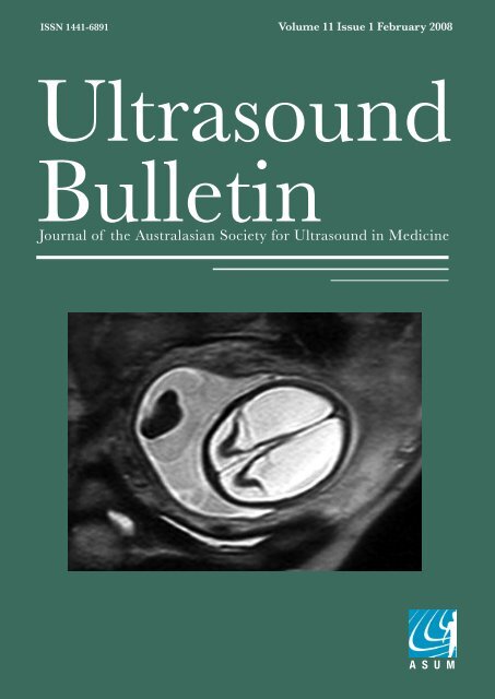
Cover image: Prenatal axial MRI of fetal brain<br />
demonstrating hydranencephaly. P 31.<br />
Editor writes that <strong>Ultrasound</strong> Bulletin is<br />
winning international readers and authors<br />
ASUM member on the experience of<br />
sonography in remote parts of Australia<br />
Pace picks up <strong>for</strong> WFUMB 2009 planning<br />
CEO rounds up what to expect in meetings and<br />
education <strong>for</strong> the year ahead<br />
Author reviews charts currently used in<br />
assessing fetal growth in Australia<br />
An exploration of the various factors associated<br />
with parental desire to discover the sex<br />
of their baby<br />
The authors draw from their own clinical experience<br />
in identifying the best methods of using<br />
interventional ultrasound in gastroenterology<br />
The authors report on the diagnosis of late<br />
gestation cranial abnormalities using MRI<br />
Second and final part of 2007 ASM abstracts<br />
The Gleaner reviews the current and best of<br />
ultrasound articles published globally<br />
All the latest ultrasound books reviewed<br />
2007 Giulia Franco Fellow Elvie Haluszkiewicz'<br />
teaching engagements took her to Central<br />
Australia and the Northern Territory<br />
Examiner writes that 2007 was a year of<br />
reinvigoration <strong>for</strong> the DMU<br />
ASUM <strong>Ultrasound</strong> Bulletin 2008 <strong>February</strong> <strong>11</strong> (1)<br />
<strong>Australasian</strong> <strong>Society</strong><br />
<strong>for</strong> <strong>Ultrasound</strong><br />
in Medicine<br />
Multidisciplinary<br />
<strong>Ultrasound</strong> Workshop<br />
28 & 29 March 2008<br />
Sydney, Australia<br />
Registration Brochure<br />
www.asummdw2008.com<br />
Convenors<br />
Dr Glenn McNally<br />
Obstetrics &<br />
Gynaecology<br />
Dr Susan Campbell<br />
Westerway<br />
General<br />
Mrs Jenifer Kidd<br />
Vascular<br />
Dr Andrew McLennan<br />
Nuchal Translucency<br />
Associated<br />
Meetings<br />
DDU Technical<br />
Seminar<br />
26 – 27 March 2008<br />
DMU Preparation<br />
Courses<br />
26 – 30 March 2008<br />
Nuchal<br />
Translucency<br />
Course<br />
27 March 2008<br />
Faculty<br />
Prof Alan Cameron<br />
Scotland<br />
Dr Ashley Robinson<br />
Canada<br />
plus a strong faculty<br />
of 40 from Australia<br />
and New Zealand<br />
Promoting Excellence<br />
in <strong>Ultrasound</strong><br />
<br />
<br />
<br />
<br />
<br />
ASUM <strong>Ultrasound</strong> Bulletin 2008 <strong>February</strong> <strong>11</strong> (1)
Provisional Program<br />
Please visit www.asummdw2008.com <strong>for</strong> further in<strong>for</strong>mation.<br />
The program is subject to change at any time without notice.<br />
* Please indicate on your registration <strong>for</strong>m which<br />
concurrent session you will be attending.<br />
Wednesday<br />
26 March 2008<br />
DMU Preparation Courses and<br />
DDU Technical Seminar<br />
▸ Physics<br />
Faculty : Roger Gent & Rob Gill<br />
Thursday<br />
27 March 2008<br />
DMU Preparation Courses and<br />
DDU Technical Seminar<br />
▸ Physics<br />
Faculty : Roger Gent & Rob Gill<br />
Nuchal Translucency<br />
Course organised by Ann Robertson<br />
(RANZCOG)<br />
Course convenor: Andrew McLennan<br />
Faculty : Jon Hyett, Andrew McLennan,<br />
Vanessa Pincham, Ann Robertson<br />
Friday<br />
28 March 2008<br />
DMU Preparation<br />
Courses<br />
Vascular<br />
Cardiac<br />
General & Obstetrics<br />
Obstetrics & Gynaecological<br />
<strong>Ultrasound</strong> Symposium<br />
▸ Neonatal Spine<br />
▸ <strong>Ultrasound</strong> Guided<br />
Paediatric Interventions<br />
▸ Obstetric <strong>Ultrasound</strong>:<br />
When? Why? How?<br />
▸ First Trimester Anomaly Detection<br />
▸ Thoracoabdominal abnormalities<br />
▸ Fetal Ocular Pathology<br />
▸ Multiple Pregnancy<br />
and Fetal Therapy<br />
▸ Early Pregnancy Complications<br />
Faculty : Ron Benzie, Alan<br />
Cameron, Danny Challis, Terry<br />
Chang, George Condous, Hans Peter<br />
Dietz, David Elwood, Jon Hyett,<br />
Greg Kesby Simon Meagher, Ashley<br />
Robinson, John Smolinec<br />
Format : Lecture Sessions<br />
General <strong>Ultrasound</strong><br />
▸ Paediatric Hip/Spine,<br />
Renal and Abdomen<br />
▸ Upper limb neuro<br />
▸ Fetal Heart<br />
▸ Calf/thigh muscles<br />
▸ Renal<br />
▸ Interventional <strong>Ultrasound</strong><br />
▸ Salivary Glands<br />
▸ Thyroid<br />
▸ Forefoot pain<br />
▸ Hip/groin<br />
▸ Testes<br />
Faculty : Matthew Andrews,<br />
Stephen Bird, Roger Gent, Jo Lennox,<br />
David McCauley, Neil Simmons<br />
Format : Live Scanning Workshops,<br />
Lecture Sessions<br />
Vascular <strong>Ultrasound</strong><br />
▸ Cerebrovascular Disease Clinical Perspectives<br />
▸ Is there a High Risk Plaque <strong>for</strong> Stroke and what’s the<br />
current Status of Carotid Stenosis Grading?<br />
▸ Carotid Endarterectomy vs Carotid Stenting<br />
▸ Duplex follow-up after Carotid Stenting<br />
▸ Peripheral Arterial Disease Clinical Perspectives<br />
▸ Ankle Brachial Indices – are they relevant?<br />
▸ Current Status of SFA angioplasty/ stenting<br />
▸ Importance of a Surveillance Program following<br />
Infrainguinal Bypass Grafting<br />
▸ Pre & Post operative Imaging <strong>for</strong> Haemodialysis Access<br />
▸ Carotid Duplex<br />
▸ Lower Extremity Vein Graft Imaging<br />
▸ Upper Limb Arteries & Veins<br />
▸ Aorto-iliac & Lower Extremity Arteries<br />
Faculty : Bernard Bourke, Alan Bray, Alison Burnett, Deb Coghlan,<br />
Jenifer Kidd, Virginia Makeham, Elizabeth Pluis, Philip Walker<br />
Format : Live Scanning Workshops, Lecture Sessions<br />
Saturday<br />
29 March 2008<br />
DMU Preparation<br />
Courses<br />
Vascular<br />
Cardiac<br />
General & Obstetrics<br />
Obstetrics & Gynaecological<br />
<strong>Ultrasound</strong> Symposium<br />
▸ Fetal Brain Development: Systerna<br />
Magna and Cerebellar Vermis<br />
Development and Anomalies<br />
▸ <strong>Ultrasound</strong> in the Delivery Suite<br />
▸ Fetal Therapy Update<br />
▸ Pediatric Surgical Overview of<br />
Thoracoabdominal abnormalities<br />
▸ Uterine Anomalies: Role of 3D/4D<br />
▸ Evaluation of Endometriosis<br />
▸ 3D/4D and Surgical Practic<br />
▸ Updating Clinicla and<br />
Molecular Genetics<br />
▸ <strong>Ultrasound</strong> and Infertility<br />
Faculty : Ron Benzie, Alan<br />
Cameron, Guy Henry, Glenn McNally,<br />
Simon Meagher, David Mowett,<br />
Andrew Ngu, Ashley Robinson<br />
Format : Lecture Sessions<br />
General <strong>Ultrasound</strong><br />
▸ Hip/groin<br />
▸ Paediatric head<br />
▸ Common Pitt falls<br />
▸ Shoulder<br />
▸ Fetal Heart<br />
▸ Abdominal vasculature<br />
▸ Neck/Salivary<br />
▸ Wrist/hand/elbow<br />
▸ Scrotum<br />
▸ Forefoot Pain<br />
▸ Hernias<br />
▸ Abdomen – Biliary Tree<br />
▸ Ankle<br />
Faculty : Stephen Bird, Roger Gent, Rob<br />
McGregor, Delwyn Nicholls, Ann Quinton,<br />
Neil Simmons, Robin Tantau<br />
Format : Live Scanning Workshops,<br />
Lecture Sessions<br />
Vascular <strong>Ultrasound</strong><br />
▸ Aneurysmal Disease Clinical Perspectives<br />
▸ Evolution and Current Status of Aortic Grafts <strong>for</strong> Repair of AAA<br />
▸ Duplex <strong>Ultrasound</strong> <strong>for</strong> Endoleak Detection<br />
and Aortic Endograft Assessment<br />
▸ Renal and Mesenteric Disease<br />
▸ Renal and Mesenteric Imaging – Optimisation is everything<br />
▸ Venous Disease Clinical Perspectives<br />
▸ What’s new in DVT management and follow up<br />
▸ Varicose veins and per<strong>for</strong>ator disease –<br />
What the surgeon needs to know<br />
▸ Upper Extremity Venous Thrombosis – U/S Diagnosis and follow up<br />
▸ Aortic Stent Graft Imaging<br />
▸ Renal & Mesenteric Artery Imaging<br />
▸ DVT Imaging<br />
▸ Venous Incompetence Imaging<br />
Faculty : Alison Burnett, Kathryn Busch, Deb Coghlan, Debbie<br />
Hamilton, John Harris, Jenifer Kidd, Andrew Lennox, Philip Walker<br />
Format : Live Scanning Workshops, Lecture Sessions<br />
Sunday<br />
30 March 2008<br />
DMU Preparation<br />
Courses<br />
Vascular<br />
Cardiac<br />
General & Obstetrics<br />
ASUM <strong>Ultrasound</strong> Bulletin 2008 <strong>February</strong> <strong>11</strong> (1)<br />
ASUM extends a warm welcome to you at upcoming ASUM meetings<br />
<strong>Australasian</strong> <strong>Society</strong><br />
<strong>for</strong> <strong>Ultrasound</strong><br />
in Medicine<br />
Multidisciplinary<br />
<strong>Ultrasound</strong> Workshop<br />
26 – 30 March 2008<br />
Sydney, Australia<br />
<br />
<br />
<br />
<br />
<br />
<br />
<br />
<br />
<br />
Convenors<br />
Dr Glenn McNally<br />
Obstetrics & Gynaecology<br />
and Point of Care Course<br />
Associated<br />
Meetings<br />
DDU Technical<br />
Seminars<br />
26 – 27 March 2008<br />
Promoting Excellence<br />
in <strong>Ultrasound</strong><br />
Dr Susan Campbell<br />
Westerway<br />
General<br />
Mrs Jenifer Kidd<br />
Vascular<br />
DMU Preparation<br />
Courses<br />
26 – 30 March 2008<br />
Nuchal Translucency<br />
Course<br />
27 March 2008<br />
Meeting Offi ce<br />
ICMS Pty Ltd<br />
Locked Bag Q4002<br />
QVB Post Offi ce<br />
Sydney NSW 1230<br />
Australia<br />
ASUM Head Offi ce<br />
PO Box 943<br />
Crows Nest NSW 1585<br />
Sydney, Australia<br />
Telephone: +61 2 9438 2078<br />
Facsimile: +61 2 9438 3686<br />
Email: asum@asum.com.au<br />
Website: www.asum.com.au<br />
ASUM CEO<br />
Dr Caroline Hong<br />
<br />
Telephone: +61 2 9290 3366<br />
Facsimile: +61 2 9290 2444<br />
ASUM Education Manager<br />
Mr Keith Henderson<br />
www.asum.com.au<br />
Upcoming ASUM Meetings<br />
ASUM Multidisciplinary Workshop 2008<br />
Sydney, Australia 5 - 9 March 2008<br />
Go to www.asum.com.au <strong>for</strong> more details<br />
ASUM 38th Annual Scientific Meeting 2008<br />
Auckland, New Zealand 18 - 21 September 2008<br />
Go to www.asum.com.au <strong>for</strong> more details<br />
World Federation <strong>for</strong> <strong>Ultrasound</strong> in<br />
Medicine and Biology 2009<br />
Sydney, Australia 30 August- 3 September 2009<br />
Go to www.asum.com.au <strong>for</strong> more details<br />
ASUM Contacts<br />
Chief Executive Offi cer Dr Caroline Hong<br />
Email: carolinehong@asum.com.au<br />
Education Manager Mr Keith Henderson<br />
Email: khenderson@asum.com.au<br />
ASUM Head Office<br />
PO Box 943 Crows Nest NSW 1585 Sydney, Australia<br />
Tel: +61 2 9438 2078 Fax: +61 2 9438 3686<br />
Email: asum@asum.com.au Website: www.asum.com.au<br />
Promoting Excellence in <strong>Ultrasound</strong>
EDITORIAL<br />
Authors in Ireland, Denmark, Holland and Australia add an <br />
international flavour to our journal<br />
Our first issue of the New Year has an<br />
international flavour, with research<br />
and reviews from Ireland, Denmark,<br />
Holland and Australia. There is also<br />
an interesting letter to the editor with<br />
excellent advice <strong>for</strong> sonographers<br />
and sonologists.<br />
Jacqueline Cartmill, now in<br />
Belfast, and her colleagues at the<br />
University of Sydney present three<br />
Prof Ron Benzie<br />
fetal intracranial anomalies where<br />
MRI was used in addition to ultrasound <strong>for</strong> prenatal diagnosis.<br />
As we all know, working out what the actual lesion in the<br />
fetal head is can be a challenge. If we have access to MRI<br />
in a tertiary centre, we should consider its use. In the cases<br />
presented, it certainly helped parental decision-making.<br />
Lisa Hui has written a thorough review of Australian charts<br />
<strong>for</strong> assessing fetal growth. This is a complex area and more<br />
studies are needed. The author concludes her excellent summation<br />
of current knowledge with recommendations, which<br />
we would do well to consider implementing in our practices.<br />
Those of you who did not manage to attend the 37th<br />
Annual Scientific Meeting at Cairns last year will find Part<br />
2 of the abstracts of interest (Part 1 was published in the<br />
December 2007 issue).<br />
Our CADUCEUS (Collaborative <strong>Australasian</strong> Danish<br />
Undertaking <strong>for</strong> Continued Excellence in <strong>Ultrasound</strong>) connection<br />
has again borne fruit. Torben Lorentzen and Christian<br />
Nolsøe, both recent ASUM visiting speakers, provide a review<br />
of interventional ultrasound in gastroenterology. This is a rapidly<br />
expanding area and undoubtedly we will see more clinical<br />
applications <strong>for</strong> its use.<br />
And from Holland, Eveline Bauman and Lisanne Hollander<br />
and colleagues here and in the University of Nijmegan found<br />
that there are several factors which affect parental desire to<br />
find out their baby’s gender be<strong>for</strong>e birth. We might think that<br />
the answer to the question ‘Why do you want to know baby’s<br />
sex’ is a simple one, however, it has uncovered factors we<br />
might not have thought about.<br />
The two lead authors were final year medical students<br />
when they conducted their research and it was their first<br />
such project. Doubtless, they learned a lot about <strong>for</strong>mulating<br />
and testing a hypothesis, as well as doing rigorous<br />
literature searches and writing up the final article. We hope<br />
the experience will have encouraged them to continue a<br />
research interest.<br />
WFUMB 2009 Congress<br />
At the time of writing it is only 18 months until the WFUMB<br />
2009 Congress. I would urge you to go to the website www.<br />
wfumb2009.com and see <strong>for</strong> yourself the innovative program<br />
being created by the Committee from ASUM, which is hosting<br />
this major event – the Sydney ‘Olympics of the <strong>Ultrasound</strong><br />
World’! If you would like to participate in any way please<br />
contact us.<br />
Prof Ron Benzie<br />
Letter to the editor – Bush sonography, flying solo<br />
For a sonographer working alone in a small rural hospital,<br />
life can be both confronting and rewarding.<br />
You are responsible <strong>for</strong> diagnostic scans when there are<br />
no other imaging modalities available. The nearest major<br />
centre with a CT may be hundreds of kilometers away. This<br />
can mean seriously disrupting families plus the added cost of<br />
transport. If a diagnosis can be made in a small hospital environment<br />
a lot of precious time and money can be saved.<br />
I started ultrasound in country western Victoria and<br />
south-east South Australia and spent 12 years on a very steep<br />
learning curve. Although I undertook the RMIT and ASUM<br />
DMU courses, my main focus was hands-on scanning. The<br />
medical staff had the notion that if you could hold a transducer<br />
you were automatically a competent sonographer. This<br />
was a serious expectation that had to be overcome.<br />
In the ensuing years, I encountered ruptured spleens and<br />
kidneys from sporting mishaps, torsion of an ovarian cyst in<br />
a 10-year-old when the clinical diagnosis was appendicitis<br />
and numerous other medical emergencies.<br />
There were many memorable times as well. I was once<br />
called during the early hours of the morning to the labour<br />
ward to scan <strong>for</strong> fetal presentation only to stay <strong>for</strong> a happy<br />
uneventful birth.<br />
Sadly, sometimes the outcomes were not so good and it<br />
was always difficult to be caught up in the highs and lows of<br />
people’s lives in a small rural community.<br />
At present I am employed at a busy suburban medical clinic<br />
equipped with state-of-the-art CT and ultrasound machines<br />
including all the bells and whistles. Life is certainly easier but I<br />
will never stop appreciating where I started to learn my trade.<br />
The top survival tips in a small country hospital are as<br />
follows:<br />
n Do not try and pretend to know what you really do not.<br />
That is, know your limitations.<br />
n If you are struggling to obtain a good diagnostic scan <strong>for</strong><br />
various reasons, say so.<br />
n Always spend time to expand your knowledge base by<br />
reading and undertaking courses and attending conferences<br />
whenever possible.<br />
n Use experts in major hospitals or ultrasound clinic. They<br />
are always more than happy to give you some direction and<br />
advice and are simply a phone call away.<br />
In summary, work closely with the physicians, it’s all<br />
about teamwork.<br />
Chairmaine Burdett<br />
ASUM <strong>Ultrasound</strong> Bulletin 2008 <strong>February</strong> <strong>11</strong> (1)
Pushing the boundaries<br />
Philips iU22 <strong>Ultrasound</strong> System with Vision<br />
2008 brings you expanded volumetric capabilities and new<br />
imaging solutions <strong>for</strong> technically difficult patients, advanced<br />
musculoskeletal imaging and clinically outstanding obstetric<br />
ultrasound. The new interventional and contrast tools, as well<br />
as new approaches to workflow efficiencies makes Philips iU22<br />
your total solution <strong>for</strong> ultrasound imaging.<br />
To learn more, contact Philips Healthcare on 1800 251 400<br />
(Aust), or 0800 251 400 (NZ).<br />
www.philips.com/healthcare
THE EXECUTIVE<br />
Pace picks up <strong>for</strong> WFUMB 2009 planning<br />
Matthew Andrews<br />
On behalf of the ASUM Council and<br />
Staff, I would like to wish all ASUM<br />
members a very happy 2008. This<br />
year will be crucial <strong>for</strong> the <strong>Society</strong>.<br />
In addition to its usual wide range of<br />
activities and services, the organisation<br />
of the World Federation of <strong>Ultrasound</strong><br />
in Medicine (WFUMB) 2009 World<br />
Congress, being hosted by ASUM in<br />
Sydney, will be consolidated.<br />
British Medical <strong>Ultrasound</strong><br />
<strong>Society</strong><br />
I was recently <strong>for</strong>tunate to take part<br />
in the ASUM/BMUS Presidential<br />
Exchange Program, in which I was<br />
invited to represent ASUM at the<br />
BMUS Annual Scientific Meeting.<br />
This program was established several<br />
years ago to promote cooperation and<br />
the exchange of scientific and organisational<br />
knowledge between the two<br />
societies.<br />
BMUS has a similar membership<br />
profile to ASUM, with the bulk of<br />
members being sonographers and radiologists.<br />
Membership numbers are also<br />
similar to ASUM.<br />
Physicists appear to play a greater<br />
role in BMUS currently than they do in<br />
ASUM. Dr Kevin Martin, the current<br />
BMUS President, is a physicist and the<br />
President-elect, Ms Julie Walton is a<br />
sonographer.<br />
The BMUS meeting is rotated<br />
around Britain and in 2007 was held in<br />
Harrogate, approximately three hours<br />
train journey north of London. Similar<br />
to the ASUM ASM, the BMUS meeting<br />
is held over three days, but within<br />
the working week, with no weekend<br />
component. The meetings have similar<br />
numbers of registrants and similar<br />
sized trade components.<br />
Features of the scientific program<br />
included several veterinary ultrasound<br />
sessions, with dogs being scanned, an<br />
interactive quiz session and an extremely<br />
interesting History of <strong>Ultrasound</strong><br />
talk by Dr Paul Allen, Past President of<br />
BMUS, which featured some ASUM<br />
members.<br />
There were lunchtime trade seminars,<br />
where vendors displayed their latest<br />
technologies. The scientific presentations,<br />
both oral and poster, were wideranging<br />
and of a very high standard.<br />
<strong>Volume</strong> acquisition ultrasound talks<br />
were impressive, where a whole volume<br />
is scanned and images then processed<br />
and obtained on workstation in any<br />
plane required. This would appear to<br />
have the potential to revolutionise the<br />
way ultrasound is practised.<br />
My presentation was part of a seminar<br />
discussing the future of ultrasound<br />
practice, including the roles of sonographers,<br />
sonologists and clinicians per<strong>for</strong>ming<br />
ultrasound, where I gave the<br />
<strong>Australasian</strong> perspective.<br />
The <strong>for</strong>um allowed me the opportunity<br />
to showcase ASUM’s Certificate<br />
of Clinician Per<strong>for</strong>med <strong>Ultrasound</strong><br />
(CCPU), which was very well received<br />
by both BMUS and clinicians in attendance.<br />
BMUS expressed interest in<br />
exploring the CCPU concept further<br />
and will liaise with ASUM.<br />
I would like to acknowledge the<br />
very kind hospitality provided to me by<br />
BMUS including the BMUS staff, who<br />
were extremely helpful.<br />
I am confident my visit will be<br />
of benefit to ASUM and I look <strong>for</strong>ward<br />
to welcoming Dr Martin when<br />
he reciprocates by attending this year’s<br />
ASUM meeting in Auckland. I would<br />
also like to acknowledge the <strong>for</strong>esight<br />
of my predecessors in establishing this<br />
exchange program.<br />
Dr Matthew Andrews<br />
Help us promote WFUMB<br />
2009 Sydney<br />
ASUM welcomes members’ assistance<br />
in promoting the Congress.<br />
To help spread the message,<br />
members who are attending and<br />
presenting at meetings overseas are<br />
encouraged to include promotional<br />
slides <strong>for</strong> WFUMB 2009 in their<br />
presentation<br />
ASUM <strong>Ultrasound</strong> Bulletin 2008 <strong>February</strong> <strong>11</strong> (1)<br />
Snap open <strong>for</strong> quality.<br />
Snap closed <strong>for</strong> protection.<br />
Aquasonic ® 100, the world standard <strong>for</strong> medical ultrasound, now has a new proprietary<br />
Snap-Cap with valve, providing unparalleled benefits to both user and patient.<br />
Designed <strong>for</strong> One Handed Operation: Engineered to Eliminate Drips and “Draw<br />
Back.”<br />
Exclusive self-sealing silicone valve instantly cuts off the flow of gel.<br />
• Eliminates drawing product back into the bottle, thus reducing the potential<br />
<strong>for</strong> cross-contamination<br />
• Maintains a clean and safe work environment by preventing drips and<br />
product residue<br />
• Provides precise unimpeded flow control from the new larger<br />
aperture and valve<br />
Easy to use One-Handed Snap-Cap keeps the nozzle and aperture protected from<br />
the work environment.<br />
• Open and close the cap with one hand and maintain position and procedure continuity<br />
• Protect the nozzle from old gels that can often collect on the surface of<br />
ultrasound equipment<br />
• AND no more lost red tips thanks to the permanently attached cap<br />
Welcome our new<br />
Snap-Cap to your pratice…<br />
Invite a safer and more<br />
efficient workplace.<br />
ISO 13485:2003
THE EXECUTIVE<br />
COUNCIL 2005–2007<br />
CEO’s message<br />
EXECUTIVE<br />
President<br />
Matthew Andrews Vic<br />
Medical Councillor<br />
President Elect<br />
Ron Benzie NSW<br />
Medical Councillor<br />
Honorary Secretary<br />
Roslyn Savage Qld<br />
Sonographer Councillor<br />
Honorary Treasurer<br />
Andrew Ngu Vic<br />
Medical Councillor<br />
MEMBERS<br />
Medical Councillors<br />
John Crozier NSW<br />
Roger Davies SA<br />
Simon Meagher Vic<br />
Monica Pahuja Vic<br />
Sonographer Councillors<br />
Stephen Bird SA<br />
Margaret Condon Vic<br />
Kaye Griffiths NSW<br />
Michelle Pedretti WA<br />
ASUM Head Office<br />
Chief Executive Officer<br />
Caroline Hong<br />
Education Manager<br />
Keith Henderson<br />
All correspondence should be<br />
directed to:<br />
The Chief Executive Officer<br />
<strong>Australasian</strong> <strong>Society</strong> <strong>for</strong><br />
<strong>Ultrasound</strong> in Medicine<br />
Level 2, 5<strong>11</strong> Pacific Highway<br />
St. Leonards NSW 2065 Australia<br />
email asum@asum.com.au<br />
www..asum.com.au<br />
Dr Caroline Hong<br />
New Year’s resolutions<br />
The New Year started <strong>for</strong> me with a<br />
number of key resolutions: to include<br />
a more work-life-family balance; more<br />
exercise; eating healthily; and remembering<br />
to make time <strong>for</strong> celebrations,<br />
big and small.<br />
I hope you are happy with the way<br />
the year 2008 has begun <strong>for</strong> you.<br />
Last year was a good year <strong>for</strong><br />
ASUM as we all worked hard to use<br />
our time and resources effectively<br />
to provide member services and to<br />
advance the <strong>Society</strong>. I am pleased to<br />
report that the ASUM membership as<br />
at 31st December 2007 has increased<br />
by 14% compared to 31st December<br />
2006.<br />
WFUMB 2009 major sponsors<br />
We are filled with gratitude <strong>for</strong> the<br />
confidence and support that the major<br />
global ultrasound companies have<br />
shown to ASUM, in our recent dealings<br />
with them.<br />
Over the years and in the last few<br />
months, I have been involved, together<br />
with various members of the ASUM<br />
Executive and the WFUMB Organising<br />
Committee, in discussions with the<br />
trade about sponsorship opportunities<br />
<strong>for</strong> ASUM meetings. At the same time,<br />
we have also been talking to them<br />
about sponsorship opportunities <strong>for</strong> the<br />
WFUMB 2009 World Congress.<br />
We are pleased to report that, as<br />
of 21st January 2008, Toshiba, GE<br />
Healthcare, Medison and Philips have<br />
been confirmed as our four major<br />
sponsors.<br />
We also value the strong<br />
support of Siemens, a gold supporter<br />
of ASUM meetings <strong>for</strong> some time. The<br />
global office has indicated support at<br />
WFUMB 2009, with the level of support<br />
to be determined soon.<br />
On behalf of the <strong>Society</strong>, staff,<br />
members and Council, we gratefully<br />
thank the major sponsors <strong>for</strong> their<br />
early commitment of support, almost<br />
two years ahead of the Congress. This<br />
is a demonstration of the successful<br />
partnership we have with our corporate<br />
supporters, to ensure a strong working<br />
relationship <strong>for</strong> the most ambitious<br />
project ever taken on by ASUM.<br />
WFUMB 2009 sponsorship<br />
opportunities<br />
There are many sponsorship opportunities<br />
that can be specifically tailored<br />
to individual company needs. If any<br />
member is interested or knows of anyone<br />
who might be interested in being a<br />
sponsor or exhibitor, or wishes to suggest<br />
and refer potential sponsors/supporters,<br />
please email carolinehong@<br />
asum.com.au or alternatively to sponsorship@icms.com.au.<br />
ASUM also welcomes members’<br />
assistance in promoting the Congress.<br />
To help spread the message, members<br />
who are attending and presenting<br />
at meetings overseas are encouraged<br />
to include promotional slides <strong>for</strong><br />
WFUMB 2009 in their presentation.<br />
We thank the members who already<br />
have responded and assisted in this<br />
promotion.<br />
If anyone wishes to help in this<br />
manner, please email carolinehong@<br />
asum.com.au to obtain a set of the<br />
promotional slides or <strong>for</strong> more in<strong>for</strong>mation.<br />
We are regularly updating<br />
the WFUMB 2009 Congress website<br />
as our planning and organising<br />
progress. The address is www.<br />
wfumb2009.com.<br />
ASUM MDW 2008 Sydney<br />
By popular demand, the ASUM<br />
Multidisciplinary Workshops (MDW)<br />
ASUM <strong>Ultrasound</strong> Bulletin 2008 <strong>February</strong> <strong>11</strong> (1)<br />
THE EXECUTIVE<br />
will be on again soon, from 26th–30th<br />
March 2008. The MDW incorporates<br />
the DMU and DDU preparation courses.<br />
The MDW will be held at the Hilton<br />
Sydney Hotel immediately after the<br />
Easter holidays.<br />
We are indebted to Dr Glenn<br />
McNally, Sue Campbell Westerway,<br />
Jeni Kidd, Dr Rob Gill and Dr Andrew<br />
McLennan, all of whom have worked<br />
tirelessly with the ASUM Secretariat<br />
and in particular, Keith Henderson, to<br />
present to our members a quality program<br />
of workshops. These workshops<br />
are very popular and fill up quickly.<br />
This year, we have invited two very<br />
special overseas presenters, Prof Alan<br />
Cameron and Dr Ashley Robinson.<br />
Prof Cameron is a Consultant<br />
Obstetrician and Head of Fetal<br />
Medicine at the Queen Mother’s<br />
Hospital, Glasgow. His specialist areas<br />
are prenatal diagnosis and fetal therapy.<br />
He trained in Glasgow, Newcastle and<br />
Canada and has published widely on<br />
the subject of high-risk pregnancy.<br />
Prof Cameron is the Scottish members’<br />
representative on the Council of<br />
the Royal College of Obstetricians and<br />
Gynaecologists. He is also a member<br />
of Council of the Royal College of<br />
Midwives, a founder member of the<br />
British Maternal and Fetal <strong>Society</strong> and<br />
a member of the International Fetal<br />
Medicine and Surgery <strong>Society</strong>.<br />
He is a reviewer <strong>for</strong> the British<br />
and American Journals of Obstetrics<br />
and Gynaecology and <strong>for</strong> the Journal<br />
of <strong>Ultrasound</strong> in Obstetrics and<br />
Gynaecology. He is an editor of the<br />
first international CD text of fetal ultrasound.<br />
His current research interest is the<br />
development of in utero keyhole surgical<br />
techniques in an attempt to improve<br />
the outcome <strong>for</strong> babies with congenital<br />
mal<strong>for</strong>mations.<br />
Dr Robinson did his radiology<br />
training in Manchester, UK, followed<br />
by three fellowships in general paediatric<br />
radiology, paediatric interventional<br />
radiology, both at the Hospital <strong>for</strong> Sick<br />
Children in Toronto, Canada, then ultrasound<br />
at the University of Cali<strong>for</strong>nia,<br />
San Francisco. He is currently a radiologist<br />
at the Children’s and Women’s<br />
Hospital of British Columbia, Canada,<br />
specialising in fetal imaging and pediatric<br />
intervention.<br />
His awards include the Derek<br />
Harwood-Nash Award from the<br />
American <strong>Society</strong> of Pediatric<br />
Neuroradiology, the Ella Preiskel Prize<br />
in Pediatric Radiology from the Royal<br />
College of Radiologists (UK) and<br />
the John Kirkpatrick Award from the<br />
<strong>Society</strong> of Pediatric Radiology.<br />
Once again we are indebted to<br />
Toshiba, Philips, GE Healthcare and<br />
Siemens <strong>for</strong> their ongoing support and<br />
agreeing to be gold sponsors at this<br />
workshop.<br />
Accommodation at the Hilton<br />
Sydney Hotel, as the preferred conference<br />
hotel, has been block booked <strong>for</strong><br />
this workshop. There is a limited room<br />
block so please make sure you click<br />
‘Book online’ via the ‘Accommodation’<br />
section on the website at www.asum<br />
mdw2008.com to get the specially<br />
negotiated ASUM rates <strong>for</strong> 2008.<br />
Registration brochures and online<br />
registration <strong>for</strong> all the various disciplines<br />
are now available at www.asum<br />
mdw2008.com:<br />
n 2008 ASUM Multidisciplinary<br />
Workshop 28th–29th March 2008<br />
n ASUM Annual O & G <strong>Ultrasound</strong><br />
Symposium 28th–29th March<br />
2008<br />
n 2008 DMU Preparation Course<br />
26th–30th March 2008<br />
n Nuchal Translucency Course 27th<br />
March 2008<br />
Log in NOW <strong>for</strong> complete registration<br />
details on www.asummdw2008.<br />
com.<br />
For all enquiries about your registration,<br />
please email asummdw2008@<br />
asum.com.au.<br />
For general ASUM enquiries, email<br />
asum@asum.com.au.<br />
The multidisciplinary workshops<br />
fill up on a first come first serve basis.<br />
Please register early to avoid disappointment.<br />
ASUM 2008 ASM Auckland<br />
ASUM thanks GE Healthcare,<br />
Siemens, Philips and Toshiba <strong>for</strong><br />
their ongoing support and agreeing<br />
to be gold sponsors of the ASUM<br />
2008 ASM. This meeting will be held<br />
from 18th–21st September 2008 at the<br />
Skycity Auckland Convention Centre,<br />
Auckland, New Zealand. A special<br />
thanks also goes to GE Healthcare <strong>for</strong><br />
sponsorship of a keynote speaker, Prof<br />
Bernard Benoit.<br />
For enquiries about sponsorship<br />
and the trade exhibition, email admin@<br />
mianz.co.nz or contact the ASUM CEO<br />
at carolinehong@asum.com.au directly<br />
<strong>for</strong> specially tailored packages.<br />
A great program – ‘Into the Next<br />
Dimension’ – has been created by<br />
Dr David Rogers and the Organising<br />
Committee. Look out <strong>for</strong> regular<br />
updates at www.asum2008.com.au.<br />
The <strong>for</strong>mat of the meeting will<br />
include a skills day on Thursday 18th<br />
September, followed by a three-day<br />
event from Friday 19th September to<br />
Sunday 21st September <strong>for</strong> the main<br />
part of the meeting. Members are<br />
encouraged to submit their proffered<br />
papers and poster abstracts no later<br />
than Friday 16th May 2008.<br />
The registration brochure is ready<br />
and is being distributed with this issue<br />
of the <strong>Ultrasound</strong> Bulletin.<br />
Early registration rates close on<br />
Sunday 20th July 2008, so book early<br />
to benefit from the reduced rate and also<br />
to secure your accommodation requirements.<br />
Special rates can be negotiated<br />
<strong>for</strong> company, hospital, institution and<br />
private group bookings paid in full and<br />
in group blocks. Overseas members of<br />
affiliated societies of WFUMB are also<br />
welcome to register at member rates.<br />
These requests should be directed to<br />
carolinehong@asum.com.au.<br />
Auckland, popularly known as the<br />
City of Sails, is the biggest city in New<br />
Zealand. It is an attractive city with<br />
many things to do and see. The ASM<br />
is a wonderful opportunity to combine<br />
attendance, participation or exhibition<br />
at the meeting with a New Zealand<br />
holiday. You may wish to start planning<br />
your stay in Auckland as a visitor<br />
and conference delegate by viewing<br />
www.aucklandnz.com. More in<strong>for</strong>mation<br />
about the ASUM 2008 meeting<br />
venue at Skycity Auckland Convention<br />
Centre can be seen at www.skycity.<br />
co.nz.<br />
ISUM seeks speakers <strong>for</strong><br />
Jakarta meeting<br />
An email broadcast was sent out to<br />
members in December 2007, with a<br />
request from the Indonesian <strong>Society</strong><br />
<strong>for</strong> <strong>Ultrasound</strong> in Medicine (ISUM)<br />
<strong>for</strong> speakers to present at the <strong>Society</strong>’s<br />
meeting in Jakarta on 21st–22nd<br />
November.<br />
The ASUM Executive had approved<br />
support <strong>for</strong> ISUM by sending one or<br />
two speakers to present at the meeting.<br />
The 6th Asian Breast Diseases<br />
Association (ABDA) Teaching Course<br />
will be held in conjunction with the<br />
17th Annual Meeting of the Indonesian<br />
<strong>Society</strong> of <strong>Ultrasound</strong> in Medicine and<br />
10 ASUM <strong>Ultrasound</strong> Bulletin 2008 <strong>February</strong> <strong>11</strong> (1)
2nd Annual Meeting of the Indonesian<br />
<strong>Society</strong> of Oncology Imaging.<br />
This meeting will be held at the<br />
Novotel Hotel, Jarkarta. Any member<br />
interested in presenting at the meeting<br />
should contact Dr Caroline Hong,<br />
ASUM CEO, at asum@asum.com.au<br />
<strong>for</strong> more details. Conditions apply.<br />
DMU registration closes on <br />
31st March<br />
As of 31st December 2007, 1534<br />
sonographers and doctors had been<br />
awarded the Diploma of Medical<br />
Ultrasonography (DMU), since the<br />
Diploma was introduced in 1979.<br />
The 2008 examination registration<br />
date closes on 31st March. Candidates<br />
should contact ASUM at dmu@asum.<br />
com.au if they have any enquiries.<br />
To assist in preparing <strong>for</strong> the examinations,<br />
candidates are encouraged to<br />
enrol <strong>for</strong> the DMU preparation courses,<br />
which will run from 26th–30th March<br />
2008 at the Hilton Sydney. Register<br />
online at www.asummdw2008.com.<br />
ASUM is proud to have established<br />
the DMU in 1979. The Diploma was<br />
the first certification established by<br />
ASUM <strong>for</strong> sonographers. It is a selfdirected<br />
program of study leading to<br />
the postgraduate DMU examination.<br />
The main prerequisite to be eligible <strong>for</strong><br />
the DMU is to have a Bachelor degree<br />
in any discipline.<br />
There are two parts to the examination.<br />
The Part I Examination consists<br />
of two written multiple-choice papers,<br />
each of two hours duration. Part I<br />
examinations are usually held on the<br />
last Saturday in July and on the first<br />
Saturday in November each year.<br />
It is important to understand that<br />
a DMU candidate does not have to<br />
be employed in sonography to sit <strong>for</strong><br />
the Part I Examination, but will be<br />
required to have at least two years<br />
of ultrasound scanning experience<br />
to be eligible to sit <strong>for</strong> the Part II<br />
Examination. In<strong>for</strong>mation about the<br />
2008 DMU Handbook, regulations,<br />
syllabus, application <strong>for</strong>m and enrolment<br />
criteria are available in the DMU<br />
section of the ASUM website at www.<br />
asum.com.au.<br />
DMU (Asia)<br />
As of 31st December 2007, 22 sonographers,<br />
who completed their training<br />
at Vision College in Kuala Lumpur<br />
since the College was established in<br />
2005, have been awarded the Diploma<br />
of Medical Ultrasonography (Asia).<br />
The level of interest is increasing<br />
each year, with a recent decision from<br />
the Malaysian Government to allow<br />
Vision College to accept enrolment of<br />
students from outside Malaysia.<br />
The DMU (Asia) was established<br />
by ASUM in 2005 through an agreement<br />
between ASUM and Vision<br />
College, a private educational body.<br />
The DMU (Asia) standards satisfy the<br />
diploma accreditation requirements in<br />
Malaysia <strong>for</strong> sonographer qualification<br />
<strong>for</strong> practice in Asia. Vision College<br />
provides the training <strong>for</strong> students who<br />
are enrolled in the DMU (Asia) and<br />
the Diploma is awarded by ASUM to<br />
the candidates who have successfully<br />
completed and passed the examinations<br />
conducted by ASUM approved<br />
examiners.<br />
DDU<br />
As of 31st December 2007, the DDU<br />
has been awarded to 535 medical specialists,<br />
since it was established in<br />
1976.<br />
Registrations <strong>for</strong> the 2008 examinations<br />
will close on 17th March 2008.<br />
Candidates should contact ASUM at<br />
ddu@asum.com.au if they have any<br />
enquiries.<br />
The DDU was established in 1976<br />
by ASUM. It was the first and remains<br />
the only diploma certification <strong>for</strong> doctors<br />
in diagnostic ultrasound.<br />
ASUM is recognised by the government<br />
and industry bodies as the peak<br />
body <strong>for</strong> ultrasound and the ASUM<br />
DDU remains a highly sought after<br />
qualification.<br />
In<strong>for</strong>mation about the 2008<br />
Handbook, regulations, syllabus, application<br />
<strong>for</strong>m and enrolment criteria are<br />
in the DDU section on the ASUM<br />
website at www.asum.com.au.<br />
The Part I Examination <strong>for</strong> 2008<br />
will be held on Monday 12th May.<br />
Applications will close on Monday<br />
17th March 2008. More in<strong>for</strong>mation<br />
about the DDU examinations and<br />
important dates can be obtained in the<br />
DDU section of the ASUM website.<br />
CCPU is popular<br />
As of 31st December 2007, two doctors<br />
have successfully attained the CCPU<br />
(Emergency Medicine). At the time of<br />
writing, four CCPU (Neonatal) and one<br />
CCPU (O&G) are about to be conferred<br />
and approved by the ASUM Council.<br />
The CCPU is gaining popularity<br />
and is making progress, with valuable<br />
input from distinguished members of<br />
the <strong>Society</strong>. To date, 131 doctors have<br />
registered <strong>for</strong> the CCPU.<br />
Members may recall that in 2006,<br />
the CCPU became the first certification<br />
<strong>for</strong> medical specialists who are not<br />
imaging specialists <strong>for</strong> targeted ultrasound<br />
at the point of care. The CCPU<br />
(O&G), CCPU (Emergency Medicine)<br />
and CCPU (Surgical Practice) are possible<br />
only with the willing support and<br />
assistance of many key members of the<br />
<strong>Society</strong>, who have put in many hours<br />
towards developing the curriculum,<br />
courses and certification process.<br />
Organisations providing ultrasound<br />
courses can apply to ASUM <strong>for</strong> appropriate<br />
recognition towards the CCPU,<br />
if the necessary criteria are met. For<br />
example, several of the AIU courses<br />
are accredited by ASUM as meeting<br />
the requirements of the CCPU. More<br />
in<strong>for</strong>mation is available in the CCPU<br />
section of the ASUM website at www.<br />
asum.com.au.<br />
Dr Coll Fisher 1935–2008<br />
We were sad to learn of the passing<br />
of Dr Coll Fisher, who died after<br />
New Year’s Day. Coll was a popular<br />
obstetrician and was at the centre of<br />
many changes in the care of pregnant<br />
women. He was a loyal member of<br />
ASUM from 1976 to 1998.<br />
He had been an expert in the use of<br />
ultrasound since the days when grey<br />
scale ultrasound was first developed<br />
by Bill Garret and George Kossoff in<br />
Australia.<br />
On behalf of the <strong>Society</strong>, we mark<br />
the passing of a great man and send<br />
condolences to the family and all<br />
those whose lives were touched by this<br />
extraordinary doctor.<br />
Dr Caroline Hong<br />
Chief Executive Officer<br />
carolinehong@asum.com.au<br />
Changing jobs?<br />
Changing address?<br />
Be sure to tell the <strong>Society</strong><br />
email asum@asum.com.au<br />
tel + 61 2 9438 2078<br />
ASUM <strong>Ultrasound</strong> Bulletin 2008 <strong>February</strong> <strong>11</strong> (1)<br />
<strong>11</strong>
DIAGNOSTIC ULTRASOUND<br />
ASUM <strong>Ultrasound</strong> Bulletin <strong>February</strong> 2008; <strong>11</strong> (1): 12–18<br />
Australian charts <strong>for</strong> assessing fetal growth: <br />
a review<br />
Lisa Hui<br />
Division of Maternal Fetal Medicine, Royal Hospital <strong>for</strong> Women, Barker St, Randwick NSW 2031, Australia.<br />
Correspondence to Lisa Hui. Email lisahui77@gmail.com<br />
Introduction<br />
Monitoring fetal growth is one of the basic goals of antenatal<br />
care, <strong>for</strong> low-risk and high-risk women alike. While the<br />
‘large <strong>for</strong> gestational age’ fetus is at increased risk of perinatal<br />
complications, the burden of adverse outcomes is overwhelmingly<br />
carried by the pathologically small fetus. Fetal<br />
growth restriction is accompanied by dramatically increased<br />
perinatal mortality, with mortality being eight times higher<br />
when weight is below the 10th centile and nearly 20 times<br />
higher when weight is below the 3rd centile 1 . In addition to<br />
increased mortality, both short term 2,3 and long term morbidity<br />
4,5 are increased in infants affected by intrauterine growth<br />
restriction.<br />
The <strong>Ultrasound</strong> Bulletin has previously published a<br />
review on the accuracy of sonographic estimation of fetal<br />
weight that discusses the sources of error in determining<br />
whether or not a given fetal size is ‘normal’ 6 . This paper<br />
seeks to further examine the centile charts in current use<br />
in Australia and to describe their derivation. The multitude<br />
of biometry and birthweight charts can cause confusion<br />
if a fetus is compared against different standards during<br />
pregnancy and this has the potential to cause inappropriate<br />
management decisions. Finally, the rising interest in individualised<br />
birthweight standards and their role in developing<br />
intrauterine growth curves will also be covered.<br />
Definitions of SGA/FGR<br />
Various growth percentile thresholds <strong>for</strong> defining ‘small <strong>for</strong><br />
gestational age’ (SGA) babies have been used, but the most<br />
commonly accepted standard is the 10th centile 7 <strong>for</strong> estimated<br />
fetal weight (EFW) or abdominal circumference (AC).<br />
However, it is important to remember that babies below the<br />
10th centile are a heterogeneous group comprising of constitutionally<br />
small normal babies and those with true growth<br />
restriction with the accompanying increase in morbidity and<br />
mortality. Identifying those SGA babies at increased risk of<br />
true fetal growth restriction (FGR) involves incorporating<br />
other measures of fetal well-being such as amniotic fluid<br />
volume, fetal arterial and venous Doppler measurements and<br />
cardiotography 8,9 .<br />
<strong>Ultrasound</strong> estimation of fetal size<br />
There are several discrete steps in the process of ultrasound<br />
estimation of fetal growth. Each step is subject to error. An<br />
acceptable margin of error <strong>for</strong> the final EFW is generally<br />
accepted to be ± 15%. These steps are:<br />
1 Accurate assessment of dates;<br />
2 Fetal measurements: two-dimensional measurement of<br />
biometry;<br />
3 Mathematical calculation of an estimated fetal weight;<br />
4 Charting fetal size – EFW and/or biometry – against<br />
population standards <strong>for</strong> gestation and determining the<br />
corresponding percentile band; and<br />
5 Comparing serial measurements if available, to determine<br />
whether growth velocity is being maintained along<br />
the appropriate centile curve.<br />
Determining intrauterine weight percentile<br />
The ideal chart should be created from a representative sample<br />
of the local population. These growth charts can be constructed<br />
from birthweight data <strong>for</strong> preterm and term infants,<br />
or from standards derived from ultrasound measurements of<br />
fetuses. A third method has been recently investigated using<br />
customised term birthweights to develop intrauterine growth<br />
curves.<br />
Australian birthweight charts<br />
Until the late 1990s, the most commonly used standards<br />
of growth in Australia were derived from hospital-based<br />
studies of infants born in Melbourne. The first publications<br />
were produced by Kitchen in 1968 10 and Betheras in<br />
1969 <strong>11</strong> . Explicit in the publication of Betheras’ charts was<br />
the caveat that the use of birthweights to define normal<br />
growth at preterm gestations contains an inherent fault: ‘the<br />
obstetrical complication which may have precipitated the<br />
premature termination of pregnancy may have affected fetal<br />
growth.’ These early charts are disadvantaged by the less<br />
accurate dating methods, with early ultrasound dating being<br />
uncommon. Betheras’ chart included women if there was<br />
‘reasonable certainty’ that the EDC was correct, and if the<br />
infant had a weight and head circumference (HC) measured<br />
‘shortly after birth’. These early charts also have very small<br />
numbers of infants at extremely preterm gestations, and in<br />
Betheras’ case, only commence from 28 weeks gestation.<br />
There have been significant changes in ethnic composition<br />
and socioeconomic factors in the Australian population<br />
since the publication of Betheras’ chart, which was based<br />
almost entirely on women of Anglo-Saxon origin. Kitchen<br />
updated early data from 1968 and in 1983 produced revised<br />
intrauterine growth curves from livebirth data 12 . Birthweight<br />
curves from 24 to 42 weeks were produced from a combination<br />
of data from live births at the Royal Women’s Hospital<br />
in Melbourne in 1979 and from previous publications on<br />
those born
Australian charts <strong>for</strong> assessing fetal growth – a review<br />
Fig.2: Hadlock's chart.<br />
Fig.1: Beeby's population based birthweight percentile chart.<br />
newborn infants’ growth characteristics correctly.<br />
One of the first published population-based charts was<br />
produced in Western Australian in 1989 by Kliewer, et al. 14 .<br />
They extracted data from the WA Midwives’ Notification<br />
System, which recorded details of all births of 20 weeks gestation<br />
or more, or birthweight of 400 g or more from 1980<br />
to 1986. Gestational age was calculated from last menstrual<br />
period. The charts were not specifically designed to act<br />
as standards of intrauterine growth, but to compare health<br />
outcomes between indigenous and non-indigenous babies.<br />
They found that the distributions of both birthweight and<br />
gestational age were shifted downward in Indigenous infants<br />
compared with white infants, a trend that un<strong>for</strong>tunately<br />
persists today. These charts were also gender-specific, with<br />
separate charts <strong>for</strong> male and female babies, recognising the<br />
importance of sex on fetal size.<br />
In 1996, Beeby published the first NSW population-based<br />
birthweight percentile charts, which was also the largest<br />
Australian population-based study at the time. This study<br />
included data from 422 139 live born singletons held by the<br />
NSW Midwives Data Collection and from King George V<br />
Hospital 15 . Separate charts <strong>for</strong> male and female infants were<br />
developed. The charts followed the general trend of increasing<br />
birthweights over time and had generally higher percentiles<br />
than previously published charts.<br />
The first Australian birthweight percentiles published<br />
using national data were published in 1999 by Roberts and<br />
Lancaster 16 . They aimed to develop national birthweight<br />
percentiles by gestational age (GA) <strong>for</strong> male and female<br />
singleton infants and then to compare BW distributions by<br />
GA <strong>for</strong> indigenous and non-indigenous infants. Using data<br />
from the National Perinatal Statistics Unit (NPSU), 761 902<br />
births were analysed and birthweight percentiles <strong>for</strong> indigenous<br />
and non-indigenous Australian-born women were<br />
made. They included only singleton live infants delivered<br />
by Australian-born mothers in the non-indigenous charts<br />
in order to reduce environmental factors associated with<br />
migrant populations. In keeping with the WA data, indigenous<br />
infants were more likely to be classified as SGA and<br />
preterm than non-indigenous infants. More specifically,<br />
Fig.3: Customised chart.<br />
birthweights of indigenous infants diverged from that of the<br />
non-indigenous population after 34 weeks. By 40 weeks,<br />
the median birthweight <strong>for</strong> male indigenous babies was 160 g<br />
below that of non-indigenous males and birthweight <strong>for</strong><br />
female indigenous infants was 130 g below non-indigenous<br />
females. More than 17% of indigenous infants born at term<br />
were classified as SGA. Separate charts <strong>for</strong> indigenous<br />
births were published, but the authors cautioned that they<br />
should not be interpreted as separate population norms until<br />
the effect of population and environmental differences could<br />
be ascertained.<br />
While the authors acknowledged that birthweight percentiles<br />
do not necessarily represent fetal growth standards,<br />
the 10th centile on these charts are the current standard used<br />
by the NPSU <strong>for</strong> defining fetal growth restriction in the perinatal<br />
mortality data collection system.<br />
Advantages of birthweight charts<br />
n Standards can be derived from very large numbers and<br />
are there<strong>for</strong>e statistically robust;<br />
n Population-based data provides a more demographically<br />
representative sample to be obtained compared with hospital<br />
or private practice populations;<br />
ASUM <strong>Ultrasound</strong> Bulletin 2008 <strong>February</strong> <strong>11</strong> (1)
Lisa Hui<br />
Table 1: Australian birthweight charts compared with the Hadlock EFW chart.<br />
Author Date Population n GA range<br />
Kitchen 1968<br />
Betheras 1969<br />
Kitchen 1983<br />
Kliewer 1989<br />
Guaran 1994<br />
Royal Women’s Hospital,<br />
Melbourne<br />
Private paediatric<br />
practice, Melbourne<br />
Royal Women’s Hospital,<br />
Melbourne<br />
WA<br />
population data<br />
Mercy Hospital,<br />
Melbourne<br />
Gender<br />
specific<br />
Australian Birthweight Charts<br />
10th C 40w<br />
(g)<br />
50th C 40w<br />
(g)<br />
90th C 40w<br />
(g)<br />
2637 23–44 N 2600 3430 4000<br />
7818 28–44 N 2726 3371 4025<br />
3120 24–42 N 2980 3507 4040<br />
158729 20–44+ Y<br />
Beeby 1996 NSW population 422,139 22–43 Y<br />
Roberts 1999<br />
National, Australianborn<br />
population<br />
3050 (M)<br />
2925 (F)<br />
3585 (M)<br />
3430 (F)<br />
4150 (M)<br />
3980 (F)<br />
49,429 22–44 N 3000 3515 4090<br />
761,902 20–44 Y<br />
Sonographic Weight Standard<br />
3046 (M)<br />
2932 (F)<br />
3070 (M)<br />
2950 (F)<br />
3576 (M)<br />
3436 (F)<br />
3600 (M)<br />
3450 (F)<br />
4154 (M)<br />
3993 (F)<br />
4170 (M)<br />
4000 (F)<br />
Hadlock 1991 USA white middle class 392 10–40 N 3004 3619 4234<br />
n Many population-based birthweight charts are gender<br />
specific; however this in<strong>for</strong>mation is often lacking in<br />
clinical practice and makes their application difficult.<br />
The convention is to assume female gender if the sex is<br />
unknown; and<br />
n Using locally-derived birthweight charts facilitates clinical<br />
consistency with the newborn assessment by the<br />
paediatricians.<br />
Disadvantages of birthweight charts<br />
The fundamental problem with the use of birthweight charts<br />
is the assumption that average preterm birthweights can<br />
properly be used to determine normal intrauterine fetal size.<br />
Preterm infants must be assumed to be affected by some pathological<br />
process leading to early delivery, and are known to be<br />
disproportionately affected by intrauterine growth restriction.<br />
A study of the neonatal outcomes of 7898 preterm neonates<br />
found that conventional birthweight standards <strong>for</strong> preterm<br />
infants contained weights from a large proportion of FGR<br />
babies and this lead to non-recognition of fetal growth restriction<br />
in preterm infants 17 . This raises the interesting question<br />
of whether preterm neonates themselves should be assessed<br />
against EFW-based intrauterine growth curves from normal<br />
fetuses rather than birthweight standards 18 .<br />
The accuracy of gestational age assignment may be<br />
harder to ascertain in large state-wide data collection than in<br />
smaller hospital-based studies.<br />
Sonographic standards <strong>for</strong> biometry<br />
<strong>Ultrasound</strong>-derived standards <strong>for</strong> fetal measurements began<br />
to be developed in the 1980s, pioneered by the work of<br />
Hadlock 19–21 , whose charts and <strong>for</strong>mulae continue to be in<br />
widespread use over 20 years later. These curves are created<br />
by mathematically modelling data collected on a large number<br />
of fetuses after excluding pregnancies with uncertain<br />
dates or with known risk factors <strong>for</strong> abnormal growth such<br />
as multiple gestation, fetal anomaly and maternal diseases.<br />
There are many differences in the methodology and study<br />
populations in the numerous published charts, resulting in<br />
many variations of 10th and 90th centile boundaries <strong>for</strong><br />
normal fetal growth. Some sonographic standards are based<br />
on longitudinal studies of individual fetal growth, but the<br />
vast majority, including Hadlock charts, use cross-sectional<br />
analysis of data.<br />
There are important methodological distinctions between<br />
developing charts <strong>for</strong> determining fetal size <strong>for</strong> known gestation<br />
and charts developed <strong>for</strong> dating pregnancies according<br />
to fetal size. Furthermore, while cross-sectional data is suitable<br />
<strong>for</strong> determining fetal size, longitudinal study is more<br />
appropriate <strong>for</strong> assessing fetal growth patterns. The ideal<br />
design <strong>for</strong> defining normal intrauterine growth curves would<br />
be prospectively gathered measurements from serial scans of<br />
well-dated normal pregnancies; this has yet to be per<strong>for</strong>med<br />
in an Australian population. The correct methodology <strong>for</strong><br />
creating centile curves <strong>for</strong> fetal size is rather different and<br />
has been discussed in detail by Altman and Chitty 22 , who<br />
published their own reference charts in 1994 23–25 .<br />
Prior to 2001, ASUM recommended the use of Hadlock<br />
charts <strong>for</strong> head circumference (HC) 20 and femur length (FL) 21<br />
and Australian charts from de Crespigny <strong>for</strong> bi-parietal diameter<br />
(BPD) 26 . The Hadlock charts were constructed from data<br />
collected on predominately white middle-class Americans in<br />
the 1980s. De Crespigny's BPD chart from 1989 was based<br />
on measurements on 677 patients at the Royal Women’s<br />
Hospital, Melbourne. This was designed primarily as a dating<br />
chart; however the authors note the wide normal range<br />
of BPD in late pregnancy and state that <strong>for</strong> gestations from<br />
33 weeks the chart values are intended to predict the growth<br />
in fetal head size from a known gestation.<br />
Australian sonographic standards <strong>for</strong> fetal measurements<br />
In 2000, Westerway published the first set of Australian<br />
ultrasonic fetal measurement charts 27 and these were subsequently<br />
adopted by ASUM as the national standard 28 .<br />
4 ASUM <strong>Ultrasound</strong> Bulletin 2008 <strong>February</strong> <strong>11</strong> (1)
Australian charts <strong>for</strong> assessing fetal growth – a review<br />
Table 2: Commonly used sonographic weight standards.<br />
Author Date Country Subjects n Parameters<br />
Hadlock 1982 USA > 95% Caucasian middle class 400 HC<br />
Hadlock 1982 USA Caucasian middle-class 338 FL<br />
Deter 1982 USA Caucasian middle class 252 HC, AC<br />
Hadlock 1982 USA Caucasian middle class 533<br />
BPD<br />
de Crespigny 1989 Aust Royal Women’s Hospital, Melb 677 BPD<br />
Hadlock 1991 USA Caucasian middle class 392 EFW (Hadlock C <strong>for</strong>mula)<br />
Chitty 1994 UK London teaching hospital 594 BPD, HC, OFD, CI, CA<br />
Chitty 1994 UK London teaching hospital 425 AC<br />
Chitty 1994 UK London teaching hospital 649 FL<br />
Westerway 2000 Aust NSW wide, 26 practices, Multicultural 3800 CRL, BPD, OFD, HC, AC FL, HL<br />
Schluter 2004 Aust Brisbane private ultrasound practice 20555 BPD, FL, AC, HC<br />
This study of 3800 pregnancies aimed to establish normal<br />
growth curves from an Australian multicultural population<br />
<strong>for</strong> fetuses between 6 and 40 weeks gestation. Public<br />
and private patients of over 70 different nationalities were<br />
scanned in 28 ultrasound practices throughout country and<br />
regional NSW. One hospital, the Royal Hospital <strong>for</strong> Women<br />
in Sydney, provided over 700 of the participants. The inclusion<br />
criteria were: singleton gestation 5–40 weeks, known<br />
last menstrual period (LMP), no fetal abnormalities seen at<br />
ultrasound, and no maternal disease known to affect fetal<br />
size. The indications <strong>for</strong> the scans from which the data was<br />
obtained included: dating, routine anomaly scan, or nuchal<br />
translucency scan. Third trimester scans were only included<br />
if they were <strong>for</strong> fetal lie, placental location or maternal reassurance<br />
in an attempt to exclude those pregnancies in which<br />
there was a clinical concern regarding fetal growth. Data<br />
collection was cross sectional from <strong>11</strong> weeks.<br />
When compared with existing charts, Westerway found<br />
that the mean BPD was not significantly different from the<br />
De Crespigny chart, nor the FL different from Hadlock; but<br />
that the mean HC was significantly different compared with<br />
Hadlock 20 , and the AC statistically different to Deter 29 .<br />
This publication represented the first attempt to produce<br />
fetal measurement graphs using modern ultrasound technology<br />
in a multicultural Australian population, but its adoption<br />
by ASUM was met with a mixed response. In particular,<br />
a group of Victorian sonologists voiced concerns with the<br />
new charts, questioning the methodology and stating that<br />
they did not correlate with clinical experience and other<br />
published standards 30 . Westerway responded to these concerns,<br />
and also challenged those dissatisfied with the current<br />
ASUM standards to produce their own charts.<br />
A Brisbane group subsequently published another set of<br />
charts in 2004. Schulter, et al. used patients seen in a private<br />
ultrasound practice in Brisbane to construct population-specific<br />
charts of fetal biometry <strong>for</strong> <strong>11</strong>–41 weeks gestation 31 .<br />
Women attending between 1993 and 2003 were included<br />
if their GA was <strong>11</strong>–41 weeks, and they had a known LMP<br />
and regular cycle. The usual exclusion of fetal abnormalities<br />
and maternal disease was applied. Separate scans of 20 555<br />
pregnancies from 17 660 women were used to derived reference<br />
ranges <strong>for</strong> BPD, FL, AC and HC. This represented the<br />
largest prospective study in Australia and the authors confidently<br />
offered them <strong>for</strong> adoption by ASUM to replace the<br />
Westerway charts. The same group produced a reanalysis of<br />
this data to produce a fetal dating chart three years later 32 .<br />
Again, there were mixed responses to this new attempt<br />
to create standards based on Australian data. Among the<br />
concerns were an apparent deviation of the AC from international<br />
standards, a suspected bias from menstrual dating and<br />
the lack of reported demographic characteristics 33 .<br />
Sonographic standards <strong>for</strong> EFW<br />
In contrast to the several attempts to create Australian<br />
sonographic standards <strong>for</strong> biometry, there has been no<br />
Australian chart published <strong>for</strong> estimated fetal weight. The<br />
1991 Hadlock publication, A sonographic weight standard<br />
<strong>for</strong> in utero analysis of fetal growth 34 , was the first national<br />
study in the USA to devise an in utero fetal weight standard<br />
with ultrasound and this chart continues to be in widespread<br />
use internationally, including in Australia. This study<br />
population consisted of 392 white middle-class women with<br />
certain menstrual dates, seen between 10 and 41 weeks <strong>for</strong><br />
ultrasound. The EFW was calculated from the HC, BPD, AC<br />
and FL according to the Hadlock C <strong>for</strong>mula 35 . Comparison<br />
of predicted term weights with those observed at delivery<br />
was very good: <strong>for</strong> a fetus at 40 weeks, the 50th centile EFW<br />
was 3619 g, the observed 50th centile <strong>for</strong> BW was 3686 g.<br />
The Hadlock chart is the one currently incorporated into<br />
the Viewpoint software program, commonly used <strong>for</strong> reporting<br />
in Australian fetal medicine units. The biometry standards<br />
in Viewpoint also come from Hadlock and Deter’s work.<br />
Of note is that none of the Australian publications on<br />
biometry standards recommend a specific <strong>for</strong>mula <strong>for</strong> EFW.<br />
This is due to the difficulty in identifying a single <strong>for</strong>mula<br />
that outper<strong>for</strong>ms others <strong>for</strong> various populations and gestational<br />
ages. ASUM’s position is that ‘No <strong>for</strong>mula <strong>for</strong> estimating<br />
fetal weight has achieved an accuracy which enables<br />
us to recommend its use’ 28 . However, this ignores the fact<br />
that in clinical practice most of the attention is focussed on<br />
the final EFW, not the individual fetal measurements. Thus,<br />
while some clinicians may consider individual biometry<br />
results, particularly the AC, many will skim over these<br />
and base their decisions on the final EFW and percentile<br />
range. Many clinicians assume that the final EFW centile<br />
is somehow related to the centiles ranges of the individual<br />
fetal measurements, when in fact it does not. Hence, the notinfrequent<br />
situation when the percentiles of the biometric<br />
measurements and the final EFW do not correlate (e.g. all<br />
biometry within normal range <strong>for</strong> local biometry charts, but<br />
ASUM <strong>Ultrasound</strong> Bulletin 2008 <strong>February</strong> <strong>11</strong> (1)
Lisa Hui<br />
Table 3: Features of a well-designed study <strong>for</strong> determining fetal size <strong>for</strong> gestation (Altman DG, Chitty LS. Charts of fetal size: 1. Methodology.<br />
Br J Obstet Gynaecol 1994; 101 (1): 29–34).<br />
Sample selection<br />
n Data should be prospectively collected specifically <strong>for</strong> the<br />
purpose of developing a reference range;<br />
n Each fetus should only be measured once;<br />
n Date of measurement should be randomised so that<br />
approximately equal numbers are measured <strong>for</strong> each week<br />
of gestation;<br />
n Data collected late in pregnancy should not include clinically<br />
indicated ultrasounds;<br />
n Further sampling near term may be required to obtain<br />
sufficient numbers at late gestation;<br />
n Reference data should relate to normal fetuses: ie an<br />
unselected population;<br />
n Acceptable exclusions include: congenital anomalies,<br />
maternal conditions that affect fetal growth (DM or renal<br />
disease), multiple pregnancy, uncertain LMP, ultrasound and<br />
menstrual age at 18-22 weeks differing by more than 10<br />
days; and<br />
n Cross-sectional data recommended to develop reference<br />
centiles <strong>for</strong> fetal size (as opposed to centiles <strong>for</strong> growth<br />
where serial measurements may be more appropriate).<br />
an EFW that appears small when plotted on the Hadlock<br />
EFW chart).<br />
While the debate regarding the best biometry standards<br />
<strong>for</strong> an Australian population continues, in many respects the<br />
outcome remains irrelevant so long as clinicians and patients<br />
place so much emphasis on the EFW.<br />
Which <strong>for</strong>mula should we use? The ideal <strong>for</strong>mula <strong>for</strong><br />
estimating fetal weight would show very little bias (
Australian charts <strong>for</strong> assessing fetal growth – a review<br />
way? There are several well-identified physiological factors<br />
affecting birthweight; namely fetal sex, maternal height and<br />
weight, ethnicity and parity. Research ef<strong>for</strong>ts led by Gardosi<br />
in the UK have produced software programs to customise<br />
birthweight references <strong>for</strong> individual women according to<br />
these physiological factors (but not pathologicial factors<br />
such as smoking or extremes in BMI). These customised<br />
charts have been investigated <strong>for</strong> both birthweight 45–47 and<br />
fetal growth assessment 48,49 and were the subject of a systematic<br />
review in 2005 50 .<br />
Data to date has been mainly confined to retrospective<br />
analysis of infants according to assignment as growth<br />
restricted by conventional population-based charts or by<br />
customised chart. Customised birthweight standards have<br />
been shown to more accurately identify infants at increased<br />
risk of perinatal mortality and morbidity when compared<br />
with conventional local birthweight centiles in the UK 51 ,<br />
Sweden 45 , Spain 47 , The Netherlands 52 , New Zealand 53 and<br />
France 52 . Recent Australian interest in developing local programs<br />
have lead to coefficients being published by two separate<br />
groups in Brisbane 54 and Sydney 55 . However, caution<br />
should be used when considering ethnic coefficients <strong>for</strong> the<br />
indigenous population as produced by the Brisbane group as<br />
there is almost certainly a strong contribution of socio-economic<br />
factors to perinatal outcomes and fetal growth.<br />
The second function of the customised ‘term optimised<br />
weight’ (TOW) is to develop growth curves <strong>for</strong> use when<br />
per<strong>for</strong>ming ultrasound estimates of fetal size during pregnancy<br />
56 . Once the TOW is calculated, proportionality curves<br />
derived from Hadlock’s EFW chart are used to generate a<br />
fetal growth curve. This curve contains the assumption that<br />
33% of the TOW should be achieved at 28 weeks, 50% by<br />
31 weeks, 67% by 34 weeks and 100% by 40 weeks. The<br />
normal range around the median curve is calculated by using<br />
the coefficient of variation of birthweight at term (<strong>11</strong>%),<br />
allowing the normal range to become smaller as the median<br />
is reduced. The advantage of this fetal weight curve is that<br />
it avoids the negative skewness of birthweight distributions<br />
contained in birthweight charts.<br />
The concept of making individual references <strong>for</strong> fetal<br />
growth is a very attractive and promising approach. The<br />
Royal College of Obstetricians and Gynaecologists has recommended<br />
the use of customised growth charts in its clinical<br />
guideline on the management of the SGA infant 9 . However,<br />
there is not yet any evidence from prospective studies that<br />
they improve perinatal outcomes, though a retrospective study<br />
did find them to be more predictive of SGA at birth, operative<br />
delivery <strong>for</strong> fetal distress and NICU admission than conventional<br />
birthweight charts 28 . This study also concluded that the<br />
10th centile remains an appropriate cut-off to identify those at<br />
risk of adverse outcomes due to size when using customised<br />
intrauterine growth charts. Randomised controlled studies are<br />
needed to determine the effect of using customised growth<br />
charts on perinatal and maternal outcomes.<br />
One of the major hurdles to adopting customised growth<br />
curves is the cost and logistical challenges in implementation.<br />
Multiple service providers would need to be involved in the<br />
implementation process, and probably each patient would need<br />
to keep a hand-held record of her own fetal growth chart. Further<br />
evidence regarding their effect on substantial outcomes will be<br />
necessary to mobilise all stakeholders to support their use.<br />
Conclusion<br />
The task of per<strong>for</strong>ming ultrasound assessment of fetal size<br />
is a complex one with many pitfalls, inherent inaccuracies<br />
and methodological controversies. While ASUM has implemented<br />
a standard obstetric chart to ensure uni<strong>for</strong>m reporting<br />
of fetal measurements, this set of references does not contain<br />
a recommendation <strong>for</strong> a particular EFW <strong>for</strong>mula, nor does it<br />
provide an EFW curve. Currently, most practices use either<br />
the Hadlock chart, or state or national birthweight charts; a<br />
few centres continue to use hospital-based figures. As long as<br />
this lack of an Australian standard <strong>for</strong> EFW continues, inconsistencies<br />
in reporting will continue to occur.<br />
Recommendations <strong>for</strong> practice<br />
n Examine own practice and understand the various charts<br />
and <strong>for</strong>mulae installed on your machines and computers;<br />
n Report consistently with the same charts to avoid errors<br />
in assessing interval growth;<br />
n Report the percentile band or SD range <strong>for</strong> each measurement<br />
rather than the ‘equivalent GA’;<br />
n Provide the reference charts with the fetal measurements<br />
and/or EFW plotted on them at the end of the report;<br />
n Biometry and/or EFW from any previous scans from<br />
your practice should also be plotted and included in the<br />
current report to demonstrate whether growth velocity is<br />
being maintained along the appropriate percentile curve;<br />
and<br />
n Provide the literature references <strong>for</strong> the percentile charts<br />
and EFW <strong>for</strong>mula in your report.<br />
References<br />
1 Scott KE, Usher R. Fetal malnutrition: its incidence, causes, and<br />
effects. Am J Obstet Gynecol; 94: 951–63.<br />
2 McIntire DD, Bloom SL, Casey BM, Leveno KJ. Birthweight in relation<br />
to morbidity and mortality among newborn infants. New Engl J<br />
Med 1999; 340: 1234–8.<br />
3 Garite TJ, Clark R, Thorp JA. Intrauterine growth restriction increases<br />
morbidity and mortality among premature neonates. Am J Obstet<br />
Gynecol 2004; 191: 481–7.<br />
4 Levy-Marchal C, Jaquet D. Long-term metabolic consequences of<br />
being born small <strong>for</strong> gestational age. Pediatric Diabet 2004; 5: 147–53.<br />
5 St Clair D, Xu M, Wang P, Yu Y, Fang Y, Zhang F, et al. Rates of<br />
adult schizophrenia following prenatal exposure to the Chinese famine<br />
of 1959–1961. JAMA 2005; 294: 557–62.<br />
6 Edwards A. In the balance: the accuracy of sonographic estimation of<br />
fetal weight. ASUM <strong>Ultrasound</strong> Bulletin 2001; 4: 3–6.<br />
7 Chang TC, Robson SC, Boys RJ, Spencer JA. Prediction of the small<br />
<strong>for</strong> gestational age infant: which ultrasonic measurement is best?<br />
Obstet Gynecol 1992; 80: 1030–8.<br />
8 Baschat AA. Arterial and venous Doppler in the diagnosis and<br />
management of early onset fetal growth restriction. Earl Hum Devel<br />
2005;81: 877–87.<br />
9 Royal College of Obstetricians and Gynaecologists. The Investigation<br />
and Management of the Small-<strong>for</strong>-gestational-age fetus. RCOG<br />
Guideline No 31. 2002.<br />
10 Kitchen WH. The relationship between birthweight and gestational age<br />
in an Australian hospital population. Aust Paedatr J 1968; 4: 29–37.<br />
<strong>11</strong> Betheras FR, White JG, Betheras GW. Intrauterine growth in an<br />
Australian population. Aust NZ J Obstet Gynaecol 1969; 9: 3–61.<br />
12 Kitchen WH, Robinson HP, Dickinson AJ. Revised intrauterine<br />
growth curves <strong>for</strong> an Australian hospital population. Aust Paediat J<br />
1983; 19: 157–61.<br />
13 Guaran RL, Wein P, Sheedy M, Walstab J, Beischer NA. Update of<br />
growth percentiles <strong>for</strong> infants born in an Australian population. Aust<br />
NZ J Obstet Gynaecol 1994; 34: 39–50.<br />
ASUM <strong>Ultrasound</strong> Bulletin 2008 <strong>February</strong> <strong>11</strong> (1)
Lisa Hui<br />
14 Kliewer EV, Stanley FJ. Aboriginal and white births in Western<br />
Australia, 1980–1986. Part I: Birthweight and gestational age. Med J<br />
Aust 1989; 151(9): 493–502.<br />
15 Beeby PJ, Bhutap T, Taylor LK. New South Wales population-based<br />
birthweight percentile charts. J Paediat Child Heal 1996; 32: 512–8.<br />
16 Roberts CL, Lancaster PA. Australian national birthweight percentiles<br />
by gestational age. Med J Aust 1999; 170: <strong>11</strong>4–8.<br />
17 Cooke RW. Conventional birthweight standards obscure fetal growth<br />
restriction in preterm infants. Arch Dis Child 2007; 92: F189–92.<br />
18 Ehrenkranz RA. Estimated fetal weights versus birthweights: should<br />
the reference intrauterine growth curves based on birthweights be<br />
retired? Arch Dis Child 2007; 92: F161–2.<br />
19 Hadlock FP, Deter RL, Harrist RB, Park SK. Fetal biparietal diameter:<br />
a critical re-evaluation of the relation to menstrual age by means<br />
of real-time ultrasound. J <strong>Ultrasound</strong> Med 1982; 1: 97–104.<br />
20 Hadlock FP, Deter RL, Harrist RB, Park SK. Fetal head circumference:<br />
relation to menstrual age. AJR 1982; 138: 649–53.<br />
21 Hadlock FP, Harrist RB, Deter RL, Park SK. Fetal femur length as<br />
a predictor of menstrual age: sonographically measured. AJR 1982;<br />
138: 875–8.<br />
22 Altman DG, Chitty LS. Charts of fetal size: 1. Methodology. Br J<br />
Obstet Gynecol 1994; 101: 29–34.<br />
23 Chitty LS, Altman DG, Henderson A, Campbell S. Charts of fetal<br />
size: 2. Head measurements. Br J Obstet Gynecol 1994; 101: 35–43.<br />
24 Chitty LS, Altman DG, Henderson A, Campbell S. Charts of fetal<br />
size: 3. Abdominal measurements. Br J Obstet Gynecol <strong>11</strong>94; 101:<br />
125–31.<br />
25 Chitty LS, Altman DG, Henderson A, Campbell S. Charts of fetal<br />
size: 4. Femur length. Br J Obstet Gynecol 1994; 101: 132–5.<br />
26 de Crespigny LC, Speirs AL. A new look at biparietal diameter. A NZ<br />
J Obstet Gynaecol 1989; 29: 4.<br />
27 Westerway SC, Davison A, Cowell S. Ultrasonic fetal measurements:<br />
new Australian standards <strong>for</strong> the new millennium. A NZ J Obstet<br />
Gynaecol 2000; 40: 297–302.<br />
28 <strong>Australasian</strong> <strong>Society</strong> <strong>for</strong> <strong>Ultrasound</strong> in Medicine. Statement on normal<br />
ultrasonic fetal measurements. ASUM Policies and Statements<br />
2001.<br />
29 Deter RL, Harrist RB, Hadlock FP, Carpenter RJ. Fetal head and<br />
abdominal circumferences: II. A critical re-evaluation of the relationship<br />
to menstrual age. J Clin <strong>Ultrasound</strong> 1982; 10: 365–72.<br />
30 Nisbet D, Robinson H, Halliday J, de CL. <strong>Australasian</strong> <strong>Society</strong> of<br />
<strong>Ultrasound</strong> in Medicine (ASUM) Policy Statement on normal ultrasonic<br />
fetal measurements. A NZ J Obstet Gynaecol 2002; 42: 101–3.<br />
31 Schluter PJ, Pritchard G, Gill MA. Ultrasonic fetal size measurements<br />
in Brisbane, Australia. Australas Radiol 2004; 48: 480–6.<br />
32 Schluter PJ, Pritchard G, Gill MA. Using ultrasonic fetal size<br />
measurements to estimate gestational age in Brisbane, Australia.<br />
Australas Radiol 2007; 51: 46–52.<br />
33 Mongelli M, Benzie R. RE: <strong>Ultrasound</strong> and fetal size measurements<br />
in Brisbane, Australia. Australas Radiol 2005; 49: 441.<br />
34 Hadlock FP, Harrist RB, Martinez-Poyer J. In utero analysis of fetal<br />
growth: a sonographic weight standard. Radiol 1991; 181: 129–33.<br />
35 Hadlock FP, Harrist RB, Sharman RS, Deter RL, Park SK. Estimation<br />
of fetal weight with the use of head, body, and femur measurements<br />
– a prospective study. Am J Obstet Gynecol 1985; 151: 333–7.<br />
36 Anderson NG, Jolley IJ, Wells JE. Sonographic estimation of fetal<br />
weight: comparison of bias, precision and consistency using 12 different<br />
<strong>for</strong>mulae. <strong>Ultrasound</strong> Obstet Gynecol 2007; 913–4.<br />
37 Rose BI, McCallum WD. A simplified method <strong>for</strong> estimating fetal<br />
weight using ultrasound measurements. Obstet Gynecol 1987; 69:<br />
671–5.<br />
38 Warsof SL, Gohari P, Berkowitz RL, Hobbins JC. The estimation of<br />
fetal weight by computer-assisted analysis. Am J Obstet Gynecol 1977;<br />
128: 881–92.<br />
39 Shepard MJ, Richards VA, Berkowitz RL, Warsof SL, Hobbins JC. An<br />
evaluation of two equations <strong>for</strong> predicting fetal weight by ultrasound.<br />
Am J Obstet Gynecol 1982; 142: 47–54.<br />
40 Hadlock FP, Harrist RB, Carpenter RJ, Deter RL, Park SK.<br />
Sonographic estimation of fetal weight. The value of femur length<br />
in addition to head and abdomen measurements. Radiol 1984; 150:<br />
535–40.<br />
41 Woo JS, Wan CW, Cho KM. Computer-assisted evaluation of ultrasonic<br />
fetal weight prediction using multiple regression equations with and<br />
without the fetal femur length. J <strong>Ultrasound</strong> Med 1985; 4: 65–7.<br />
42 Scott F, Beeby P, Abbott J, Edelman D, Boogert A. New <strong>for</strong>mula <strong>for</strong><br />
estimating fetal weight below 1000 g: comparison with existing <strong>for</strong>mulas.<br />
J <strong>Ultrasound</strong> Med 1996; 15: 669–72.<br />
43 de Jong CL, Francis A, van Geijn HP, Gardosi J. Fetal growth rate and<br />
adverse perinatal events. <strong>Ultrasound</strong> Obstet Gynecol 1999; 13: 86–9.<br />
44 Mongelli M, Ek S, Tambyrajia R. Screening <strong>for</strong> fetal growth restriction:<br />
a mathematical model of the effect of time interval and ultrasound<br />
error. Obstet Gynecol 1998; 92: 908–12.<br />
45 Clausson B, Gardosi J, Francis A, Cnattingius S. Perinatal outcome<br />
in SGA births defined by customised versus population-based birthweight<br />
standards. Br J Obstet Gynecol 2001; 108: 830–4.<br />
46 de Jong CL, Gardosi J, Dekker GA, Colenbrander GJ, van Geijn HP.<br />
Application of a customised birthweight standard in the assessment<br />
of perinatal outcome in a high risk population. Br J Obstet Gynecol<br />
1998; 105: 531–5.<br />
47 Figueras F, Figueras J, Meler E, Eixarch E, Coll O, Gratacos E, et al.<br />
Customised birthweight standards accurately predict perinatal morbidity.<br />
Arch Dis Child 2007; 92: F277–80.<br />
48 De Jong CL, Francis A, Van Geijn HP, Gardosi J. Customized fetal<br />
weight limits <strong>for</strong> antenatal detection of fetal growth restriction.<br />
<strong>Ultrasound</strong> Obstet Gynecol 2000; 15: 36–40.<br />
49 Mongelli M, Gardosi J. Reduction of false-positive diagnosis of fetal<br />
growth restriction by application of customized fetal growth standards.<br />
Obstet Gynecol 1996; 88: 844–8.<br />
50 Gelbaya TA, Nardo LG. Customised fetal growth chart: a systematic<br />
review. J Obstet Gynaecol 2005; 25: 445–50.<br />
51 Gardosi J, Chang A, Kalyan B, Sahota D, Symonds EM. Customised<br />
antenatal growth charts. Lancet; 339: 283–7.<br />
52 Ego A, Subtil D, Grange G, Thiebaugeorges O, Senat MV, Vayssiere<br />
C, et al. Customized versus population-based birthweight standards<br />
<strong>for</strong> identifying growth restricted infants: a French multicenter study.<br />
Am J Obstet Gynecol; 194: 1042–9.<br />
53 McCowan LM, Harding JE, Stewart AW. Customized birthweight<br />
centiles predict SGA pregnancies with perinatal morbidity. Br J<br />
Obstet Gynaecol 2005; <strong>11</strong>2: 1026–33.<br />
54 Pain S, Chang AM, Flenady V, Chan FY. Customised birthweight:<br />
coefficients <strong>for</strong> an Australian population and validation of the model.<br />
A NZ J Obstet Gynaecol 2006; 46: 388–94.<br />
55 Mongelli M, Figueras F, Francis A, Gardosi J. A customized birthweight<br />
centile calculator developed <strong>for</strong> an Australian population. A<br />
NZ J Obstet Gynaecol 2007; 47: 128–31.<br />
56 Gardosi J. Customized fetal growth standards: rationale and clinical<br />
application. Sem Perinatol 2004; 28: 33–40.<br />
8 ASUM <strong>Ultrasound</strong> Bulletin 2008 <strong>February</strong> <strong>11</strong> (1)
ASUM <strong>Ultrasound</strong> Bulletin <strong>February</strong> 2008; <strong>11</strong> (1): 19–24<br />
DIAGNOSTIC ULTRASOUND<br />
What factors are associated with parental desire <br />
to find out the sex of their baby?<br />
E.S. Bauman 1 , L. N. Hollander 1 , D.E.V. Fauchon 1 , A.J. Eggink 2 , F.K. Lotgering 2 , Ron J. Benzie 1<br />
1<br />
Christopher Kohlenberg Department of Perinatal <strong>Ultrasound</strong>, Nepean Hospital, University of Sydney, NSW, Australia.<br />
2<br />
Department of Obstetrics and Gynaecology, Radboud University, Nijmegen Medical Centre, The Netherlands.<br />
Correspondence to RJ Benzie. Email BenzieR@wahs.nsw.gov.au<br />
Key words: fetal sex, fetal gender, sex preference, ultrasound.<br />
Abstract<br />
Objectives: The main purpose of this study was to determine which factors are associated with parental<br />
desire to find out the sex of their baby, or not, be<strong>for</strong>e birth and to explore the reasons that parents find<br />
important in making this decision. Methods: A total of 200 questionnaires were filled in at the Nepean<br />
Hospital, Western Sydney. Questions were asked about the mothers’ demographic factors, her partner<br />
if applicable, this pregnancy, ultrasound examinations, pregnancy history, childbirth plans, maternal<br />
and paternal sex preference and desire to find out the baby’s sex. At the final question scores had to<br />
be given <strong>for</strong> reasons that may have played a role in the parents’ decision to find out or not to find out<br />
the sex of their unborn child. Results: 64% of the mothers and 55.1% of the fathers wanted to know<br />
the sex of the baby. Factors related to maternal desire to find out the sex of the baby are; low maternal<br />
and paternal education level, low household income, having previous children, having a sex preference,<br />
having no partner, knowing the sex of previous children be<strong>for</strong>e birth, planning to move based on the<br />
sex of the baby, not having more children in the future and not planning to breastfeed. The reason that<br />
scored the highest percentage (66.4%) <strong>for</strong> being an important motive to find out the sex of the baby<br />
was ‘curiosity’. Conclusions: Several factors are associated with maternal desire to find out the sex of<br />
the baby be<strong>for</strong>e birth.<br />
Introduction<br />
During pregnancy, most women will have at least one<br />
ultrasound examination, which is the first opportunity <strong>for</strong><br />
the expectant mother to get an image of her unborn child.<br />
<strong>Ultrasound</strong> is a widely accepted method to detect potential<br />
abnormalities in fetal growth and development but is also the<br />
most common way to find out the sex of the baby. Attempts<br />
in the past to predict fetal sex by fetal heart rate, severity of<br />
morning sickness or shape of the maternal abdomen have<br />
been proven to have no scientific validity 1 .<br />
Recent research proves it is possible to find out the sex<br />
of the fetus by ultrasound with a 98–100% accuracy from<br />
12–14 weeks of gestation 2–6 .<br />
Having an ultrasound examination involves a decision to<br />
be made by the parents, i.e. do they want to know whether<br />
they will be having a boy or a girl or will they keep the sex of<br />
the baby a surprise until birth? Although this choice affects<br />
every expectant parent, little is known about the process of<br />
reasoning which leads to a decision. For a number of parents<br />
the need to know their baby’s sex is clear; <strong>for</strong> instance a<br />
family history of X-linked disease where determination of<br />
female gender excludes the disease while male gender gives<br />
the fetus a 50% risk of inheriting the disorder 4 . For some<br />
parents the preference <strong>for</strong> having a son is so strong they<br />
certainly want to find out their baby’s sex. This is especially<br />
important in countries like India and China where expecting<br />
a girl still leads to sex selective abortion 7 . However, <strong>for</strong> the<br />
majority of parents, the reason will not be that obvious.<br />
Consequences of knowing the sex of the baby be<strong>for</strong>e<br />
birth and having a sex preference have been studied in the<br />
past. One study showed that parents who knew the sex of the<br />
baby be<strong>for</strong>e birth had significantly lower scores on the Fetal<br />
Attachment Scale, even when having a baby of the preferred<br />
sex 8 . Having a sex preference and knowing the sex of the<br />
baby leads to a higher percentage of obstetric difficulties<br />
and feelings of depression, somatisation and anxiety when<br />
having a baby of the undesired sex 1,9 .<br />
The only recent study of factors that are associated with<br />
parents’ desire to know the fetal sex was published by Shipp,<br />
et al. in the USA in 2004. They concluded that specific<br />
demographic and socio-economic characteristics predicted<br />
whether or not parents chose to know the sex of their unborn<br />
child 10 .<br />
The main purpose of this research, done among first trimester<br />
gravidas, was to determine which factors are associated<br />
with parental desire to find out or not find out the sex of<br />
their baby be<strong>for</strong>e birth. Furthermore, it explored the reasons<br />
that parents find most important in making their decision.<br />
Determining the factors that are involved in parents’ decision<br />
to find out the sex of their unborn child, or not, can help<br />
health care employees to anticipate problems that may occur<br />
by telling parents the sex of the baby be<strong>for</strong>e birth.<br />
Methods<br />
A total of 200 questionnaires were filled in at the Nepean<br />
Hospital, University of Sydney, in Penrith, Australia. The<br />
questionnaire consisted of 40 questions; three open-ended, 35<br />
multiple choice and two five-point numeric scale question.<br />
The three open-ended questions asked <strong>for</strong> maternal and<br />
paternal age and postal code.<br />
ASUM <strong>Ultrasound</strong> Bulletin 2008 <strong>February</strong> <strong>11</strong> (1)
E.S. Baumann, L.N. Hollander, David E.V. Fauchon, A.J. Eggink, F.K. Lotgering, Ron J. Benzie<br />
The multiple-choice questions concerned:<br />
1 Demographic factors; background, religion, relationship<br />
status, education, main daily activity, household<br />
income;<br />
2 Partner (if applicable); paternal education, main daily<br />
activity and duration of relationship;<br />
3 This pregnancy; planning of pregnancy and method of<br />
conception;<br />
4 Previous ultrasound examinations; number of ultrasounds<br />
be<strong>for</strong>e current examination and whether problems have<br />
been discovered earlier;<br />
5 Pregnancy history; previous children, miscarriage,<br />
whether previous children had the same father and if<br />
the mother knew the sex of her previous children be<strong>for</strong>e<br />
birth;<br />
6 Childbirth plans; breastfeeding, naming the baby, future<br />
childbirth and if parents are planning a move or house<br />
renovation based on the sex of the baby;<br />
7 Maternal and paternal desire to find out the sex of the<br />
baby, and if yes; would they have an extra ultrasound just<br />
to find out the sex and would the parents disclose the sex<br />
of the baby to other people; and<br />
8 Maternal and paternal sex preference.<br />
In the five-point numeric scale questions, scores had to<br />
be given <strong>for</strong> reasons that may have played a role in the parents’<br />
decision to discover the sex of their unborn child or not<br />
to. At this question, the parents could also write down a personal<br />
reason that was important to them. The questionnaires<br />
were handed out to the women attending <strong>for</strong> an ultrasound<br />
examination and as a result the paternal in<strong>for</strong>mation is filled<br />
in as perceived by the expectant mother.<br />
The research sample consisted of 200 patients who came<br />
<strong>for</strong> a routine nuchal translucency (NT) scan between <strong>11</strong> and<br />
13 weeks gestation at the perinatal ultrasound department.<br />
The NT scan is offered to all pregnant women in Australia<br />
and is an ultrasound risk assessment based on the measurement<br />
of a subcutaneous accumulation of fluid visible behind<br />
the neck of the fetus <strong>11</strong> . The collection of data was per<strong>for</strong>med<br />
from <strong>February</strong> until April 2007.<br />
Analysis of data was per<strong>for</strong>med using SPSS <strong>for</strong> Windows<br />
(SPSS Inc Chicago, IL, USA) Basic descriptive statistics<br />
were calculated. The influence of multiple factors on maternal<br />
desire to know the sex of the baby was tested using the<br />
Pearson’s Chi-square test. Statistical-significance was set at<br />
P
What factors are associated with parental desire to find out the sex of their baby?<br />
Table 2: Maternal sex preference based on previous children.<br />
Sex of children<br />
Prefer a girl<br />
n (%)<br />
Prefer a boy<br />
n (%)<br />
No sex preference<br />
n (%)<br />
Total<br />
n (100%)<br />
Equal sex or no previous children 12 (14.5) 5 (6.0) 66 (79.5) 83<br />
One girl and no boys 2 (6.3) 6 (18.8) 24 (75.0) 32<br />
One boy and no girls 7 (17.5) 1 (2.5) 32 (80.0) 40<br />
Unequal sex of children 16 (13.7) <strong>11</strong> (9.4) 90 (76.9) <strong>11</strong>7<br />
between the level of maternal and paternal education and<br />
the expectant mothers desire to find out the sex of the baby;<br />
74.8% of the mothers with a ‘school only’ education wanted<br />
to know the sex of the baby versus 49.1% of the university<br />
educated mothers (P = 0.002). When the expectant father<br />
had a ‘school only’ education, 70.2% of the mothers wanted<br />
to know the sex in comparison with 42.9% <strong>for</strong> university<br />
educated fathers (P = 0.015). As would be expected, income<br />
and education are significantly associated with each other in<br />
this research sample (P = 0.010) there<strong>for</strong>e, the same association<br />
is found <strong>for</strong> household income; a significantly higher<br />
percentage of mothers in the low-income class wanted to<br />
know the sex of the baby (P = 0.009).<br />
Finding out the sex of the baby and sex preference<br />
Overall, 64% of the mothers and 55.1% of the fathers<br />
wanted to know the sex of the baby. Of the parents, 29% did<br />
not agree with each other about finding out the sex of the<br />
baby. Of the mothers, 22% had a sex preference, of which<br />
14% preferred a girl and 8% preferred a boy. Of the fathers,<br />
29.6% had a sex preference, of whom 10.2% preferred a girl<br />
and 19.4% preferred a boy.<br />
Of all mothers wanting to know the sex of the baby<br />
29.7% had a sex preference and of all mothers not wanting<br />
to know the sex only 8.3% had a sex preference.<br />
Maternal sex preference based on previous children is<br />
shown in Table 2. A higher preference <strong>for</strong> a girl is noticeable<br />
when the expectant mother had no previous children or<br />
children with equal sex. The percentage of expectant mothers<br />
having a sex preference decreases 0.5% when having<br />
one boy and no girls and increases to 4.5% when having one<br />
girl and no boys and 2.6% when having unequal numbers of<br />
boys and girls, compared to having no children or an equal<br />
number of boys and girls. Having a sex preference increases<br />
the percentage of expectant mothers wanting to know the sex<br />
from 57.7% without having a preference to 86.4% <strong>for</strong> wanting<br />
to know and having a preference (P = 0.00).<br />
Current pregnancy<br />
Of the expectant parents, 69% had planned their pregnancy,<br />
of which, 85% got pregnant within 12 months and 6.6%<br />
needed medical intervention to get pregnant. Planning of<br />
pregnancy, the time needed to conceive or the way of conception<br />
did not influence the parental desire to find out the<br />
sex of the baby, 100% of all expectant mothers without a<br />
partner (6.5% of participants) wanted to know the sex of<br />
the baby be<strong>for</strong>e birth in comparison with 61.5% of expectant<br />
mothers with a partner. Being pregnant without having<br />
a partner is there<strong>for</strong>e significantly associated with desire to<br />
know the child’s sex (P = 0.005).<br />
<strong>Ultrasound</strong> examinations<br />
Of the Australian expectant mothers, 45% had had no previous<br />
scan per<strong>for</strong>med during their current pregnancy when<br />
visiting the hospital <strong>for</strong> NT; 35.5% had had one scan done<br />
be<strong>for</strong>e and 19.5% had had more than one. In 1.8%, problems<br />
were discovered with the baby on previous ultrasounds.<br />
None of these factors proved to make a difference in the<br />
maternal desire to know the baby’s sex.<br />
Pregnancy history<br />
Of the participants, 65.5% had one or more previous children,<br />
and of these 79.5% indicated that the previous children<br />
had the same father as their current baby. Having one or<br />
more children increases the percentage of expectant mothers<br />
wanting to know the sex of the baby from 50.7% <strong>for</strong> mothers<br />
without children to 71.0% <strong>for</strong> expectant mothers with<br />
previous children (P = 0.005). Further, 43.9% of the women<br />
with children found out the sex of all their previously born<br />
children be<strong>for</strong>e birth, 18.9% knew in a part of their pregnancies<br />
and 37.1% did not know the sex. If the mother knew the<br />
sex of all her previous children, 82.8% wanted to know the<br />
sex of this baby again, if they knew partially 96% wanted to<br />
know and if they did not know 44.9% changed their mind<br />
and wanted to know the sex during this pregnancy. Knowing<br />
the sex of previous children be<strong>for</strong>e birth is there<strong>for</strong>e significantly<br />
influencing maternal desire (P = 0.000).<br />
Women having a previous miscarriage (29.1% of the<br />
participants) wanted to know the sex 69% of the time compared<br />
with 61.7% of women without previous miscarriage<br />
(P = 0.330).<br />
Childbirth plans<br />
Expectant mothers planning not to breastfeed their baby<br />
(24% of all participants) wanted to know the sex of the<br />
baby more often (P = 0.013). Mothers planning to breastfeed<br />
wanted to know the sex in 58.6% of cases and this<br />
increased to 76.9% when the mother is planning not to<br />
breastfeed. Of the expectant mothers, 51.5% had thought of<br />
a name <strong>for</strong> their unborn child. The majority of mothers who<br />
had thought of a name <strong>for</strong> the child had thought of alternatives<br />
<strong>for</strong> both genders (38%). When the expectant mother<br />
had only been thinking about one name, slightly more<br />
mothers had thought only of a girl’s name (8.5%) rather<br />
than a boy’s name (5%). Naming the baby is not associated<br />
with wanting to know the sex of the baby less or more often<br />
(P = 0.974). Of the parents, 8.5% were planning to move<br />
based on the sex of the baby and a majority did want to<br />
know the sex in 88.2% v. 60.8% of cases when not planning<br />
to move (P = 0.030). Another factor significantly associated<br />
with wanting to know the sex was a lack of desire <strong>for</strong><br />
ASUM <strong>Ultrasound</strong> Bulletin 2008 <strong>February</strong> <strong>11</strong> (1)<br />
21
E.S. Baumann, L.N. Hollander, David E.V. Fauchon, A.J. Eggink, F.K. Lotgering, Ron J. Benzie<br />
Table 3: Reasons <strong>for</strong> wanting to know.<br />
Reason<br />
Important<br />
(%)<br />
Neutral (%) Unimportant (%)<br />
Cannot wait 40.9 42.7 16.4<br />
Curiosity 66.4 24.5 9.1<br />
Prepare older siblings A 55.4 26.5 19.0<br />
Want no surprises 20.1 36.7 43.1<br />
Shopping 56.3 22.7 20.9<br />
Partner wants to know B 49.4 25.3 25.3<br />
Sex preference C 36.7 43.3 20.0<br />
Naming the child 34.9 33.9 31.2<br />
Just want to know 60.5 29.4 10.1<br />
Emotional attachment/ bonding 22.2 41.7 36.1<br />
Planning/preparation 63.0 20.4 16.7<br />
A<br />
Only scores of women with children were used<br />
B<br />
Only scores of women were the partner wanted to know were used<br />
C<br />
Only scores of women with a sex preference were used.<br />
Table 4: Reasons <strong>for</strong> not wanting to know.<br />
Reason<br />
Important<br />
(%)<br />
Neutral<br />
(%)<br />
Unimportant<br />
(%)<br />
Surprise at birth 87.7 9.2 3.1<br />
Does not matter what the sex<br />
of the baby is<br />
56.5 27.4 16.1<br />
Partner doesn’t want to know A 51.0 25.5 23.4<br />
Add to excitement/ more fun 61.5 13.8 4.6<br />
Care only that the baby is healthy 95.3 3.1 4.7<br />
A<br />
Only scores of the partner didn’t want to know were used<br />
more children (P = 0.001). Parents wanting more children<br />
in the future wanted to learn the sex of the baby less often<br />
(49.4%) than parents considering having more children<br />
(67.2%) or not wanting more children (78.5%).<br />
Reasons <strong>for</strong> wanting to know<br />
Table 3 shows the reasons <strong>for</strong> wanting to know. The three<br />
reasons that scored the highest percentage <strong>for</strong> being important<br />
<strong>for</strong> finding out the sex of the baby <strong>for</strong> the expectant<br />
mothers were; ‘Curiosity’ (66.4%), ‘Planning/preparation’<br />
(63.0%) and ‘Just want to know’ (60.5%). None of the participants<br />
wrote down a personal reason.<br />
Reasons <strong>for</strong> not wanting to know<br />
Table 4 shows the reasons <strong>for</strong> not wanting to know. The<br />
three reasons which scored the highest percentage <strong>for</strong> being<br />
important <strong>for</strong> not finding out the sex of the baby were; ‘Care<br />
only that the baby is healthy’ (95.3%), ‘Surprise at birth’<br />
(87.7%) and ‘Add to excitement/more fun’ (61.5%).<br />
Five expectant mothers gave an extra personal reason<br />
<strong>for</strong> not wanting to know the sex of the baby, <strong>for</strong> example;<br />
‘Husband’s excitement of telling me when the baby is born’,<br />
‘We can’t keep our mouths shut’ and ‘A baby is a blessing<br />
in any <strong>for</strong>m’.<br />
Conclusion<br />
The intention of this study was to determine factors that are<br />
associated with parental desire to find out the sex of their<br />
baby or not be<strong>for</strong>e birth and explore the reasons that parents<br />
find most important in making their decision. This study has<br />
been able to confirm several factors that are significantly<br />
related to the desire to find out the sex of the baby be<strong>for</strong>e<br />
birth <strong>for</strong> expectant mothers.<br />
The reason that scored the highest percentage <strong>for</strong> being<br />
important <strong>for</strong> finding out the sex of the baby was ‘Curiosity’<br />
(66.4%). The reason that scored the highest percentage<br />
<strong>for</strong> being important <strong>for</strong> not finding out the sex of the baby<br />
<strong>for</strong> the expectant mothers was; ‘Care only that the baby is<br />
healthy’ (95.3%).<br />
The factors associated with a desire to find out the sex of<br />
the baby <strong>for</strong> expectant mothers are; low maternal and paternal<br />
education level, low income, having previous children,<br />
having a sex preference, having no partner, knowing the sex<br />
of previous children be<strong>for</strong>e birth, planning to move based on<br />
the sex of the baby, not having more children in the future<br />
and not planning to breastfeed. Table 5 shows the factors<br />
associated with desire to know and the OR.<br />
The reason why a low education level and low income<br />
are associated factors is hard to explain but confirms the<br />
conclusions from previous research 10,12 . The factor ‘not planning<br />
to breastfeed’ is related to the factor ‘low education’,<br />
because parents with lower socio-economic status bottlefeed<br />
more in comparison to highly-educated mothers 13 .<br />
The same clarification can be used <strong>for</strong> the factor having no<br />
partner since 100% of the participants without a partner had<br />
a low level of education.<br />
22 ASUM <strong>Ultrasound</strong> Bulletin 2008 <strong>February</strong> <strong>11</strong> (1)
What factors are associated with parental desire to find out the sex of their baby?<br />
Table 5: Factors associated with maternal desire to know.<br />
Factor P value OR (95% CI)<br />
Maternal low education level ‘school only’ 0.001 2.84 (1.56–5.16)<br />
Paternal low education level ‘school only’ 0.008 2.25 (1.23–4.09)<br />
Having previous children 0.005 2.38 (1.30–4.35)<br />
Having a sex preference
What factors are associated with parental desire to find out the sex of their baby?<br />
Psychological and Obstetrical Responses of Mothers following<br />
Antenatal Fetal Sex Identification. J Obstet Gynaecol Res 1999; 25:<br />
43–50.<br />
10 Shipp TD , Shipp DZ, Bromley B, Sheahan R, Cohen A, Lieberman<br />
E, Benacerraf B. What Factors Are Associated with Parents’ Desire<br />
To Know the Sex of Their Unborn Child? Birth 2004; 31: 272–79.<br />
<strong>11</strong> Health Council of the Netherlands; Prenatal Screening; Down’s<br />
syndrome, neural tube defects. The Hague: Health Council of the<br />
Netherlands, 2004; publication no. 2004/06.<br />
12 Gudex C, Nielsen BL, Madsen M. Why women want prenatal ultrasound<br />
in normal pregnancy. <strong>Ultrasound</strong> Obstet Gynecol 2006; 27:<br />
145–50.<br />
13 Humphreys AS, Thompson NJ, Miner KR; Intention to Breastfeed<br />
in Low-Income Pregnant Women: The Role of Social Support and<br />
Previous Experience, Birth 1998; 25: 3:169–174.<br />
14 Walker MK and Conner GK. Fetal sex preference of second-trimester<br />
gravidas. J Nurse-Midwif 1993; 38: <strong>11</strong>0–13.<br />
15 Harrington K, Armstrong V, Freeman J, Aquilina J and Campbell S.<br />
Fetal sexing by ultrasound in the second trimester: maternal preference<br />
and professional ability. <strong>Ultrasound</strong> Obstet Gynecol 1996; 8:<br />
318–21.<br />
16 Lea JH. Psychological progression through pregnancy, A Model <strong>for</strong><br />
Sonographer-Patient Interaction. J Diag Med Sonog 1985; 1: 55–8.<br />
24 ASUM <strong>Ultrasound</strong> Bulletin 2008 <strong>February</strong> <strong>11</strong> (1)
ASUM <strong>Ultrasound</strong> Bulletin <strong>February</strong> 2008; <strong>11</strong> (1): 25–29<br />
DIAGNOSTIC ULTRASOUND<br />
Interventional ultrasound – general principles and<br />
applications in gastroenterology<br />
Torben Lorentzen 1 and Christian Nolsøe 2<br />
1<br />
Department of Gastroenterologic Surgery, Copenhagen University Hospital at Herlev, 2 Department of Radiology,<br />
Copenhagen University Hospital at Koege, Denmark.<br />
Correspondence to Torben Lorentzen. Email tlo@dadlnet.dk<br />
Fig. 1a<br />
Equipment in interventional US. 1a Needle guide made of metal is attached to the transducer, which is inside a sterile cover. A large cutting needle<br />
(mounted in the biopsy gun) is inserted in the needle guide that can be adjusted <strong>for</strong> different sizes of needles and catheters. 1b Ready <strong>for</strong> a liver<br />
biopsy. The needle will follow the puncture line seen on the monitor, (modelphoto).<br />
Fig. 1b<br />
Introduction<br />
<strong>Ultrasound</strong> (US) imaging, because it is in real time, is an<br />
unsurpassed imaging guide <strong>for</strong> interventional procedures. It<br />
is rapid and convenient, all puncture directions are possible<br />
and it allows continuous visualisation of the needle tip during<br />
a needle insertion. There is no ionising radiation and the<br />
equipment is mobile and relatively inexpensive. Because of<br />
these advantages, US and US-guided interventional procedures<br />
have gained widespread use.<br />
Interventional procedures are minimally invasive, less<br />
time consuming and gentle alternatives to, or replacements<br />
<strong>for</strong>, many surgical procedures and have consequently had a<br />
great impact on the management of numerous diseases. In<br />
this paper, we focus on the general principles of interventional<br />
US and give some examples of diagnostic and therapeutic<br />
applications in gastroenterology.<br />
The typical US examination in gastroenterology includes<br />
abdominal imaging of the liver, gall bladder, pancreas, GItract,<br />
spleen and retroperitoneum. The liver and pancreas<br />
might be examined in more detail with a surgical approach<br />
using intraoperative US (IUS), laparoscopic US (LUS), or<br />
endoscopic US (EUS). Furthermore, ano-rectal diseases and<br />
pelvic fluid collections might be examined with transrectal<br />
or transvaginal US. Both the ‘classic’ abdominal US as<br />
well as the surgically US approach can guide interventional<br />
procedures.<br />
Guidance technique<br />
A needle guide can be attached to the transducer as seen in<br />
Fig. 1a. The needle guides are either sterilised disposable<br />
utensils or reusable devices that need to be sterilised between<br />
procedures. Using a needle guide provides safer control of<br />
the needle during insertion, but at the cost of reduced flexibility<br />
of needle manipulation and limited degree of freedom<br />
regarding direction of puncture.<br />
The transducer produces two-dimensional images of the<br />
scanned object, which is a three-dimensional structure. As<br />
the first step in the interventional procedure, the transducer<br />
is moved over the area of interest until the scanning sector<br />
traverses the target, which is then visualised on the monitor<br />
(Fig. 2). When the needle guide button on the scanner display<br />
is activated, the puncture line appears on the monitor<br />
(Fig. 1b). The transducer is then moved until the puncture<br />
line goes through the target, which implies that a needle<br />
inserted through the attached needle guide will be able to<br />
hit the target.<br />
The point where the needle will penetrate the skin is<br />
marked with ink and local anaesthesia is applied. Then, the<br />
needle guide is mounted on the transducer and, depending<br />
on the size of the device to be inserted, a small skin incision<br />
may be made. The planned intervention can now be<br />
per<strong>for</strong>med. If necessary, consecutive needle passes may be<br />
carried out through the same incision.<br />
ASUM <strong>Ultrasound</strong> Bulletin 2008 <strong>February</strong> <strong>11</strong> (1)<br />
25
Torben Lorentzen and Christian Nolsøe<br />
Fig. 2: Principle of US-guided puncture. (A) Target not visualised. (B ) Transducer moved and target is seen on the monitor. (C) Transducer tilted until<br />
the target is transected by puncture line. (D) Needle inserted through the needle guide attached to the transducer, and the target is hit.<br />
Fig. 3a Fig. 3b Fig. 3c Fig. 3d<br />
Free hand technique. 3a Oblique needle insertion in the correct scanning plane. 3b Corresponding US image of biopsy phantom with needle on its way<br />
towards the target (a grape), the entire needle shaft visualised. 3c Oblique needle insertion perpendicular to the scanning plane. 3d Corresponding US<br />
image of the needle above the target (target not hit). Notice that only the part of the needle traversing the scanning plane is visualised as an echogenic<br />
spot. One must there<strong>for</strong>e be sure this spot represents the needle tip be<strong>for</strong>e a biopsy is taken.<br />
With the ‘free hand’ technique there is no physical connection<br />
between needle and transducer and there<strong>for</strong>e no<br />
limitations exist regarding point of needle insertion or angle<br />
of puncture. There is no puncture line on the monitor to<br />
guide the procedure, and the needle may be inserted from<br />
any direction parallel or perpendicular to the scanning plane,<br />
whichever solution is most suitable according to the situation<br />
at hand (Fig. 3). Only the part of the needle that is in<br />
the scanning plane can be seen on the monitor. This implies<br />
that the entire needle shaft should usually be visualised on<br />
the US image if the needle is inserted from the end of the<br />
transducer parallel to the scanning plane, whereas with a<br />
needle insertion from the side of the transducer, perpendicular<br />
to the scanning plane, the needle is only seen as a single<br />
or double echo spot at the point where it traverses the scanning<br />
plane. This makes it technically more delicate – and<br />
sometimes difficult – to per<strong>for</strong>m a perpendicular ‘free hand’<br />
procedure. If the full path of the needle is not visualised,<br />
care should be taken that the needle does not traverse any<br />
structures that may cause complications if punctured.<br />
Some will speak strongly in favour of either the ‘free<br />
hand’ or the ‘needle guide’ technique and disregard the<br />
other. In our opinion, this is the wrong attitude.<br />
Both the ‘needle guide’ and the ‘free hand’ technique are<br />
excellent tools, with advantages and drawbacks. When per<strong>for</strong>ming<br />
biopsy of a small lesion seated deep in the liver, a<br />
needle guide is the obvious choice, but if the target is a large<br />
superficial lesion, the ‘free hand’ technique may be just as<br />
safe and also quicker to use. If the lesion is not only superficial<br />
but small and located in a region which is difficult to<br />
access with full contact between transducer and skin, e.g. a<br />
lymph node in the supraclavicular region – the ‘free hand’<br />
technique may be the only option (physically there may not<br />
be room <strong>for</strong> the needle guide or it may be impossible to<br />
make the puncture line go through the lesion). Thus, generally<br />
speaking, the two techniques should not be looked on<br />
as conflicting with each other but rather as potential alternatives<br />
that one can choose between.<br />
Equipment <strong>for</strong> biopsy<br />
Fine needle/large needle<br />
A fine needle is defined as a needle with an outer diameter<br />
less than 1 mm and, consequently, any needle with a diameter<br />
equal to or more than 1 mm is defined as a large (coarse)<br />
needle.<br />
Cytology/histology<br />
The biopsy needle <strong>for</strong> cytology is a simple cannula (fine<br />
needle size) connected to a 10 cc syringe (Fig. 4). During<br />
the biopsy using fine needle aspiration cytology (FNAC),<br />
the needle is moved back and <strong>for</strong>th inside the tumour while<br />
26 ASUM <strong>Ultrasound</strong> Bulletin 2008 <strong>February</strong> <strong>11</strong> (1)
Interventional ultrasound – general principles and applications in gastroenterology<br />
Fig. 4: A 0.8 mm intramuscular cannula mounted on a 10 cc syringe<br />
<strong>for</strong> FNAC. The two above needles (mounted in biopsy gun) are cutting<br />
needles <strong>for</strong> histologic biopsy. The upper needle has a diameter of 2.0<br />
mm (large needle size) and the middle needle has a diameter of 0.9 mm<br />
(fine needle size).<br />
tumour cells are aspirated. After the biopsy, the tumour cells<br />
are smeared on a mounting glass <strong>for</strong> air drying or fixating<br />
be<strong>for</strong>e further processing by the pathologist. FNAC is often<br />
fast and easy to per<strong>for</strong>m. The pathologic evaluation of the<br />
cellular morphology can provide in<strong>for</strong>mation about presence<br />
of malignant cells, however, specific tumour classification is<br />
often not possible.<br />
For the histologic biopsy, a dedicated cutting needle is<br />
mounted into a dedicated biopsy gun (Figs. 1a and 4). A<br />
spring-load mechanism can remove a tissue core with high<br />
power and velocity, which ensures a tissue core of high<br />
quality. After the procedure, the tissue core is fixed into <strong>for</strong>malin.<br />
The pathologic evaluation provides in<strong>for</strong>mation about<br />
tissue architecture. Since special stainings, tumour markers,<br />
and immunohistochemical technique can be used, specific<br />
tumour classification is often possible. Cutting needles are<br />
typically in the range of 0.9 mm to 2 mm and, there<strong>for</strong>e,<br />
include both fine needles and large needles (Fig. 4).<br />
Sterility<br />
All interventional procedures should be per<strong>for</strong>med using an<br />
aseptic technique, however, not all cases need full draping<br />
and transducer cover to comply with this concept. FNAC or<br />
diagnostic fluid aspiration carried out with the ‘free hand’<br />
technique can be completed without the transducer ever<br />
touching the needle. With the transducer in place over the<br />
area of interest, the chosen site of skin puncture is cleaned<br />
of superfluous gel and sterilised with alcohol/chlorhexidine.<br />
The needle can now be directed towards the target without<br />
contact with the transducer, but still under full US guidance.<br />
Cases involving multiple needle passes or catheter placement,<br />
or cases more ‘complicated’ in other ways as well<br />
as all cases per<strong>for</strong>med with the ‘needle guide’ technique<br />
require draping of the patient, use of sterile US gel, and<br />
sterile transducer surface either obtained by using a sterile<br />
transducer cover (Fig. 1a) or by soaking the entire transducer<br />
in alcohol/chlorhexidine be<strong>for</strong>e the intervention. Please note<br />
that most transducers cannot be fully submerged.<br />
Patient preparation and in<strong>for</strong>mation<br />
In<strong>for</strong>mation given to the patient prior to an interventional<br />
procedure as well as preparation and follow-up depends on<br />
the type of procedure. Specific guidelines <strong>for</strong> every case<br />
cannot be issued, but we will relate some of the recommendations<br />
we have used <strong>for</strong> a number of years.<br />
In our institutions, as a general rule, a fine needle biopsy<br />
(cytology or histology) is per<strong>for</strong>med without requiring any<br />
blood tests or having blood available <strong>for</strong> transfusion. The<br />
patient need not be fasting and post-biopsy observation is<br />
not required, which implies that the procedure can be carried<br />
out on an outpatient basis. Pre-medication is not required,<br />
and the biopsy may be per<strong>for</strong>med using local anaesthesia<br />
or perhaps even without anaesthesia in the case of a<br />
simple FNAC with one or two needle passes. The patient<br />
is in<strong>for</strong>med verbally and through written correspondence in<br />
accordance with medico-legal legislation about the nature<br />
of the procedure, the possible discom<strong>for</strong>t related to it, the<br />
potential risk of complications and how to react in the case<br />
of unexpected symptoms.<br />
Patients with known coagulation disorders or patients<br />
suspected of having a higher risk of bleeding require more<br />
restrictive guidelines. In our institutions, the following<br />
guidelines are followed: in-hospital patients fast <strong>for</strong> six<br />
hours prior to procedure, a blood test on coagulative system<br />
less than three days old with INR 40 000 per microlitre and six hours post-procedural<br />
observation are required. For the sake of simplicity, these<br />
recommendations could also apply to patients undergoing<br />
large needle biopsy or procedures requiring the use of large<br />
catheters or other utensils.<br />
Most procedures can be per<strong>for</strong>med under local anaesthesia<br />
and without sedation. Some patients may be so nervous<br />
that sedation or even general anaesthesia is required.<br />
Prolonged or complicated cases, as in most RF ablations,<br />
are best carried out under general anaesthesia, which is also<br />
the method used when per<strong>for</strong>ming interventional procedures<br />
in paediatrics.<br />
Complications<br />
Haemorrhage after a fine needle biopsy is rare, and the<br />
mortality rate is very low, approximately 0.03% 1,2 . Cases of<br />
death have been reported after biopsy of every abdominal<br />
organ, but the highest complication rates and mortality rates<br />
are reported after hepatic or pancreatic biopsies 3 .<br />
We have found that the bigger the needle, the higher<br />
the complication rate. The diagnosis of medical liver diseases<br />
(such as cirrhosis) often requires a very large (cutting)<br />
needle (14 gauge). Significant bleeding (requiring blood<br />
transfusion) into the peritoneal cavity after such a biopsy is<br />
reported in 0.2% of cases, and the mortality rate is reported<br />
to be 0.04% 4 of cases.<br />
Tumour seeding in the needle tract is somewhat controversial<br />
5 . Several casuistic reports have been published which<br />
may leave the reader with the impression of a fairly frequent<br />
and potentially dangerous complication rate 6 . Because of the<br />
problems with getting complete follow-up, the actual number<br />
is difficult to determine. In a review of eight large studies,<br />
the incidence varied between 0.003 and 0.036% 3 . It there<strong>for</strong>e<br />
appears as if tumour seeding is rare, with a frequency comparable<br />
to that of other major complications. This minimal<br />
risk obviously must be weighed against the major gain in<br />
patient treatment obtained by achieving a biopsy.<br />
Diagnostic applications in gastroenterology<br />
A biopsy is often indicated when a tumor-like lesion is discovered<br />
on imaging. If the patient has a history of cancer, a<br />
lesion in the liver or retrioperitoneum is probably a metastasis,<br />
and it is usually sufficient to per<strong>for</strong>m a FNAC to demonstrate<br />
ASUM <strong>Ultrasound</strong> Bulletin 2008 <strong>February</strong> <strong>11</strong> (1)<br />
27
Torben Lorentzen and Christian Nolsøe<br />
Fig. 5: FNAC from a liver metastasis, the patient had a primary colon<br />
cancer. The tip of the aspiration needle (echogenic spots) is seen at the<br />
upper rim of the tumor. Since free-hand technique was used, no puncture<br />
line is seen. Cytologic examination showed adenocarcinoma cells.<br />
Fig. 6: Test puncture from a fluid collection in the left liver lobe in a<br />
patient suspected of an abdominal abscess. A 0.8 mm intramuscular<br />
cannula mounted on a 10 cc syringe as seen on Fig. 4 was used and pus<br />
could be aspirated.<br />
Fig. 7a Fig. 7b Fig. 7c<br />
2 mm pigtail trocar catheter <strong>for</strong> one-step use in the treatment of fluid collections. 7a The tip of the catheter during insertion, 7b the catheter consists<br />
of three parts: Inner stylet (left), needle (middle), and outer catheter (right), 7c when the needle including the inner stylet has been removed, the<br />
catheter takes a pigtail shape.<br />
the presence of metastatic deposit (Fig. 5). If, however,<br />
the patient has no history of cancer, a lesion in the liver or<br />
retroperitoneum must be tumor-classified, which requires a<br />
histologic biopsy (cutting needle of 0.9 mm fine needle size<br />
or 1.2 mm large needle size). A primary pancreatic cancer can<br />
be diagnosed with FNAC, however, some pathologists prefer<br />
a histologic biopsy.<br />
A fluid collection of unknown origin might be indicative<br />
of an abscess or a cystic tumor depending on the US<br />
appearance and the clinical situation. A test puncture using<br />
the FNAC technique can determine the nature of such a fluid<br />
collection, and the aspirated fluid can be sent <strong>for</strong> microbiologic<br />
and/or cytologic evaluation (Fig. 6).<br />
If a medical liver disease such as cirrhosis is to be diagnosed<br />
and properly classified, a large histologic biopsy in<br />
the range 1.6 mm to 2 mm is per<strong>for</strong>med. In this case, no<br />
specific lesion is to be targeted, but US helps to guide <strong>for</strong> a<br />
safer biopsy of liver tissue.<br />
Therapeutic applications in gastroenterology<br />
Drainage of fluid collections like ascites and pleural effusion<br />
can in most cases easily and safely be per<strong>for</strong>med by inserting<br />
a small (2 mm) one-step catheter under US guidance.<br />
Catheters may be pig-tail type and equipped with an internal<br />
string <strong>for</strong> internal loop fixation or may use a balloon <strong>for</strong><br />
internal fixation. A one-step catheter consists of an outer<br />
non-cutting needle with a trocar stylet <strong>for</strong> penetration and<br />
a catheter pulled over the outer needle (Fig. 7). Once the<br />
needle tip is seen inside the fluid to be drained, the stylet<br />
is withdrawn and if fluid is flowing out freely, the catheter<br />
is inserted by gently pushing it <strong>for</strong>ward, while the needle is<br />
kept in a fixed position. If the fluid is not under pressure,<br />
it may be necessary to use a syringe or a suction system to<br />
drain it.<br />
Percutaneous drainage of abdominal abscesses (i.e.<br />
periappendicular abscesses, liver abscesses, and gall bladder<br />
empyemas) has become the method of choice since it was<br />
introduced more than 20 years ago 7 . Drainage of an abscess<br />
is per<strong>for</strong>med following the same guidelines as outlined above<br />
regarding US guidance and the puncture itself. Generally, an<br />
abscess should be treated by inserting a catheter, emptying<br />
the pus by suction and irrigating with sterile saline until the<br />
aspirate becomes clear. A relatively thin catheter with an<br />
outer diameter of 2 mm to 3 mm is often sufficient. The procedure<br />
of aspiration and irrigation should be repeated several<br />
times per day till the patient is cured.<br />
If the abscess is small, (less than 3 cm), or if US cannot<br />
rule out overlying bowel loops, drainage should be<br />
per<strong>for</strong>med with a needle by applying the same rules <strong>for</strong><br />
emptying and irrigation. In case a large abscess is positioned<br />
deeply underneath superimposed intestines, a catheter may<br />
be inserted and aspiration and irrigation per<strong>for</strong>med this<br />
way, but the catheter must be withdrawn after the procedure<br />
to avoid fistula <strong>for</strong>mation. Follow-up US should include<br />
28 ASUM <strong>Ultrasound</strong> Bulletin 2008 <strong>February</strong> <strong>11</strong> (1)
Interventional ultrasound – general principles and applications in gastroenterology<br />
Fig. 8a<br />
Fig. 8b<br />
Percutaneous gastrostomy. 8a longitudinal section of upper abdomen where the liver is seen to the left and the empty stomach is seen anterior to the<br />
aorta, 8b the stomach has been distended with 300 cc of tap water via a nasogastric tube and a 1.2 mm lumbar needle has been introduced into the<br />
gastric lumen as the first step in the procedure.<br />
repeated puncture and drainage, if necessary.<br />
Percutaneous gastrostomy guided by US and fluoroscopy<br />
has been shown to be a safe technique with a high success<br />
rate in patients with difficulties in swallowing (Fig. 8).<br />
The percutaneous endoscopic gastrostomy and the fluoroscopic<br />
guided gastrostomy does not demonstrate the needle<br />
puncturing the stomach in the beginning of the procedure,<br />
and the liver or bowel loops may be punctured accidentally.<br />
US, on the other hand, can safely display this part of the<br />
procedure 8 .<br />
Conclusion<br />
Interventional US has many applications not mentioned in<br />
this paper and, without doubt, will continue to inspire new<br />
users to develop new procedures in the future <strong>for</strong> the benefit<br />
of the patients and the medical community 9–12 .<br />
Editor’s note<br />
The sterilisation procedures described in this article differ<br />
from those normally practiced in Australia and New<br />
Zealand. ASUM’s current guidelines are published at www.<br />
asum.com.au.<br />
References<br />
1 Giorgio A , Tarantino L, de Stefano G, et al. Complications after<br />
interventional sonography of focal liver lesions: a 22-year singlecenter<br />
experience. J <strong>Ultrasound</strong> Med 2003; 22: 193–205.<br />
2 Nolsøe CP, Nielsen L, Torp-Pedersen S, Holm HH. Major complications<br />
and death due to interventional ultrasonography: A review of<br />
8000 cases. J Clin <strong>Ultrasound</strong> 1990; 18: 179–184.<br />
3 Buscarini E. Review of interventional ultrasound in the abdomen:<br />
safety first. Ultraschall in Med 2004; 24: <strong>11</strong>–15.<br />
4 McGill DB, Rakela J, Zinmeister AR et al. A 21 year experience<br />
with major hemorrhage after percutaneous liver biopsy. Gastroenterol<br />
1990; 99: 1396–400.<br />
5 Tarantino L, Francica G, Esposito F, et al. Seeding from hepatocellular<br />
carcinoma after percutaneous ablation: color Doppler ultrasound<br />
findings. Abdom Imaging 2006; 31: 69–77.<br />
6 Ohlsson B, Nilsson J, Stenram U, Akerman M, Tranberg KG.<br />
Percutaneous fine-needle aspiration cytology in the diagnosis and<br />
management of liver tumours. Br J Surg 2002; 89: 757–62.<br />
7 Sonnenberg E, Wittich GR, Goodacre BW, et al. Percutaneous<br />
abscess drainage: update. World J Surg 2001; 25: 362–69.<br />
8 Lorentzen T, Nolsøe CP, Adamsen S. Percutaneous radiologic gastrostomy<br />
with a simplified gastropexy technique under ultrasonographic<br />
and fluoroscopic guidance: experience in 154 patients. Acta Radiol<br />
2007; 48: 13–19.<br />
9 Seitz K, Judmaier G. The extended repertoire of sonography: contrast<br />
enhancement, radio frequency ablation and puncture. Ultraschall in<br />
Med 2007; 28: 158–160.<br />
10 Skjoldbye B, Bachmann NM. Contrast enhanced ultrasonography and<br />
US-guided interventions. Ultraschall in Med 2006; 27: 4–7.<br />
<strong>11</strong> Stang A, Keles H, Seydewitz C, et al. Percutanous and intraoperative<br />
ultrasound-guided radiofrequency ablation of hepatic tumours.<br />
Ultraschall in Med 2007; 28: 181–88.<br />
12 Ewertsen C, Grossjohann HS, Nielsen MB. Image fusion involving<br />
ultrasound. Ultraschall in Med 2006; 27: 128–29.<br />
ASUM <strong>Ultrasound</strong> Bulletin 2008 <strong>February</strong> <strong>11</strong> (1)<br />
29
CASE STUDY<br />
ASUM <strong>Ultrasound</strong> Bulletin <strong>February</strong> 2008; <strong>11</strong> (1): 30–35<br />
Fetal intracranial abnormalities in the third <br />
trimester – MRI as a useful diagnostic tool<br />
Jacqueline Cartmill 1 , Ann Quinton 1 , Michael Peek 2<br />
1<br />
Nepean Centre <strong>for</strong> Perinatal Care and Research, Level 5 South Block, Nepean Hospital, Penrith, New South Wales 2750, Australia.<br />
2<br />
The University of Sydney, Nepean Centre <strong>for</strong> Perinatal Care and Research, Level 5 South Block, Nepean Hospital, Penrith,<br />
New South Wales 2050, Australia.<br />
Correspondence to Mrs Ann Quinton. Email aquinton@med.usyd.edu.au.<br />
Fig 1: Sagittal transvaginal (TV) fetal brain USS at 26 weeks 6 days<br />
demonstrating anechoic space posteriorly.<br />
Fig. 3: Axial TV USS demonstrating preserved falx and difficulty in visualising<br />
brain structures due to USS reverberation at 30 weeks gestation.<br />
Introduction<br />
<strong>Ultrasound</strong> has many advantages as an imaging modality as it<br />
is relatively inexpensive, does not use ionising radiation, and<br />
high resolution images are obtained allowing dynamic real time<br />
assessment. It is widely employed in prenatal diagnosis and the<br />
assessment of fetal anomalies, however, sonographic evaluation<br />
of the fetal central nervous system (CNS) can be hindered<br />
by the non-specific appearance of some abnormalities, and by<br />
technical factors such as maternal obesity and advanced gestational<br />
age 1 . Developments in the field of magnetic resonance<br />
imaging (MRI), particularly the use of ultra fast image acquisition<br />
which minimises fetal motion artefact, have led to its use<br />
as a non-invasive, complementary technique in situations where<br />
Fig. 2: Axial TV USS demonstrating bilateral anechoic areas posteriorly<br />
and preserved frontal brain structures at 30 weeks gestation.<br />
the extent of the fetal intracranial anomaly cannot be accurately<br />
determined on ultrasound scan (USS).<br />
We report three cases of fetal intracranial abnormalities<br />
detected late in pregnancy where sonographic visualisation<br />
of the structural anatomy of the CNS in the third trimester<br />
was found to be inconclusive, and MRI was then used in an<br />
attempt to clarify the diagnosis.<br />
Case 1 – Hydranencephaly<br />
A 41-year-old woman in her second pregnancy, booked<br />
late <strong>for</strong> antenatal care and had a routine fetal anatomy scan<br />
per<strong>for</strong>med at 26 weeks 6 days gestation. An intra-cranial<br />
abnormality was strongly suspected, although structures<br />
were difficult to visualise, even with transvaginal scanning,<br />
due to fetal position and gestational age (Figs. 1, 2, 3).<br />
Amniocentesis was per<strong>for</strong>med and reported a normal 46XY<br />
(male) karyotype and negative polymerase chain reaction<br />
(PCR) <strong>for</strong> cytomegalovirus (CMV) and toxoplasmosis. A<br />
fetal MRI was per<strong>for</strong>med, which demonstrated absence of<br />
normal cortical mantle apart from a thin rim of tissue in<br />
the inferior aspect of the frontal lobe and the inferomedial<br />
aspect of the temporal lobes. The thalami, falx, cerebellum<br />
and brainstem appeared to be intact. These findings were<br />
consistent with hydranencephaly (Figs. 4, 5, 6).<br />
Following multidisciplinary discussion regarding the<br />
poor prognosis, the patient requested termination of pregnancy.<br />
Fetocide was carried out with intracardiac potassium<br />
chloride injection at 30 weeks gestation, followed by<br />
induction of labour. A stillborn male infant was delivered<br />
weighing 1240 g. Postmortem examination confirmed the<br />
diagnosis of hydranencephaly.<br />
0 ASUM <strong>Ultrasound</strong> Bulletin 2008 <strong>February</strong> <strong>11</strong> (1)
Fetal intracranial abnormalities in the third trimester – MRI as a useful diagnostic tool<br />
Fig. 4: Prenatal axial MRI of fetal brain demonstrating hydranencephaly.<br />
Fig. 5: Prenatal coronal MRI demonstrating hydranencephaly.<br />
Fig. 6: Prenatal sagittal MRI demonstrating hydranencephaly. Note<br />
preservation of inferior aspect of frontal lobe and infero-medial aspect<br />
of temporal lobes of brain.<br />
Fig. 7: USS demonstrating mild lateral ventriculomegaly.<br />
Fig. 8: Coronal USS demonstrating absent left cerebral cortex.<br />
Fig. 9: Axial USS demonstrating absent left cerebral cortex.<br />
Case 2 – Schizencephaly<br />
A 16-year-old female in her first pregnancy, presented<br />
late <strong>for</strong> antenatal care. A routine fetal anatomy scan per<strong>for</strong>med<br />
at 29 weeks 2 days gestation demonstrated apparent<br />
absence of the cerebral cortex on the left side of the<br />
fetal brain, with mild right-sided lateral ventriculomegaly<br />
(12 mm) (Figs. 7, 8, 9). Amniocentesis was per<strong>for</strong>med<br />
and reported a normal 46XY (male) karyotype, and<br />
ASUM <strong>Ultrasound</strong> Bulletin 2008 <strong>February</strong> <strong>11</strong> (1)
Jaqueline Cartmill, Ann Quinton, Michael Peek<br />
Fig. 10: Axial prenatal MRI demonstrating unilateral left 'porencephalic<br />
cyst'.<br />
Fig. 12: Sagittal prenatal MRI demonstrating preserved antero-medial<br />
left frontal lobe.<br />
Fig. <strong>11</strong>: Coronal prenatal MRI demonstrating mild ventriculomegaly<br />
negative PCR <strong>for</strong> CMV and toxoplasmosis. Fetal MRI<br />
reported a unilateral left porencephalic cyst, consistent<br />
with intrauterine ischaemic insult and resultant infarction<br />
in the left middle cerebral artery territory. There was<br />
ventriculomegaly of the normally <strong>for</strong>med right cerebral<br />
hemisphere, which was felt to be most likely secondary<br />
to some degree of global ischaemia (Figs 10, <strong>11</strong>, 12).<br />
Following consultation with a paediatric neurologist, it<br />
was felt that the most likely outcome would consist of<br />
contralateral weakness and sensory loss, with mild to<br />
moderate intellectual impairment, the severity of which<br />
was difficult to estimate. Multidisciplinary discussion<br />
was undertaken with the patient and she elected to continue<br />
with the pregnancy. Regular two weekly scans were<br />
per<strong>for</strong>med throughout the remainder of the pregnancy<br />
and did not demonstrate any significant changes in the<br />
appearance of intracranial structures. The patient developed<br />
preeclampsia at 37 weeks 5 days gestation and a<br />
live male infant was delivered by emergency caesarean<br />
section, weighing 3180 g with Apgars of 4 at one minute<br />
and 7 at five minutes.<br />
On initial neurologic examination, there was mild hypotonia<br />
in all limbs with normal reflexes. Intermittent orogastric<br />
tube feeds were required at first, however, sucking gradually<br />
improved and full breast feeds were achieved on day nine.<br />
Genetic consultation noted a few dysmorphic features (large<br />
mouth, broad nasal bridge, small upturned nose and hypertelorism).<br />
A repeat karyotype was normal. Eye check showed<br />
bilaterally small optic discs with normal retinas.<br />
Postnatal MRI of the baby, per<strong>for</strong>med at seven days,<br />
demonstrated a large left sided cystic space lined by grey<br />
matter and an absent septum pellucidum, consistent with a<br />
large open-lip schizencephaly. There was also a lipoma adjacent<br />
to the right caudate head. There was no evidence of any<br />
structural abnormality on the right side and the remaining<br />
brain parenchyma was normal. The overall appearance was<br />
felt to be most likely due to an early vascular insult in the left<br />
middle cerebral artery territory (Figs 13, 14, 15).<br />
The infant was discharged on day <strong>11</strong> of life, and follow<br />
up was arranged with a paediatric neurologist, an ophthalmologist<br />
and genetic services.<br />
Case 3 – Intracranial haemorrhage<br />
A 31-year-old woman, in her second pregnancy, presented at<br />
29 weeks 4 days gestation with upper abdominal pain. The<br />
pregnancy had otherwise been uneventful. An abdominal<br />
ultrasound was per<strong>for</strong>med, and as an incidental finding, fetal<br />
intracranial anatomy appeared to be abnormal with head<br />
measurements significantly increased.<br />
<strong>Ultrasound</strong> in our unit at 30 weeks 1 day gestation<br />
demonstrated features highly suggestive of fetal intracranial<br />
haemorrhage (Figs 16, 17), with a markedly enlarged head<br />
size above the 95th centile (biparietal diameter of <strong>11</strong>5.5<br />
mm and head circumference of 413.5 mm). Liquor volume<br />
was also increased (amniotic fluid index of 29.6 cm with<br />
maximum vertical pool of 12.3 cm), which was postulated<br />
to be the reason <strong>for</strong> the initial presentation with abdominal<br />
discom<strong>for</strong>t. Fetal intracranial anatomy was extremely difficult<br />
to visualise adequately.<br />
Blood was sent <strong>for</strong> investigation of feto-maternal alloimmune<br />
thrombocytopenia (FMAIT), CMV and toxoplasmosis<br />
serology and thrombophilia screen, all of which were negative.<br />
In view of the polyhydramnios, the patient was commenced<br />
on oral sulindac 200 mg TDS. Amniocentesis was<br />
2 ASUM <strong>Ultrasound</strong> Bulletin 2008 <strong>February</strong> <strong>11</strong> (1)
Fetal intracranial abnormalities in the third trimester – MRI as a useful diagnostic tool<br />
Fig. 13: Coronal postnatal MRI demonstrating left sided open-lipped<br />
schizencephaly.<br />
Fig.14: Axial postnatal MRI demonstrating left sided cystic mass.<br />
Fig.15: Coronal postnatal MRI demonstrating cystic mass posteriorly.<br />
per<strong>for</strong>med and reported a normal 46XY (male) karyotype<br />
and negative PCR <strong>for</strong> CMV and toxoplasmosis. Due to the<br />
difficulties with visualising intracranial structures and in<br />
order to determine whether there was any other underlying<br />
aetiology, a fetal brain MRI was per<strong>for</strong>med. The MRI<br />
images were reviewed by both paediatric radiological and<br />
neurosurgical teams and the appearances were felt to be<br />
consistent with a large, inoperable intracranial tumour, with<br />
the unlikely possibility of this being a bleed per se (Figs 18,<br />
19, 20).<br />
Following multidisciplinary discussion and counselling,<br />
planned preterm delivery by caesarean section was<br />
per<strong>for</strong>med in view of the rapidly enlarging head size, with<br />
subsequent palliative care of the neonate. A live male infant<br />
weighing 3280 g was delivered at 31 weeks 5 days gestation<br />
with subsequent peaceful demise at 3 hours, 32 minutes of<br />
age. Head circumference at birth was 48.5 cm, with no other<br />
dysmorphic features noted on external examination.<br />
Postmortem examination was carried out with findings<br />
of severe hydrocephalus with a large subarachnoid haemorrhagic<br />
mass adjacent to the falx cerebri in the region of the<br />
sagittal sinus posteriorly.<br />
Discussion<br />
Compared to USS, MRI techniques have less spatial resolution,<br />
lack real time imaging capabilities, are costly and<br />
less versatile 2 . However, the advantages of MRI are that<br />
it provides a larger diagnostic window allowing total fetal<br />
Fig. 16: Prenatal USS demonstrating large intracranial mass with fine low<br />
level echoes suggestive of a haemorrhage.<br />
Fig. 17: Colour Doppler USS demonstrating no flow within the mass.<br />
imaging with excellent resolution of tissue composition. It<br />
provides visualisation of a precise anatomical plane, allows<br />
easy interpretation of several planes and permits computeraided<br />
reconstruction of multiple planes following a single<br />
study 2 including three-dimensional modelling of the fetal<br />
brain in utero 3 . MRI does not use ionising radiation and at<br />
present, there is no evidence that short term exposure to<br />
electromagnetic fields harms developing fetuses 4,5 .<br />
In using MRI to assess CNS abnormalities of the fetus,<br />
many of the technical artefacts of sound wave transmission<br />
ASUM <strong>Ultrasound</strong> Bulletin 2008 <strong>February</strong> <strong>11</strong> (1)
Jaqueline Cartmill, Ann Quinton, Michael Peek<br />
Fig. 18: Prenatal MRI demonstrating mass occupying almost the entire<br />
cranium.<br />
Fig. 20: Prenatal MRI. Note size of head relative to body.<br />
are eliminated 6 . Difficulties experienced with USS include<br />
poor visualisation of the non-dependent cerebral hemisphere<br />
due to reverberation from the calvarial wall. Sonography of<br />
the posterior fossa anatomy may be impaired by shadowing<br />
from the fetal skull, particularly in the third trimester 1 , and<br />
fetal positioning can pose problems with obtaining coronal<br />
and sagittal views on USS 6 .<br />
Compared to ultrasound, MRI has been widely reported<br />
to be most useful in evaluating ventriculomegaly (in particular,<br />
anomalies occurring in conjunction with ventricular dilatation,<br />
which account <strong>for</strong> most morbidity and mortality) 7,8 ,<br />
abnormalities of the posterior fossa or corpus callosum, and<br />
in the detection of parenchymal or migrational disorders<br />
which are too subtle to be visualised sonographically 1,9,10 .<br />
MRI also permits diagnosis of encephalomalacia prenatally,<br />
with increased sensitivity compared with USS, which has<br />
important implications in counselling and management in<br />
Fig. 19: Prenatal MRI demonstrating large intra-cranial haemorrhage.<br />
cases of intra-uterine death of a monozygotic twin 1 .<br />
One study by Malinger, et al. has challenged the concept<br />
of MRI as a useful adjunct to ultrasound in the assessment<br />
of fetal CNS abnormalities. They found that in 42 fetuses<br />
referred with suspected intracranial anomalies, detailed neurosonography<br />
per<strong>for</strong>med by a dedicated neurosonographer<br />
in the setting of a Fetal Neurology Clinic was equal to MRI<br />
in the diagnosis of fetal brain anomalies <strong>11</strong> .<br />
In all three of our cases, the fetal intracranial abnormality<br />
was not detected until the third trimester (in two of the cases<br />
this was as a result of women presenting late <strong>for</strong> antenatal<br />
care), which made sonographic imaging of CNS anatomy<br />
difficult. In all cases, the ultrasound findings were available<br />
to the MRI personnel, and neither maternal sedation nor<br />
muscular blockade of the fetus were per<strong>for</strong>med.<br />
In the case of hydranencephaly, unfavourable fetal<br />
position contributed significantly to the problems we<br />
encountered with visualisation of intracranial structures<br />
and was mainly responsible <strong>for</strong> the referral <strong>for</strong> MRI. The<br />
accuracy of diagnosis achieved with MRI had a major<br />
impact on counselling and the subsequent decision to<br />
terminate the pregnancy.<br />
In the case of schizencephaly, additional in<strong>for</strong>mation<br />
from the MRI enabled us to obtain multidisciplinary input<br />
from a paediatric neurologist, thus providing the patient with<br />
more accurate counselling regarding likely outcome and<br />
prognosis. Although the postnatal MRI diagnosis of schizencephaly<br />
differed from the antenatal report of porencephalic<br />
cyst, these conditions are thought to have a similar underlying<br />
pathophysiology, occurring as a result of ischaemic insult<br />
in the territory of the middle cerebral artery. Schizencephaly<br />
is thought to occur as a result of early brain injury during the<br />
second trimester and is differentiated from porencephaly by<br />
the absence of scar tissue around the defect. Porencephaly<br />
is caused by local ischaemia after 26 weeks of gestational<br />
age. The injured brain tissue subsequently dissolves,<br />
leaving a fluid filled cavity or cyst, which can resemble<br />
the cleft seen in schizencephaly. These two conditions<br />
may be distinguished by the fact that in schizencephaly, the<br />
cyst cavity is lined by grey matter, whereas in porencephaly<br />
4 ASUM <strong>Ultrasound</strong> Bulletin 2008 <strong>February</strong> <strong>11</strong> (1)
Fetal intracranial abnormalities in the third trimester – MRI as a useful diagnostic tool<br />
the cyst is lined by white matter. This case highlights the<br />
limitations of USS and MRI prenatally, and demonstrates<br />
how it may not be possible to make a definitive diagnosis<br />
until after birth.<br />
In the case of massive subarachnoid haemorrhage, the<br />
resulting compression of the fetal brain made ultrasound<br />
assessment of CNS anatomy difficult, and MRI was per<strong>for</strong>med<br />
to ascertain whether the haemorrhage was the<br />
primary event responsible <strong>for</strong> the rapidly enlarging head<br />
size, or whether the bleed was secondary to some other<br />
underlying abnormality such as a tumour. The antenatal<br />
MRI images were subsequently reviewed by paediatric<br />
radiologists and neurosurgeons, and the area of haemorrhage<br />
(which was subsequently confirmed on post-mortem<br />
examination) was mistakenly reported as an intracranial<br />
tumour. This case demonstrates the advantages of real time<br />
imaging and dynamic assessment achieved with ultrasound,<br />
and the importance of combining in<strong>for</strong>mation obtained from<br />
all imaging modalities to increase diagnostic accuracy.<br />
In summary, fetal MRI has been reported as a valuable diagnostic<br />
aid when investigating intracranial pathology, and it had<br />
a major impact on the decision making processes and eventual<br />
pregnancy outcomes in all three cases we have reported. Fetal<br />
MRI has only recently been undertaken in our institution and<br />
perhaps this at least partially accounts <strong>for</strong> why there was a<br />
minor diagnostic discrepancy in one case (schizencephaly) and<br />
misdiagnosis in another (intracranial haemorrhage).<br />
Acknowledgements<br />
The authors gratefully acknowledge the assistance of<br />
the medical imaging department at Nepean Hospital and<br />
Professor Albert Lam at Westmead Children’s hospital.<br />
2 Aguirre Vila-Coro A, Dominguez R. Intrauterine diagnosis of hydranencephaly<br />
by magnetic resonance. Mag Res Imaging 1989; 7: 105–7.<br />
3 Ismail KMK, Ashworth JR, Martin WL, Chapman S, McHugo J,<br />
Whittle MJ, Kilby MD. Fetal magnetic resonance imaging in prenatal<br />
diagnosis of central nervous system abnormalities: three-year experience.<br />
J Mat Fet Neonat Med 2002; 12: 185–90.<br />
4 Wolff S, Crooks LE, Brown P, Howard R, Painter RB. Tests <strong>for</strong> DNA<br />
and chromosomal damage induced by nuclear magnetic resonance<br />
imaging. Radiology 1980; 136: 707–10.<br />
5 Baker PN, Johnson IR, Harvey PR, Gowland PA, Mansfield P. A threeyear<br />
follow-up of children imaged in utero using echo planar magnetic<br />
resonance. Am J Obstet Gynecol 1994; 170: 32–3.<br />
6 Twickler DM, Magee KP, Caire J, Zaretsky M, Fleckenstein JL, Ramus<br />
RM. Second-opinion magnetic resonance imaging <strong>for</strong> suspected fetal<br />
central nervous system abnormalities. Am J Obstet Gynecol 2003; 188<br />
(2): 492–96.<br />
7 Patel MD, Filly AL, Hersh DR, Goldstein RB. Isolated mild fetal cerebral<br />
ventriculomegaly: Clinical course and outcome. Radiology 1994;<br />
192: 759–64.<br />
8 Vintzileos AM, Campbell WA, Weinbaum PJ, Nochimson DJ. Perinatal<br />
management and outcome of fetal ventriculomegaly. Obstet Gynecol<br />
1987; 69: 5–<strong>11</strong>.<br />
9 Whitby E, Paley MN, Davies N, Sprigg A, Griffiths PD. Ultrafast magnetic<br />
resonance imaging of central nervous system abnormalities in<br />
utero in the second and third trimester of pregnancy: comparison with<br />
ultrasound. Br J Obstet Gynecol 2001; 108: 519–26.<br />
10 Whitby EH, Paley MNJ, Sprigg A, Rutter S, Davies NP, Wilkinson<br />
ID, Griffiths PD. Comparison of ultrasound and magnetic resonance<br />
imaging in 100 singleton pregnancies with suspected brain abnormalities.<br />
Br J Obstet Gynecol 2004; <strong>11</strong>1: 784–92.<br />
<strong>11</strong> Malinger G, Ben-Sira L, Lev D, Ben-Aroya Z, Kidron D, Lerman-<br />
Sagie T. Fetal brain imaging: a comparison between magnetic resonance<br />
imaging and dedicated neurosonography. <strong>Ultrasound</strong> Obstet<br />
Gynecol 2004; 23: 333–40.<br />
References<br />
1 Levine D, Barnes PD, Madsen JR, Abbott J, Mehta T, Edelman RR.<br />
Central nervous system abnormalities assessed with prenatal magnetic<br />
resonance imaging. Obstet Gynecol 1999; 94: 10<strong>11</strong>–19.<br />
ASUM <strong>Ultrasound</strong> Bulletin 2008 <strong>February</strong> <strong>11</strong> (1)
Advertisement<br />
Gaining greater<br />
accuracy and efficiency<br />
Realizing the benefits of volume ultrasound imaging<br />
Who/where<br />
Dr. Simon Elliott<br />
Freeman Hospital,<br />
Newcastle upon Tyne, U.K.<br />
Challenge<br />
Freeman Hospital was looking to<br />
advance past two dimensional imaging<br />
and take on new technologies that<br />
would allow them to scan more<br />
patients, with greater accuracy.<br />
Solution<br />
From clinical benefits, to improvement<br />
in operational efficiencies, Philips<br />
volume imaging is changing the<br />
way Freeman Hospital’s radiology<br />
department works.<br />
Over the past three decades, ultrasound has<br />
undergone dramatic shifts in technology. There<br />
are many drivers influencing this evolution<br />
including the need <strong>for</strong> rapid throughput, greater<br />
efficiency within the radiology department,<br />
tighter department budgets and the need to<br />
focus on care cycle in disease management. As<br />
a result, clinicians are demanding more from<br />
ultrasound as they look to improve workflow<br />
and diagnostic capability.<br />
Philips has responded to these market changes<br />
with volume imaging, a newer approach to the<br />
way that ultrasound data is acquired, visualized,<br />
and quantified. <strong>Volume</strong> imaging enhances<br />
workflow in the radiology department, allowing<br />
physicians to do more in a given period of<br />
time. Additionally, it aids disease management<br />
through advances in image visualization.<br />
Freeman Hospital, in Newcastle upon<br />
Tyne, U.K., is an 800-bed tertiary referral<br />
center and teaching hospital with specialties<br />
including renal, liver, pancreas, heart and lung<br />
transplantation work. The bulk of the hospital’s<br />
general ultrasound work is abdominal and small<br />
parts imaging.<br />
“We’ve been using two-dimensional ultrasound<br />
<strong>for</strong> as long as anyone can remember,” said Dr.<br />
Simon Elliott, consultant radiologist at Freeman<br />
Hospital. “However, ultrasound is now facing<br />
a lot of new challenges. At Freeman Hospital,<br />
we need to scan more patients with greater<br />
accuracy, particularly against other modalities<br />
like CT and MR.”<br />
“One thing we’ve been looking <strong>for</strong> is a model<br />
where we can acquire data with a great deal<br />
of security so that we know we’ve got all the<br />
tissue that we want on the scan and then review<br />
it at a later date just as we do with CT and<br />
MR,” continued Dr. Elliott. “<strong>Volume</strong> ultrasound<br />
has enabled us to get some way into that<br />
sort of work, plus it’s a modality with which<br />
radiologists are already familiar.”<br />
What is volume imaging?<br />
In conventional ultrasound, the operator<br />
acquires a series of two dimensional (2D)<br />
static images and real-time clips to evaluate a<br />
region of interest. At minimum, structures are<br />
viewed in two orthogonal (usually sagittal and<br />
transverse) planes, although other intermediate<br />
planes are often imaged as well. However, 2D<br />
images and clips do not permit examination<br />
of structures in planes that cannot be directly<br />
interrogated by the ultrasound beam, nor do<br />
they provide 3D representations of anatomy.<br />
“The biggest impact <strong>for</strong> me of using<br />
volume imaging is its efficiency.”<br />
6 ASUM <strong>Ultrasound</strong> Bulletin 2008 <strong>February</strong> <strong>11</strong> (1)
Advertisement<br />
“We now see a 50 percent overall<br />
reduction in the total exam time.”<br />
MPR of IVC right renal vein<br />
<strong>Volume</strong> imaging is a more efficient method<br />
<strong>for</strong> acquiring optimal 2D images. Physicians<br />
have the ability to obtain a variety of views<br />
that are richer than conventional imaging.<br />
Additionally, its greater accuracy improves<br />
the evaluation of relational anatomy by<br />
allowing radiologists to analyze surface<br />
anatomical structures and assess global<br />
motion and functional in<strong>for</strong>mation.<br />
Improvements in efficiency and<br />
throughput equal faster scan time,<br />
positive impact on bottom line<br />
The ability to obtain volumes and review<br />
and manipulate those images at a later<br />
time is helping Freeman Hospital realize a<br />
reduction in overall examination time.<br />
“We’ve seen significant reductions in the<br />
time the patients spend on the exam table<br />
and the actual scan time,” said Dr. Elliott.<br />
“The question then is ‘are we spending<br />
a lot of time manipulating the images?’<br />
Having looked at all of our data, we’ve<br />
found that we are not spending a lot of<br />
time manipulating images. Even if you<br />
take into account not only the scan time,<br />
but also the patient’s time on the table,<br />
manipulation and measurement time, there<br />
is still an overall 50 percent reduction<br />
across all the studies that I’ve looked at,<br />
in the total exam time. This offers huge<br />
advantages in the way we work.”<br />
Dr. Elliott also cites the quicker scan times<br />
in helping improve the hospital’s patient<br />
throughput, which means a positive impact<br />
on the bottom line.<br />
“What we’re finding in terms of efficiency<br />
in the rooms is that we can have somebody<br />
doing a quick volume acquisition, another<br />
per<strong>for</strong>m the interpretation, while<br />
somebody else is getting another patient<br />
onto the bed and scanned. So we actually<br />
have an overlap now, which is enabling us<br />
to increase throughput in that one room.<br />
The acquisition phase of exams has the<br />
greatest potential <strong>for</strong> positive workflow<br />
impact. Conventional workflow requires<br />
the acquisition of 2D image after 2D image,<br />
followed by printing and archiving of the<br />
images. With volume imaging, volume<br />
interrogation follows volume acquisition,<br />
making it possible to shorten<br />
acquisition time.<br />
“We’d been using 3D imaging, seeing<br />
images of surface rendering, when I<br />
suddenly realized that we could actually<br />
take the volume of data and use it <strong>for</strong><br />
something we’d been doing every day in<br />
2D measuring,” said Dr. Elliott. “In one<br />
particular case, a simple abdominal aortic<br />
aneurism, I realized we had the potential<br />
to acquire volume data and do very simple<br />
measurements in a fraction of the time.”<br />
In addition to changing the way his<br />
department works, Dr. Elliott also cites<br />
the efficiency of volume imaging as a real<br />
benefit of the technology.<br />
“The biggest impact <strong>for</strong> me of using volume<br />
imaging is its efficiency. I can scan more<br />
patients in a unit of time and then review<br />
the data later. I can acquire the data<br />
instead of having to analyze it and measure<br />
images on the machine. I can also review<br />
data another day after the patient is long<br />
gone. So it offers the potential <strong>for</strong> me to<br />
change the way I work.”<br />
“It also has an impact in the UK where the<br />
sonographers are scanning and reporting<br />
independently or I’ve got trainees who<br />
are scanning,” continued Dr. Elliott. “A<br />
good example of that would be out of<br />
hours. There’s always that doubt with<br />
two-dimensional ultrasound when you’re<br />
scanning in fixed planes that something<br />
may have been missed. With 3D imaging,<br />
I know that they’ve acquired that data and<br />
we can go back and review that later<br />
as a team.<br />
Patient benefits<br />
<strong>Volume</strong> imaging also has a significant<br />
benefit to patients, who no longer endure<br />
lengthy scan times or the need <strong>for</strong> call<br />
backs as the result of a missed scan.<br />
“The first thing patients will probably<br />
notice, and ours do, is that some scans<br />
take a fraction of the time,” said Dr. Elliott.<br />
“We’re seeing a mean reduction between<br />
80 and 90 percent in time that the patient<br />
spends on the exam table. We operate<br />
a number of one-stop clinics, so patients<br />
coming to see a surgeon will come to<br />
see us <strong>for</strong> the ultrasound. For example,<br />
patients attending <strong>for</strong> abdominal aortic<br />
aneurism assessment are coming in <strong>for</strong><br />
surveillance scans and can then go straight<br />
to the clinic. Now, scan time is just a very<br />
small portion of the patient’s visit.<br />
ASUM <strong>Ultrasound</strong> Bulletin 2008 <strong>February</strong> <strong>11</strong> (1)
Advertisement<br />
“There are other areas such as critical care<br />
and neonatal work, pediatric work, where<br />
you really do want to acquire the data as<br />
quickly as possible and put the patient or<br />
the children through as little trauma as<br />
possible,” continued Dr. Elliott. “We can<br />
be up to intensive care, per<strong>for</strong>m the scans<br />
very quickly, with minimal intrusion on the<br />
patient and on their care.”<br />
Clinical impacts<br />
Advances in quantification technology<br />
now allow radiologists to per<strong>for</strong>m true<br />
volume measurements of structures such<br />
as cysts and tumors. <strong>Volume</strong> imaging allows<br />
clinicians to assess normal and abnormal<br />
structures to aid in patient management<br />
and therapy planning by viewing structures<br />
in new scan planes.<br />
“A good example of what volume imaging<br />
can provide would be identifying the<br />
location of a gall stone, which is critical<br />
prior to surgery or patient’s treatment<br />
plan to be able to identify where the stone<br />
might be sitting. With 3D imaging, we<br />
can acquire that volume block and then<br />
lock onto that stone as our central pivot<br />
point in that image and rotate around<br />
the stone. What we get is a picture of<br />
where that stone is sitting related to the<br />
adjacent structures; in one very early case<br />
we weren’t sure if a stone was embedded<br />
in the cystic duct. What we found with<br />
manipulation of the volume data was the<br />
stone was actually sitting in the common<br />
duct and blocking both left and right<br />
intrahepatic ducts, a finding which had<br />
an immediate effect on that patient’s<br />
management. That crucial diagnostic<br />
plane was one that is unachievable with<br />
conventional 2D ultrasound.”<br />
Another key application is an automated<br />
volume renal ultrasound exam. For this<br />
exam, the user gathers a volume of the<br />
kidney, slices the volume to obtain sagittal<br />
views, transverse views, and even coronal<br />
views, which are important in viewing<br />
the hilus of the kidney. Radiologists could<br />
go a step further to detect the edges of<br />
the kidney and any other abnormality<br />
and then automatically generate volume<br />
measurements.<br />
Imaging advances<br />
<strong>Volume</strong> imaging has the capability to play<br />
a critical role in oncology as clinicians<br />
are able to assess tumors with greater<br />
accuracy. The real-time visualization of<br />
ultrasound is also providing improved<br />
guidance <strong>for</strong> non-invasive tumor<br />
ablation procedures.<br />
“We can learn a certain amount from<br />
doing 2D measurements, particularly on<br />
things like tumors, but we know now<br />
that that’s not telling us the whole story<br />
about the tumor,” said Dr. Elliott. “One of<br />
the greatest assessments of knowing the<br />
response to chemotherapy or any other<br />
therapy is the reduction in tumor bulk,<br />
and if we can measure tumor volume, we<br />
can get a more accurate assessment of<br />
that response to therapy. Of course the<br />
advantages over other modalities, even if<br />
using contrast or not, are that this is very<br />
rapid, it’s not using ionizing radiation, it<br />
can be done very quickly after any other<br />
procedures have been done. I think volume<br />
imaging’s role, not only in initial tumor<br />
assessment, but in response to therapy<br />
will increase.”<br />
In conclusion, Dr. Elliott lauds volume<br />
imaging in changing the way he and his<br />
department work.<br />
“The more I use volume ultrasound, the<br />
more I find that it’s redefining the work<br />
that we’ve been doing <strong>for</strong> years. We’re<br />
looking at new ways of working, which I<br />
think is very important with the pressures<br />
on ultrasound as a modality within overall<br />
cross-sectional imaging. We’ve got a model<br />
here that works—I believe it is the next<br />
big thing in ultrasound.”<br />
“That crucial diagnostic plane<br />
was one that is unachievable<br />
with conventional 2D<br />
ultrasound.”<br />
Contact us today<br />
Visit www.philips.com/pushingtheboundaries<br />
<strong>for</strong> more in<strong>for</strong>mation. Or contact us to see<br />
the iU22 at work in your lab:<br />
Tel: 1800 251 400 (Aust) or<br />
0800 251 400 (NZ)<br />
Email: pmsa.contactus@philips.com<br />
8 ASUM <strong>Ultrasound</strong> Bulletin 2008 <strong>February</strong> <strong>11</strong> (1)
ASUM <strong>Ultrasound</strong> Bulletin <strong>February</strong> 2008; <strong>11</strong> (1): 39–43<br />
ASM 2007 ABSTRACTS<br />
Abstracts 37th Annual Scientific Meeting <br />
Cairns, 2007: Part 2 – Part 1 published 2007 vol 10 (4)<br />
410 <strong>Ultrasound</strong> of chronic liver disease<br />
Richard B Allan, Flinders Medical Centre, Australia<br />
Objective<br />
To review the use of ultrasound in chronic liver disease.<br />
Chronic liver disease is a common disease and the eighth<br />
leading cause of death in the developed world. Scanning the<br />
liver can be technically difficult due a variety of problems<br />
and the use of small foot print transducers, tissue harmonic<br />
imaging and spatial compounding will all improve image<br />
quality.<br />
<strong>Ultrasound</strong> is relatively insensitive to mild cirrhosis and<br />
there is a poor correlation between the ultrasound appearances<br />
and histological severity. The ultrasound appearances<br />
of cirrhosis are variable but a coarse, heterogeneous echo<br />
pattern is commonly seen. Surface irregularities may be seen<br />
and alteration of the liver architecture due to regenerating<br />
nodules may be present. Appearances may be complicated<br />
by the presence of fatty infiltration. Increased attenuation<br />
and increased echogenicity may be seen if there is an acute<br />
fatty component to liver disease. While fatty liver has variable<br />
appearances these generally fit into easily recognised<br />
patterns.<br />
Common complications of chronic liver disease are<br />
portal hypertension (PHT) and hepatic malignancy. PHT is<br />
a common sequela of chronic liver disease and ultrasound is<br />
used extensively in its diagnosis and monitoring. Of patients,<br />
1–5% will develop HCC and ultrasound is commonly used<br />
as a screening modality.<br />
<strong>Ultrasound</strong> contrast media are used extensively in Europe<br />
and Asia and offer major improvements in the accuracy of<br />
ultrasound. Un<strong>for</strong>tunately, these agents are not available <strong>for</strong><br />
general use in Australia and great opportunities <strong>for</strong> improved<br />
assessment are not being utilised.<br />
4<strong>11</strong> Peyronie’s disease: the long and the short of it<br />
Michelle K Pedretti, Australia<br />
Objective<br />
This presentation will cover the causes, risk factors, signs,<br />
symptoms and treatment variations of Peyronie’s disease.<br />
Sonographic demonstration and appearances will be presented,<br />
although Peyronie’s s disease is generally a clinical<br />
diagnosis.<br />
Cases of Peyronie’s disease have been documented from<br />
as early as 1687. First described by Francois de la Peyronie,<br />
a French surgeon, in 1743. Peyronie’s disease had previously<br />
been classified as a <strong>for</strong>m of sexual incompetence but is now<br />
recognised as a component of erectile dysfunction.<br />
Peyronie’s disease is a benign condition that affects<br />
approximately 1% of the male population. It is most commonly<br />
seen in males between the ages of 45 and 60 years of age. It<br />
can, however, occur in the young and elderly male population.<br />
Thought to have genetic links, its exact nature and etiology<br />
remain uncertain. The condition may spontaneously resolve<br />
in 1–2 years from onset. Surgical intervention is required <strong>for</strong><br />
those in whom the condition does not resolve.<br />
A hard lump, comprised of plaque, in the tunica albuginea<br />
of the penis characterises Peyronie’s disease. The plaque<br />
is thought to originate as local inflammation and develop<br />
into a hardened scar. Plaque development may be on any surface<br />
of the penile shaft, though more commonly is located<br />
on the superior surface of the penis. With penile erection the<br />
plaque causes the penis to bend and can be associated with<br />
pain and distortion.<br />
412 Interventional ultrasound: the basics<br />
Matthew W Andrews, ASUM President, Australia<br />
Objectives<br />
Knowledge of the ultrasound principles and skills underpinning<br />
all ultrasound-guided interventional ultrasound<br />
procedures.<br />
The initial common pathway of all ultrasound-guided<br />
procedures involves the placement of a needle tip into a target<br />
within the body. Once the needle has been placed, a wide<br />
variety of procedures can then be per<strong>for</strong>med.<br />
This talk reviews the current status of the multiple steps<br />
and factors which all contribute to successful needle placement.<br />
Examples of particular procedures will be demonstrated.<br />
The construction of phantoms <strong>for</strong> developing skills<br />
will also be described.<br />
413 Liver Doppler<br />
Richard B Allan, Flinders Medical Centre, Australia<br />
Objective<br />
To review the portal venous system and outline the most<br />
effective methods <strong>for</strong> investigating common abnormalities<br />
with ultrasound.<br />
The liver has complex vascular arrangements and a good<br />
understanding of the anatomy and physiology is essential<br />
when per<strong>for</strong>ming a Doppler ultrasound assessment.<br />
Liver Doppler is a technically challenging examination<br />
due to the difficulties encountered in scanning the diseased<br />
liver and the abnormal flow states often present. Sound<br />
techniques utilising a thorough understanding of the technical<br />
parameters of Doppler ultrasound is necessary to obtain<br />
clinically useful in<strong>for</strong>mation.<br />
The most common pathological processes found in the<br />
portal venous system are portal vein thrombosis (PVT) and<br />
portal hypertension (PHT).<br />
PVT can be accurately diagnosed with ultrasound with a<br />
reported sensitivity of 93% and specificity of 99%.<br />
<strong>Ultrasound</strong> is also used extensively to diagnose PHT<br />
and a number of ultrasound findings have been described<br />
although these have very variable per<strong>for</strong>mances.<br />
While commonly used, both portal vein dilatation and<br />
reversal of flow in the portal vein are relatively poor indicators<br />
of PHT with sensitivities of only 40% and 1–8%<br />
respectively.<br />
The presence of portosystemic collaterals is the most<br />
sensitive indicator of PHT with a sensitivity of 70–83%.<br />
Identification of collaterals is aided by their predictable<br />
positions and clues from flow patterns found in the por-<br />
ASUM <strong>Ultrasound</strong> Bulletin 2008 <strong>February</strong> <strong>11</strong> (1)
ASM 2007 Abstracts<br />
tal system. The most commonly seen with ultrasound are<br />
paraumbilical, splenorenal and gastroesophageal collaterals.<br />
A systematic evaluation <strong>for</strong> collaterals is the most reliable<br />
method <strong>for</strong> the ultrasound diagnosis of PHT.<br />
414 The pivotal role of ultrasound in assisted reproduction<br />
Robert Miller, Gynaecologist, Australia<br />
This presentation will review the major role that ultrasound<br />
plays in the management of infertility, where the ultrasound<br />
vaginal transducer has replaced the digital examination<br />
– almost.<br />
The preliminary investigation of infertility, both to measure<br />
and assess the ovarian and endometrial biophysical<br />
profile, as well as looking <strong>for</strong> abnormality.<br />
The assistance that ultrasound gives to superovulation<br />
monitoring and assisted reproduction treatment. Then to<br />
help sort out the potential complications and outcomes of<br />
such treatment; that can raise some interesting diagnostic<br />
conundrums.<br />
415 Update in fetal surgery<br />
Yves Ville, Université Paris, France<br />
Timely diagnosis of many congenital anomalies has paved<br />
the way <strong>for</strong> developing or re-assessing indications and<br />
efficacy of fetal treatment, particularly fetal surgery. Fetal<br />
surgery is usually envisaged when the natural history of a<br />
fetal condition is either lethal or leads to severe postnatal<br />
impairment.<br />
In Europe, open fetal surgery became poorly accepted<br />
because of its invasiveness and the high incidence of postoperative<br />
premature labour and rupture of the fetal membranes.<br />
Since the 1990s, advanced video-endoscopic surgery has<br />
set the ground <strong>for</strong> endoscopic fetal surgery. The outcome of a<br />
randomised clinical trial demonstrating that fetoscopic laser<br />
coagulation of chorionic plate vessels is the most effective<br />
treatment <strong>for</strong> twin-to-twin transfusion syndrome (TTTS) has<br />
started the development of endoscopic fetal therapy.<br />
Operating on the fetus itself is yet another challenge.<br />
Clinical fetal surgery programs were virtually non-existent<br />
in Europe until minimally invasive fetoscopic surgery, mainly<br />
within the Eurofoetus program, made such operations<br />
clinically possible as well as maternally acceptable.<br />
At present, most experience has been gathered with fetal<br />
tracheal occlusion as a therapy <strong>for</strong> severe congenital diaphragmatic<br />
hernia. Whereas minimal access seems to solve the<br />
problem of preterm labour, all procedures remain invasive,<br />
and carry a risk to the mother and a substantial risk of preterm<br />
prelabour rupture of the membranes (PPROM), but preventive<br />
and therapeutic modalities may become available.<br />
Therapeutic modalities involving needle access to the<br />
fetus have allowed the guidance of probes into the fetal<br />
heart with the potential of reverting potentially poor natural<br />
history of a severe reversing of the main outflow tracts.<br />
Preliminary results seem to confirm the feasibility of the<br />
procedure although selection criteria need to be refined and<br />
long-term outcome identified. The relatively long history<br />
of fetal shunting procedures has primarily led to deceptive<br />
results. However, the re-appraisal of fetal indications<br />
based on a combination of multiple imaging techniques and<br />
biochemistry may prove useful at selecting cases in which<br />
vesico-amniotic shunting will be beneficial.<br />
Open fetal surgery is a practice mainly confined to the<br />
United States and implies a more invasive approach to the<br />
mother. Open surgery <strong>for</strong> congenital diaphragmatic hernia<br />
has proven unsuccessful and indications are likely to remain<br />
scarce unless treatment of fetal spina bifida proves successful<br />
at decreasing postnatal neurological morbidity.<br />
The probabilistic nature of fetal medicine and surgery<br />
calls <strong>for</strong> the specialists in this field themselves to clarify<br />
ethical issues and, often, dilemmas surrounding fetal therapy.<br />
One can distinguish between at least four main components<br />
within this field that will be reviewed including<br />
potential opposition between potential benefit to the fetus<br />
and the woman abortion preferences when therapy fails,<br />
the obligation of competence does account <strong>for</strong> the difference<br />
between experimental medicine and a real therapeutic<br />
perspective, the transition from innovation to standard of<br />
care <strong>for</strong> maternal-fetal surgery. All issues mentioned above<br />
are now confronted to a greater mobility of women/patients<br />
who more often seek advice or care from another country/<br />
legal system. Ethics should not be left aside when moral /<br />
ethical/legal obligations are increasingly being overruled by<br />
economically or intellectually based principles providing<br />
unequal access to care.<br />
501 Imaging of the Essure Permanent Birth Control Device:<br />
a review<br />
Frances E Miceli, Nepean Hospital, Australia<br />
Objective<br />
To evaluate the diagnostic ability of 3D transvaginal ultrasound<br />
with hysterosonography to assess the placement and<br />
function of the Essure device compared to radiography.<br />
Materials and methods<br />
Review of the current literature and our experience with this<br />
procedure.<br />
Conclusion<br />
3D transvaginal ultrasound with hysterosonography is a suitable<br />
method to assess this device and may be preferable to<br />
radiography.<br />
502 Periodic intermittent absent and reversed end diastolic<br />
flow in the umbilical artery in monochorionic twin <br />
pregnancy<br />
Lesleigh S Baker, Monash Medical Centre, Australia<br />
Monochorionic twin pregnancy represents a complex haemodynamic<br />
phenomenon. Arterio-arterial anastomoses<br />
(AAA) are common in monochorionic placentas with only a<br />
small percentage of the anastomoses causing haemodynamic<br />
disturbances. AAA result in the collision of two opposing<br />
umbilical artery blood flows. AAAs are more commonly<br />
seen in the setting of selective IUGR and in placentas with<br />
a short distance between cord insertions. They represent a<br />
direct vascular communication between the umbilical arteries<br />
arising from each fetus.<br />
<strong>Ultrasound</strong> examination shows a characteristic periodic<br />
<strong>for</strong>m of intermittent, absent, or intermittent reversed end<br />
diastolic flow in the umbilical artery. This is seen as repeated<br />
short periods, usually only lasting a few seconds, of absent<br />
or reversed end diastolic flow, followed by a brief return to<br />
<strong>for</strong>ward distolic flow.<br />
This presentation describes two cases of classic intermittent<br />
umbilical artery Doppler changes due to the phenomenon<br />
of AAA.<br />
40 ASUM <strong>Ultrasound</strong> Bulletin 2008 <strong>February</strong> <strong>11</strong> (1)
ASM 2007 Abstracts<br />
503 The safety meter tool: a positive per<strong>for</strong>mance <br />
measurement system <strong>for</strong> sonographer and patient <br />
occupational health and safety<br />
Grant R Bradly, Paula L Robson, Erina Diagnostic Imaging,<br />
Australia<br />
Background<br />
At our new imaging centre, sonographers and management<br />
devoted considerable resources to ensure the safety and<br />
wellbeing of staff and the patients in our care. Benchmarks<br />
<strong>for</strong> best safety practice were derived from The NSW Work<br />
Cover Authority and Sonographer Practice Guidelines.<br />
However, these agencies did not propose avenues <strong>for</strong><br />
implementation or pathways <strong>for</strong> auditing, education and<br />
management.<br />
A validated safety meter tool developed <strong>for</strong> manufacturing<br />
industries promised to meet these objectives. To our<br />
knowledge, it is the first time the Safety Meter Tool has been<br />
implemented in a healthcare setting.<br />
Objective<br />
To implement a safety measurement system that can be<br />
easily used, encourages participation and meets regulatory<br />
requirements.<br />
Process<br />
Staff consultation <strong>for</strong> design, participation and implementation;<br />
review of healthcare injury statistics to identify areas<br />
<strong>for</strong> safety improvement <strong>for</strong> sonographers; development of<br />
categories representing safety concern and criterion indicative<br />
of correct work practices, process and outcome indicators;<br />
incorporation of a scoring sheet <strong>for</strong> point of inspection;<br />
implementation of a corrective action sheet enabling scoring<br />
against a Hazpac Matrix and auditing of actions required<br />
within a given timeframe. Design of an Excel documentation<br />
system.<br />
Results<br />
Staff consultation and participation in the safety meter<br />
design and development increased safety awareness and<br />
a keen desire to advance occupational health and safety<br />
(OH&S) in ultrasound practice.<br />
Conclusions<br />
Staff willingness to participate and the similarity of many<br />
categories and criterion will enable safety assessments in<br />
areas other than ultrasound.<br />
It is anticipated that dissemination of departmental<br />
OH&S per<strong>for</strong>mance via charts will encourage positive competition<br />
between areas.<br />
504 Sonographic diagnosis of fetal lipoma: a case report<br />
Jacqueline L Cartmill, Australia, Ann Quinton, Nepean Centre <strong>for</strong><br />
Perinatal Care and Research, NSW, Michael J Peek, University of<br />
Sydney, NSW, Australia<br />
Introduction<br />
We present the antenatal sonographic findings of a case of<br />
fetal lipoma. A 28-year-old woman in her first pregnancy<br />
was referred with a solid left posterolateral fetal abdominal<br />
mass detected on routine anatomy scan at 17 + 5 weeks gestation.<br />
The mass measured 25 x 19 x 19 mm and appeared<br />
to be separate from intraabdominal organs and the spine.<br />
Serial ultrasounds initially showed an increase in size of the<br />
mass with a feeder vessel arising from the left iliac artery.<br />
The mass appeared highly vascular with low resistance,<br />
high velocity flow. As the pregnancy progressed, the mass<br />
became increasingly difficult to differentiate on ultrasound<br />
and appeared as a thickening of the skin.<br />
The patient had a <strong>for</strong>ceps delivery at term of a live male<br />
infant in good condition, weighing 3470 g.<br />
Postnatally, the left sided fetal mass appeared as a raised<br />
area measuring 60 x 50 x 10 mm, which was partially mobile<br />
and did not seem to be attached to the skin or underlying<br />
structures. <strong>Ultrasound</strong> showed an isoechoic well-defined<br />
area subcutaneously measuring 39 x 18 x 40 mm, which<br />
was felt to be a lipoma. The spinal cord and intracranial<br />
structures appeared normal. Neurosurgical follow-up with<br />
MRI was arranged.<br />
Discussion<br />
Establishing a diagnosis is important <strong>for</strong> accuracy of prenatal<br />
counselling, as lipomas usually have a favourable outcome.<br />
Differential diagnoses in this case included haemangioma,<br />
neural tube defect (although this was excluded as the<br />
lesion was separate to the spine and intracranial structures<br />
appeared normal) and neoplastic lesion.<br />
505 Fetal intracranial abnormalities in the third trimester<br />
– MRI as a useful diagnostic tool<br />
Jacqueline L Cartmill, Australia, Ann Quinton, Nepean Centre<br />
<strong>for</strong> Perinatal Care and Research, Michael J Peek, University of<br />
Sydney, NSW Australia<br />
Case 1 Hydranencephaly<br />
A 41-year-old woman in her second pregnancy booked<br />
late <strong>for</strong> antenatal care and had a routine fetal anatomy scan<br />
per<strong>for</strong>med at 26 + 6 weeks gestation. Intracranial structures<br />
were difficult to visualise, even with transvaginal scanning,<br />
due to fetal position and gestational age. Amniocentesis<br />
reported a normal 46XY (male) karyotype, and negative<br />
PCR <strong>for</strong> CMV and toxoplasmosis.<br />
A fetal MRI was per<strong>for</strong>med, which demonstrated findings<br />
consistent with hydranencephaly. Following discussion<br />
regarding the poor prognosis, the patient requested termination<br />
of pregnancy. Fetocide was carried out with intracardiac<br />
KCl injection at 30 + 0 weeks gestation, followed by<br />
induction of labour. A stillborn male infant was delivered<br />
weighing 1240 g. Postmortem examination confirmed the<br />
diagnosis of hydranencephaly.<br />
Case 2 Unilateral left porencephalic cyst<br />
A 16-year-old female in her first pregnancy, presented<br />
late <strong>for</strong> antenatal care. A routine fetal anatomy scan per<strong>for</strong>med<br />
at 29 + 2 weeks gestation demonstrated apparent<br />
absence of cerebral cortex on the left side of the fetal brain.<br />
Amniocentesis reported a normal 46XY (male) karyotype,<br />
and negative PCR <strong>for</strong> CMV and toxoplasmosis. Fetal MRI<br />
reported a unilateral left porencephalic cyst and right hemispheric<br />
brain atrophy. Following consultation with a paediatric<br />
neurologist regarding the likely prognosis, the patient<br />
elected to continue with the pregnancy.<br />
Discussion<br />
Fetal MRI has been reported as a valuable diagnostic aid<br />
when investigating intracranial pathology. It provided us<br />
with an accurate anatomical assessment in both cases and<br />
had a major impact on the decision-making processes and<br />
eventual pregnancy outcomes.<br />
ASUM <strong>Ultrasound</strong> Bulletin 2008 <strong>February</strong> <strong>11</strong> (1)<br />
41
ASM 2007 Abstracts<br />
506 Antenatal diagnosis of cloacal exstrophy<br />
Jacqueline L Cartmill, Australia, Ann Quinton, Nepean Centre<br />
<strong>for</strong> Perinatal Care and Research, Michael J Peek, University of<br />
Sydney, NSW, Australia<br />
Introduction<br />
We present a case of cloacal exstrophy diagnosed on antenatal<br />
ultrasound.<br />
Case report<br />
A 31-year-old woman in her second pregnancy was referred<br />
following detection of a fetal omphalocoele at a nuchal<br />
translucency scan. <strong>Ultrasound</strong> per<strong>for</strong>med at 16 + 3 weeks<br />
demonstrated a moderate omphalocoele containing bowel.<br />
The liver, gall bladder and stomach appeared normally<br />
situated. It was difficult to visualise the region between<br />
the omphalocoele and the perineum. Normal bladder and<br />
genitalia could not be adequately visualised. Amniocentesis<br />
confirmed a normal male karyotype (46XY).<br />
A further ultrasound per<strong>for</strong>med at 17 + 5 weeks demonstrated<br />
findings highly suggestive of cloacal exstrophy. The<br />
bladder was not visualised and there was a large infraumbilical<br />
omphalocoele with appearances typical of a prolapsed<br />
ileum (‘elephant trunk’). The patient was counselled regarding<br />
the findings and requested termination of pregnancy.<br />
Postmortem examination of the fetus confirmed cloacal<br />
exstrophy with omphalocoele and imper<strong>for</strong>ate anus (likely<br />
representing omphalocoele-exstrophy-imper<strong>for</strong>ate anus-spinal<br />
defects (OEIS) complex).<br />
Discussion<br />
Cloacal exstrophy is a rare complex anomaly caused by<br />
a defect in the <strong>for</strong>mation of the urorectal septum, which<br />
occurs in 1/200 000 births. This case demonstrates how an<br />
omphalocoele with unusual sonographic features may represent<br />
a more serious underlying defect with a less favourable<br />
prognosis. The ‘elephant trunk’ appearance of the prolapsed<br />
terminal ileum has previously been reported, and it was this<br />
feature that assisted us in making the diagnosis of cloacal<br />
exstrophy antenatally.<br />
507 Intraabdominal lymphangiohaemangiomas<br />
Amaranthi Y De Silva, Monash Medical Centre, Vic, Australia,<br />
Surekha Kumbla, Australia<br />
Objective<br />
To discuss a rare case of recurrent and multiple intussusceptions<br />
due to intraabdominal lymphangiohaemangiomas.<br />
Lymphangiohaemangiomas are rare, and we present a<br />
case of extensive systemic lymphangiohaemangioma presenting<br />
as recurrent intussceptions.<br />
Neonatal intussusception is a common condition, however,<br />
intussusception occurring in the older child requires<br />
evaluation <strong>for</strong> a lead point.<br />
Our patient presented with two episodes of recurrent<br />
abdominal pain and vomiting on the background of multiple<br />
subcutaneous lymphangiohaemangiomas, and was found to<br />
have multiple intussusceptions on both occasions.<br />
<strong>Ultrasound</strong> examination demonstrated a complex, multiloculated<br />
cystic mass anterior to the left kidney and two<br />
intussusceptions with similar looking smaller lesions acting<br />
as lead points. Correlative ultrasound examination of the<br />
left flank soft tissue mass demonstrated similar appearance.<br />
These findings were similar to CT findings at the patient’s<br />
previous presentation.<br />
Although, lymphangiohaemangiomas are rare, they can<br />
occur intra-abdominally and, if located in the bowel wall,<br />
may present with intussusception.<br />
508 Epiploic appendagitis: a case report<br />
Tim D Huynh, Austin Health, Vic, Australia<br />
Objective<br />
To demonstrate the use of ultrasound in the diagnosis of<br />
epiploic appendagitis.<br />
Epiploic appendagitis is a benign self limited inflammatory<br />
condition secondary to the infarction of an epiploic<br />
appendage, due to spontaneous torsion or venous thrombosis.<br />
It normally presents as a sudden onset of focal abdominal<br />
pain and most commonly occurs in the left lower quadrant.<br />
This is a case where ultrasound was used in conjunction<br />
with CT in the diagnosis of epiploic appendagitis. The<br />
patient was a 25-year-old male who presented with left<br />
lower quadrant abdominal pain. His blood tests were normal<br />
and his pain was controlled with analgesia. The question was<br />
whether he had an abdominal hernia.<br />
<strong>Ultrasound</strong> of this patient demonstrated an echogenic<br />
ovoid structure, deep to the anterior abdominal wall, directly<br />
beneath the area of focal pain. No vascularity was demonstrated<br />
within the structure. No hernia was seen. A subsequent<br />
CT was also per<strong>for</strong>med which demonstrated an area<br />
of increased density of fat, with a hyperdense rind, adjacent<br />
to the descending colon. These appearances were consistent<br />
with the diagnosis of an epiploic appendagitis.<br />
509 The normative ranges of amniotic fluid single deepest<br />
pocket, two-diameter pocket and amniotic fluid index in an<br />
Asian population<br />
Lai Mok, Sook-Ling Lee, Stephanie Fook-Chong, Singapore<br />
General Hospital, Singapore, Wei-Ching Tan, Hak-Koon Tan,<br />
Singapore<br />
Introduction<br />
Ultrasonography serves as a useful modality <strong>for</strong> the evaluation<br />
of amniotic fluid volume, but different techniques<br />
of evaluation may produce significantly different results.<br />
As amniotic fluid volume may be associated with adverse<br />
outcomes in pregnancy, the identification of an accurate<br />
technique <strong>for</strong> its evaluation is important.<br />
Objective<br />
To establish the normal ranges of single deepest pocket, twodiameter<br />
pocket and amniotic fluid index throughout gestation.<br />
Method<br />
Two-hundred-and-eighty patients with gestational ages<br />
between 14 and 40 weeks were recruited <strong>for</strong> this crosssectional<br />
study. Inclusion criteria included patients with<br />
well-established dates (confirmed by earlier ultrasound)<br />
and singleton, non-anomalous fetuses. Two sonographers<br />
per<strong>for</strong>med the cases. The protocol <strong>for</strong> examination was as<br />
follows:<br />
i) The single deepest pocket was identified and measured;<br />
ii) The 2-diameter pocket was calculated by multiplying the<br />
depth and the width of the single largest pocket; and<br />
iii) The amniotic fluid index was obtained by dividing the<br />
abdomen into four quadrants, measuring the largest<br />
pocket in each quadrant and summing these values.<br />
Linear regression models were fitted to the data <strong>for</strong><br />
analysis. Polynomial regression models were utilised to<br />
42 ASUM <strong>Ultrasound</strong> Bulletin 2008 <strong>February</strong> <strong>11</strong> (1)
ASM 2007 Abstracts<br />
predict the normal values of amniotic fluid index, single<br />
deepest pocket and two-diameter pocket.<br />
Results<br />
Mean maternal age was 30.6 years The cohort included 70%<br />
Chinese, 22% Malay and 8% Indian. The predictive values<br />
of oligohydramnios or polyhydramnios from the three techniques<br />
were significantly different.<br />
Conclusion<br />
Normal ranges from the three semi-quantitative techniques<br />
were established.<br />
510 An effective technique <strong>for</strong> antral follicle count<br />
Cheng Phoon, Sook-Ling Lee, Roland Chieng, Singapore General<br />
Hospital, Singapore, Su-Ling Yu, Singapore<br />
Introduction<br />
Antral follicle count has been found to be a useful predictor<br />
of pregnancy outcome in patients undergoing in-vitro<br />
fertilistaion (IVF).<br />
Objective<br />
To evaluate the usefulness of 3D inversion mode <strong>for</strong> antral<br />
follicle count.<br />
Method<br />
Fifty-two patients undergoing IVF were recruited. They had<br />
antral follicle count on day 2 of the menstrual cycle. Two<br />
techniques were utilised:<br />
1) Conventional transvaginal two-dimensional (2D) scanning.<br />
Follicles were counted and classified into the following<br />
groups: (a) less than 4 mm, (b) 4–7 mm, (c) 8–<strong>11</strong> mm.<br />
2) On completion of the evaluation in (1), a three-dimensional<br />
(3D) volume data was collected on a GE Voluson<br />
730 Expert ultrasound system. The patient was sent<br />
home be<strong>for</strong>e analysis of the 3D data was per<strong>for</strong>med.<br />
Inversion mode was utilised on the rendered image, the<br />
chroma was set on ‘copper’ tone. The follicles were<br />
counted by rotating along the X and Y axes. They were<br />
classified according to the groupings in (1).<br />
Results<br />
With the conventional 2D transvaginal technique, the examination<br />
time ranged from 5–10 minutes <strong>for</strong> each ovary. The<br />
timing required <strong>for</strong> the 3D inversion mode technique was<br />
almost the same, but the patient need not be present when<br />
the counting was in progress. This significantly reduced the<br />
discom<strong>for</strong>t <strong>for</strong> the patient and the stress on the sonographer<br />
Conclusion<br />
3D inversion mode is an effective technique <strong>for</strong> antral follicle<br />
count.<br />
DDU FEES AND DATES 2008<br />
Applicants must be a member of ASUM to sit the DDU<br />
examination. Application <strong>for</strong>ms <strong>for</strong> both the DDU exam and ASUM<br />
membership may be downloaded from our website at www.<br />
asum.com.au<br />
DDU FEES 2008<br />
Part I A$990.00 (includes GST) Examination fee <strong>for</strong> ASUM<br />
members sitting in Australia<br />
Part I A$900.00 (ex GST) Examination fee <strong>for</strong> ASUM members<br />
sitting in NZ<br />
Part II A$1760.00 (includes GST) Examination fee<br />
Part II A$330.00 (includes GST) Casebook fee<br />
Part II Part II Candidates sitting exam <strong>for</strong> second time, oral portion<br />
only A$880.00 (includes GST) Examination fee <strong>for</strong> oral<br />
portion of exam to approved candidates only<br />
DDU DATES 2008<br />
2008 Part I<br />
The Part I Applications close on Monday 17th March 2008<br />
The Part I Examination will be held on Monday 12th May 2008<br />
2008 PART II<br />
Casebooks <strong>for</strong> 2008 Part II DDU Examination must be submitted by<br />
Monday 14th January 2008 and accompanied by the prescribed<br />
fee of A$330.00 <strong>for</strong> all participants.<br />
The Part II Applications close on Monday 17th March 2008.<br />
The Written Examination <strong>for</strong> Part II will be held on Monday 12th<br />
May 2008.<br />
The Oral Exam <strong>for</strong> Cardiology candidates only will be in MELBOURNE<br />
on Thursday 19th June 2008.<br />
The Oral Examination <strong>for</strong> Part II candidates (excluding Cardiology)<br />
will be held in SYDNEY on Saturday 21st June 2008.<br />
RESULTS<br />
ALL examination results <strong>for</strong> both Part 1 and Part 2 candidates will<br />
be mailed to candidates at the same time, two weeks after the<br />
final Part 2 exams (4th July 2008).<br />
DMU FEES AND DATES 2008<br />
DMU FEES 2008<br />
DMU Enrolment (once only fee) $A326.00 + GST = $A358.60<br />
DMU Part I APP $ A326.00 + GST = $A358.60<br />
DMU Part I PHY $ A326.00 + GST = $A358.60<br />
DMU Part II Written $A540.00 + GST = $A594.00<br />
DMU Part II Oral $A540.00 + GST = $A594.00<br />
DMU Part II Practical $A800.00 + GST = $A880.00<br />
SUPPLEMENTARY EXAMINATIONS<br />
DMU Part I Supplementary APP $A326.00 + GST = $A358.60<br />
DMU Part I Supplementary PHY $A326.00 + GST = $A358.60<br />
DMU DATES 2008<br />
Part I and Part II Applications Open (1st December 2007)<br />
Part I and Part II Applications Close (31st January 2008)<br />
Part I and Part II Late Application Close (31st March 2008)<br />
Application <strong>for</strong> Exemption Close (31st March 2008)<br />
DMU PREPARATION COURSE<br />
Sydney (26th March to 30th March)<br />
DMU Part I Written Examination 26th July 2008<br />
DMU Part II Written Examination 26th July 2008<br />
DMU Part II Oral Examination Period – September 2008<br />
DMU Part II Practical Examination Period – August 2008<br />
DMU Part I Supplementary Written Exam – 1st November 2008<br />
Changing jobs?<br />
Changing address?<br />
Be sure to tell the <strong>Society</strong><br />
email asum@asum.com.au<br />
tel + 61 2 9438 2078<br />
ASUM <strong>Ultrasound</strong> Bulletin 2008 <strong>February</strong> <strong>11</strong> (1)<br />
43
Advertisement<br />
An ultrasound solution<br />
<strong>for</strong> the technically<br />
difficult patient<br />
Who/Where<br />
St. Paul’s Hospital<br />
Downtown Vancouver<br />
British Columbia, Canada<br />
Cathy Fix, RDMS<br />
<strong>Ultrasound</strong> Supervisor,<br />
St. Paul’s Hospital<br />
Clinical Instructor,<br />
Department of UBC in Radiology<br />
Gaining penetration, resolution and diagnostic confidence<br />
St. Paul’s Hospital is an acute care, academic and research hospital located in<br />
downtown Vancouver, B.C. With over 500 acute care beds in use, and home to<br />
many world-class medical and surgical programs, the hospital serves both the local<br />
community and patients from across B.C. Its downtown location brings many of<br />
Vancouver’s tourists and visitors to its doors <strong>for</strong> care. St. Paul’s also has a<br />
longstanding history of providing care to disadvantaged populations.<br />
Challenge<br />
Significant increases in the number<br />
of failed ultrasound exams as a result<br />
of higher rates of obesity, limited<br />
acoustical access and fatty liver disease.<br />
Solution<br />
The Philips C5-1 transducer. Through<br />
state-of-the-art, clinically proven<br />
PureWave crystal technology, the<br />
C5-1 provides unparalleled image<br />
clarity. Tissue aberration correction<br />
technology and special algorithms<br />
compensate <strong>for</strong> the change in the<br />
speed of sound when imaging through<br />
adipose tissue.<br />
Obesity impeding ultrasound<br />
examinations<br />
Like others in the global healthcare industry,<br />
St. Paul’s Hospital’s ultrasound practitioners<br />
are seeing a significant increase in the<br />
number of failed ultrasound exams as a<br />
result of higher rates of obesity, limited<br />
acoustical access and fatty liver disease.<br />
Failed ultrasound exams are leading to<br />
higher costs of healthcare due to delayed or<br />
equivocal diagnoses, the need <strong>for</strong> additional<br />
exams, and a decrease in patient throughput.<br />
The increased work-related musculoskeletal<br />
disorders (WRMSDs) among sonographers<br />
as the result of lengthier, more difficult<br />
procedures is also a factor. Staff shortages<br />
caused by WRMSDs can mean a loss in<br />
revenue with an increase in medical bills,<br />
payout <strong>for</strong> workers’ compensation claims<br />
and new staff recruitment.<br />
“Approximately 20 to 50 percent of our case<br />
load involves technically difficult patients,”<br />
said Cathy Fix, ultrasound supervisor, clinical<br />
instructor with the Department of UBC<br />
in Radiology, St. Paul’s Hospital. “This has<br />
a significant impact to our lab in having to<br />
spend more time on each patient, making<br />
fewer exams possible. This increase in<br />
difficulty is also placing more physical strain<br />
on our sonographers, which can lead to<br />
injury, as well as creating the need <strong>for</strong> more<br />
tests since the ultrasound cannot easily<br />
penetrate to gather clear images.”<br />
Many patients are referred <strong>for</strong> CT or MR<br />
exams when imaging with ultrasound does<br />
not provide diagnostic data, delaying the<br />
diagnosis and adding to healthcare costs.<br />
44 ASUM <strong>Ultrasound</strong> Bulletin 2008 <strong>February</strong> <strong>11</strong> (1)
“Approximately 20 to 50 percent of our case<br />
load involves technically difficult patients”<br />
Advertisement<br />
Improved image quality means more<br />
accurate diagnoses<br />
To help address these challenges, St. Paul’s recently<br />
upgraded its lab to include two Philips iU22 ultrasound<br />
systems featuring the new C5-1 transducer. The C5-1 is<br />
designed specifically to address challenges associated with<br />
imaging technically difficult patients. Through state-of-theart,<br />
clinically proven PureWave crystal technology, the<br />
C5-1 transducer provides exceptionally clear imaging to<br />
help ultrasound practitioners save valuable time and obtain<br />
greater diagnostic confidence.<br />
The C5-1’s new tissue aberration correction technology<br />
compensates <strong>for</strong> the speed of sound as it passes through<br />
adipose tissue, improving penetration when imaging<br />
technically difficult patients. As a result, sonographers<br />
are getting better detail, better contrast resolution, less<br />
speckle, and reduction in side lobes or reverberation.<br />
“Eighty percent of our work here at St. Paul’s is abdominal<br />
work,” said Fix. “Of that 80 percent, 25 to 30 percent<br />
can be technically difficult, meaning not only the size of<br />
the patient, but also the kind of pathologies we see in the<br />
liver. For example, we see a lot of hepatitis patients and a<br />
lot of patients with very fatty livers. From an ultrasound<br />
perspective, these targets can be very technically<br />
challenging because they attenuate the sound.<br />
“About 20 percent of our work flow is obstetrics <strong>for</strong> which<br />
we do detailed 18-week and dating scans,” said Fix. “With<br />
the C5-1, I now have staff fighting to get into that particular<br />
room because the quality of the echo in viewing the heart<br />
is so much better. The valves are crisper; the chambers<br />
are better—so much so that we picked up two congenital<br />
anomalies we might otherwise have missed be<strong>for</strong>e. In one<br />
case scanned earlier, we had a feeling there was something<br />
wrong with the heart. After further evaluation by imaging<br />
with the C5-1, it became clearly evident that there was a<br />
transposition of the great vessels, which is a huge impact to<br />
the patient. We now have more confidence when scanning<br />
detail obstetrics, especially the fetal heart, as well as<br />
imaging the fibroid uterus.”<br />
Fix also cites the improved image quality in saving time<br />
typically spent by radiologists needing to rescan patients.<br />
“We show all of our cases to our radiologists prior to<br />
letting our patients go. In a lot of our cases they have to<br />
come in and scan the patients themselves as the images do<br />
not display the pathology that we describe, especially with<br />
technically challenging patients. With the C5-1, not only is<br />
our technical confidence high, the image quality is better,<br />
which has reduced the number of rescans required by our<br />
radiologist since they have a better sense of the<br />
underlying pathology.”<br />
“We found with the C5-1 we don’t have to work the probe<br />
as hard,” added Fix, “we’re getting anterior [images] from<br />
the front of the liver to the back wall penetration as well<br />
as good pixel size. In other words, we have really good<br />
contrast and spatial resolution. This is critical when we’re<br />
looking at livers that have hepatitis or fatty infiltration,<br />
since we are looking <strong>for</strong> primary cancers in the liver. To<br />
make these diagnoses, we need penetration and good<br />
spatial and contrast resolution and the C5-1 has those.”<br />
Reducing time imaging technically difficult patients<br />
Patient throughput is important to a hospital’s bottom<br />
line. The C5-1 is helping St. Paul’s sonographers to save<br />
time on each exam, while obtaining high-quality images on<br />
technically challenging patients.<br />
Obstetrical work is another area in which image quality is<br />
crucial in making diagnoses. Fix says the C5-1 has helped<br />
staff diagnose conditions that may otherwise have<br />
been missed.<br />
ASUM <strong>Ultrasound</strong> Bulletin 2008 <strong>February</strong> <strong>11</strong> (1)<br />
45
Advertisement<br />
“When I ask our sonographers what they think of the new<br />
probe, their replies are ‘I don’t have to work as hard with<br />
the probe, and I get the image I want with less stress on my<br />
shoulder from not having to push as hard, and less pressure on<br />
my hand from not having to optimize the system’ meaning we<br />
should see fewer repetitive strain injuries.”<br />
Staff at St. Paul’s Hospital, Department of UBC in Radiology.<br />
“We do about 160 exams a day. When you have technically<br />
challenging patients you’re taking more time to optimize<br />
the system to get the best image resolution, which really<br />
affects the flow of our department and reduces patient<br />
throughput. With the C5-1, we have that penetration<br />
so we’re spending less time than normal on system<br />
optimization. We also noticed a big reduction in our<br />
artifacts, including silo-artifacts. So those factors really<br />
help give us better patient imaging and better quality<br />
examinations <strong>for</strong> our radiologists when they’re preparing<br />
their reports.<br />
“We also have a more efficient patient workflow pattern<br />
and less wait time <strong>for</strong> our patients as we are able to acquire<br />
more diagnostic exams in less time,” reported Fix. In<br />
Fix’s department, ultrasound has become the first line of<br />
imaging, based on the image clarity they’ve been able to<br />
achieve with the iU22 system and C5-1 transducer.<br />
Ergonomic benefits reduce sonographer strain<br />
Philips C5-1’s ability to obtain exceptionally high quality<br />
images means sonographers don’t have to press as hard<br />
during exams, reducing stress on their bodies.<br />
“When you have technically challenging patients you’re<br />
using your shoulder more and pushing harder to get to the<br />
back wall of the liver,” said Fix. “With the C5-1, you’ve got<br />
the penetration you need so you’re not pressing as hard,<br />
there<strong>for</strong>e reducing the ergonomic issues associated with<br />
the increased pressure on the shoulder and wrist.<br />
Fewer referrals to other exams<br />
Fix said the C5-1 gives them the diagnostic confidence<br />
to reduce or eliminate referrals to other examinations.<br />
“We recently had a case in which a patient presented with<br />
quarry right upper quadrant pain. In scanning the patient with<br />
another machine, it appeared that the wall of the gall bladder<br />
looked abnormal, but we weren’t sure. We then imaged the<br />
patient with the C5-1 and it became clearly evident that there<br />
was a small degree of calcium in the wall of the gall bladder.<br />
Using our other machine, we wouldn’t have picked that up.<br />
If we had not imaged that patient with the C5-1, that patient<br />
probably would have gone <strong>for</strong> CT.<br />
“In another case, two of our radiologists reported that the<br />
image quality of the C5-1 helped them confirm a hemangioma<br />
in the posterior segment of the right lobe that was much better<br />
visualized with the C5-1. This prevented the patient from<br />
going to CT.”<br />
Overall, Fix and her staff have realized significant benefits using<br />
the C5-1 in obtaining high quality images that provide more<br />
in<strong>for</strong>mation on the anatomy of technically challenging patients<br />
than they could obtain be<strong>for</strong>e.<br />
“The C5-1 is best <strong>for</strong> penetration, spatial and contrast<br />
resolution, which <strong>for</strong> us is the best of everything because<br />
it gives us high diagnostic quality. The spatial and contrast<br />
resolutions are superb and it has all the features that make a<br />
good probe work. I would like all my systems to have a C5-1, as<br />
I think that’s going to be the future.”<br />
Contact us today<br />
Visit www.philips.com/pushingtheboundaries <strong>for</strong> more<br />
in<strong>for</strong>mation. Or contact us to see the iU22 at work in your lab:<br />
Tel: 1800 251 400 (Aust) or 0800 251 400 (NZ)<br />
Email: pmsa.contactus@philips.com<br />
“We found with the C5-1 we don’t<br />
have to work the probe as hard”<br />
46 ASUM <strong>Ultrasound</strong> Bulletin 2008 <strong>February</strong> <strong>11</strong> (1)
REVIEWS<br />
Scanning the journals<br />
Antenatal diagnosis of placenta previa accreta in patients<br />
with previous caesarean scar<br />
Paparaj R P et al. J Obstet Gynaecol Res 2007; 33: 431–37.<br />
This Malaysian team studied 21 patients with previous caesarean<br />
scar, partial or total placenta praevia diagnosed after<br />
28 weeks gestation. Gray scale B-mode transabdominal<br />
sonography and colour Doppler transvaginal scans were<br />
done on each patient. Criteria suggestive of placenta praevia<br />
accreta on gray scale were:<br />
n Loss of retroplacental hypoechoic zone;<br />
n Multiple lakes representing dilated vessels extending<br />
from the placenta through the myometrium;<br />
n Thinning or disruption of the uterine serosa-bladder wall<br />
interface; and<br />
n Focal elevation of tissue with placental echogenicity<br />
beyond uterine serosa.<br />
n Color and power Doppler criteria included:<br />
n Dilated vascular channels with diffuse lacunar flow;<br />
n Abnormal vessels linking placenta to bladder; and<br />
n Dilated subplacental vascular channels with pulsatile<br />
venous flows over the cervix.<br />
At least one of these features was found in seven<br />
patients (33%) with a mean gestational age of 29 weeks at<br />
diagnosing.<br />
Hypervascularity linking placenta to bladder in color<br />
Doppler was the most reliable sign (7/7) with the presence of<br />
multiple lakes seen with grayscale being less reliable (6/7).<br />
They found that the transabdominal approach was<br />
superior to the transvaginal.<br />
This is an important study highlighting the importance of<br />
ultrasonographic diagnosis of this maternal life-threatening<br />
condition.<br />
Prenatal ultrasound diagnosis of vasa praevia and analysis<br />
of risk factors<br />
Baulies S, et al. Prenat Diag 2007; 27: 595–99.<br />
And another article on a potentially lethal condition, this<br />
time of the fetus. All cases of vasa praevia were diagnosed<br />
during the second trimester scan and so they had no prenatal<br />
deaths related to this condition. They also emphasise the<br />
importance of checking placental cord insertion at the fetal<br />
morphology scan, as marginal or velamentous insertions are<br />
risk factors <strong>for</strong> vasa praevia.<br />
Paradoxical movement of abdominal contents – a real-time<br />
sonographic finding indicating a congenital diaphragmatic<br />
hernia<br />
Sista AK and Filly RA. J <strong>Ultrasound</strong> Med 2007;<br />
26: 1617–19.<br />
The authors describe paradoxical movement of the intra<br />
abdominal contents in congenital diaphragmatic hernia. The<br />
video clips available at www.jultrasoundmed.org illustrate<br />
the movement of intra-abdominal contents during fetal<br />
breathing, especially on inspiration.<br />
Real time observation and recording in this difficult<br />
diagnostic area might be invaluable in arriving at a correct<br />
diagnosis.<br />
Prevention of spontaneous preterm birth: the role of sonographic<br />
cervical length in identifying patients who may<br />
benefit from progesterone treatment<br />
Romero R, et al. <strong>Ultrasound</strong> Obstet Gynaecol 2007;<br />
30: 675–86.<br />
This editorial is an exhaustive review of the topic. It merits<br />
reading because it rein<strong>for</strong>ces the fact that measuring cervical<br />
length is a valuable part of the mid trimester ultrasound<br />
examination. Preterm birth is still an enigma. We can help<br />
provide in<strong>for</strong>mation as to the likely risk <strong>for</strong> preterm delivery<br />
in primigravidae when we check cervical lengths at the fetal<br />
morphology scan. It is reassuring that ASUM guideline D2<br />
includes sonographic evaluation of the cervix at the fetal<br />
morphology scan.<br />
A Review of findings in fetal cardiac section drawings part<br />
1: the four-chamber view<br />
Jeanty P, et al. J <strong>Ultrasound</strong> Med 2007; 26: 1601–10.<br />
It is not often that we see an article in an ultrasound journal<br />
with no sonographic images. But this presentation had as<br />
its goal a review of some common and some rare fetal heart<br />
anomalies using drawings to illustrate the main features.<br />
Fetal cardiac scanning is difficult and any help the<br />
general sonographer can get is welcome. This is the first of<br />
a three-part series and is well worth reading carefully. If you<br />
come across an unusual finding on the four-chamber view it<br />
could help with the differential diagnosis be<strong>for</strong>e referral to<br />
the paediatric cardiologist.<br />
Are metastases really hypovascular in the arterial phase?<br />
Murphy-Lavalee J, et al. J <strong>Ultrasound</strong> Med 2007;<br />
26: 1545–56.<br />
The question is a good one. Received wisdom from contrastenhanced<br />
CT and MR work would seem to suggest that liver<br />
metastases are hypovascular. This work from Toronto using<br />
contrast-enhanced ultrasonography (CEUS) shows that most<br />
hepatic metastases show arterial hypervascularity and rapid<br />
complete wash-out. The technique might be helpful in<br />
differentiating mestastases from primary liver tumours. And<br />
in the future CEUS might have a place in management of<br />
patients after drug therapy.<br />
Comparing differential tissue harmonic imaging with tissue<br />
harmonic and fundamental gray scale imaging of the liver<br />
Chiou SY, et al. J <strong>Ultrasound</strong> Med 2007; 26: 1557–63.<br />
While I did not understand the physics (especially the<br />
inclusion of the Khokhlov – Zabolotskaya – Kuznetsov<br />
equation) it appears that differential tissue harmonic<br />
imaging (DTHI) and tissue harmonic imaging do better than<br />
fundamental sonography <strong>for</strong> liver scanning. In particular, the<br />
lateral resolution of DTHI was superior and better penetration<br />
was achieved. This argues well <strong>for</strong> improvements in image<br />
quality and may be especially useful in obese patients.<br />
The Gleaner<br />
ASUM <strong>Ultrasound</strong> Bulletin 2008 <strong>February</strong> <strong>11</strong> (1)<br />
47
REVIEWS<br />
Book reviews<br />
Clinical Sonography: A Practical Guide<br />
Authors/Eds Roger C Sanders and<br />
Tom Winter<br />
Lippincott Williams and Wilkins<br />
Approx Cost $A140<br />
This text will be familiar to sonographers,<br />
being a prescribed text <strong>for</strong><br />
a number of local ultrasound courses.<br />
The 4th edition, in which Roger<br />
Sanders shares principle authorship<br />
with Thomas Winter, and 27 other<br />
contributors, has been published nine<br />
years after the third edition, and developments<br />
in the profession, and in ultrasound<br />
technology and systems have<br />
been reflected in the text.<br />
As is remarked on in the preface<br />
n Attributes of a sonographer are<br />
increasingly being defined, as befits<br />
a growing profession<br />
n Criteria <strong>for</strong> accreditation of ultrasound<br />
laboratories have been<br />
defined<br />
n Guidelines <strong>for</strong> virtually all sonographic<br />
examinations have been<br />
laid out; and<br />
n The <strong>Society</strong> of Diagnostic Medical<br />
Sonographers has approved a code<br />
of ethics.<br />
Reference to new technologies has<br />
been made throughout, with mention<br />
being made to higher frequency transducers<br />
(up to 20 MHz), 3D imaging,<br />
harmonics, compound imaging,<br />
extended field of view imaging and<br />
PACS.<br />
The book aims to provide practical<br />
guidance to sonographers and radiologists.<br />
It is divided into 62 chapters and<br />
44 appendices, an increase from the 57<br />
and 36, respectively, of the 1998 edition.<br />
There are chapters on physics and instrumentation,<br />
abdominal imaging, small<br />
parts, obstetrics and gynecology, pediatrics<br />
and neonates, vascular, musculoskeletal,<br />
procedures preliminary reporting,<br />
malpractice, and accreditation.<br />
New chapters have been included<br />
on ankle and feet, malpractice accreditation<br />
and ergonomics.<br />
Rather than approach a topic<br />
anatomical region by region, where<br />
pathologies are listed and discussed,<br />
the authors have looked at the presentation,<br />
placing the patient and<br />
the pathology into an actual clinical<br />
context, there<strong>for</strong>e, we have chapters<br />
entitled Right Upper Quadrant Pain,<br />
First Trimester Bleed, and Pain and<br />
Swelling in the Limbs<br />
Each chapter contains a brief list<br />
of ultrasound abbreviation, keywords<br />
and definitions and a list of relevant<br />
lab values. It is then divided as follows:<br />
The Clinical Problem, Anatomy,<br />
Technique, Pathology, Pitfalls, Where<br />
to look, and a suggested reading list,<br />
in short, each of the components one<br />
would need to complete a successful<br />
diagnostic examination<br />
The inclusion of laboratory values<br />
(LFTS and their differential diagnoses,<br />
renal function tests, thyroid function<br />
test, etc), was particularly pleasing,<br />
given that many departments now have<br />
more ready access to this in<strong>for</strong>mation<br />
via their computer systems, and<br />
a familiarity with abnormal pathology<br />
results can only enhance our ability to<br />
tailor the examination to the clinical<br />
problem.<br />
To paraphrase the author, such additional<br />
in<strong>for</strong>mation elevates the average<br />
sonographer to ‘Top Flight’ – one<br />
who continues to exercise intellectual<br />
curiosity.<br />
Some chapters have been almost<br />
completely rewritten, in particular, there<br />
is a superb revision of the Breast chapter,<br />
with reference to AT Stavros (Breast<br />
<strong>Ultrasound</strong>, Lippincott Williams and<br />
Wilkins 2004), also the Neonatal head,<br />
Spine, Pediatric Abdominal Masses and<br />
Carotid chapters.<br />
A notable change from the previous<br />
edition has been the inclusion of<br />
ultrasound images. These replace the<br />
diagrams previously used and include<br />
800 black and white, and 48 full color<br />
images.<br />
All of the chapters have been<br />
enhanced by the addition of these<br />
images, the obstetric and gynecology<br />
chapters in particular.<br />
The musculoskeletal chapters were<br />
beautifully presented with transducer<br />
placement and pathology diagrams<br />
and photographs, high resolution sonograms,<br />
even intraoperative images<br />
accompanying the text.<br />
There is so much more to admire<br />
about this text, despite a very small<br />
number of the ultrasound images<br />
being a little dark, and some of the<br />
color image pages being bound out<br />
of sequence. The Couinaud Bismuth<br />
designation of segmental liver anatomy<br />
was described, but an actual diagram<br />
would have been useful.<br />
A notable part is the Appendices. In<br />
the previous edition, they were printed<br />
at the back and included biometric<br />
tables, as well as very detailed AIUM<br />
guidelines <strong>for</strong> completing most examinations,<br />
including equipment, scanning<br />
protocols, and documentation. All very<br />
useful, especially <strong>for</strong> the student. They<br />
are mentioned throughout the book,<br />
but were only available through an<br />
online student resource centre.<br />
The idea, as suggested by the author,<br />
was that the student could assess the<br />
resource centre at any time and print<br />
them up as needed. This could only be<br />
accessed by logging on and entering a<br />
12 unit code on the inside page of the<br />
cover, which was revealed by scratching<br />
off a concealed panel – meaning<br />
the faculty resources are restricted to<br />
the adopter of the text.<br />
I was able to successfully log on to<br />
the site, but was denied access, despite<br />
repeated attempts using the code in<br />
the review copy I was given. I am not<br />
sure how students using copies borrowed<br />
from libraries would be able to<br />
use this system, given the restrictions<br />
described.<br />
This minor issue aside, Clinical<br />
Sonography is a text one will return to<br />
again and again, simply because of<br />
its day-to-day usefulness. It gives the<br />
technical in<strong>for</strong>mation, the lab workup,<br />
scan techniques and the differential<br />
diagnoses.<br />
It advocates an approach whereby<br />
the patient is looked on as a whole<br />
rather than pathology in isolation.<br />
Judy Lees (GDU)<br />
Royal Melbourne Hospital<br />
Help us promote<br />
WFUMB 2009 Sydney<br />
ASUM welcomes members’ assistance<br />
in promoting the Congress.<br />
To help spread the message,<br />
members who are attending and<br />
presenting at meetings overseas<br />
are encouraged to include promotional<br />
slides <strong>for</strong> WFUMB 2009 in<br />
their presentation<br />
48 ASUM <strong>Ultrasound</strong> Bulletin 2008 <strong>February</strong> <strong>11</strong> (1)
ExpAnd<br />
yoUr horizons<br />
Australia • Middle East • UK<br />
ChoiceOne are a leading Australian Medical Imaging Agency<br />
offering you amazing world-wide work options. Thanks to over<br />
15 years international medical recruitment experience we can<br />
put you in touch with opportunities in Australia, the United<br />
Kingdom, saudi Arabia and the UAE.<br />
Options available <strong>for</strong> Sonographers and Radiologists<br />
Talk to us now and let ChoiceOne send you packing.<br />
1300 655 060 (Australia wide)<br />
email: international@choiceone.com.au<br />
www.choiceone.com.au<br />
ASUM <strong>Ultrasound</strong> Bulletin 2008 <strong>February</strong> <strong>11</strong> (1)<br />
49
ASA PERTH<br />
2008<br />
2007 Giulia Franco<br />
Teaching Fellowship<br />
report<br />
EDUCATION<br />
16-18 May 2008<br />
Perth Convention & Exhibition Centre<br />
The 15th National Conference of the<br />
Australian Sonographers Association<br />
REGISTRATIONS NOW OPEN<br />
Join us in Perth <strong>for</strong> the largest annual education and<br />
networking meeting of sonographers in Australia.<br />
Register now to ensure workshop selections.<br />
Early bird registration closes Friday 14 March 2008.<br />
For further in<strong>for</strong>mation please contact:<br />
Australian Sonographers Association<br />
National Office,<br />
PO Box 709<br />
Moorabbin VIC 3189<br />
P: 03 9585 2996<br />
F: 03 9585 2331<br />
E: events@A-S-A.com.au<br />
There is now advanced Doppler capability on all ultrasound<br />
equipment and a thorough ultrasound examination of any<br />
anatomy or pathology involves documentation of vascularity.<br />
It was <strong>for</strong> this reason, that a vascular sonographer was<br />
chosen by ASUM <strong>for</strong> the Toshiba Giulia Franco Teaching<br />
Fellowship, 2007.<br />
I am a DMU (vascular) sonographer at Royal Perth<br />
Hospital. I have worked in the public health system and also<br />
been heavily involved in sonographer education in WA <strong>for</strong><br />
many years. Working with patients with vascular disease, particularly<br />
diabetics and indigenous patients, I see first-hand the<br />
devastating effect on individuals as well as their families.<br />
My professional interest in vascular ultrasound includes the<br />
ultrasound service our department provides to renal patients on<br />
dialysis. Imaging of the dialysis fistula is a complex and time<br />
consuming exercise, which is crucial to patient management.<br />
Providers of health care in the remote areas visited by<br />
the Travelling Fellow are more than familiar with these<br />
types of ultrasound service requirements. The relevance<br />
of the presentations and workshops, which were first<br />
delivered in Alice Springs, ensured a committed and<br />
enthusiastic response from the sonographers there. The<br />
presentations covered a variety of vascular applications,<br />
including the arterio-venous fistula, which was also the<br />
subject of one of the workshops.<br />
A first time visit to Alice Springs with such an insight<br />
into the immediacy of health care in isolation, made this a<br />
valuable and memorable experience <strong>for</strong> me and I enjoyed<br />
meeting Virginia Loy and her team, who made me most<br />
welcome. Their dedication and professionalism despite the<br />
isolation impressed me.<br />
I next travelled to Townsville and isolation of a different<br />
sort. Here, there is a greater diversity in both patient demographic<br />
and service providers. The presentations and workshops<br />
were none-the-less topical and I welcomed the opportunity<br />
to meet with so many colleagues. I visited both the Mater<br />
and Townsville Hospitals, with a joint meeting as well.<br />
The final segment will be in Darwin in 2008.<br />
As the Giulia Franco Teaching Fellow, It has been my<br />
privilege and pleasure to share my knowledge, experiences,<br />
and fascination <strong>for</strong> Doppler ultrasound. It has been a unique<br />
experience and the highlight of my professional career.<br />
I would like to thank ASUM and Toshiba <strong>for</strong> this opportunity,<br />
and also the sonographers who went out of their way<br />
to accommodate me.<br />
Elvie Haluszkiewicz<br />
Changing jobs?<br />
Changing address?<br />
Be sure to tell the <strong>Society</strong><br />
email asum@asum.com.au<br />
tel + 61 2 9438 2078<br />
0 ASUM <strong>Ultrasound</strong> Bulletin 2008 <strong>February</strong> <strong>11</strong> (1)
REPORTS<br />
DMU 2007 report<br />
2007 has been a year in which the<br />
DMU Board of Examiners has begun<br />
a major project to improve and reinvigorate<br />
the DMU processes. This<br />
restructuring represents generational<br />
change in the DMU and will result in<br />
candidates experiencing a much more<br />
structured and guided learning and<br />
examination process.<br />
The restructuring of Part 1 examinations<br />
is completed and will be implemented<br />
in the 2008 exam cycle, while<br />
the improved Part 2 learning and examination<br />
process will be ready <strong>for</strong> implementation<br />
in the 2009 exam cycle.<br />
The previous DMU recommended<br />
texts and suggested reading lists have<br />
been replaced by a much shorter list<br />
of relevant, modern core texts supported<br />
by specialist texts to cover niche<br />
knowledge requirements. The DMU<br />
curriculum has also been reviewed and<br />
substantially updated to reflect current<br />
knowledge requirements and maintain<br />
consistency with the new recommended<br />
texts.<br />
In 2008, <strong>for</strong> the first time, DMU<br />
Part 1 candidates will have access to<br />
comprehensive learning guides, which<br />
provide valuable assistance by outlining<br />
the detail and depth of knowledge<br />
that is required. The learning guides are<br />
available <strong>for</strong> General, Cardiac, Vascular<br />
and Obstetric DMU Part 1 candidates.<br />
Incorporated in the learning guides are<br />
clear learning objectives and suggested<br />
learning activities to assist the learning<br />
process and allow the candidate to test<br />
their own knowledge.<br />
Part 2 recommended texts, syllabi<br />
and learning guides are currently being<br />
reviewed and developed <strong>for</strong> implementation<br />
in 2009.<br />
Another significant event which<br />
will impact upon the DMU is the<br />
decision of ASAR to <strong>for</strong>mally adopt a<br />
new Program Accreditation Guidelines<br />
(PAG) document which the DMU will<br />
have to comply with <strong>for</strong> reaccreditation<br />
in 2008. ASAR has made a number<br />
of significant changes to the PAG<br />
and during 2008 the DMU Board of<br />
Examiners will make further modifications<br />
to the DMU processes in order<br />
to fulfill our re-accreditation requirements.<br />
The restructure that has already<br />
been completed <strong>for</strong> Part 1 and the<br />
planned restructure of Part 2 will be<br />
DMU Cardiac<br />
Andrew Hall NZ<br />
Simon Undrill Vic<br />
Lisa Courtney NSW<br />
Nicholas Palmieri Vic<br />
Christine Wong NSW<br />
Katie Maslin WA<br />
Linda Passfield SA<br />
Emily Reed Vic<br />
Julia Zantvoort SA<br />
Anthony Morris WA<br />
James Harley Vic<br />
Glenn Hastings Vic<br />
Daniel Colombini WA<br />
Michael Gorman WA<br />
DMU Diploma awarded 2007<br />
undertaken to facilitate the re-accreditation<br />
of the DMU by ASAR.<br />
I would like to thank Margo<br />
Gill, Rob Gill, Lucia Pemble, Keith<br />
Henderson and Raghib Ahmed <strong>for</strong><br />
their tireless ef<strong>for</strong>ts in coordinating<br />
the Part 1 review process. I would also<br />
like to thank the many individuals who<br />
gave of their time to assist behind the<br />
scenes producing material <strong>for</strong> this project.<br />
The generosity of the team working<br />
on this project <strong>for</strong> ASUM and more<br />
broadly the sonography profession is<br />
breathtaking.<br />
2007 saw a total of 62 new candidates<br />
entering the DMU process by<br />
attempting the Part 1 Examination.<br />
From this group of 62 new student<br />
sonographers 55 (88%) were successful<br />
and will now go on to hone their<br />
clinical skills and gather theoretical<br />
knowledge in preparation <strong>for</strong> the Part<br />
2 Examination.<br />
Part 2 Examinations are now available<br />
in a modular <strong>for</strong>mat with candidates<br />
choosing to attempt all or just<br />
some of the components during the<br />
exam cycle. A total of 50 candidates<br />
attempted the written examinations,<br />
48 attempted the practical exams and<br />
52 attempted the oral examinations.<br />
From this group, it is my pleasure to<br />
welcome 42 new Accredited Medical<br />
Sonographers holding the ASUM<br />
DMU to the profession of sonography.<br />
Kristy Dawson NT<br />
Emilie Rasheed SA<br />
Keren Glasson NZ<br />
Stuart Cox Vic<br />
Freya Lees Qld<br />
Renelle Nisbet NZ<br />
Susan Perkins NZ<br />
DMU General<br />
Jenene Green NSW<br />
Jessica White NSW<br />
Juliet Watson NZ<br />
Joy Hunt NZ<br />
Lucienne Mckinnon NZ<br />
Stuart Mcgregor NZ<br />
Joanne Mewes NZ<br />
I would like to thank all the volunteers<br />
who helped with the examinations<br />
during the 2007 examination<br />
cycle. Without the help of our excellent<br />
and loyal team of volunteer examiners,<br />
the DMU could not exist. I encourage<br />
anyone who is able to help in some<br />
way with the DMU to contact the<br />
ASUM office as the more volunteers<br />
we have the smoother and more flexible<br />
the process becomes.<br />
If you are in a position where you<br />
can put a little back into your profession<br />
by helping the credentialling of<br />
the next generation of sonographers,<br />
please do. It is a very satisfying and<br />
enjoyable process. Those who have<br />
been involved experience great personal<br />
professional development from<br />
the process as well as great friendships<br />
with like minded professional sonographers<br />
who are willing to share their<br />
knowledge with the future of their<br />
profession.<br />
Finally I would like to congratulate<br />
Margaret Condon on her appointment<br />
as the new Chairperson of the<br />
DMU Board of Examiners. Margaret<br />
is a long standing and highly regarded<br />
DMU Board of Examiners member<br />
and ASUM Federal Councilor making<br />
her the perfect person to take the DMU<br />
process <strong>for</strong>ward in 2008.<br />
Stephen Bird<br />
Melanie Younger NSW<br />
Amy Mcgill NZ<br />
Sally Begley NSW<br />
Sarah SeagerNZ<br />
Elliot Bruce NZ<br />
Joanna Frost NZ<br />
Jacqueline Kok NSW<br />
DMU Vascular<br />
Ross Christie NZ<br />
Aletta Landman NZ<br />
Huw Jones Qld<br />
Jacqueline Flavell Vic<br />
Daniel Traves Qld<br />
Jamie MaunderQld<br />
Kirsten Fowler Qld<br />
ASUM <strong>Ultrasound</strong> Bulletin 2008 <strong>February</strong> <strong>11</strong> (1)<br />
51
Advertisement<br />
Saving time while<br />
increasing revenue<br />
University of Colorado Hospital increased<br />
productivity with efficient protocol driven<br />
ultrasound exams<br />
Who/where<br />
Julia A. Drose, BA, RDMS, RDCS, RVT<br />
Associate Professor<br />
Department of Radiology<br />
Chief Sonographer<br />
Division of Diagnostic <strong>Ultrasound</strong><br />
University of Colorado<br />
Health Sciences Center<br />
Denver<br />
Challenge<br />
Increase productivity and improve<br />
efficiencies in per<strong>for</strong>ming patient<br />
ultrasound exams<br />
Solution<br />
The new ‘Protocols’ feature on the<br />
Philips iU22 ultrasound system<br />
Top medical professionals, superior medicine<br />
and progressive change make the University of<br />
Colorado Hospital one of the leading hospitals<br />
in the nation. Consistently ranked among the<br />
top hospitals in the country by U.S. News &<br />
World Report’s annual survey of “America’s Best<br />
Hospitals,” University of Colorado Hospital<br />
is internationally respected <strong>for</strong> its exceptional<br />
teams of medical specialists.<br />
In keeping with the University of Colorado’s<br />
mission of being a state-of-the-art teaching and<br />
research hospital by delivering comprehensive<br />
medical care, the <strong>Ultrasound</strong> Division<br />
sought to increase productivity and improve<br />
efficiencies in per<strong>for</strong>ming patient ultrasound<br />
exams.<br />
Previously, the department had in place a<br />
procedure manual that ensured all images<br />
necessary <strong>for</strong> accreditation and billing of a<br />
specific exam were being acquired. However,<br />
there were a number of issues within the<br />
department related to consistency in image<br />
acquisition, annotation, and calculation of<br />
measurements. Each sonographer had his<br />
or her own way of acquiring images, which<br />
ranged from the order in which images were<br />
taken to the number of images acquired <strong>for</strong> a<br />
specific exam. As a result, valuable PACS space<br />
was wasted, doctors questioned the accuracy<br />
of some images, and the lack of consistency in<br />
acquired images was difficult <strong>for</strong> clinicians to<br />
ascertain the data being presented. This also<br />
made pulling images <strong>for</strong> accreditation cases<br />
problematic since there was inconsistency in the<br />
acquired images.<br />
Annotation and calculation of measurements<br />
also were a challenge. Sonographers were<br />
annotating in a variety of ways, meaning<br />
radiologists were spending time deciphering<br />
codes, as well as evaluating and trying to<br />
compare different views of the pathology. The<br />
location of measurements and the number<br />
of measurements taken also varied among<br />
sonographers.<br />
Since the University of Colorado Hospital,<br />
Division of <strong>Ultrasound</strong> has a large sonographer<br />
training program, inconsistency among<br />
sonographers also proved very confusing to<br />
our students. The 14 sonographers on staff<br />
presented a broad variety of interpretations<br />
regarding what the procedure manual was<br />
requiring. This made it difficult <strong>for</strong> the students<br />
to understand exactly what constituted a<br />
complete and correct exam.<br />
“Philips’ Protocols feature is helping University of Colorado<br />
Hospital meet its primary objective to deliver comprehensive,<br />
quality medical care.”<br />
52 ASUM <strong>Ultrasound</strong> Bulletin 2008 <strong>February</strong> <strong>11</strong> (1)
Advertisement<br />
“With Protocols, sonographers<br />
now know exactly which images<br />
are necessary as the system guides<br />
them through a list of images <strong>for</strong><br />
a specific exam and automatically<br />
annotates the data.”<br />
Abdominal exams are streamlined at the University of Colorado. Using Protocols, each<br />
sonographer sees the required views in the on-screen display.<br />
In January 2007, the University of Colorado Results at University of Colorado<br />
implemented Philips <strong>Ultrasound</strong>’s iU22 In the beginning, there were concerns<br />
Protocols feature to standardize our throughout the department related to an<br />
operations and eliminate inconsistencies outside entity dictating how exams were<br />
within our department. By implementing to be per<strong>for</strong>med. However, Philips’<br />
Protocols, radiologists and sonographers Protocols system is customizable –<br />
now have a tool to ensure consistency of allowing us to incorporate our existing<br />
exams, more accurate annotation, greater procedures into the system. Once we<br />
accuracy and ease of use in acquisition and showed sonographers how Protocols<br />
diagnosis, time savings in individual exams, would save them time, we achieved 100%<br />
as well as a tool that follows industry and acceptance within a few days.<br />
accreditation guidelines.<br />
With Protocols, sonographers now know<br />
The Protocols technique<br />
exactly which images are necessary as the<br />
The Protocols feature is designed to system guides them through a list of images<br />
enable consistency from patient to patient <strong>for</strong> a specific exam and automatically<br />
and across the department. When the annotates the data. This has decreased<br />
Protocols feature is launched, an on-screen our overall scan time – and some exams<br />
display shows a list of required views. At by up to 50%. The Protocols feature has<br />
the University of Colorado, we entered eliminated the need to rescan patients due<br />
our specific protocols <strong>for</strong> abdominal, to <strong>for</strong>gotten images, which used to occur<br />
vascular, and OB/GYN exams. These approximately 10% of the time.<br />
protocols are utilized by everyone in the<br />
department when using the iU22 systems, With abdominal scans, we have reduced<br />
which ensures consistency of imaging when the time spent per<strong>for</strong>ming the exam by<br />
scanning patients.<br />
38%. This has been achieved by having a list<br />
Department gains<br />
• Consistency<br />
• Fewer missed views<br />
• Reduced PACS space<br />
• Shorter exams<br />
• More patient focus<br />
readily available on the system that reminds<br />
the sonographer which images have been<br />
acquired and which images still need to<br />
be acquired. The Protocols feature can be<br />
set to ask <strong>for</strong> as many images as you deem<br />
appropriate <strong>for</strong> an individual organ. For<br />
example, our protocol manual stated three<br />
longitudinal images of each kidney (lateral,<br />
mid and medial) and three transverse<br />
images (superior, mid and inferior).<br />
However, sonographers would frequently<br />
take many more scans because they liked<br />
how a specific patient imaged, they weren’t<br />
sure if they had already acquired a certain<br />
image, or they just felt like it. With the<br />
Protocols feature they are prompted to<br />
acquire only the required images and then<br />
move on. However, it is also possible<br />
to pause the program and take as many<br />
additional images as necessary when<br />
pathology is encountered. The number of<br />
unnecessary images acquired has dropped<br />
substantially.<br />
ASUM <strong>Ultrasound</strong> Bulletin 2008 <strong>February</strong> <strong>11</strong> (1)<br />
53
Advertisement<br />
Exam<br />
Time savings<br />
Abdominal 38%<br />
Vascular 52%<br />
OB 43%<br />
The <strong>Ultrasound</strong> Department staff at the University of Colorado Hospital.<br />
The same is true with our vascular imaging,<br />
where we have realized a 52% reduction<br />
in exam time. The Protocols feature is<br />
always right in front of you to remind you<br />
what images have been acquired and which<br />
images are still needed. The Protocols<br />
feature has also been helpful in reminding<br />
staff which Doppler measurements we<br />
require, as well as the level of the vessel a<br />
specific measurement should be taken. This<br />
is particularly useful to our students who<br />
are often overwhelmed with the amount<br />
of in<strong>for</strong>mation they are expected to retain<br />
regarding specific requirements of each<br />
exam type.<br />
OB has been a little more challenging<br />
since a fetus doesn’t necessarily cooperate<br />
in letting us acquire images in a specific<br />
order consistently. Despite the inherent<br />
challenges in acquiring fetal images, we<br />
have realized a 43% reduction in exam time<br />
and find that the Protocols feature acts as<br />
a useful tool in reminding sonographers<br />
which images still need to be taken.<br />
An additional benefit is the reduced<br />
number of keystrokes as the result of the<br />
automatic annotation. We anticipate that<br />
this feature will help reduce sonographers’<br />
repetitive motion injuries of the hand,<br />
wrist, elbow and/or neck, as well as<br />
decrease the time it takes to type a word<br />
or locate the annotation feature on<br />
a machine.<br />
Accreditation procedures<br />
Though our department’s procedure<br />
manual addressed accreditation guidelines,<br />
these guidelines were not consistently met<br />
as the result of varying scanning styles.<br />
The iU22 Protocols will be an essential<br />
element to our lab in meeting accreditation<br />
requirements and assuring quality control.<br />
Sonographers now know exactly which<br />
views are required <strong>for</strong> specific exams<br />
and annotation is automatically entered<br />
when a given view is accepted. We will<br />
no longer have to review numerous cases<br />
looking <strong>for</strong> those appropriate to submit <strong>for</strong><br />
accreditation, only to find that a required<br />
image or measurement was not obtained.<br />
Bottom-line business benefits<br />
As a result of the time savings realized,<br />
the University of Colorado has realized a<br />
20% increase in patients seen each day. We<br />
anticipate that our workload will be further<br />
increased as we acquire more systems<br />
capable of per<strong>for</strong>ming this feature.<br />
This time savings also means a stronger<br />
focus on quality patient time, as Protocols<br />
automate many of the time-consuming,<br />
repetitious moves.<br />
Philips’ Protocols feature is helping<br />
the University of Colorado Hospital<br />
meet its primary objective to deliver<br />
comprehensive, quality medical care. The<br />
increase in revenue realized by increasing<br />
the number of patients seen makes<br />
justifying the investment in the<br />
new technology that much easier.<br />
About the Author:<br />
Julia A. Drose, BA, RDMS, RDCS, RVT,<br />
is Manager, Divisions of <strong>Ultrasound</strong> and<br />
Prenatal Diagnosis & Genetics, Associate<br />
Professor, Department of Radiology,<br />
University of Colorado Hospital.<br />
Contact us today<br />
Visit www.philips.com/pushingtheboundaries<br />
<strong>for</strong> more in<strong>for</strong>mation. Or contact us to<br />
see the iU22 at work in your lab:<br />
Tel: 1800 251 400 (Aust) or<br />
0800 251 400 (NZ)<br />
Email: pmsa.contactus@philips.com<br />
54 ASUM <strong>Ultrasound</strong> Bulletin 2008 <strong>February</strong> <strong>11</strong> (1)
CALENDAR<br />
Calendar of ultrasound events<br />
ASUM CONTACTS<br />
PO Box 943, Crows Nest<br />
NSW 1585 Australia<br />
tel +61 2 9438 2078<br />
fax +61 2 9438 3686<br />
1st Mar 2008<br />
CCPU Course Early Pregnancy<br />
St Vincent Hospital, Melbourne<br />
Contact ASUM CCPU<br />
Coordinator<br />
1st Mar 2008<br />
CCPU Advanced Course<br />
– Acute Gynaecology and 1st<br />
Trimester Bleeding<br />
Venue Melbourne TBA<br />
Contact ASUM CCPU<br />
Coordinator<br />
15th Mar 2008<br />
CCPU Basic Emergency<br />
Venue Melbourne TBA<br />
Contact ASUM CCPU<br />
Coordinator<br />
26th–30th Mar 2008<br />
ASUM Multidisciplinary<br />
Workshop 2008<br />
Venue Sydney, Australia<br />
26th–27th Mar 2008<br />
DDU Technical Seminar<br />
(Physics)<br />
Venue Sydney, Australia<br />
Contact ASUM DDU Coordinator<br />
26th–30th Mar 2008<br />
DMU Preparation Course<br />
Venue Sydney, Australia<br />
Corporate members<br />
Contact ASUM DMU<br />
Coordinator<br />
27th Mar 2008<br />
Nuchal translucency Course<br />
Venue Sydney, Australia<br />
Contact ASUM<br />
28th–29th Mar 2008<br />
O&G Symposium<br />
Venue Sydney, Australia<br />
Contact ASUM<br />
26th July 2008 <br />
ASUM DMU Part I & Part<br />
II Written Examination<br />
Provisional<br />
Venue as allocated. Candidates<br />
receive individual notification<br />
Contact DMU Coordinator<br />
18th–21th September 2008<br />
ASUM Annual Scientific<br />
Meeting 2008<br />
Venue SkyCity Auckland<br />
Convention Centre<br />
New Zealand<br />
Contact ASUM<br />
2009<br />
30th Aug–3rd Sept 2009<br />
ASUM hosts WFUMB 2009<br />
World Congress <br />
Sydney Australia<br />
Venue Sydney Convention and<br />
Exhibition Centre<br />
Contact Dr Caroline Hong<br />
ASUM CEO<br />
Practical <strong>Ultrasound</strong> Training<br />
With the AIU<br />
Educational Opportunities<br />
Abound in 2008<br />
Sample of upcoming programs:<br />
n March 17th & 18th 3D Obstetric <strong>Ultrasound</strong><br />
n March 19th & 20th <strong>Ultrasound</strong> in Renal Dialysis<br />
n April 7th–<strong>11</strong>th <strong>Ultrasound</strong> in O&G Workshop<br />
n April 21st–23rd <strong>Ultrasound</strong> <strong>for</strong> Surgeons<br />
n May 19th–30th New Entrant Sonographer FastTrack<br />
n May 12th–14th Advanced O&G Workshop<br />
Learning new skills? Expanding current skills?<br />
Do so at the AIU and have some fun at the same time!<br />
Check the website www.aiu.edu.au <strong>for</strong> more<br />
If you haven’t received your annual booklet just<br />
give us a call or email to let us know.<br />
Find out more, contact us:<br />
on-line www.aiu.edu.au<br />
Email: info@aiu.edu.au<br />
Phone: (07) 5526 6655<br />
Fax: (07) 5526 6041<br />
Bambach Saddle Seat Pty Ltd<br />
Sue Johnston<br />
tel 02 9939 8325<br />
email<br />
sjohnston@bambach.com.au<br />
www.bambach.com.au<br />
Bristol-Myers Squibb<br />
Medical Imaging<br />
Wayne Melville<br />
tel 02 9701 9108<br />
mob: 0409 985 0<strong>11</strong><br />
email<br />
wayne.melville@bms.com<br />
www.bms.com<br />
Central Data Networks <br />
Pty Ltd<br />
Robert Zanier<br />
tel 1300 722 632<br />
mob0407 069 307<br />
email info@cdn.com.au<br />
www.cdnpacs.com<br />
CR Kennedy<br />
Graham Hines<br />
tel 03 9823 1515<br />
email gshines@crkennedy.com.au<br />
www.crkennedy.com.au<br />
GE Healthcare<br />
Stephanie Mason<br />
tel 02 9846 4000<br />
email<br />
stephanie.mason@ge.com<br />
General Manager Kevin Potter<br />
www.gemedicalsystems.com<br />
Hahn Healthcare Recruitment<br />
Anne De Silva<br />
tel 02 8081 4319<br />
email<br />
anne@hahnhealthcare.com.au<br />
www.hahnhealthcare.com.au<br />
Healthsite Recruitment<br />
Australia<br />
Ian Stewart<br />
tel 07 5445 4604<br />
email<br />
ian.stewart@healthsiterecruitment.com<br />
www.healthsiterecruitment.com<br />
Inderlec Medical Systems<br />
Pty Ltd<br />
Jeff Gibson<br />
tel 1300 364 336<br />
email jeff@inderlec.com.au<br />
www.inderlec.com.au<br />
Meditron Pty Ltd<br />
Michael Fehrmann<br />
tel 03 9879 6200<br />
email info@meditron.com.au<br />
www.meditron.com.au<br />
Peninsular Vascular<br />
Diagnostics<br />
Claire Johnston<br />
tel 03 9781 5001<br />
email pvd@vascularsurgeon.biz<br />
Philips Medical Systems<br />
Australasia Pty Ltd<br />
Ian Schroen<br />
tel 1800 251 400<br />
email ian.schroen@philips.com<br />
CEO Harry van Dyk<br />
www.medical.philips.com<br />
Queensland X-Ray<br />
James Abbott<br />
tel 07 3343 9466<br />
email<br />
James.abbott@qldxray.com.au<br />
www.qldxray.com.au<br />
Siemens Ltd – Medical<br />
Solutions<br />
Nick Kapsimallis<br />
tel 02 9491 5863<br />
email<br />
nick.kapsimallis@siemens.com<br />
Marketing General Manager<br />
Cameron Marcuccio<br />
www.medical.siemens.com<br />
Sonosite Australasia Pty Ltd<br />
Matthew Tucker<br />
tel 1300 663 516<br />
email<br />
matt.tucker@sonosite.com<br />
www.sonosite.com<br />
Symbion Imaging<br />
Mark Mooney<br />
tel 02 9005 7702<br />
email mark.mooney@symbionhealth.com<br />
www.symbionhealth.com<br />
Toshiba (Aust) Pty Ltd<br />
Medical Division<br />
Louise Archer<br />
tel 02 9887 8041<br />
email larcher@toshiba-tap.com<br />
General Manager Nick Swaan<br />
www.toshiba.com.au<br />
ASUM invites<br />
suppliers of medical equipment,<br />
services and consumables<br />
to join the <strong>Society</strong>.<br />
Call Dr Caroline Hong<br />
tel +61 2 9438 2078<br />
<strong>for</strong> intormation<br />
ASUM <strong>Ultrasound</strong> Bulletin 2008 <strong>February</strong> <strong>11</strong> (1)<br />
55
GUIDELINES<br />
Guidelines <strong>for</strong> authors<br />
Authors are invited to submit papers<br />
<strong>for</strong> publication in the categories<br />
described below. Final responsibility<br />
<strong>for</strong> accepting material lies with the<br />
Editor, and the right is reserved to<br />
introduce changes necessary to ensure<br />
con<strong>for</strong>mity with the editorial standards<br />
of the <strong>Ultrasound</strong> Bulletin.<br />
Original research<br />
Manuscripts will be subject to expert<br />
referee prior to acceptance <strong>for</strong> publication.<br />
Manuscripts will be accepted<br />
on the understanding that they are<br />
contributed solely to the <strong>Ultrasound</strong><br />
Bulletin.<br />
Quiz cases<br />
A case study presented as a quiz, involving<br />
no more than three or four images<br />
and a paragraph briefly summarising<br />
the clinical history as it was known at<br />
the time. It will pose two or three questions,<br />
and a short explanation.<br />
Case reports<br />
Case reports are more substantial presentations<br />
resembling short scientific<br />
papers which illustrate new in<strong>for</strong>mation,<br />
or a new or important aspect of<br />
established knowledge.<br />
Review articles<br />
Review articles are original papers, or<br />
articles reviewing significant areas in<br />
ultrasound and will normally be illustrated<br />
with relevant images and line<br />
drawings. Unless specifically commissioned<br />
by the Editor, articles will<br />
be subject to expert referee prior to<br />
acceptance <strong>for</strong> publication.<br />
Forum articles<br />
Members are invited to contribute<br />
short articles expressing their observations,<br />
opinions and ideas. Forum articles<br />
should not normally exceed 1000<br />
words. They will not be refereed but<br />
will be subject to editorial approval.<br />
Calendar items<br />
Organisers of meetings and educational<br />
events relevant to medical ultrasound<br />
are invited to submit details <strong>for</strong><br />
publication. Each listing must contain:<br />
activity title, dates, venue, organising<br />
body and contact details including<br />
name, address, telephone and facsimile<br />
numbers (where available) and email<br />
address (where available). Notices will<br />
not usually be accepted <strong>for</strong> courses run<br />
by commercial organisations.<br />
Corporate news<br />
Corporate members are invited to publish<br />
news about the company, including<br />
structural changes, staff movements<br />
and product developments. Each corporate<br />
member may submit one article<br />
of about 200 words annually. Logos,<br />
illustrations and tables cannot be published<br />
in this section.<br />
Format<br />
Manuscripts should be submitted in<br />
triplicate in print and on PC <strong>for</strong>matted<br />
diskette as MS Word documents.<br />
Images must be supplied separately<br />
and not embedded. PowerPoint presentations<br />
are not accepted.<br />
l Font size: maximum 12 pt, minimum<br />
10 pt<br />
l Double spacing <strong>for</strong> all pages<br />
l Each manuscript should have the<br />
following:<br />
Title page, abstract, text, references,<br />
tables, legends <strong>for</strong> illustrations.<br />
l Title page should include the:<br />
Title of manuscript, the full names<br />
of the authors listed in order of their<br />
contribution to the work, the department<br />
or practice from which the work<br />
originated, and their position.<br />
Corresponding author’s name, contact<br />
address, contact telephone number<br />
and facsimile number (where available)<br />
<strong>for</strong> correspondence.<br />
l Abbreviations may be used after<br />
being first written in full with abbreviation<br />
in parentheses.<br />
l References should be cited using the<br />
Vancouver style, numbered according to<br />
the sequence of citation in the text, and<br />
listed in numerical order in the bibliography.<br />
Examples of Vancouver style:<br />
1 In-text citation Superscript. If at<br />
the end of a sentence the number(s)<br />
should be placed be<strong>for</strong>e the full<br />
stop or comma.<br />
2 Journal article Britten J, Golding<br />
RH, Cooperberg PL. Sludge balls<br />
to gall stones. J <strong>Ultrasound</strong> Med<br />
1984; 3: 81–84.<br />
3 Book: Strunk W Jr, White EB.<br />
The elements of style (3rd ed.).<br />
New York: Macmillan, 1979.<br />
4. Book section Kriegshauser JS,<br />
Carroll BA. The urinary tract. In:<br />
Rumack CM, Wilson SR, Charboneau<br />
JW, eds. Diagnostic <strong>Ultrasound</strong>. St<br />
Louis, 1991: 209–260.<br />
Abstract<br />
Manuscripts <strong>for</strong> feature articles and<br />
original research must include an<br />
abstract not exceeding 200 words,<br />
which describes the scope, major findings<br />
and principal conclusions. The<br />
abstract should be meaningful without<br />
reference to the main text.<br />
Images<br />
Images may be submitted as hard copy<br />
(in triplicate) or in digital <strong>for</strong>mat. Images<br />
sent must have all personal and hospital<br />
or practice identifiers removed. Do not<br />
embed images in text. Separate images<br />
are required <strong>for</strong> publication purposes.<br />
A figure legend must be provided<br />
<strong>for</strong> each image. Hard copy images<br />
should be presented as glossy print or<br />
original film. Any labelling should be<br />
entered on the front of the glossy print<br />
using removable labels. Send one copy<br />
of illustrations without labelling as<br />
this can be added electronically prior<br />
to publication. On the back of the print<br />
include the author’s name, figure number<br />
and a directional arrow indicating<br />
the top of the print.<br />
Digitised graphics should be supplied<br />
as JPG or TIFF files on PC<br />
<strong>for</strong>matted 3.5” diskette or CD, which<br />
must be clearly labelled with the<br />
author’s name and the names of the<br />
image files.<br />
Copyright<br />
Authors are required to provide assurance<br />
that they own all property rights<br />
to submitted manuscripts, and to transfer<br />
to ASUM the right to freely reproduce<br />
and distribute the manuscript.<br />
Address <strong>for</strong> submissions<br />
By email to editor@asum.com.au<br />
or by mail to:<br />
The Editor <strong>Ultrasound</strong> Bulletin<br />
PO Box 943 Crows Nest<br />
NSW 1585 Australia<br />
Authors must provide current email<br />
address, telephone number and street<br />
address.<br />
2008 <br />
<strong>Ultrasound</strong> Bulletin<br />
Publication Dates<br />
Submission<br />
Deadline<br />
May 08 Aug 08 Nov 08<br />
31 Mar 30 June 29 Sep<br />
Post Date 16 May 8 Aug 14 Nov<br />
56 ASUM <strong>Ultrasound</strong> Bulletin 2008 <strong>February</strong> <strong>11</strong> (1)
18 reasons (at least) why<br />
you should join ASUM<br />
ASUM is a unique professional society dedicated to excellence<br />
in ultrasound and the professionals who work in this vital<br />
and constantly evolving medical specialty.<br />
c<br />
c<br />
c<br />
c<br />
c<br />
c<br />
c<br />
c<br />
1 Professional qualifications<br />
Diploma in Medical <strong>Ultrasound</strong> (DMU)<br />
Diploma in Diagnostic <strong>Ultrasound</strong> (DDU)<br />
Certificate in Clinician Per<strong>for</strong>med<br />
<strong>Ultrasound</strong> (CCPU)<br />
2 <strong>Ultrasound</strong> training<br />
The new ASUM College of <strong>Ultrasound</strong><br />
accepts its first students in 2006<br />
3 Online education<br />
Free online physics education <strong>for</strong><br />
DMU and DDU candidates<br />
4 Online clinical handbook<br />
A reference collection of images, cases<br />
and differential diagnoses<br />
5 Educational resources<br />
Extensive library of ultrasound videos,<br />
CDs and DVDs<br />
6 Policies and statements<br />
Guidelines, updates and worksheets<br />
used by policy makers<br />
7 MOSIPP<br />
Recording of CPD/CME points<br />
8 Professional advancement<br />
Speaking opportunities at meetings in<br />
Australia, New Zealand and Asia<br />
c<br />
c<br />
c<br />
c<br />
c<br />
c<br />
<strong>11</strong> Service to medical ultrasound<br />
ASUM welcomes ultrasound professionals<br />
to its Council and committees<br />
12 Attend ASUM meetings at<br />
reduced rates<br />
Members enjoy special registration fee<br />
discounts <strong>for</strong> the Annual Scientific<br />
Meeting and Multidisciplinary<br />
Workshops<br />
13 Professional, quality networking<br />
Connect with your colleagues and<br />
ultrasound systems suppliers at<br />
meetings and workshops and through<br />
high quality networks<br />
14 Free website employment advertising<br />
Advertising <strong>for</strong> staff on the ASUM<br />
website is free to ASUM members<br />
15 <strong>Ultrasound</strong> Bulletin delivered to<br />
your door<br />
The quarterly ASUM <strong>Ultrasound</strong> Bulletin<br />
is highly regarded both <strong>for</strong> its medical<br />
ultrasound articles and professional<br />
news content<br />
16 Professional indemnity insurance<br />
Peace of mind <strong>for</strong> sonographer<br />
members <strong>for</strong> a modest annual<br />
premium<br />
c<br />
9 Published author<br />
Publish your research in the ASUM<br />
<strong>Ultrasound</strong> Bulletin<br />
c<br />
17 Special home loan rates from AMP<br />
AMP is one of Australia’s biggest home<br />
lenders. ASUM members qualify <strong>for</strong><br />
special home loan rates<br />
c<br />
10 Research Grants<br />
ASUM supports research which extends<br />
knowledge of clinical ultrasound<br />
c<br />
18 Drive <strong>for</strong> less with Hertz<br />
ASUM Members qualify <strong>for</strong> discounted<br />
Hertz car hire rates<br />
<strong>Australasian</strong> <strong>Society</strong> <strong>for</strong> <strong>Ultrasound</strong> in Medicine<br />
Level 2, 5<strong>11</strong>, Pacific Highway St Leonards<br />
Sydney NSW 2065 Australia<br />
tel +61 2 438 2078 fax +61 29438 3686<br />
email asum@asum.com.au
Monitors that see<br />
the diagnosis right through.<br />
ASUM ULTRASOUND BULLETIN VOLUME <strong>11</strong> ISSUE 1 FEBRUARY 2008<br />
30,000 hours realtime panel luminescence<br />
and X-light technology.<br />
The NEC MultiSync MD Series of medical grade LCD Monitors have been designed exclusively <strong>for</strong><br />
the demanding needs of the diagnostic professional. Using state of the art equipment during<br />
production, each NEC MD series monitor is calibrated to the DICOM display function <strong>for</strong> luminance<br />
and to a desired whitepoint (colour of the white). NEC’s X-Light TM technology has the unique ability to<br />
control and adjust the luminance and whitepoint via an integrated fast feedback internal backlight<br />
sensor, which continuously monitors and realigns these settings to maintain the factory calibration<br />
throughout the entire life of the monitor. X-Light technology also alleviates the colour shift of the<br />
backlight to the yellow spectrum, a common issue with other monitors. This benefi t is achieved by<br />
keeping the whitepoint constant, while white light intensity remains the same.<br />
These two critical features provide the basis <strong>for</strong> excellent diagnostic quality.<br />
Call 131 632 from anywhere in Australia or visit www.nec.com.au


