

Volume 11 Issue 1 (February) - Australasian Society for Ultrasound ...
Volume 11 Issue 1 (February) - Australasian Society for Ultrasound ...
Volume 11 Issue 1 (February) - Australasian Society for Ultrasound ...

Create successful ePaper yourself
Turn your PDF publications into a flip-book with our unique Google optimized e-Paper software.
CASE STUDY<br />
ASUM <strong>Ultrasound</strong> Bulletin <strong>February</strong> 2008; <strong>11</strong> (1): 30–35<br />
Fetal intracranial abnormalities in the third <br />
trimester – MRI as a useful diagnostic tool<br />
Jacqueline Cartmill 1 , Ann Quinton 1 , Michael Peek 2<br />
1<br />
Nepean Centre <strong>for</strong> Perinatal Care and Research, Level 5 South Block, Nepean Hospital, Penrith, New South Wales 2750, Australia.<br />
2<br />
The University of Sydney, Nepean Centre <strong>for</strong> Perinatal Care and Research, Level 5 South Block, Nepean Hospital, Penrith,<br />
New South Wales 2050, Australia.<br />
Correspondence to Mrs Ann Quinton. Email aquinton@med.usyd.edu.au.<br />
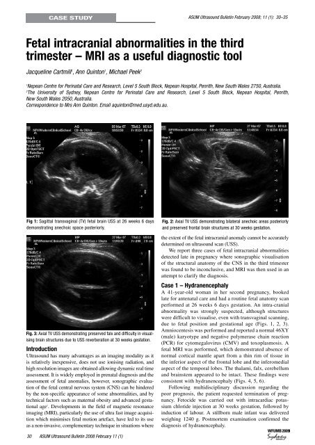
Fig 1: Sagittal transvaginal (TV) fetal brain USS at 26 weeks 6 days<br />
demonstrating anechoic space posteriorly.<br />
Fig. 3: Axial TV USS demonstrating preserved falx and difficulty in visualising<br />
brain structures due to USS reverberation at 30 weeks gestation.<br />
Introduction<br />
<strong>Ultrasound</strong> has many advantages as an imaging modality as it<br />
is relatively inexpensive, does not use ionising radiation, and<br />
high resolution images are obtained allowing dynamic real time<br />
assessment. It is widely employed in prenatal diagnosis and the<br />
assessment of fetal anomalies, however, sonographic evaluation<br />
of the fetal central nervous system (CNS) can be hindered<br />
by the non-specific appearance of some abnormalities, and by<br />
technical factors such as maternal obesity and advanced gestational<br />
age 1 . Developments in the field of magnetic resonance<br />
imaging (MRI), particularly the use of ultra fast image acquisition<br />
which minimises fetal motion artefact, have led to its use<br />
as a non-invasive, complementary technique in situations where<br />
Fig. 2: Axial TV USS demonstrating bilateral anechoic areas posteriorly<br />
and preserved frontal brain structures at 30 weeks gestation.<br />
the extent of the fetal intracranial anomaly cannot be accurately<br />
determined on ultrasound scan (USS).<br />
We report three cases of fetal intracranial abnormalities<br />
detected late in pregnancy where sonographic visualisation<br />
of the structural anatomy of the CNS in the third trimester<br />
was found to be inconclusive, and MRI was then used in an<br />
attempt to clarify the diagnosis.<br />
Case 1 – Hydranencephaly<br />
A 41-year-old woman in her second pregnancy, booked<br />
late <strong>for</strong> antenatal care and had a routine fetal anatomy scan<br />
per<strong>for</strong>med at 26 weeks 6 days gestation. An intra-cranial<br />
abnormality was strongly suspected, although structures<br />
were difficult to visualise, even with transvaginal scanning,<br />
due to fetal position and gestational age (Figs. 1, 2, 3).<br />
Amniocentesis was per<strong>for</strong>med and reported a normal 46XY<br />
(male) karyotype and negative polymerase chain reaction<br />
(PCR) <strong>for</strong> cytomegalovirus (CMV) and toxoplasmosis. A<br />
fetal MRI was per<strong>for</strong>med, which demonstrated absence of<br />
normal cortical mantle apart from a thin rim of tissue in<br />
the inferior aspect of the frontal lobe and the inferomedial<br />
aspect of the temporal lobes. The thalami, falx, cerebellum<br />
and brainstem appeared to be intact. These findings were<br />
consistent with hydranencephaly (Figs. 4, 5, 6).<br />
Following multidisciplinary discussion regarding the<br />
poor prognosis, the patient requested termination of pregnancy.<br />
Fetocide was carried out with intracardiac potassium<br />
chloride injection at 30 weeks gestation, followed by<br />
induction of labour. A stillborn male infant was delivered<br />
weighing 1240 g. Postmortem examination confirmed the<br />
diagnosis of hydranencephaly.<br />
0 ASUM <strong>Ultrasound</strong> Bulletin 2008 <strong>February</strong> <strong>11</strong> (1)


