

Volume 11 Issue 1 (February) - Australasian Society for Ultrasound ...
Volume 11 Issue 1 (February) - Australasian Society for Ultrasound ...
Volume 11 Issue 1 (February) - Australasian Society for Ultrasound ...

Create successful ePaper yourself
Turn your PDF publications into a flip-book with our unique Google optimized e-Paper software.
Fetal intracranial abnormalities in the third trimester – MRI as a useful diagnostic tool<br />
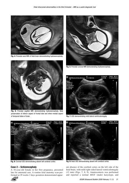
Fig. 4: Prenatal axial MRI of fetal brain demonstrating hydranencephaly.<br />
Fig. 5: Prenatal coronal MRI demonstrating hydranencephaly.<br />
Fig. 6: Prenatal sagittal MRI demonstrating hydranencephaly. Note<br />
preservation of inferior aspect of frontal lobe and infero-medial aspect<br />
of temporal lobes of brain.<br />
Fig. 7: USS demonstrating mild lateral ventriculomegaly.<br />
Fig. 8: Coronal USS demonstrating absent left cerebral cortex.<br />
Fig. 9: Axial USS demonstrating absent left cerebral cortex.<br />
Case 2 – Schizencephaly<br />
A 16-year-old female in her first pregnancy, presented<br />
late <strong>for</strong> antenatal care. A routine fetal anatomy scan per<strong>for</strong>med<br />
at 29 weeks 2 days gestation demonstrated apparent<br />
absence of the cerebral cortex on the left side of the<br />
fetal brain, with mild right-sided lateral ventriculomegaly<br />
(12 mm) (Figs. 7, 8, 9). Amniocentesis was per<strong>for</strong>med<br />
and reported a normal 46XY (male) karyotype, and<br />
ASUM <strong>Ultrasound</strong> Bulletin 2008 <strong>February</strong> <strong>11</strong> (1)


