Volume 11 Issue 1 (February) - Australasian Society for Ultrasound ...
Volume 11 Issue 1 (February) - Australasian Society for Ultrasound ...
Volume 11 Issue 1 (February) - Australasian Society for Ultrasound ...
Create successful ePaper yourself
Turn your PDF publications into a flip-book with our unique Google optimized e-Paper software.
Advertisement<br />
“We now see a 50 percent overall<br />
reduction in the total exam time.”<br />
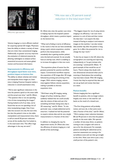
MPR of IVC right renal vein<br />
<strong>Volume</strong> imaging is a more efficient method<br />
<strong>for</strong> acquiring optimal 2D images. Physicians<br />
have the ability to obtain a variety of views<br />
that are richer than conventional imaging.<br />
Additionally, its greater accuracy improves<br />
the evaluation of relational anatomy by<br />
allowing radiologists to analyze surface<br />
anatomical structures and assess global<br />
motion and functional in<strong>for</strong>mation.<br />
Improvements in efficiency and<br />
throughput equal faster scan time,<br />
positive impact on bottom line<br />
The ability to obtain volumes and review<br />
and manipulate those images at a later<br />
time is helping Freeman Hospital realize a<br />
reduction in overall examination time.<br />
“We’ve seen significant reductions in the<br />
time the patients spend on the exam table<br />
and the actual scan time,” said Dr. Elliott.<br />
“The question then is ‘are we spending<br />
a lot of time manipulating the images?’<br />
Having looked at all of our data, we’ve<br />
found that we are not spending a lot of<br />
time manipulating images. Even if you<br />
take into account not only the scan time,<br />
but also the patient’s time on the table,<br />
manipulation and measurement time, there<br />
is still an overall 50 percent reduction<br />
across all the studies that I’ve looked at,<br />
in the total exam time. This offers huge<br />
advantages in the way we work.”<br />
Dr. Elliott also cites the quicker scan times<br />
in helping improve the hospital’s patient<br />
throughput, which means a positive impact<br />
on the bottom line.<br />
“What we’re finding in terms of efficiency<br />
in the rooms is that we can have somebody<br />
doing a quick volume acquisition, another<br />
per<strong>for</strong>m the interpretation, while<br />
somebody else is getting another patient<br />
onto the bed and scanned. So we actually<br />
have an overlap now, which is enabling us<br />
to increase throughput in that one room.<br />
The acquisition phase of exams has the<br />
greatest potential <strong>for</strong> positive workflow<br />
impact. Conventional workflow requires<br />
the acquisition of 2D image after 2D image,<br />
followed by printing and archiving of the<br />
images. With volume imaging, volume<br />
interrogation follows volume acquisition,<br />
making it possible to shorten<br />
acquisition time.<br />
“We’d been using 3D imaging, seeing<br />
images of surface rendering, when I<br />
suddenly realized that we could actually<br />
take the volume of data and use it <strong>for</strong><br />
something we’d been doing every day in<br />
2D measuring,” said Dr. Elliott. “In one<br />
particular case, a simple abdominal aortic<br />
aneurism, I realized we had the potential<br />
to acquire volume data and do very simple<br />
measurements in a fraction of the time.”<br />
In addition to changing the way his<br />
department works, Dr. Elliott also cites<br />
the efficiency of volume imaging as a real<br />
benefit of the technology.<br />
“The biggest impact <strong>for</strong> me of using volume<br />
imaging is its efficiency. I can scan more<br />
patients in a unit of time and then review<br />
the data later. I can acquire the data<br />
instead of having to analyze it and measure<br />
images on the machine. I can also review<br />
data another day after the patient is long<br />
gone. So it offers the potential <strong>for</strong> me to<br />
change the way I work.”<br />
“It also has an impact in the UK where the<br />
sonographers are scanning and reporting<br />
independently or I’ve got trainees who<br />
are scanning,” continued Dr. Elliott. “A<br />
good example of that would be out of<br />
hours. There’s always that doubt with<br />
two-dimensional ultrasound when you’re<br />
scanning in fixed planes that something<br />
may have been missed. With 3D imaging,<br />
I know that they’ve acquired that data and<br />
we can go back and review that later<br />
as a team.<br />
Patient benefits<br />
<strong>Volume</strong> imaging also has a significant<br />
benefit to patients, who no longer endure<br />
lengthy scan times or the need <strong>for</strong> call<br />
backs as the result of a missed scan.<br />
“The first thing patients will probably<br />
notice, and ours do, is that some scans<br />
take a fraction of the time,” said Dr. Elliott.<br />
“We’re seeing a mean reduction between<br />
80 and 90 percent in time that the patient<br />
spends on the exam table. We operate<br />
a number of one-stop clinics, so patients<br />
coming to see a surgeon will come to<br />
see us <strong>for</strong> the ultrasound. For example,<br />
patients attending <strong>for</strong> abdominal aortic<br />
aneurism assessment are coming in <strong>for</strong><br />
surveillance scans and can then go straight<br />
to the clinic. Now, scan time is just a very<br />
small portion of the patient’s visit.<br />
ASUM <strong>Ultrasound</strong> Bulletin 2008 <strong>February</strong> <strong>11</strong> (1)


