

Volume 11 Issue 1 (February) - Australasian Society for Ultrasound ...
Volume 11 Issue 1 (February) - Australasian Society for Ultrasound ...
Volume 11 Issue 1 (February) - Australasian Society for Ultrasound ...

You also want an ePaper? Increase the reach of your titles
YUMPU automatically turns print PDFs into web optimized ePapers that Google loves.
Advertisement<br />
“With Protocols, sonographers<br />
now know exactly which images<br />
are necessary as the system guides<br />
them through a list of images <strong>for</strong><br />
a specific exam and automatically<br />
annotates the data.”<br />
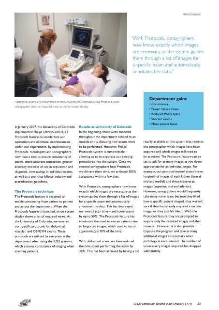
Abdominal exams are streamlined at the University of Colorado. Using Protocols, each<br />
sonographer sees the required views in the on-screen display.<br />
In January 2007, the University of Colorado Results at University of Colorado<br />
implemented Philips <strong>Ultrasound</strong>’s iU22 In the beginning, there were concerns<br />
Protocols feature to standardize our throughout the department related to an<br />
operations and eliminate inconsistencies outside entity dictating how exams were<br />
within our department. By implementing to be per<strong>for</strong>med. However, Philips’<br />
Protocols, radiologists and sonographers Protocols system is customizable –<br />
now have a tool to ensure consistency of allowing us to incorporate our existing<br />
exams, more accurate annotation, greater procedures into the system. Once we<br />
accuracy and ease of use in acquisition and showed sonographers how Protocols<br />
diagnosis, time savings in individual exams, would save them time, we achieved 100%<br />
as well as a tool that follows industry and acceptance within a few days.<br />
accreditation guidelines.<br />
With Protocols, sonographers now know<br />
The Protocols technique<br />
exactly which images are necessary as the<br />
The Protocols feature is designed to system guides them through a list of images<br />
enable consistency from patient to patient <strong>for</strong> a specific exam and automatically<br />
and across the department. When the annotates the data. This has decreased<br />
Protocols feature is launched, an on-screen our overall scan time – and some exams<br />
display shows a list of required views. At by up to 50%. The Protocols feature has<br />
the University of Colorado, we entered eliminated the need to rescan patients due<br />
our specific protocols <strong>for</strong> abdominal, to <strong>for</strong>gotten images, which used to occur<br />
vascular, and OB/GYN exams. These approximately 10% of the time.<br />
protocols are utilized by everyone in the<br />
department when using the iU22 systems, With abdominal scans, we have reduced<br />
which ensures consistency of imaging when the time spent per<strong>for</strong>ming the exam by<br />
scanning patients.<br />
38%. This has been achieved by having a list<br />
Department gains<br />
• Consistency<br />
• Fewer missed views<br />
• Reduced PACS space<br />
• Shorter exams<br />
• More patient focus<br />
readily available on the system that reminds<br />
the sonographer which images have been<br />
acquired and which images still need to<br />
be acquired. The Protocols feature can be<br />
set to ask <strong>for</strong> as many images as you deem<br />
appropriate <strong>for</strong> an individual organ. For<br />
example, our protocol manual stated three<br />
longitudinal images of each kidney (lateral,<br />
mid and medial) and three transverse<br />
images (superior, mid and inferior).<br />
However, sonographers would frequently<br />
take many more scans because they liked<br />
how a specific patient imaged, they weren’t<br />
sure if they had already acquired a certain<br />
image, or they just felt like it. With the<br />
Protocols feature they are prompted to<br />
acquire only the required images and then<br />
move on. However, it is also possible<br />
to pause the program and take as many<br />
additional images as necessary when<br />
pathology is encountered. The number of<br />
unnecessary images acquired has dropped<br />
substantially.<br />
ASUM <strong>Ultrasound</strong> Bulletin 2008 <strong>February</strong> <strong>11</strong> (1)<br />
53


