

Volume 11 Issue 1 (February) - Australasian Society for Ultrasound ...
Volume 11 Issue 1 (February) - Australasian Society for Ultrasound ...
Volume 11 Issue 1 (February) - Australasian Society for Ultrasound ...

You also want an ePaper? Increase the reach of your titles
YUMPU automatically turns print PDFs into web optimized ePapers that Google loves.
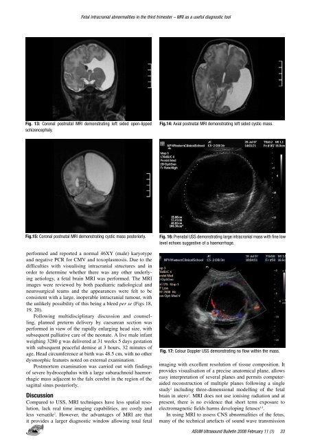
Fetal intracranial abnormalities in the third trimester – MRI as a useful diagnostic tool<br />
Fig. 13: Coronal postnatal MRI demonstrating left sided open-lipped<br />
schizencephaly.<br />
Fig.14: Axial postnatal MRI demonstrating left sided cystic mass.<br />
Fig.15: Coronal postnatal MRI demonstrating cystic mass posteriorly.<br />
per<strong>for</strong>med and reported a normal 46XY (male) karyotype<br />
and negative PCR <strong>for</strong> CMV and toxoplasmosis. Due to the<br />
difficulties with visualising intracranial structures and in<br />
order to determine whether there was any other underlying<br />
aetiology, a fetal brain MRI was per<strong>for</strong>med. The MRI<br />
images were reviewed by both paediatric radiological and<br />
neurosurgical teams and the appearances were felt to be<br />
consistent with a large, inoperable intracranial tumour, with<br />
the unlikely possibility of this being a bleed per se (Figs 18,<br />
19, 20).<br />
Following multidisciplinary discussion and counselling,<br />
planned preterm delivery by caesarean section was<br />
per<strong>for</strong>med in view of the rapidly enlarging head size, with<br />
subsequent palliative care of the neonate. A live male infant<br />
weighing 3280 g was delivered at 31 weeks 5 days gestation<br />
with subsequent peaceful demise at 3 hours, 32 minutes of<br />
age. Head circumference at birth was 48.5 cm, with no other<br />
dysmorphic features noted on external examination.<br />
Postmortem examination was carried out with findings<br />
of severe hydrocephalus with a large subarachnoid haemorrhagic<br />
mass adjacent to the falx cerebri in the region of the<br />
sagittal sinus posteriorly.<br />
Discussion<br />
Compared to USS, MRI techniques have less spatial resolution,<br />
lack real time imaging capabilities, are costly and<br />
less versatile 2 . However, the advantages of MRI are that<br />
it provides a larger diagnostic window allowing total fetal<br />
Fig. 16: Prenatal USS demonstrating large intracranial mass with fine low<br />
level echoes suggestive of a haemorrhage.<br />
Fig. 17: Colour Doppler USS demonstrating no flow within the mass.<br />
imaging with excellent resolution of tissue composition. It<br />
provides visualisation of a precise anatomical plane, allows<br />
easy interpretation of several planes and permits computeraided<br />
reconstruction of multiple planes following a single<br />
study 2 including three-dimensional modelling of the fetal<br />
brain in utero 3 . MRI does not use ionising radiation and at<br />
present, there is no evidence that short term exposure to<br />
electromagnetic fields harms developing fetuses 4,5 .<br />
In using MRI to assess CNS abnormalities of the fetus,<br />
many of the technical artefacts of sound wave transmission<br />
ASUM <strong>Ultrasound</strong> Bulletin 2008 <strong>February</strong> <strong>11</strong> (1)


