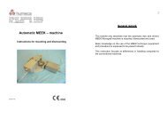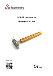
General company brochure with info of all products - Humeca
General company brochure with info of all products - Humeca
General company brochure with info of all products - Humeca

You also want an ePaper? Increase the reach of your titles
YUMPU automatically turns print PDFs into web optimized ePapers that Google loves.
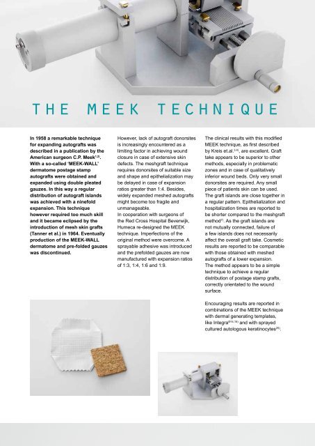
the meek technique<br />
In 1958 a remarkable technique<br />
for expanding autografts was<br />
described in a publication by the<br />
American surgeon C.P. Meek 1,2) .<br />
With a so-c<strong>all</strong>ed ‘MEEK-WALL’<br />
dermatome postage stamp<br />
autografts were obtained and<br />
expanded using double pleated<br />
gauzes. In this way a regular<br />
distribution <strong>of</strong> autograft islands<br />
was achieved <strong>with</strong> a ninefold<br />
expansion. This technique<br />
however required too much skill<br />
and it became eclipsed by the<br />
introduction <strong>of</strong> mesh skin grafts<br />
(Tanner et al.) in 1964. Eventu<strong>all</strong>y<br />
production <strong>of</strong> the MEEK-WALL<br />
dermatome and pre-folded gauzes<br />
was discontinued.<br />
However, lack <strong>of</strong> autograft donorsites<br />
is increasingly encountered as a<br />
limiting factor in achieving wound<br />
closure in case <strong>of</strong> extensive skin<br />
defects. The meshgraft technique<br />
requires donorsites <strong>of</strong> suitable size<br />
and shape and epithelialization may<br />
be delayed in case <strong>of</strong> expansion<br />
ratios greater than 1:4. Besides,<br />
widely expanded meshed autografts<br />
might become too fragile and<br />
unmanageable.<br />
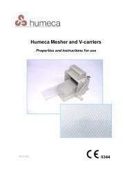
In cooperation <strong>with</strong> surgeons <strong>of</strong><br />
the Red Cross Hospital Beverwijk,<br />
<strong>Humeca</strong> re-designed the MEEK<br />
technique. Imperfections <strong>of</strong> the<br />
original method were overcome. A<br />
sprayable adhesive was introduced<br />
and the prefolded gauzes are now<br />
manufactured <strong>with</strong> expansion ratios<br />
<strong>of</strong> 1:3, 1:4, 1:6 and 1:9.<br />
The clinical results <strong>with</strong> this modified<br />
MEEK technique, as first described<br />
by Kreis et.al. 3,4) , are excellent. Graft<br />
take appears to be superior to other<br />
methods, especi<strong>all</strong>y in problematic<br />
zones and in case <strong>of</strong> qualitatively<br />
inferior wound beds. Only very sm<strong>all</strong><br />
donorsites are required. Any sm<strong>all</strong><br />
piece <strong>of</strong> patients skin can be used.<br />
The graft islands are close together in<br />
a regular pattern. Epithelialization and<br />
hospitalization times are reported to<br />
be shorter compared to the meshgraft<br />
method 7) . As the graft islands are<br />
not mutu<strong>all</strong>y connected, failure <strong>of</strong><br />
a few islands does not necessarily<br />
affect the over<strong>all</strong> graft take. Cosmetic<br />
results are reported to be comparable<br />
<strong>with</strong> those obtained <strong>with</strong> meshed<br />
autografts <strong>of</strong> a lower expansion.<br />
The method appears to be a simple<br />
technique to achieve a regular<br />
distribution <strong>of</strong> postage stamp grafts,<br />
correctly orientated to the wound<br />
surface.<br />
Encouraging results are reported in<br />
combinations <strong>of</strong> the MEEK technique<br />
<strong>with</strong> dermal generating templates,<br />
like Integra ®14,15) and <strong>with</strong> sprayed<br />
cultured autologous keratinocytes 25) .<br />
4