General company brochure with info of all products - Humeca
General company brochure with info of all products - Humeca
General company brochure with info of all products - Humeca

You also want an ePaper? Increase the reach of your titles
YUMPU automatically turns print PDFs into web optimized ePapers that Google loves.
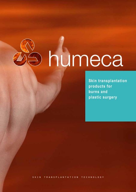
Skin transplantation<br />
<strong>products</strong> for<br />
burns and<br />
plastic surgery<br />
s k i n t r a n s p l a n t a t i o n t e c h n o l o g y<br />
1
mission<br />
Our mission is to provide medical experts <strong>with</strong> materials and equipment for optimum treatment <strong>of</strong> their patients<br />
and to <strong>of</strong>fer them the highest level <strong>of</strong> support and service. Our goal is continuous improvement <strong>of</strong> our <strong>products</strong>,<br />
the source <strong>of</strong> which we believe is intensive communication <strong>with</strong> experts in the field.<br />
2
MEEK<br />
4<br />
<strong>company</strong> pr<strong>of</strong>ile<br />
The <strong>Humeca</strong> consulting group was<br />
founded in 1981 in Enschede, The<br />
Netherlands.<br />
Initi<strong>all</strong>y the group focussed on product<br />
development, trouble-shooting and<br />
research in the field <strong>of</strong> biomedical<br />
engineering and polymer technology.<br />
By contract the customers became<br />
the owners <strong>of</strong> the newly developed<br />
<strong>products</strong> or innovations and <strong>Humeca</strong><br />
was hired for its knowledge and<br />
creativeness.<br />
In 1984 <strong>Humeca</strong> started to<br />
cooperate <strong>with</strong> surgeons <strong>of</strong> the Red<br />
Cross Hospital in Beverwijk, The<br />
Netherlands. The goal was to redevelop<br />
and modernize the MEEK-<br />
WALL micrograft technique for skin<br />
transplantation in burns and plastic<br />
surgery. The American surgeon<br />
C.P. Meek laid the foundation <strong>of</strong><br />
this method in 1958 already, but the<br />
technique required too much skill and<br />
it was discontinued after a few years.<br />
In 1993 the modified MEEK technique<br />
was re-introduced at the European<br />
Burns Association (EBA) congress in<br />
Brighton, UK.<br />
Contrary to former projects<br />
<strong>Humeca</strong> developed the modified<br />
MEEK technique on its own risk.<br />
Consequently <strong>Humeca</strong> is the<br />
owner <strong>of</strong> the knowledge and the<br />
characteristics and production<br />
technology <strong>of</strong> the components <strong>of</strong><br />
this unique and ingenious skin<br />
transplantation method. Thousands<br />
<strong>of</strong> patients have been treated <strong>all</strong> over<br />
the world <strong>with</strong> excellent results and<br />
opinion leaders in the field <strong>of</strong> burns<br />
and plastic surgery published about<br />
it. The MEEK technique has gained<br />
its place as one <strong>of</strong> the standard skin<br />
grafting methods.<br />
Later on <strong>Humeca</strong> introduced more<br />
<strong>products</strong> for skin grafting. At first the<br />
cordless, battery driven dermatomes<br />
D42 and D80 were developed,<br />
followed by a mesher and the<br />
carriers that belong to it. <strong>Humeca</strong><br />
also supplies a range <strong>of</strong> blades<br />
for different types and brands <strong>of</strong><br />
dermatomes. More recently <strong>Humeca</strong><br />
is involved in attempts to <strong>of</strong>fer<br />
surgeons in low-income countries<br />
durable and low-cost techniques<br />
for burn treatment, like the SOBER<br />
dermatome and the “Dr. Charles”<br />
mesher. .<br />
Since the early nineties <strong>Humeca</strong> is a<br />
sm<strong>all</strong>, but constant and valued player<br />
in the field <strong>of</strong> burn care and plastic<br />
surgery. The <strong>products</strong> are sold via<br />
a network <strong>of</strong> distributors <strong>all</strong> over the<br />
world.<br />
All <strong>Humeca</strong> <strong>products</strong> meet the Medical Device<br />
Directive (MDD) 93/42/EEC, as ammended<br />
by 2007/47/CE and therefore have the CE<br />
h<strong>all</strong>mark.<br />
<strong>Humeca</strong> is an ISO 13485:2003 certified<br />
<strong>company</strong>.<br />
Dermatomes<br />
10<br />
V-Carriers<br />
14<br />
Meshgraft<br />
20<br />
Order<br />
22<br />
3
the meek technique<br />
In 1958 a remarkable technique<br />
for expanding autografts was<br />
described in a publication by the<br />
American surgeon C.P. Meek 1,2) .<br />
With a so-c<strong>all</strong>ed ‘MEEK-WALL’<br />
dermatome postage stamp<br />
autografts were obtained and<br />
expanded using double pleated<br />
gauzes. In this way a regular<br />
distribution <strong>of</strong> autograft islands<br />
was achieved <strong>with</strong> a ninefold<br />
expansion. This technique<br />
however required too much skill<br />
and it became eclipsed by the<br />
introduction <strong>of</strong> mesh skin grafts<br />
(Tanner et al.) in 1964. Eventu<strong>all</strong>y<br />
production <strong>of</strong> the MEEK-WALL<br />
dermatome and pre-folded gauzes<br />
was discontinued.<br />
However, lack <strong>of</strong> autograft donorsites<br />
is increasingly encountered as a<br />
limiting factor in achieving wound<br />
closure in case <strong>of</strong> extensive skin<br />
defects. The meshgraft technique<br />
requires donorsites <strong>of</strong> suitable size<br />
and shape and epithelialization may<br />
be delayed in case <strong>of</strong> expansion<br />
ratios greater than 1:4. Besides,<br />
widely expanded meshed autografts<br />
might become too fragile and<br />
unmanageable.<br />
In cooperation <strong>with</strong> surgeons <strong>of</strong><br />
the Red Cross Hospital Beverwijk,<br />
<strong>Humeca</strong> re-designed the MEEK<br />
technique. Imperfections <strong>of</strong> the<br />
original method were overcome. A<br />
sprayable adhesive was introduced<br />
and the prefolded gauzes are now<br />
manufactured <strong>with</strong> expansion ratios<br />
<strong>of</strong> 1:3, 1:4, 1:6 and 1:9.<br />
The clinical results <strong>with</strong> this modified<br />
MEEK technique, as first described<br />
by Kreis et.al. 3,4) , are excellent. Graft<br />
take appears to be superior to other<br />
methods, especi<strong>all</strong>y in problematic<br />
zones and in case <strong>of</strong> qualitatively<br />
inferior wound beds. Only very sm<strong>all</strong><br />
donorsites are required. Any sm<strong>all</strong><br />
piece <strong>of</strong> patients skin can be used.<br />
The graft islands are close together in<br />
a regular pattern. Epithelialization and<br />
hospitalization times are reported to<br />
be shorter compared to the meshgraft<br />
method 7) . As the graft islands are<br />
not mutu<strong>all</strong>y connected, failure <strong>of</strong><br />
a few islands does not necessarily<br />
affect the over<strong>all</strong> graft take. Cosmetic<br />
results are reported to be comparable<br />
<strong>with</strong> those obtained <strong>with</strong> meshed<br />
autografts <strong>of</strong> a lower expansion.<br />
The method appears to be a simple<br />
technique to achieve a regular<br />
distribution <strong>of</strong> postage stamp grafts,<br />
correctly orientated to the wound<br />
surface.<br />
Encouraging results are reported in<br />
combinations <strong>of</strong> the MEEK technique<br />
<strong>with</strong> dermal generating templates,<br />
like Integra ®14,15) and <strong>with</strong> sprayed<br />
cultured autologous keratinocytes 25) .<br />
4
A 42x42 mm (1.65x1.65")<br />
cork plate...<br />
is covered <strong>with</strong> a piece <strong>of</strong><br />
split skin autograft<br />
Also sm<strong>all</strong> graft pieces<br />
can be used<br />
The graft is cut into 196<br />
3x3 mm squares<br />
meek surgery<br />
After the epidermal side <strong>of</strong> graft is sprayed <strong>with</strong> an<br />
adhesive, the graft is transferred to a prefolded gauze<br />
At operation wounds to be grafted<br />
are excised down to the underlying<br />
fascia and haemostasis is secured.<br />
A square piece <strong>of</strong> cork measuring<br />
42x42 mm (1.65x1.65") <strong>with</strong><br />
thickness 2.5 mm (0.1") is covered<br />
<strong>with</strong> a split skin autograft, dermal<br />
size down. Sm<strong>all</strong>er graft remnants<br />
are also suitable by placing them<br />
on the cork plate like a ‘puzzle’.<br />
The cork, covered <strong>with</strong> graft, is<br />
then placed in a special MEEK<br />
cutting machine.<br />
The machine contains 13 par<strong>all</strong>el<br />
circular blades. The cork plate <strong>with</strong><br />
the graft on it is passed through the<br />
machine, where the rotating blades<br />
cut through the graft, but not through<br />
the cork. Thus the graft is cut into 14<br />
stripes, 3 mm (0.12") wide. After the<br />
first pass, the cork is rotated 90° and<br />
passed through the machine once<br />
more, thus cutting the graft into<br />
14x14 = 196 square pieces<br />
measuring 3x3 mm (0.12x0.12").<br />
The upper (epidermal) surface <strong>of</strong> the<br />
graft is then sprayed <strong>with</strong> an adhesive<br />
dressing spray and <strong>all</strong>owed to dry<br />
and become tacky. The cork plate,<br />
covered <strong>with</strong> graft and adhesive<br />
is then pressed onto a prefolded<br />
polyamide gauze, which is folded on<br />
an aluminium foil backing into<br />
14x14 square pleats, the size <strong>of</strong><br />
which corresponds to the size <strong>of</strong><br />
the cuts in the graft. Then the cork<br />
is gently removed, leaving the graft<br />
islands adhering to the gauze. The<br />
gauze is pulled out by firm traction<br />
on <strong>all</strong> four sides, until the pleats<br />
become completely unfolded. Fin<strong>all</strong>y<br />
the aluminium backing is peeled <strong>of</strong>f,<br />
to leave the expanded gauze <strong>with</strong> the<br />
separated adherent autograft islands<br />
ready for transplantation.<br />
After trimming the margins, or folding<br />
them double down, the gauze is<br />
applied, graft side down, to the<br />
wound bed and secured <strong>with</strong> surgical<br />
staples. After about 6 days the grafts<br />
have grown sufficiently into the<br />
wound bed to <strong>all</strong>ow removal <strong>of</strong> the<br />
gauze, leaving the autograft islands in<br />
situ on the wound. The grafts are then<br />
covered <strong>with</strong> a non-adherent sheeting<br />
to prevent any movement during daily<br />
dressing changes. After a further<br />
5-6 days the sheeting is removed.<br />
Daily dressings are continued until<br />
epithelialization is complete.<br />
Only in cases <strong>of</strong> a large expansion<br />
factor (1:9), it is recommended<br />
to cover the MEEK grafts <strong>with</strong> an<br />
overlay <strong>of</strong> <strong>all</strong>ografts, meshed 1:1.5<br />
(a procedure c<strong>all</strong>ed ‘sandwich<br />
grafting’). At lower expansions ratio,<br />
the use <strong>of</strong> <strong>all</strong>ografts is not necessary.<br />
Alternatively cultured autologous<br />
keratinocytes can be applied to<br />
accelerate wound closure at large<br />
expansions.<br />
Expansion is realized by unfolding the gauze,<br />
first in one direction...<br />
... and then in the other direction<br />
5
donorsite area<br />
Burn on a shoulder, gauzes 1:6 applied<br />
In 1988, R. Peeters and A. Hubens<br />
<strong>of</strong> the Stuivenberg Hospital,<br />
Antwerp, Belgium, proved that the<br />
actual expansion ratio <strong>of</strong> a meshed<br />
graft is much less than the given<br />
one 5) . They compared the expanded<br />
graft surface area to the original<br />
non-expanded graft surface area.<br />
Based on their findings we calculated<br />
the graft surface area required to<br />
cover a 100 cm 2 (0.11 ft 2 ) burn wound<br />
<strong>with</strong> a meshed graft and compared it<br />
to the area required when using the<br />
MEEK technique.<br />
The result is shown in the graph.<br />
The vertical axis represents the graft<br />
surface, while the horizontal axis<br />
shows the expansion ratio.<br />
Conclusion<br />
The MEEK technique (<strong>with</strong> true<br />
expansion ratios from 1:3 to 1:9)<br />
requires only about half <strong>of</strong> the graft<br />
surface compared to the meshgraft<br />
method.<br />
Consequently, in comparison <strong>with</strong> the<br />
MEEK technique, the application <strong>of</strong><br />
widely meshed grafts requires more<br />
grafting procedures in case <strong>of</strong> severe<br />
burns. A high graft take, less spillage<br />
and a better principle <strong>of</strong> expansion<br />
contribute to the over<strong>all</strong> opinion that<br />
the MEEK technique is the most<br />
effective way to deal <strong>with</strong> the patients’<br />
skin. In an over<strong>all</strong> view, the MEEK<br />
method turns out to be less time<br />
consuming than meshing, especi<strong>all</strong>y<br />
in case <strong>of</strong> large burns 21) .<br />
Same burn, 6 weeks post burn<br />
White spots indicate anti-bacterial cream<br />
G r a f t s s u r f a c e c m 2<br />
100<br />
90<br />
80<br />
70<br />
60<br />
50<br />
40<br />
30<br />
20<br />
10<br />
0<br />
Graft surface required to cover a 100 cm 2 (0.11 ft 2 ) burn<br />
Meshgraft<br />
MEEK<br />
1 1,5 3 4 6 9<br />
Same burn, 6 months post burn<br />
6
Axilla and upper arm 21 days post burn,<br />
before operation<br />
meek case<br />
Gauze applied to the wound (upper arm)<br />
Gauze secured <strong>with</strong> staples<br />
By dr. A.W.F.P. Vloemans, surgeon<br />
at Red Cross Hospital Beverwijk,<br />
The Netherlands<br />
An 84 years old woman was admitted<br />
in our burns centre because <strong>of</strong> a burn<br />
wound she sustained in the kitchen.<br />
While taking a kettle from the gas<br />
stove her nightgown caught fire. At<br />
first she did not notice, but after a<br />
while she saw her clothes burning.<br />
She put out the flames and cooled<br />
the burns under the shower.<br />
On admission we saw a healthy<br />
obese woman <strong>with</strong> burns at the<br />
right upper arm, axilla and thorax.<br />
The total body surface area burned<br />
amounted 11%; about 9 % full<br />
thickness. The borders between full<br />
thickness and partial thickness were<br />
not sharp.<br />
The medical history recorded<br />
insulin dependant diabetes and a<br />
stroke. As medication she also used<br />
acetylsalicylic acid.<br />
Topical treatment consisted <strong>of</strong> daily<br />
application <strong>of</strong> Silver Sulfadiazin 1%<br />
cream dressings. Resuscitation was<br />
applied by means <strong>of</strong> an isotonic<br />
saline solution and fluids ad libitum.<br />
Insulin medication was continued<br />
based on blood sugar levels and also<br />
treatment <strong>with</strong> acetylsalicylic acid<br />
was continued.<br />
As an early tangential excision was<br />
expected to cause a great blood loss<br />
and as the distinction between partial<br />
and full thickness was not clear, a<br />
secondary excision and grafting after<br />
healing <strong>of</strong> the partial thickness burns<br />
was performed.<br />
Operation took place on post<br />
burn day 21; five days earlier<br />
treatment <strong>with</strong> acetylsalicylic acid<br />
was discontinued. The wound bed<br />
consisted <strong>of</strong> granulation tissue and<br />
remainders <strong>of</strong> necrotic dermis. Debris<br />
was excised using a dermatome.<br />
Haemostasis was performed by<br />
means <strong>of</strong> the application <strong>of</strong> an<br />
adrenalin solution to the excised<br />
wound bed.<br />
A thin split thickness skin graft was<br />
harvested from the right upper leg.<br />
Ten Meek gauzes 1:6 were prepared<br />
and applied to the wound. After<br />
fixation <strong>with</strong> staples the wound was<br />
covered <strong>with</strong> gauzes soaked in a<br />
silvernitrate solution. The donorsite<br />
was dressed <strong>with</strong> polyurethane foam<br />
dressing.<br />
After 6 days the MEEK gauzes were<br />
removed from the wound. The graft<br />
take appeared to be about 80%.<br />
Two weeks after the first operation,<br />
parts <strong>of</strong> the wound where the graft<br />
had not taken because <strong>of</strong> insufficient<br />
excision at the first operation, were<br />
excised again and grafted <strong>with</strong> Meek<br />
gauzes 1:4. Parts <strong>of</strong> the wound <strong>with</strong><br />
a good graft take, but insufficient<br />
outgrowth <strong>of</strong> epithelium were also<br />
re-grafted.<br />
After the second operation only minor<br />
defects remained, requiring only daily<br />
application <strong>of</strong> a topical antibacterial<br />
dressing. Five weeks post burn the<br />
patient was released from hospital.<br />
Gauzes and parts <strong>of</strong> gauzes on the wound<br />
Initial result after the first transplantation<br />
Re-grafting: a gauze is cut into pieces<br />
to graft sm<strong>all</strong>er areas on the thorax<br />
7
Standard hand driven MEEK machine<br />
<strong>with</strong> single cutting block<br />
Motor driven MEEK machine<br />
<strong>with</strong> block shape motor<br />
Motor driven MEEK machine<br />
<strong>with</strong> cylindrical shape motor<br />
meek <strong>products</strong><br />
Cutting machine<br />
The cutting machine contains 13<br />
circular blades mounted on a cutting<br />
axis. The cork plate <strong>with</strong> the graft is<br />
placed in a cutting block that moves<br />
under a bridge during cutting. Both<br />
the drive <strong>of</strong> the blades and the drive<br />
<strong>of</strong> the cutting block can be performed<br />
by pneumatic motor or by hand.<br />
The motor drive <strong>of</strong> the blades is<br />
exchangeable <strong>with</strong> the hand drive<br />
and both can be supplied as separate<br />
sets. The drive <strong>of</strong> the cutting block is<br />
not exchangeable; it is either motor<br />
driven or hand driven. The hand drive<br />
<strong>of</strong> the cutting block can be provided<br />
<strong>with</strong> gearwheels for faster run <strong>of</strong> the<br />
block. For pneumatic drive a pressure<br />
<strong>of</strong> 5-7 bar (72-100 psi) is required.<br />
The blades are coated <strong>with</strong> a durable,<br />
wear resistant ceramic layer. The<br />
cutting axis can simply be replaced.<br />
Individual blades can also be<br />
replaced.<br />
Gauzes<br />
The pre-folded MEEK gauze consists<br />
<strong>of</strong> an aluminium backing and a<br />
polyamide fabric, slightly adhering<br />
to each other. After expansion<br />
the aluminium foil is disposed <strong>of</strong>,<br />
while the fabric <strong>with</strong> the grafts is<br />
applied to the wound. Each gauze<br />
is supplied <strong>with</strong> a 42x42x2.5 mm<br />
(1.65x1.65x0.1") cork plate and sterile<br />
packed in a peel pouch. Standard<br />
packages are 10 and 40 pieces in a<br />
box. Available expansion ratios are<br />
1:3, 1:4, 1:6 and 1:9.<br />
Single cutting block<br />
Adhesive dressing spray<br />
The adhesive is a spray bottle <strong>with</strong><br />
a content <strong>of</strong> 200 ml (6.8 fl.oz). One<br />
bottle is enough to spray 200-250<br />
gauzes.<br />
Ordering <strong>info</strong>rmation<br />
Ordering <strong>info</strong> for <strong>all</strong> <strong>Humeca</strong> ®<br />
<strong>products</strong> can be obtained from page<br />
22 and 23 <strong>of</strong> this <strong>brochure</strong>.<br />
Double cutting block<br />
The machine is supplied <strong>with</strong> a single<br />
or a double cutting block. The double<br />
cutting block enables simultaneous<br />
cutting <strong>of</strong> two cork plates. Single and<br />
double blocks are exchangeable.<br />
The machine is entirely steam<br />
sterilized, except for the motor.<br />
A sterilization case is available.<br />
Serrated wedge (‘cam’)<br />
Adhesive spray<br />
Separate hand drive set<br />
8<br />
MEEK prefolded gauze <strong>with</strong> cork plate
Motor driven MEEK machine –<br />
automatic version <strong>with</strong> two motors<br />
meek features<br />
• Very sm<strong>all</strong> donorsites<br />
• Large expansion ratios (up to 1:9) possible<br />
• Any sm<strong>all</strong> skin fragments can be used; no long strips required<br />
• Graft islands are close together in a regular pattern, resulting in fast and uniform epithelialization<br />
• All graft islands are correctly orientated (dermal side down) on the wound bed, resulting in excellent graft take<br />
• Failure <strong>of</strong> a few islands does not affect the over<strong>all</strong> graft take<br />
• Actual expansion ratio equals theoretical expansion ratio<br />
• Cosmetic results are comparable <strong>with</strong> meshgrafts <strong>of</strong> a lower expansion<br />
• Grafts adhere to a fabric and are therefore very easy to manipulate when applying them to the wound<br />
meek literature<br />
1 Meek CP, Successful microdermagrafting using the Meek-W<strong>all</strong> microdermatome – Am. Surg. vol. 29, pp. 61 (1958)<br />
2 Meek CP, Extensive severe burn treated <strong>with</strong> enzymatic debridement and microdermagrafting – American Surgeon, vol. 29, no. 1, pp. 61-64 (1963)<br />
3 Kreis RW, Mackie DP, Vloemans AWFP, Hermans RP, Hoekstra MJ, Widely expanded postage stamp skin grafts using a modified Meek technique in combination <strong>with</strong> an <strong>all</strong>ograft overlay –<br />
Burns, vol. 19 (2), pp. 142-145 (1993)<br />
4 Kreis RW, Mackie DP, Hermans RP, Vloemans AWFP, Expansion techniques for skin grafts: comparison between mesh and Meek island (sandwich-)grafts – Burns, vol. 20 (1), pp. S39-S42 (1994)<br />
5 Peeters R, Hubens A, The mesh skin graft – true expansion rate – Burns, vol. 14 (3), pp. 239-240 (1988)<br />
6 Raff T, Hartmann B, Wagner H, Germann G, Experience <strong>with</strong> the modified MEEK technique – Acta Chirurgiae Plasticae, vol. 38 (4) pp. 142-146 (1996)<br />
7 Zermani Rita, Zarabini Andrea, Trivisonno Angelo, Micrografting in the treatment <strong>of</strong> severely burned patients – Burns, vol. 23 (7/8), pp. 604-607 (1997)<br />
8 Hermans RP, Kreis R, Micrografting – Revival <strong>of</strong> an old technique – Annals <strong>of</strong> Burns and Fire Disasters, vol. 10 (1) (1997)<br />
9 Vloemans AFPM, Micrografts versus Meshgrafts – Casus<br />
10 Hadjiiski O, Method <strong>of</strong> micrografting in treatment <strong>of</strong> large area full-thickness burns – Annals <strong>of</strong> Burns and Fire Disasters, vol. 13 (3), pp. 155-158 (2000)<br />
11 Lari AR, Gang RK, Expansion technique for skin grafts (Meek technique) in the treatment <strong>of</strong> severely burned patients – Burns, vol. 27, pp. 61-66 (2001)<br />
12 Grenier de Cardenal D, Bey E, Lambert F, Duhamel P, Chaine A, Giraud O, Cantaloube D, Le procédé <strong>Humeca</strong> chez le grand brûlé: Difficultés – Brûlures, vol. III (1), pp. 34-37 (2002)<br />
13 Papp A, Härmä M, Case report: A collagen based dermal substitue and the modified Meek technique in extensive burns – Report <strong>of</strong> three cases – Burns 29, pp. 167-171 (2003)<br />
14 Kopp J, Noah EM, Rübben A, Merk HF, P<strong>all</strong>ua N, Radical resection <strong>of</strong> giant congenital melanocytic nevus and reconstruction <strong>with</strong> Meek-graft covered Integra dermal template –<br />
Dermatologic Surgery vol. 29 (6) pp. 653-657 (2003)<br />
15 Tempelman FRH, Meek Micrografting – abstract lecture<br />
16 Tempelman FRH, Vloemans AFPM, Middelkoop E, Kreis RW., The Meek-W<strong>all</strong> Micrograft Technique. In: Surgery in Wounds, Téot L, Banwell PE, Ziegler UE, eds. Springer-Verlag Berlin Heidelberg,<br />
pp.427-434, (2004), (ISBN 3-540-22254-5, ed. 2005)<br />
17 GE Sheng-de, History <strong>of</strong> the clinic work about MEEK – Chinese Journal <strong>of</strong> Injury Repair and Wound Healing, vol. 1 (1) pp. 10-11 (2006)<br />
18 LIN Cai, CHEN Geng-xin, ZHANG Peng, et al., The MEEK technology <strong>of</strong> tiny flap graft using on extensively and deeply burning – Chinese Journal <strong>of</strong> Injury Repair and Wound Healing,<br />
vol. 1 (1) pp. 24-26 (2006)<br />
19 YANG Ding-wen, TAN Qian, WU Jie, et al., Comparison between the MEEK’s autograft and the stamp-like autograft in the clinical application –<br />
Chinese Journal <strong>of</strong> Injury Repair and Wound Healing, vol. 1 (1) pp. 30-33 (2006)<br />
20 SUN Yong-hua, ZHANG Ming-liang, ZHOU Yi-ping, et al., Transplantation <strong>of</strong> microskin Autografts and skin pulp auto-epithelium and homeoderma in the treatment <strong>of</strong> extensive full-thickness burns –<br />
Chinese Journal <strong>of</strong> Injury Repair and Wound Healing, vol. 2 (1) pp. 10-13 (2007)<br />
21 YE, Sheng-jie, PANG Shu-guang, AZHANG Wen-zhen, et al., Application experience <strong>of</strong> MEEK Skin-piece making technique on large area deep burning –<br />
Chinese Journal <strong>of</strong> Injury Repair and Wound Healing, vol. 2 (1) pp. 10-13 (2007)<br />
22 SUN, Dong-yuan, ZHANG, Hua-bin, CHEN Ji-yang, et al., Repairing <strong>of</strong> the residual wounds in major burn patients <strong>with</strong> Meek skin grafting technique –<br />
Chinese Journal <strong>of</strong> Injury Repair and Wound Healing, vol. 2 (2) pp. 113-114 (2007)<br />
23 Chun-Sheng Hsieh, Jen-Yu Schuong, W.S. Huang, Ted T. Huang, Five years’ experience <strong>of</strong> the modified Meek technique in the management <strong>of</strong> extensive burns – Burns, vol. 34, pp. 350-354 (2008)<br />
24 Lumenta DB, Kamolz LP, Frey M., Adult burn patients <strong>with</strong> more than 60% TBSA involved – MEEK and other techniques to overcome restricted skin harvest availability –<br />
The Viennese Concept – Journal <strong>of</strong> Burn Care & Research, March/April, pp. 231-242 (2009)<br />
25 S.E. James, S. Booth, P.M. Gilbert et. Al., Sprayed cultured autologous keratinocytes used alone or in combination <strong>with</strong> meshed autografts to accelerate wound closure in difficult-to-heal burns patients –<br />
Burns vol. 36, pp e10-e20 (2010).<br />
9
dermatomes<br />
<strong>Humeca</strong> designed two cordless<br />
dermatomes, the D42 and the D80.<br />
The D42 is the result <strong>of</strong> an attempt to<br />
design a sm<strong>all</strong> and lightweight battery<br />
operated dermatome. The head <strong>of</strong><br />
the dermatome is 64 mm (2.52") only<br />
and it cuts a graft width <strong>of</strong> max. 42<br />
mm (1.65"), which can be reduced<br />
to 36 and 30 mm (1.41 and 1.18")<br />
by means <strong>of</strong> width reducing clamps<br />
(other cutting widths are optional).<br />
Through the use <strong>of</strong> lightweight<br />
materials, the total weight was limited<br />
to 985 grams only, which is quite<br />
exceptional for a battery-operated<br />
dermatome.<br />
This very manoeuvrable instrument<br />
is an excellent tool in paediatric and<br />
general plastic surgery, especi<strong>all</strong>y<br />
for primary excision and harvesting<br />
grafts from curved surfaces. The<br />
cordless design assures optimum<br />
mobility. Besides it facilitates to<br />
harvest the 42x42 mm (1.65x1.65")<br />
grafts required for <strong>Humeca</strong>’s MEEK<br />
technique.<br />
The sm<strong>all</strong> diameter grip <strong>of</strong> this<br />
dermatome holds a rechargeable 7.4<br />
Volts Li-Ion battery <strong>with</strong> a capacity<br />
<strong>of</strong> 1200 mAh for approx. 45 minutes<br />
non-stop cutting.<br />
The larger D80 dermatome was<br />
developed for more general use. It<br />
cuts a width <strong>of</strong> max. 80 mm (3.15"),<br />
which can be reduced to 65, 50 and<br />
35 mm (2.56, 1.97 and 1.38") by<br />
means <strong>of</strong> width reducing clamps.<br />
The D80 dermatome has a wider<br />
head (104 mm / 4.09") and the larger<br />
grip holds a very powerful 2400<br />
mAh Li-Ion battery for more than 75<br />
minutes non-stop cutting! (These are<br />
averages, as the actual cutting time<br />
obviously depends on graft thickness,<br />
type <strong>of</strong> tissue to be cut and the<br />
general condition <strong>of</strong> the battery).<br />
If desired, the D42 dermatome can<br />
be supplied <strong>with</strong> larger grip and<br />
powerful battery too, but standard<br />
version is <strong>with</strong> 1200 mAh battery,<br />
which capacity is considered to be<br />
more than enough for such a sm<strong>all</strong><br />
dermatome.<br />
The blades <strong>of</strong> the dermatomes are<br />
precision grinded in a double facet<br />
shape for minimum resistance and<br />
uniform graft thickness. The blade<br />
moves at a maximum speed <strong>of</strong> over<br />
7000 strokes per minute (unloaded).<br />
This assures smooth cutting<br />
operation and enables also very thin<br />
grafting.<br />
Graft thickness varies from 0.0-1.2<br />
mm (0.000-0.048") and is adjusted<br />
by means <strong>of</strong> an adjusting lever <strong>with</strong><br />
readings on a thickness scale (in<br />
mm and inches). The lever is fixed<br />
at positions corresponding <strong>with</strong><br />
thickness increments <strong>of</strong> approx. 0.1<br />
mm (0.004"). Fixation <strong>of</strong> the lever<br />
prevents accidental change <strong>of</strong> graft<br />
thickness during cutting.<br />
The battery and motor cartridges are<br />
not sterilized. To prevent accidental<br />
sterilization <strong>of</strong> these cartridges,<br />
a special autoclave case was<br />
developed that can only contain<br />
the dermatome if the cartridges are<br />
removed. Before use, the non-sterile<br />
cartridges are connected to each<br />
other and put into the power shaft <strong>of</strong><br />
the dermatome by means <strong>of</strong> a sterile<br />
clamp and a sterile funnel to avoid<br />
bacterial contamination <strong>of</strong> the outside<br />
surface <strong>of</strong> the instrument.<br />
The batteries are fully charged in 2-3<br />
hours. They are protected against<br />
short-circuiting, over-charge, overdischarge<br />
and overheating by means<br />
<strong>of</strong> a protection circuit board.<br />
The <strong>Humeca</strong> D42 and D80 cordless,<br />
battery operated dermatomes<br />
10
D42 and D80 features<br />
• Extremely sm<strong>all</strong> head <strong>of</strong> D42 <strong>all</strong>ows precision cutting, especi<strong>all</strong>y in problematic zones and paediatric surgery<br />
• Cordless, battery operated and lightweight design <strong>of</strong>fers optimum manoeuvrability and mobility<br />
• Precise thickness <strong>of</strong> the graft from 0.0 to 1.2 mm (0.000 – 0.048") in 0.1 mm (0.004") increments<br />
• Graft width <strong>of</strong> 42 mm (1.65") assures optimum performance in combination <strong>with</strong> the MEEK technique<br />
• The use <strong>of</strong> width-reducing clamps on the dermatome head <strong>all</strong>ow cutting <strong>of</strong> sm<strong>all</strong>er graft widths<br />
• Battery and motor <strong>of</strong> the instrument are not sterilized, thus guaranteeing optimum durability<br />
• Thickness adjustment can be fixed to prevent accidental change <strong>of</strong> graft thickness during cutting<br />
• Safe and quick blade replacement<br />
• Powerful Li-Ion batteries <strong>with</strong> no memory effect <strong>all</strong>ow long time cutting <strong>with</strong>out intermediate charging<br />
D42 and D80 technical data<br />
Dermatome<br />
Charger for D42 / D80<br />
Total weight D42, sm<strong>all</strong> / large grip 985 / 1.115 g (35 / 39 oz) Power supply primary 100-240V / 50-60Hz<br />
Total weight D80, large grip 1.330 g (47 oz) Charge current for D42 / D80 400 / 1200 mA<br />
Weight motor cartridge 325 g (11.5 oz) Nominal output voltage 7.4 VDC<br />
Weight battery cartr. 1200 / 2400 mAh 149 / 240 g (5.3 / 8.5 oz) Time to charge empty battery 2.0 / 3.0 h<br />
Length dermatome <strong>with</strong> sm<strong>all</strong> / large grip 272 / 295 mm (10.7 / 11.6") Charger is supplied <strong>with</strong> 4 international adapters<br />
Width head type D42 / D80 64 / 104 mm (2.52 / 4.09")<br />
Maximum diameter sm<strong>all</strong> / large grip 40 / 45 mm (1.57 / 1.77") Disposable blades<br />
Standard cutting width D42 / D80 42 / 80 mm (1.65 / 3.15") Type Double facet grinded<br />
Cutting width using clamps D42 36 and 30 mm (1.41 / 1.18") Width / Thickness 19 / 0.38 mm (0.748 / 0.015”)<br />
Cutting width using clamps D80 65, 50 and 35 mm 2.56 / 1.97 / 1.38") Length D42 / D80 50 / 90 mm (1.97 / 3.54”)<br />
Graft thickness / increments 0.0-1.2 / 0.1 mm (0.000 – 0.048" / 0.004”) Material Stainless steel<br />
Stroke <strong>of</strong> blade 3.0 mm (0.118")<br />
Motor capacity / rpm 15 W / 7.030 min-1 Autoclave case<br />
Dimensions for D80 (lxwxh)<br />
375x130x52 mm (14.6x5.12x2.05”)<br />
Battery Dimensions for D42 (lxwxh) 375x90x52 mm (14.8x3.54x2.05”)<br />
Voltage / capacity battery pack, sm<strong>all</strong> 7.4V / 1200 mAh Weight <strong>of</strong> case for D42 / D80 717 / 910 g (25 / 32 oz)<br />
Voltage / capacity battery pack, large 7.4V / 2400 mAh<br />
Battery chemistry<br />
Li-Ion (no memory effect)<br />
Width reducing clamp on a D80 dermatome<br />
Motor and battery cartridge coupled<br />
Charger <strong>with</strong> support unit<br />
11
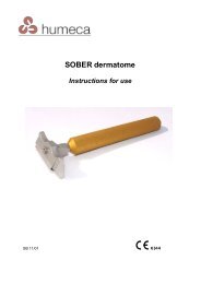
sober hand dermatome<br />
The ability to cut a split-thickness<br />
skin graft free-handedly has<br />
been regarded as a skill acquired<br />
only by long experience. Several<br />
devices have been improvised to<br />
simplify this task. However, the<br />
size and elaborate arrangements<br />
<strong>of</strong> dermatomes <strong>of</strong>ten preclude<br />
their use, especi<strong>all</strong>y if the need is<br />
in an emergency room, a clinic or<br />
at the patient’s bedside. Besides<br />
sophisticated dermatomes are<br />
costly and more adequate for<br />
harvesting larger skin grafts.<br />
In co-operation <strong>with</strong> the Dutch<br />
surgeon dr. Willem Nugteren,<br />
<strong>Humeca</strong> developed a dermatome for<br />
freehanded harvesting <strong>of</strong> a 30 mm<br />
(1¼") wide split skin graft <strong>with</strong> a predetermined<br />
thickness <strong>of</strong> about 0.25<br />
mm (0.01"). The product is c<strong>all</strong>ed the<br />
‘SOBER’ dermatome (from an English<br />
translation <strong>of</strong> the phonetic last name<br />
<strong>of</strong> the inventor).<br />
Based on his experience <strong>with</strong> surgery<br />
in third world countries, it became<br />
clear that a portable, economical and<br />
simple, yet efficient dermatome would<br />
be a very useful tool for skin grafting.<br />
As can be seen from the picture<br />
above, the shape <strong>of</strong> the SOBER<br />
dermatome was derived from a<br />
safety razor. The dermatome is held<br />
firmly against the tautly held skin at a<br />
pre-determined angle and a graft <strong>of</strong><br />
the desired length is quickly cut. No<br />
lateral movements are required.<br />
The blade in the dermatome is a<br />
double facet grinded type, similar to<br />
the <strong>Humeca</strong> ® D42 and D80 blades. It<br />
is inserted in the SOBER dermatome<br />
by loosening a screw. The tool to do<br />
this is integrated in the grip, so it can<br />
never be lost and it is always ready<br />
at hand.<br />
The SOBER dermatome <strong>of</strong>fers<br />
the surgeon a low-cost alternative<br />
to the more elaborate mechanical<br />
dermatomes, whenever sm<strong>all</strong> grafts<br />
are needed. The low price and<br />
robust, durable construction make the<br />
instrument a very suitable tool for use<br />
in third world countries and remote<br />
clinics.<br />
Donorsites on the upper leg <strong>of</strong> a child<br />
Harvesting a graft <strong>with</strong> a Sober dermatome<br />
from the lower leg<br />
Practizing SOBER dermatome on fruits<br />
SOBER dermatome set <strong>with</strong> blades and extra key,<br />
as supplied by <strong>Humeca</strong><br />
12
dermatome blades<br />
<strong>Humeca</strong> supplies a range <strong>of</strong> high<br />
quality dermatome blades for<br />
different types <strong>of</strong> dermatomes.<br />
For the <strong>Humeca</strong> ® D42, D80 and<br />
SOBER dermatomes <strong>Humeca</strong><br />
supplies symmetrical and double<br />
facet grinded blades. These blades<br />
fit in the dermatome both upper and<br />
bottom side down.<br />
<strong>Humeca</strong> supplies blades that are<br />
compatible <strong>with</strong> Aesculap ® / B.Braun ®<br />
cordless dermatomes (Acculan ® ).<br />
blades features<br />
• Precision facet grinded <strong>with</strong>out burrs for durable performance.<br />
• Separately packed in a pouch <strong>with</strong> safety sleeve.<br />
• Radiation sterilized.<br />
• Supplied in boxes <strong>of</strong> 10 pcs.<br />
• Attractively priced<br />
<strong>Humeca</strong> supplies blades that are<br />
compatible <strong>with</strong> Integra ® / Padgett ®<br />
dermatomes type B, C and S.<br />
Fin<strong>all</strong>y <strong>Humeca</strong> supplies blades<br />
that are compatible <strong>with</strong> Zimmer<br />
dermatomes, type 8801 (air) and<br />
8821 (electric).<br />
Ordering <strong>info</strong>rmation<br />
Ordering <strong>info</strong>rmation is given at page<br />
22 and 23.<br />
SOBER <strong>Humeca</strong> ® blade for Aesculap ® / B. Braun ®<br />
We emphasize that <strong>all</strong> blades mentioned here are<br />
<strong>Humeca</strong> blades and suppliers <strong>of</strong> the original brand<br />
blades cannot be held responsible for their performance.<br />
<strong>Humeca</strong> ® D80 <strong>Humeca</strong> ® blade for Padgett ®<br />
<strong>Humeca</strong> ® D42 <strong>Humeca</strong> ® blade for Zimmer ®<br />
13
v-carriers<br />
<strong>Humeca</strong> introduces a new range<br />
<strong>of</strong> grooved skin graft carriers for<br />
expansion and perforation, c<strong>all</strong>ed<br />
‘V-carriers’.<br />
The symmetrical V-shaped groove<br />
pattern <strong>of</strong> these carriers prevents<br />
unwanted sideward movement <strong>of</strong> the<br />
carrier in the mesher during cutting.<br />
The standard length <strong>of</strong> these carriers<br />
is 280 mm (11.0"), which is more<br />
than the standard length <strong>of</strong> existing<br />
carriers. Furthermore care has been<br />
taken to ensure that the groove<br />
pattern <strong>of</strong> a carrier connects exactly<br />
to that <strong>of</strong> another one. This enables<br />
cutting <strong>of</strong> extra long graft strips<br />
<strong>with</strong>out any disturbance <strong>of</strong> the mesh<br />
pattern in the graft.<br />
All V-carriers are compatible <strong>with</strong> the<br />
newly developed <strong>Humeca</strong> ® skin graft<br />
mesher. V10-type carriers are also<br />
compatible <strong>with</strong> Zimmer ® meshers<br />
and V15-type carriers are compatible<br />
<strong>with</strong> Aesculap ® / B.Braun ® meshers.<br />
V-carriers are available for<br />
expansions 1:1.5, 1:2 (type V10<br />
only) and 1:3. These are used for<br />
conventional skin meshing, where<br />
expansion <strong>of</strong> the graft surface is the<br />
main goal.<br />
For larger expansion ratios <strong>Humeca</strong><br />
developed the modified MEEK<br />
technique. Especi<strong>all</strong>y in severe burns<br />
(large total burned surface area)<br />
the MEEK technique should be the<br />
method <strong>of</strong> choice because <strong>of</strong> faster<br />
epithelialization, more efficient use<br />
<strong>of</strong> skin (better graft take and sm<strong>all</strong>er<br />
donorsites), easier handling <strong>of</strong> the<br />
graft and better final results.<br />
In addition to the V-carriers for<br />
expansion, <strong>Humeca</strong> introduces a<br />
new type <strong>of</strong> a meshgraft carrier that<br />
only perforates the graft <strong>with</strong>out<br />
the intention <strong>of</strong> expanding it: the<br />
1:1 V-carrier. Perforations in a graft<br />
are intended to achieve sufficient<br />
drainage <strong>of</strong> the wound bed in case<br />
full sheet grafts are used, in order to<br />
prevent the occurrence <strong>of</strong> seroma or<br />
haematoma under the graft.<br />
Full sheet grafts are frequently<br />
applied when skin grafting is required<br />
in cosmetic<strong>all</strong>y sensitive body parts,<br />
such as the face, the neck and<br />
the dorsal aspect <strong>of</strong> the hands, in<br />
order to avoid the appearance <strong>of</strong> an<br />
unaesthetic mesh pattern.<br />
The development <strong>of</strong> the <strong>Humeca</strong> ®<br />
V-carriers for skin grafting was<br />
sponsored by the Dutch Burns<br />
Research Institute (BRI) in Beverwijk,<br />
The Netherlands.<br />
V-carrier 1:3<br />
14
v-carriers for expansion<br />
Skin grafting is a well-known<br />
means <strong>of</strong> reconstructing a skin<br />
defect.<br />
Because a wound is re-epithelialized<br />
from the edges towards the centre,<br />
the perimeter <strong>of</strong> the graft is the<br />
only part that contributes to the<br />
epithelialization process. Expansion<br />
techniques are used to speed<br />
up that process. An expanded<br />
graft presents a larger cumulative<br />
perimeter through which epithelial<br />
outgrowth can proceed. Besides, <strong>with</strong><br />
graft expansion, larger areas <strong>of</strong> skin<br />
defects can be covered <strong>with</strong> sm<strong>all</strong>er<br />
sections <strong>of</strong> graft.<br />
One <strong>of</strong> the most popular expansion<br />
techniques, widely used in burn<br />
surgery, is the meshgraft method,<br />
introduced by Tanner and Vandeput<br />
in 1963. Meshing a graft gives it<br />
a three-dimensional flexibility that<br />
enables it to conform to irregular<br />
and concave surfaces. The slits in<br />
the graft <strong>all</strong>ow sufficient drainage <strong>of</strong><br />
fluid. At moderate expansions the<br />
results <strong>of</strong> the meshgraft technique<br />
are quite satisfying. <strong>Humeca</strong> supplies<br />
V-carriers for meshing grafts <strong>with</strong> an<br />
expansion ratio <strong>of</strong> 1:1.5, 1:2 (type<br />
V10 only) and 1:3.<br />
At larger expansions, the graft<br />
becomes more fragile and difficult to<br />
handle. Draping it correctly (dermal<br />
side down) on a wound bed <strong>with</strong>out<br />
damaging the mesh structure<br />
becomes increasingly difficult at<br />
larger expansion ratios. Besides<br />
epithelialization might be delayed<br />
due to the large distances the new<br />
epithelium has to grow. For such<br />
cases the MEEK technique is more<br />
appropriate.<br />
Graft removed from a 1:1.5 V-carrier after cutting<br />
Detail <strong>of</strong> the V-shaped groove pattern<br />
<strong>of</strong> the <strong>Humeca</strong> V-carrier<br />
Meshed graft 1:1.5 being applied to the wound<br />
Graft, meshed <strong>with</strong> a <strong>Humeca</strong> V-carrier 1:1.5 and cut to fit the size <strong>of</strong> a burn<br />
15
Example <strong>of</strong> a graft, meshed on a 1:1 <strong>Humeca</strong> ® V-carrier<br />
Example <strong>of</strong> a perforated piece <strong>of</strong> graft<br />
v-carriers for perforation<br />
The <strong>Humeca</strong> ® V-carrier 1:1 is<br />
intended for perforation purposes,<br />
<strong>with</strong>out expansion.<br />
Why perforations?<br />
Perforation is frequently applied<br />
when sheet skin grafts (either splitthickness<br />
or full-thickness) are used.<br />
Sheet grafts are perforated for the<br />
following reasons:<br />
• Drainage<br />
The slits in the graft <strong>all</strong>ow adequate<br />
drainage <strong>of</strong> fluid. Drainage prevents<br />
separation <strong>of</strong> the graft from its wound<br />
bed by haematoma or seroma. Sheet<br />
grafts are <strong>of</strong>ten used on the face<br />
and hands. It is however these two<br />
sites that have an extremely vascular<br />
bed, making the chances <strong>of</strong> the<br />
appearance <strong>of</strong> haematoma higher.<br />
Collection <strong>of</strong> fluid under a graft is said<br />
to be one <strong>of</strong> the major causes <strong>of</strong> graft<br />
loss. Consequently perforation leads<br />
to increased graft survival.<br />
• Improved cosmetic results<br />
Expanded meshed grafts are<br />
frequently criticised because <strong>of</strong> the<br />
poor appearance <strong>of</strong> the residual<br />
mesh pattern, making surgeons<br />
reluctant to consider this technique.<br />
By using a 1:1 mesh, where the<br />
graft perforations are narrowed to<br />
slits instead <strong>of</strong> holes, it is possible to<br />
prevent such a pattern to appear and<br />
get results similar to a sheet graft. It<br />
is also reported that a perforated graft<br />
gives a more matt appearance than<br />
the shiny surface <strong>of</strong> a sheet graft,<br />
which makes it more suitable when<br />
grafting the forehead.<br />
• Infection control<br />
To reduce infection occurrence.<br />
Methods to perforate grafts<br />
One <strong>of</strong> the simplest methods <strong>of</strong>ten<br />
applied in the operation theatre is<br />
piercing a sheet graft <strong>with</strong> a scalpel<br />
blade by hand. This can be done<br />
either by piercing the graft directly<br />
or by placing a piece <strong>of</strong> graft on a<br />
grooved meshgraft carrier and cutting<br />
it <strong>with</strong> a scalpel at right angles to the<br />
grooves. These methods are labour<br />
intensive, and <strong>of</strong>ten the number<br />
<strong>of</strong> slits is insufficient to guarantee<br />
enough drainage. Nevertheless the<br />
method seems adequate in case <strong>of</strong><br />
sm<strong>all</strong> grafts.<br />
Another way <strong>of</strong> perforating is to<br />
use a 1:1.5 meshed graft <strong>with</strong>out<br />
expanding it. After <strong>all</strong> theoretic<strong>all</strong>y the<br />
holes in the graft remain slits when<br />
the graft is not expanded. However<br />
it appears to be very difficult to apply<br />
such a graft to the wound bed <strong>with</strong>out<br />
disturbing the pattern <strong>of</strong> slits. During<br />
transfer <strong>of</strong> the graft to the wound,<br />
due to internal friction the slits will<br />
become holes anyway and once<br />
this occurs it is almost impossible to<br />
regain the original shape <strong>of</strong> the graft.<br />
So this method is not recommended<br />
when the final cosmetic results<br />
<strong>of</strong> the perforated graft should be<br />
comparable to the results <strong>of</strong> a sheet<br />
graft.<br />
Detail <strong>of</strong> the groove pattern <strong>of</strong> a 1:1 V-carrier<br />
Fin<strong>all</strong>y, perforations in a graft<br />
can be achieved by a procedure<br />
sometimes referred to as “Sideways<br />
meshing” or “Reversed meshing”.<br />
This procedure was first described<br />
by Davison et.al. in 1986 and it<br />
concerns crosswise cutting a 1:1.5<br />
meshgraft carrier. Such cutted parts<br />
<strong>of</strong> the carrier are then covered <strong>with</strong><br />
graft and introduced in the mesher<br />
at a sideward direction (the grooves<br />
turned 90º from normal). This results<br />
in sm<strong>all</strong>er slits that can be used as<br />
perforations. Disadvantage however<br />
is that the maximum length <strong>of</strong> the<br />
graft equals the width <strong>of</strong> the carrier<br />
(approx. 75 mm or 3 inches), while<br />
mostly larger grafts are required.<br />
To overcome the difficulties <strong>of</strong><br />
making perforations in grafts,<br />
<strong>Humeca</strong> introduced a carrier<br />
especi<strong>all</strong>y developed for this goal:<br />
the 1:1 V-carrier. When using this<br />
carrier, making perforations in a<br />
graft becomes as easy as meshing.<br />
Maximum length <strong>of</strong> the graft is 280<br />
mm or 11 inches, but even longer<br />
graft strips can be perforated when<br />
using a second carrier, from which<br />
the grooves connect exactly to the<br />
first one.<br />
The <strong>Humeca</strong> 1:1 V-carriers are<br />
available in a V10-type for <strong>Humeca</strong> ®<br />
and Zimmer ® meshers and a V15-<br />
type for <strong>Humeca</strong> ® and Aesculap ® /<br />
B.Braun ® meshers.<br />
The 1:1 perforation V-carrier was<br />
developed and clinic<strong>all</strong>y tested in<br />
cooperation <strong>with</strong> the burn centre<br />
<strong>of</strong> the University Hospital <strong>of</strong> Gent,<br />
Belgium.<br />
16
The two pictures above show the<br />
application <strong>of</strong> a perforated sheet<br />
graft on a burn on the abdomen <strong>of</strong><br />
a female patient. A perforated sheet<br />
graft autograft was fit to the size <strong>of</strong><br />
the wound. The picture on the left<br />
was taken at operation. The picture<br />
on the right shows the same burn<br />
2 weeks post-operatively. A smooth<br />
surface <strong>with</strong>out any appearance <strong>of</strong><br />
a mesh pattern in the residual scar<br />
was obtained.<br />
The two pictures at the right clearly<br />
show that the slits in a perforated<br />
graft <strong>all</strong>ow drainage <strong>of</strong> fluids through<br />
a sheet graft. When the sheet graft is<br />
not perforated, seroma and haematoma<br />
might hinder a good graft take.<br />
Full sheet grafts on fingers, showing seroma, due to insufficient drainage<br />
Graft perforated <strong>with</strong> V-carrier 1:1 on a foot. Drainage <strong>of</strong> fluids through the slits is clearly visible<br />
17
Picture showing an example <strong>of</strong> the application <strong>of</strong><br />
a 1:1.5 mesghraft on a burn <strong>of</strong> the hand, showing<br />
hypertrophic scarring and an early stage <strong>of</strong> wound<br />
contracture. The mesh pattern is clearly visible.<br />
A 1:1 perforated sheet graft would probably result in a<br />
better appearance and functionality.<br />
full- and splitthickness<br />
skin grafts<br />
Skin grafts consist <strong>of</strong> the<br />
entire epidermis and a dermal<br />
component <strong>of</strong> variable thickness. If<br />
the entire thickness <strong>of</strong> the dermis<br />
is included, an appropriate term is<br />
‘full-thickness skin graft’ (FTSG).<br />
If less then the entire dermis is<br />
included, appropriate terms are<br />
partial or ‘split-thickness skin<br />
graft’ (STSG).<br />
STSG’s are categorized further as<br />
thin (0.1-0.3 mm / 0.004-0.012"),<br />
medium-thickness (0.3-0.45 mm /<br />
0.012-0.018") or thick (0.45-0.8 mm /<br />
0.018-0.03"), based on the thickness<br />
<strong>of</strong> the harvested graft. The choice<br />
between FTSG and STSG depends<br />
on wound condition, location, size<br />
and aesthetic concerns. Splitthickness<br />
skin grafts require less<br />
ideal conditions for survival and have<br />
a much broader range <strong>of</strong> application<br />
than full-thickness skin grafts. They<br />
are for instance used to resurface<br />
large wounds, line cavities, resurface<br />
mucosal deficits, close flap donor<br />
sites and resurface muscle flaps. Fullthickness<br />
skin grafts are <strong>of</strong> limited<br />
availability. The ability to harvest<br />
them is restricted by the necessity <strong>of</strong><br />
stitching up the donor site from which<br />
they were taken. FTSG’s are ideal<br />
for repairing sm<strong>all</strong> defects where<br />
good cosmetic matching or functional<br />
restoration is important. STSG’s do<br />
have some serious disadvantages<br />
compared to FTSG’s that have to be<br />
considered:<br />
Contraction<br />
One <strong>of</strong> them concerns contraction:<br />
a skin graft begins to shrink<br />
immediately after being harvested<br />
from its donor site. This ‘primary<br />
contraction’ is about 40% in a FTSG,<br />
20% in a medium thickness STSG<br />
and about 10% in a thin STSG. So a<br />
STSG has less primary contraction<br />
than a FTSG, which is an advantage<br />
<strong>of</strong> STSG, because less tissue is<br />
needed for grafting. However, after<br />
transfer to a recipient site, the skin<br />
graft will shrink further as it heals<br />
in a process known as ‘secondary<br />
contraction’. FTSG’s tend to remain<br />
the same size (after primary<br />
contraction) and do not show any<br />
secondary contraction. STSG’s, on<br />
the other hand, contract whenever the<br />
circumstances <strong>all</strong>ow. Unless STSG’s<br />
are fixed to underlying rigid structures<br />
and cannot move, they will contract<br />
secondary. When transplanted on s<strong>of</strong>t<br />
tissue, contraction will be significant.<br />
A contracted wound is <strong>of</strong>ten tight<br />
and immobile and there is distortion<br />
<strong>of</strong> the surrounding tissue. If such a<br />
wound crosses a joint, contracture<br />
will lead to position abnormalities<br />
and inadequate motion. Therefore<br />
a FTSG is advised for such areas<br />
(hand, wrist, elbow, neck).<br />
Graft growing<br />
Once wound contraction ends, fullthickness<br />
skin grafts are able to grow<br />
<strong>with</strong> the individual, whereas splitthickness<br />
skin grafts tend to remain<br />
in a fixed contracted state and grow<br />
minim<strong>all</strong>y, if at <strong>all</strong>.<br />
Fragility<br />
STSG’s are more fragile than<br />
FTSG’s, especi<strong>all</strong>y when placed over<br />
areas <strong>with</strong> little underlying s<strong>of</strong>t tissue<br />
support.<br />
Pigmentation and other cosmetic<br />
considerations<br />
Split-thickness skin grafts tend to be<br />
abnorm<strong>all</strong>y pigmented (either pale or<br />
white) or hyperpigmented, particularly<br />
in darker-skinned individuals. Their<br />
thinness, abnormal pigmentation<br />
and frequent lack <strong>of</strong> smooth texture<br />
and hair growth, make STSG’s more<br />
functional than cosmetic. When used<br />
to resurface large burns <strong>of</strong> the face,<br />
STSG’s may yield an undesirable<br />
mask like appearance.<br />
18
v-carriers features<br />
• Compatible <strong>with</strong> existing Zimmer ® and Aesculap ® / B.Braun ® meshers<br />
• Symmetric V-pattern <strong>of</strong> grooves prevents sideward movement<br />
• Standard length 280 mm (11.0"), width 78.8 mm (3.1")<br />
• Expansion ratio 1:1.5, 1:2 (type V10 only) and 1:3<br />
• Special carrier for 1:1 perforation (drainage) <strong>of</strong> sheet grafts<br />
• Groove patterns <strong>of</strong> carriers connect to each other<br />
• Highly flexible medical grade polypropylene material<br />
• Individu<strong>all</strong>y sterile packed in peel pouch; standard boxes 10 pcs.<br />
• Attractively priced<br />
Ordering <strong>info</strong>rmation<br />
Ordering <strong>info</strong>rmation is given<br />
at page 22 and 23.<br />
19
mesher<br />
In addition to the V-carriers,<br />
<strong>Humeca</strong> developed a mesher<br />
to complete the product line for<br />
meshgrafting.<br />
The <strong>Humeca</strong> ® mesher is provided<br />
<strong>with</strong> a unique spring mechanism that<br />
prevents the blades from excessive<br />
pressure on the carrier during cutting.<br />
Due to their production process<br />
(injection molding) <strong>all</strong> meshgraft<br />
carriers have inevitable variations in<br />
thickness. When the thickness <strong>of</strong> a<br />
certain section <strong>of</strong> the carrier exceeds<br />
the maximum distance between<br />
the blades and the lower roller, the<br />
pressure exercised by the blades<br />
on the carrier surface will increase<br />
exponenti<strong>all</strong>y. Such high pressure<br />
manifests in increased friction and in<br />
the end it will damage the blades. On<br />
the other hand the mesher might not<br />
cut the graft completely at a thinner<br />
section <strong>of</strong> the carrier. To avoid such<br />
undesired phenomena, <strong>Humeca</strong><br />
provided the mesher <strong>with</strong> springs that<br />
can meet <strong>with</strong> thickness variations in<br />
carriers. The mesher can be adjusted<br />
in two positions for the V10 or the<br />
V15- types <strong>of</strong> V-carriers.<br />
During cutting the carrier is guided<br />
both at the left and the right side<br />
to assure straight movement, thus<br />
enabling exact connection <strong>of</strong> the<br />
grooves <strong>of</strong> a second carrier if applied.<br />
Unlike most conventional meshers,<br />
where the carrier is moved through<br />
the device by means <strong>of</strong> intermittent<br />
pulling a ratchet, the <strong>Humeca</strong> ®<br />
mesher is driven by the continuous<br />
rotation <strong>of</strong> a handle. A gearwheels set<br />
limits the required force. The rotation<br />
makes the meshing procedure less<br />
time consuming and the design is far<br />
more ergonomic.<br />
Side view <strong>of</strong> the <strong>Humeca</strong> ® mesher<br />
20
Bridge opened for cleaning and access to the cutting axis<br />
Cutting axis<br />
The 50 blades (diameter 36 mm /<br />
1.42") <strong>of</strong> the mesher are mounted on<br />
an axis, <strong>with</strong> an interspace <strong>of</strong> 1.5 mm<br />
(0.06") between the intersecting lines.<br />
Both the cutting axis and individual<br />
blades can be replaced if desired.<br />
Opening the bridge <strong>of</strong> the mesher<br />
<strong>all</strong>ows easy access to the cutting axis<br />
for cleaning and inspection.<br />
The mesher can be washed and<br />
steam sterilized before use. A<br />
compact stainless steel sterilization<br />
case is available.<br />
A second sm<strong>all</strong> sterilization case can<br />
hold the cutting axis.<br />
Ordering <strong>info</strong>rmation<br />
Ordering <strong>info</strong>rmation is given at page<br />
22 and 23.<br />
mesher features<br />
• Robust and durable construction<br />
• Compatible <strong>with</strong> <strong>Humeca</strong> ® V-carriers <strong>of</strong> <strong>all</strong> types (V10 and V15)<br />
• Compatible <strong>with</strong> Zimmer ® and Aesculap ® / B.Braun ® carriers<br />
• Spring mechanism prevents blades damage<br />
• Continuous rotational drive; no intermittent pulling <strong>of</strong> a ratchet<br />
• Measures lxwxh: 220x212x183 mm (8.7x8.3x7.2"). Weight: 4.4 kg (9.7 lb)<br />
• Cutting axis can easily be replaced<br />
• Individual blades can be replaced<br />
• Compact st. steel sterilization case available, lxwxh: 277x232x197 mm<br />
(10.9x9.1x7.8")<br />
Sterilization case for the <strong>Humeca</strong> mesher<br />
Sterilization case for the axis <strong>of</strong> the <strong>Humeca</strong> mesher<br />
21
For ordering, please contact your<br />
local dealer. You can find dealers via<br />
the link in www.humeca.nl<br />
If <strong>Humeca</strong> has no dealer in your<br />
territory, please contact <strong>Humeca</strong> for<br />
inquiry, preferably via email<br />
<strong>info</strong>@humeca.nl<br />
In order to provide <strong>Humeca</strong> quickly<br />
<strong>with</strong> brief and most adequate<br />
<strong>info</strong>rmation and to assure soonest<br />
reply, the preferred method is to fill<br />
in the contact form that is displayed<br />
when using the contact link in the<br />
<strong>Humeca</strong> site www.humeca.nl<br />
<strong>Humeca</strong> is a sm<strong>all</strong> organization <strong>with</strong><br />
short communication lines.<br />
This <strong>all</strong>ows very quick response and<br />
a high level <strong>of</strong> service.<br />
In case <strong>of</strong> ordering or request for<br />
quotation, please refer to the article<br />
codes given below.<br />
ordering <strong>info</strong><br />
meek micrografting<br />
Equipment<br />
3.HD/BLO<br />
MEEK cutting machine, hand driven (block model) <strong>with</strong>out cutting block<br />
3.HD/CYL<br />
MEEK cutting machine, hand driven (cylindrical model) <strong>with</strong>out cutting block<br />
3.MD/BLO<br />
MEEK cutting machine, motor driven (block model), <strong>with</strong>out cutting block<br />
3.MD/CYL<br />
MEEK cutting machine, motor driven (cylindrical model), <strong>with</strong>out cutting block<br />
3.MD/GW<br />
MEEK cutting machine, motor driven, gearwheels, <strong>with</strong>out cutting block<br />
3.MD/AUT<br />
MEEK cutting machine, automatic version (two axis motor driven)<br />
3.BL38<br />
MEEK circular blade, diameter 38 mm (1.50"). Required blade diameter depends on serial number machine<br />
3.BL39<br />
MEEK circular blade, diameter 39 mm (1.54”). Required blade diameter depends on serial number machine<br />
3.CA01<br />
MEEK cutting aid 41x41 mm (1.61x1.61")<br />
3.CB01<br />
MEEK single cutting block <strong>with</strong> cork holder<br />
3.CB02<br />
MEEK double cutting block <strong>with</strong> two cork holders<br />
3.CH01<br />
MEEK cork holder<br />
3.CP4<br />
MEEK pneumatic foot pedal <strong>with</strong> connectors and hose<br />
3.GWS<br />
MEEK gearwheel drive set<br />
3.KN13/38 MEEK cutting axis <strong>with</strong> 13 ceramic coated circular blades, diameter 38 mm (1.50")<br />
3.KN13/39 MEEK cutting axis <strong>with</strong> 13 ceramic coated circular blades, diameter 39 mm (1.54")<br />
3.MAC02<br />
MEEK sterilization case 434x254x172 mm (17.1x10.0x6.8")<br />
3.MAC03<br />
MEEK sterilization case 180x51x45 mm (7.1x2.0x1.8") for the cutting axis <strong>of</strong> the MEEK machine<br />
3.RM004<br />
MEEK pneumatic block motor <strong>with</strong> connectors and coupling<br />
3.087 MEEK cylindrical pneumatic motor 2M12<br />
3.SHD<br />
MEEK hand drive set<br />
3.SW01<br />
MEEK serrated wedge (cam)<br />
Disposables<br />
2.3/10 MEEK Micrograft gauze, expansion 1:3, <strong>with</strong> cork plate, box 10 pcs.<br />
2.4/10 MEEK Micrograft gauze, expansion 1:4, <strong>with</strong> cork plate, box 10 pcs.<br />
2.6/10 MEEK Micrograft gauze, expansion 1:6, <strong>with</strong> cork plate, box 10 pcs.<br />
2.9/10 MEEK Micrograft gauze, expansion 1:9, <strong>with</strong> cork plate, box 10 pcs.<br />
2.3/40 MEEK Micrograft gauze, expansion 1:3, <strong>with</strong> cork plate, box 40 pcs.<br />
2.4/40 MEEK Micrograft gauze, expansion 1:4, <strong>with</strong> cork plate, box 40 pcs.<br />
2.6/40 MEEK Micrograft gauze, expansion 1:6, <strong>with</strong> cork plate, box 40 pcs.<br />
2.9/40 MEEK Micrograft gauze, expansion 1:9, <strong>with</strong> cork plate, box 40 pcs.<br />
2.9190 Spray adhesive, bottle 200 ml (6.8 fl.oz).<br />
2.JG598<br />
STERILIT ® oil for surgical instruments, bottle 50 ml (1.7 fl.oz).<br />
22
humeca ® dermatomes<br />
Equipment<br />
4.D42STS<br />
D42 dermatome, complete set, 1200mAh Li-Ion battery<br />
4.D42STSx D42 dermatome, complete set, 2400mAh Li-Ion battery<br />
4.D80STS<br />
D80 dermatome, complete set, 2400mAh Li-Ion battery<br />
4.D42 D42 dermatome <strong>with</strong>out battery- and motor cartridge <strong>with</strong> sm<strong>all</strong> grip for 1200mAh battery<br />
4.D42x<br />
D42 dermatome <strong>with</strong>out battery- and motor cartridge <strong>with</strong> large grip for 2400mAh battery<br />
4.D80 D80 dermatome <strong>with</strong>out battery- and motor cartridge <strong>with</strong> large grip for 2400mAh battery<br />
4.ANS-HC<br />
Dermatome charger for Li-Ion battery <strong>of</strong> 2400 mAh <strong>with</strong> 4 international adapters<br />
4.ANS-LC<br />
Dermatome charger for Li-Ion battery <strong>of</strong> 1200 mAh <strong>with</strong> 4 international adapters<br />
4.SU01<br />
Dermatome charger support unit<br />
4.BC7.4V2400 Dermatome battery cartridge 7.4V, 2400 mAh<br />
4.BC7.4V1200 Dermatome battery cartridge 7.4V, 1200 mAh<br />
4.MCX15<br />
Dermatome motor cartridge<br />
4.D42CL30 D42 width reducing clamp 30 mm (1.18")<br />
4.D42CL36 D42 width reducing clamp 36 mm (1.42")<br />
4.D80CL35 D80 width reducing clamp 35 mm (1.38")<br />
4.D80CL50 D80 width reducing clamp 50 mm (1.97")<br />
4.D80CL65 D80 width reducing clamp 65 mm (2.56")<br />
4.D42AC1<br />
D42 sterilization case<br />
4.D80AC1<br />
D80 sterilization case<br />
4.SCL<br />
Sterile clamp<br />
4.SF01<br />
Sterile funnel for sm<strong>all</strong> diameter grip<br />
4.SF02<br />
Sterile funnel for large diameter grip<br />
4.SB01<br />
SOBER hand dermatome, complete set<br />
5.D42BL10<br />
5.D80BL10<br />
5.BL4245<br />
5.BLPG10<br />
5.BLZM10<br />
5.BLSB10<br />
2.JG598<br />
Disposables<br />
D42 dermatome blades, box 10 pcs.<br />
D80 dermatome blades, box 10 pcs.<br />
<strong>Humeca</strong> ® blade for Aesculap ® / B.Braun ® (Acculan ® ) cordless dermatomes, box 10 pcs.<br />
<strong>Humeca</strong> ® blade for Padgett ® dermatomes, model B, C and S, box 10 pcs.<br />
<strong>Humeca</strong> ® blade for Zimmer ® dermatomes, type 8801 (air) and 8821 (electric)<br />
<strong>Humeca</strong> ® blade for SOBER hand dermatome, box 10 pcs.<br />
STERILIT ® oil for surgical instruments, bottle 50 ml. (1.7 fl. oz)<br />
humeca ® v-carriers<br />
6.V10-1.0<br />
6.V10-1.5<br />
6.V10-2.0<br />
6.V10-3.0<br />
6.V15-1.0<br />
6.V15-1.5<br />
6.V15-3.0<br />
V-carrier, perforation 1:1, for <strong>Humeca</strong> and Zimmer ® meshers, box 10 pcs.<br />
V-carrier, expansion ratio 1:1.5, for <strong>Humeca</strong> and Zimmer ® meshers, box 10 pcs.<br />
V-carrier, expansion ratio 1:2, for <strong>Humeca</strong> and Zimmer ® meshers, box 10 pcs.<br />
V-carrier, expansion ratio 1:3, for <strong>Humeca</strong> and Zimmer ® meshers, box 10 pcs.<br />
V-carrier, perforation 1:1, for <strong>Humeca</strong> and Aesculap ® / B.Braun ® meshers, box 10 pcs.<br />
V-carrier, expansion ratio 1:1.5, for <strong>Humeca</strong> and Aesculap ® / B.Braun ® meshers, box 10 pcs.<br />
V-carrier, expansion ratio 1:3, for <strong>Humeca</strong> and Aesculap ® / B.Braun ® meshers, box 10 pcs.<br />
humeca ® mesher<br />
6.HM01<br />
<strong>Humeca</strong> ® mesher<br />
6.KN50/36 Cutting axis <strong>with</strong> 50 blades Ø 36 mm (1.42")<br />
6.HMAC01<br />
Sterilization case 275x230x179 mm (10.8x9.1x7.0") for <strong>Humeca</strong> ® mesher<br />
6.HMAC02<br />
Sterilization case 180x51x45 mm (7.1x2.0x1.8") for the cutting axis <strong>of</strong> the <strong>Humeca</strong> ® mesher<br />
23
www.humeca.nl<br />
s k i n t r a n s p l a n t a t i o n t e c h n o l o g y<br />
Office address<br />
Het Bijvank 251-a<br />
NL-7544 DB Enschede<br />
The Netherlands<br />
Postal address<br />
P.O.Box 40175<br />
NL-7504 RD Enschede<br />
The Netherlands<br />
<strong>Humeca</strong> BV<br />
Phone +31 53 476 26 19<br />
Fax +31 53 477 19 05<br />
E-mail <strong>info</strong>@humeca.nl<br />
24