Early Cretaceous Freshwater Fish Fauna in Kyushu, Japan
Early Cretaceous Freshwater Fish Fauna in Kyushu, Japan
Early Cretaceous Freshwater Fish Fauna in Kyushu, Japan

Create successful ePaper yourself
Turn your PDF publications into a flip-book with our unique Google optimized e-Paper software.
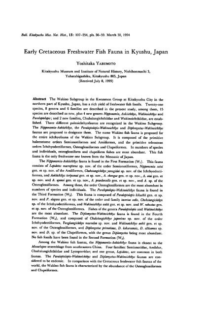
Bull. Kitakyushu Mus. Nat. Hist., 13: 107-254, pis. 36-59. March 30, 1994<br />
<strong>Early</strong> <strong>Cretaceous</strong> <strong>Freshwater</strong> <strong>Fish</strong> <strong>Fauna</strong> <strong>in</strong> <strong>Kyushu</strong>, <strong>Japan</strong><br />
Yoshitaka Yabumoto<br />
Kitakyushu Museum and Institute of Natural History, Nishihonmachi 3,<br />
Yahatahigashiku, Kitakyushu 805, <strong>Japan</strong><br />
(Received July 8, 1993)<br />
Abstract The Wak<strong>in</strong>o Subgroup <strong>in</strong> the Kwanmon Group at Kitakyushu City <strong>in</strong> the<br />
northern part of <strong>Kyushu</strong>, <strong>Japan</strong>, has a rich yield offreshwater fish fossils. Twenty-one<br />
species, 8 genera and 6 families are described <strong>in</strong> the present study, among them, 15<br />
species are described as new, plus 4 new genera Nipponamia, Aokiichthys, Wak<strong>in</strong>oichthys and<br />
Paraleptolepis; and 2 newfamilies, Chuhsiungichthiidae and Wak<strong>in</strong>oichthiidae, are estab<br />
lished. Three different paleoichthyofaunas are recognized <strong>in</strong> the Wak<strong>in</strong>o Subgroup.<br />
The Nipponamia-Aokiichthys, the Paraleptolepis-Wak<strong>in</strong>oichthys and Diplomyslus-Wak<strong>in</strong>oichthys<br />
faunas are proposed to designate them. The name Wak<strong>in</strong>o fish fauna is proposed for<br />
the entire ichthyofauna of the Wak<strong>in</strong>o Subgroup. It is composed of the primitive<br />
halecostome orders Semionotiformes and Amiiformes, and the primitive teleostean<br />
orders Ichthyodectiformes, Osteoglossiformes andClupeiformes. In numbers ofspecies<br />
and <strong>in</strong>dividuals, osteoglossiform and clupeiform fishes are most abundant. This fish<br />
fauna is the only freshwater oneknown from the Mesozoic of<strong>Japan</strong>.<br />
The Nipponamia-Aokiichthys fauna is found <strong>in</strong> the First Formation (W,). This fauna<br />
consists of Lepidotes macropterus sp. nov. of the order Semionotiformes, Nipponamia saloi<br />
gen. et sp. nov. of the Amiiformes, Chuhsiungichthysyanagidai sp. nov. of the Ichthyodecti<br />
formes, andAokiichthys toriyamai gen. et sp. nov., A. changae gen. et sp. nov., A. olai gen. et<br />
sp. nov. and A. uyenoi gen. et sp. nov., A. praedorsalis gen. et sp. nov., and A. sp. of the<br />
Osteoglossiformes. Among these, theorderOsteoglossiformes are the mostabundant <strong>in</strong><br />
numbers of species and <strong>in</strong>dividuals. The Paraleptolepis-Wak<strong>in</strong>oichthys fauna is found <strong>in</strong><br />
theThird Formation (W3). This fauna is composed ofParaleptolepis kikuchii gen. et sp.<br />
nov. and P. elegans gen. et sp. nov. of the order and family <strong>in</strong>certae sedis, Chuhsiungichthys<br />
sp.ofthe Ichthyodectiformes, and Wak<strong>in</strong>oichthys aokii gen. et sp. nov. and W. robustus gen.<br />
et sp. nov. ofthe Osteoglossiformes. <strong>Fish</strong>es ofthegenera Paraleptolepis and Wak<strong>in</strong>oichthys<br />
are the most abundant. The Diplomystus-Wak<strong>in</strong>oichthys fauna is found <strong>in</strong> the Fourth<br />
Formation (W4), and composed of Chuhsiungichthys japonicus sp. nov. of the order<br />
Ichthyodectiformes, Yungkangichthys macrodon sp. nov. and Wak<strong>in</strong>oichthys aokii gen. et sp.<br />
nov. of the Osteoglossiformes, and Diplomyslus primot<strong>in</strong>us, D. kokuraensis, D. altisomus sp.<br />
nov. and D. sp. of the Clupeiformes, with the genus Diplomyslus be<strong>in</strong>g most abundant.<br />
No fish fossils have been found <strong>in</strong> the Second Formation (W2).<br />
Among the Wak<strong>in</strong>o fish faunas, the Nipponamia-Aokiichthys fauna is closest to the<br />
Mesoclupea assemblage from southeastern Ch<strong>in</strong>a. Four families: Semionotidae, Amiidae,<br />
Chuhsiungichthiidae and Lycopteridae; and one genus, Lepidotes, are common <strong>in</strong> both<br />
faunas. The Paraleptolepis-Wak<strong>in</strong>oichthys and Diplomystus-Wak<strong>in</strong>oichthys faunas are con<br />
sidered to be endemic. In comparison with the <strong>Cretaceous</strong> freshwater fish faunas of the<br />
world, the Wak<strong>in</strong>o fish fauna is characterized by theabundance ofthe Osteoglossiformes<br />
and Clupeiformes.
108 Yoshitaka Yabumoto<br />
It is considered that the Nipponamia-Aokiichthys fauna became ext<strong>in</strong>ct by the end of<br />
the First Formation (W,) or the beg<strong>in</strong>n<strong>in</strong>g oftheSecond Formation (W2). On the basis<br />
of this study, it is hypothesized that the clupeid fishes of the Fourth Formation entered<br />
the Kowak<strong>in</strong>o-ko Lake from seaat the beg<strong>in</strong>n<strong>in</strong>g ofthis period, and took its place as the<br />
mostabundant fish, Paraleptolepis, of the Paraleptolepis-Wak<strong>in</strong>oichthys fauna.<br />
Contents<br />
Introduction 108<br />
Materials and Methods<br />
HI<br />
Localityand Horizon<br />
H3<br />
Systematic Paleontology<br />
J20<br />
Family Semionotidae 120<br />
Family Amiidae 121<br />
Family Chuhsiungichthiidae nov 130<br />
Family Lycopteridae 142<br />
Family Wak<strong>in</strong>oichthiidae nov 179<br />
Family Clupeidae 195<br />
Teleostei Order and Family <strong>in</strong>certae sedis 220<br />
Gonorynchiformes ? <strong>in</strong>certae sedis 235<br />
<strong>Fauna</strong>l Comparison 237<br />
<strong>Fauna</strong>l comparison <strong>in</strong> the Wak<strong>in</strong>o Subgroup 237<br />
Comparison with <strong>Cretaceous</strong> freshwater fish faunas <strong>in</strong> Ch<strong>in</strong>a 238<br />
Comparison with other <strong>Cretaceous</strong> freshwater fish faunas <strong>in</strong> the world 243<br />
Conclud<strong>in</strong>g Remarks 244<br />
Acknowledgments 247<br />
References 248<br />
Introduction<br />
Mesozoic lacustr<strong>in</strong>e sediments are distributed <strong>in</strong> western Honshu through north<br />
ern <strong>Kyushu</strong>, <strong>Japan</strong>. Many geologicaland paleontological studies have been done on<br />
the Mesozoic sediments <strong>in</strong> this area (Suzuki, 1893, 1906; Kochibe, 1903; Koto,<br />
1909; Ogura, 1922; Yabe, 1927; Nagao, 1929; Kobayashi, 1931; Oishi, 1933;<br />
Toriyama, 1938; Kobayashi and Ota, 1936; Kobayashi and Suzuki; 1936, 1939;<br />
Kobayashi, 1941; Matsumoto, 1949, 1951, 1954; Y. Ota, 1953, 1955, 1957, 1958,<br />
1959a-d, 1960ab; Ueda, 1957; Hase, 1958, 1960; M. Ota el al., 1979; Uyeno, 1979;<br />
Uyeno and Yabumoto, 1980; Kusumi, 1960, 1979; Ishijima, 1978, 1979; Tamura,<br />
1990; Kimura et al., 1992).<br />
The first record of non-mar<strong>in</strong>e fossils from the Mesozoic sediments <strong>in</strong> northern<br />
<strong>Kyushu</strong> was on molluscs from Wak<strong>in</strong>o <strong>in</strong> Nogata City, Fukuoka Prefecture by Nagao<br />
(1929). Kobayashi and Suzuki (1936) described five non-mar<strong>in</strong>e molluscs from<br />
Wak<strong>in</strong>o. Kobayashi and Ota (1936) designated this shell bear<strong>in</strong>g formation as the<br />
Wak<strong>in</strong>o beds and stated that the beds were correlated to the Naktong Series of South<br />
Korea, which has been considered to be referable to the Lower <strong>Cretaceous</strong> on the
<strong>Early</strong> <strong>Cretaceous</strong> <strong>Freshwater</strong> <strong>Fish</strong> <strong>Fauna</strong> <strong>in</strong> <strong>Kyushu</strong>,<strong>Japan</strong> 109<br />
basis of similarity of the fossil assemblage. Matsumoto (1951) proposed to desig<br />
nate the so-called "Inkstone Series" as the Kwanmon Group, which consists of two<br />
subgroups: the lower Wak<strong>in</strong>o Subgroup and the upper Shimonoseki Subgroup, based<br />
on the established stratigraphic column of the type area. In 1954, Matsumoto<br />
presented a general description and discussion of the Kwanmon Group.*<br />
The stratigraphy of the Kwanmon Group was established by Y. Ota (1953,<br />
1955, 1957, 1958, 1959a-d, 1960ab) and Hase (1958, 1960). The type area of the<br />
Wak<strong>in</strong>o Subgroup was reexam<strong>in</strong>ed and four formations were recognized: the Sengoku<br />
Formation, the Nyoraida Formation, the Lower Wakamiya Formation and the Upper<br />
Wakamiya Formation, <strong>in</strong> ascend<strong>in</strong>g order (Y. Ota, 1953). Y. Ota (1955, 1957,<br />
1958, 1959a) reported on four other areas <strong>in</strong> northern <strong>Kyushu</strong> and recognized<br />
equivalents for these formations. Ueda (1957) <strong>in</strong>vestigated the type area of the<br />
Shimonoseki Subgroup. Hase (1958, 1960) stated on the stratigraphy and the<br />
geologic history of the Kwanmon Group and described molluscan species from the<br />
Toyonishi and the Kwanmon Groups. Matsushita (1968) <strong>in</strong>vestigated the Kwan<br />
mon Group <strong>in</strong> Kitakyushu City. M. Ota et al. (1979) <strong>in</strong>vestigated the Wak<strong>in</strong>o<br />
Subgroup at Yamada Park, a former military arsenal <strong>in</strong> the southern district of<br />
Kokura, Kitakyushu City, and reported the stratigraphy and geologic structure <strong>in</strong><br />
detail, with a special reference to the occurrence of fish fossils.<br />
The first record of the fish fossils from the Wak<strong>in</strong>o Subgroup <strong>in</strong> the Kwanmon<br />
Group was from the First Formation (the lower formation, Wi) <strong>in</strong> southern Kokura<br />
<strong>in</strong> Kitakyushu City, Fukuoka Prefecture by Y. Ota (1955). Y. Ota (1957) reported<br />
that fish fossils were abundant <strong>in</strong> the uppermost formation (the Fourth Formation,<br />
W4) at Kokura and <strong>in</strong> 1960, he noted that "fossil fish shows an isolated occurrence <strong>in</strong><br />
a shale or associated with Estherites. Its body-side is generally parallel to the<br />
bedd<strong>in</strong>g plane" and "<strong>Fish</strong> (Manchurichthys (?) sp.) and Estherites occur rarely <strong>in</strong> the<br />
lower member (of W3) ". Hase (1958) recorded cfr. Manchurichthys sp. from the<br />
equivalent of the Lower Wakamiya Formation <strong>in</strong> Kokura, but there were no<br />
descriptions of the fossil fishes <strong>in</strong> any of the studies.<br />
In 1975, more fragments of fossil fishes were found at Kumagai, Kokura-kita-ku<br />
(Kokura Northern Ward), Kitakyushu City, by students of Mr. Takashi Sotsuka<br />
(Kitakyushu Senior High School) and were brought to Dr. Masamichi Ota of the<br />
Akiyoshidai Museum of Natural History. The fossil fish site is located on a cliff<br />
about 100 m northeast of the front gate of Yamada Park which is located almost at<br />
the center of Kitakyushu City. The site belongs to the Fourth Formation (the<br />
uppermost formation, W4) of the Wak<strong>in</strong>o Subgroup (M. Ota et al., 1979).<br />
Two excavations of the fossil fish site were made by a team of geologists and<br />
paleontologists organized by Dr. M. Ota and sponsored by the city of Kitakyushu <strong>in</strong><br />
1976 and 1977. This led to the establishment of a new museum, the Kitakyushu<br />
* There is a recent tendency that the "Kwanmon" Group is spelled as the "Kanmon" Group.
110 Yoshitaka Yabumoto<br />
Museum and Institute of Natural History, <strong>in</strong> this city. The advisor to the excava<br />
tion team was the late Dr. Ryuzo Toriyama, who was professor emeritus of <strong>Kyushu</strong><br />
University and later the first director of the Kitakyushu Museum and Institute of<br />
Natural History. The leader of the team was Dr. M. Ota who later became the<br />
second director of the Museum. As a result of these efforts, Uyeno (1979) described<br />
two new species belong<strong>in</strong>g to the clupeoid fish genus Diplomyslus: D. primot<strong>in</strong>us and D.<br />
kokuraensis. At the time of the excavations, fragments of several species of other<br />
fishes belong<strong>in</strong>g to the primitive teleostean family Lycopteridae and others were<br />
collected, along with conchostracans of the family Lioestheriidae (Kusumi, 1979) and<br />
a colony of blue-green algae, Endophycus wak<strong>in</strong>oensis (Ishijima, 1978, 1979). Among<br />
these fish fossils, one osteoglossiform fish was recognized (Yabumoto, 1989). Along<br />
with the excavations, the geology of Yamada Park was <strong>in</strong>vestigated (M. Ota et al.,<br />
1979).<br />
In 1988, Mr. Tateyu Aoki brought <strong>in</strong> some well preserved specimens from the<br />
excavation site of 1976 and 1977, and from a cliff near the Mitsubishi Material Co.<br />
Ltd. cement plant, and donated them to the Kitakyushu Museum and Institute of<br />
Natural History. These specimens are found to belong to the orders Ichthyodecti<br />
formes and Osteoglossiformes (Yabumoto and Uyeno, 1989). This led to an<br />
excavation of the cliff site at the cement plant by the Kitakyushu Museum and<br />
Institute of Natural History <strong>in</strong> 1990.<br />
In 1989, Mr. T. Aoki and Mr. Masahiro Sato found a new fish fossil site at<br />
Tokuriki, Kokura-m<strong>in</strong>ami-ku (Kokura Southern Ward) <strong>in</strong> Kitakyushu City. This<br />
fossil fish site which is located about 3.5 km south of the first fish fossil site, was under<br />
construction for hous<strong>in</strong>g. This site belongs to the First Formation (the lower<br />
formation, W|) of the Wak<strong>in</strong>o Subgroup. Mr. Aoki and Mr. Sato helped me<br />
excavat<strong>in</strong>g fish fossils at this new site and assisted <strong>in</strong> fossil preparation. These<br />
fossils belong to the Order Semionotiformes, Amiiformes, Ichthyodectiformes, and<br />
Osteoglossiformes (Yabumoto et al., 1990; Yabumoto, 1991, 1992).<br />
Mr. Naoki Kikuchi, a student at Kochi University collected some specimens<br />
from yet another fish fossil site which is located about 600m southeast of the first<br />
fossil site. This fossil site belongs to the Third Formation (the upper formation, W3)<br />
of the Wak<strong>in</strong>o Subgroup.<br />
Thus, fish fossils have been found <strong>in</strong> three formations: the First Formation (the<br />
lower formation, W^, the Third Formation (the upper formation, W3), and the<br />
Fourth Formation (the uppermost formation, W4) <strong>in</strong> the Wak<strong>in</strong>o Subgroup. These<br />
fish faunas are different from each other. In this study, the present author recog<br />
nized 21 fish species and described them as new <strong>Cretaceous</strong> freshwater fish faunas,<br />
which <strong>in</strong>clude 15 new species, 4 new genera and 2 new families. These three fish<br />
faunas <strong>in</strong> the Wak<strong>in</strong>o Subgroup are mutually compared and the farther comparison is<br />
made with other <strong>Cretaceous</strong> freshwater fish faunas <strong>in</strong> the world, especially with the<br />
fauna of Ch<strong>in</strong>a.
<strong>Early</strong> <strong>Cretaceous</strong> <strong>Freshwater</strong> <strong>Fish</strong> <strong>Fauna</strong> <strong>in</strong> <strong>Kyushu</strong>, <strong>Japan</strong> 111<br />
Materials and Methods<br />
All fossil specimens used <strong>in</strong> the present study are deposited <strong>in</strong> the Kitakyushu<br />
Museum and Institute of Natural History.<br />
Fossil specimens from the Wak<strong>in</strong>o Subgroup are poorly preserved. All of their<br />
bones were eroded and <strong>in</strong> bad condition for observation.<br />
Some specimens were immersed <strong>in</strong> a bath of 20% HC1 and 80% water <strong>in</strong> 24-48<br />
hours to etch their bones. This method was used by Grande (1987) and Grande<br />
and Lundberg (1988) for Siluriform fish from Wyom<strong>in</strong>g and was effective for some<br />
specimens. Latex peels were made from the etched fossils for observation.<br />
The outl<strong>in</strong>e of large specimens was drawn by trac<strong>in</strong>g the enlarged positive films<br />
and detailed draw<strong>in</strong>gs were made with a b<strong>in</strong>ocular microscope. Draw<strong>in</strong>gs of small<br />
specimens and parts of specimens were made with a b<strong>in</strong>oculardissect<strong>in</strong>g microscope<br />
(Wild M-8). Restoration of the skeleton was made on the basis of the whole body<br />
figure of the holotype and added characters of the paratypes.<br />
The number of vertebrae was counted from the foremost vertebra articulat<strong>in</strong>g<br />
with the cranium to the first preural centrum. S<strong>in</strong>ce the foremost vertebra was not<br />
visible <strong>in</strong> many specimens, the number of abdom<strong>in</strong>al vertebrae was estimated with<br />
the number of ribs add<strong>in</strong>g two. The number of caudal vertebrae was counted from<br />
the vertebra of the first haemal sp<strong>in</strong>e to the first preural centrum. If the first haemal<br />
sp<strong>in</strong>e was not clear, the number was counted from the vertebrae of the haemal sp<strong>in</strong>e<br />
<strong>in</strong> front of the first anal pterygiophore to the first preural centrum.<br />
For the numbers of dorsal and anal f<strong>in</strong>s, pr<strong>in</strong>cipal rays were counted. The<br />
number of branched rays and one unbranched ray of each lobe was counted for<br />
caudal f<strong>in</strong> rays.<br />
The standard length of the specimens was taken from the tip of the snout<br />
(premaxilla) to the posterior marg<strong>in</strong> of the hypural. The body depth was measured<br />
at the greatest dimension of the body. The head length was taken from the tip of the<br />
snout to the posterior marg<strong>in</strong> of the opercle. If the opercle was not preserved,it was<br />
taken from the tip of the snout to the anterior marg<strong>in</strong> of the middle of the cleithrum.<br />
The base of the dorsal and anal f<strong>in</strong>s was measured from the anterior tip of the<br />
foremost ray to the posterior tip of the last pterygiophore.<br />
Abbreviations<br />
KMNH VP—Kitakyushu Museum and Institute of Natural History, Department of<br />
Vertebrate Paleontology.<br />
Abbreviations used <strong>in</strong> text-figures.<br />
A<br />
ANG<br />
BRA<br />
CHY<br />
anal f<strong>in</strong><br />
angulo-articular (retroarticular)<br />
branchiostegal<br />
ceratohyal
112 Yoshitaka Yabumoto<br />
CLE cleithrum<br />
CRA coracoid<br />
CS caudal scale<br />
CV caudal vertebra<br />
D dorsal f<strong>in</strong><br />
DEN<br />
DS<br />
ECP<br />
ENP<br />
EPU<br />
dentary<br />
dorsal scute<br />
ectopterygoid<br />
endopterygoid<br />
epural<br />
EXT2 extrascapular 2<br />
FRO frontal<br />
G gular plate<br />
hs haemal sp<strong>in</strong>e<br />
HYO hyomandibular<br />
HYU hypural<br />
ifc <strong>in</strong>fraorbital sensory canal<br />
IFO <strong>in</strong>fraorbital<br />
ifoc <strong>in</strong>fraorbital canal<br />
INO <strong>in</strong>teropercle<br />
lc cephalic division of ma<strong>in</strong> lateral l<strong>in</strong>e<br />
MAX maxilla<br />
MET metapterygoid<br />
ns neural sp<strong>in</strong>e<br />
OPE opercle<br />
Pi pectoral f<strong>in</strong><br />
P2 pelvic f<strong>in</strong><br />
PAL palat<strong>in</strong>e<br />
PARA parasphenoid<br />
PARH parhypural<br />
PARI parietal<br />
PREM premaxilla<br />
PREO preopercle<br />
PT posttemporal<br />
PTPl first proximal pterygiophore<br />
PUl first preural vertebra<br />
QUA quadrate<br />
SAG sagitta<br />
SANG supraangular<br />
SN supraneural<br />
SUPM supramaxilla
<strong>Early</strong> <strong>Cretaceous</strong> <strong>Freshwater</strong> <strong>Fish</strong> <strong>Fauna</strong> <strong>in</strong> <strong>Kyushu</strong>, <strong>Japan</strong> 113<br />
Ul<br />
U1 a<br />
U1p<br />
U2<br />
URN<br />
VS<br />
first ural centrum<br />
anterior part of first ural centrum<br />
posterior part of first ural centrum<br />
second ural centrum<br />
uroneural<br />
ventral scute<br />
Locality and Horizon<br />
The Wak<strong>in</strong>o Subgroup is distributed from the northern part of <strong>Kyushu</strong> Island to<br />
west of Yamaguchi Prefecture. <strong>Fish</strong> fossil sites of the Wak<strong>in</strong>o Subgroup are res<br />
tricted to Kitakyushu City, Fukuoka Prefecture (Fig. 1). There are 12 localities of<br />
fish fossils.<br />
Two localities belong<strong>in</strong>g to the First Formation (the lower formation, W|) are<br />
found <strong>in</strong> Kokura-m<strong>in</strong>ami-ku (Kokura Southern Ward), Tokuriki and Dobaru. <strong>Fish</strong><br />
fossils are more abundant at Tokuriki (130°52'02*'E, 33°49'l15"N; localityno. TO-1).<br />
Some fish fossils are found at Dobaru (130°5l'00"E, 33°46'33"N; locality no. DF-1).<br />
The only locality belong<strong>in</strong>g to the Third Formation (the upper formation, W3) is<br />
at M<strong>in</strong>amigaoka (130°5l'02"E, 33°50'57*N; locality no. KM-1) <strong>in</strong> Kokura-kita-ku<br />
(Kokura Northern Ward).<br />
There are 9 localities belong<strong>in</strong>g to the Fourth Formation (the uppermost<br />
formation, W4) which are situated <strong>in</strong> Yamada Park, a former military arsenal and <strong>in</strong><br />
surround<strong>in</strong>g areas (Figs. 1 and 2). Among the localities, fish fossils are most<br />
abundant on the cliffnear the front gate of Yamada Park (130°5r46*E, 33G5ril"N;<br />
locality no. KA-0 by M. Ota et al., 1979) and on the cliff at the cement plant of<br />
Mitsubishi Material Co. Ltd. (130°5r58"E, 33°5f02'rN; locality no. KD- 34 by M.<br />
Ota et al., 1979). Some fish fossils were found <strong>in</strong> localities KA-1, KA-2, KA-6,<br />
KA-9, KA-11, KD-22, (M. Ota et al., 1979) and Sanji (130°50'35*E, 33°50'00*N;<br />
locality no. HS-1). Locality KA-0 belongs to the lower part of the Fourth Formation<br />
and the locality of KA-1 belongs to the upper part of the Fourth Formation (M. Ota<br />
et al., 1979).<br />
Y. Ota (1955, 1957) recognized four formations <strong>in</strong> the Wak<strong>in</strong>o Subgroup <strong>in</strong><br />
Kitakyushu City: the lower formation, the middle formation, the upper formation and<br />
the uppermost formation (Fig. 3). These formations were correlated respectively, to<br />
the four formations of the Wak<strong>in</strong>o Subgroup <strong>in</strong> Wak<strong>in</strong>o: the Sengoku Formation, the<br />
Nyoraida Formation, the Lower Wakamiya Formation and the Upper Wakamiya<br />
Formation (Y. Ota, 1955, 1960b). The age of the Sengoku Formation is considered<br />
to be Hauterivian to Barremian (Y. Ota, 1981). The age of the Wak<strong>in</strong>o Subgroup<br />
is considered to be Late Neocomian (Y. Ota, 1981; Matsumoto et al., 1982).<br />
Four formations <strong>in</strong> this area are described here on the basis of Y. Ota (1955,<br />
1957), Matsushita (1968), and M. Ota el al. (1979). Several names for each
114 Yoshitaka Yabumoto<br />
Y. Ota, 1953<br />
Y. Ota, 1955;<br />
1957<br />
Y. Ota, 1960<br />
Hase, 1958;<br />
1960<br />
Matsushita,<br />
1968<br />
M. Ota et al.,<br />
1979<br />
.•5<br />
w<br />
Q<br />
Wak<strong>in</strong>o<br />
(Type locality)<br />
Upper Wakamiya<br />
formation<br />
Kokura<br />
and<br />
Yahata<br />
flllPJl<br />
IIIHJP<br />
Uppermost<br />
formation<br />
North <strong>Kyushu</strong><br />
and<br />
West Yamaguchi<br />
Prefecture<br />
W4<br />
Kokura, Yahata,<br />
Tobata and<br />
Wakamatsu<br />
NIMIIIP7<br />
IIIIIIIIIF*<br />
Kitakyushu<br />
City<br />
Kokura<br />
iliii<br />
Equivalent of the The Fourth The Fourth<br />
Upper Wakamiya<br />
j> formation<br />
Formation<br />
Formation<br />
Lower Wakamiya Upper Equivalent of the The Third The Third<br />
Q.<br />
s<br />
s<br />
formation<br />
formation<br />
Wa<br />
Lower Wakamiya<br />
formation<br />
Formation<br />
Formation<br />
o<br />
c<br />
Nyoraida<br />
formation<br />
Middle<br />
formation<br />
Wz<br />
Equivalent of the<br />
Nyoraida<br />
formation<br />
The Second<br />
Formation<br />
The Second<br />
Formation<br />
Sengoku<br />
formation<br />
Lower<br />
formation<br />
Wi<br />
Equivalent of the The First The First<br />
Sengoku<br />
Formation Formation<br />
formation<br />
Fig. 3. Correlation of the formations <strong>in</strong> the Wak<strong>in</strong>o Subgroup of the Kwanmon Group.<br />
formation <strong>in</strong> this area have been used as follows: the Lower formation, the Middle<br />
formation, the Upper formation and the Upper-most (and Uppermost) formation (Y.<br />
Ota, 1957, 1960), W,, W2, W3 and W4 (Y. Ota, 1960), Equivalent of the Upper<br />
Wakamiya formation, Equivalent of the Lower Wakamiya formation, Equivalent of<br />
the Nyoraida formation and Equivalent of the Sengoku formation (Hase, 1958, 1960),<br />
and the First Formation, the Second Formation, the Third Formation and the Fourth<br />
Formation (Matsushita, 1968; M. Ota et al., 1979) <strong>in</strong> ascend<strong>in</strong>g order (Fig. 3).<br />
In this study the author tentatively adopts the local stratigraphic divisions<br />
proposed by Matsushita (1968) for the Kitakyushu area.
<strong>Early</strong> <strong>Cretaceous</strong> <strong>Freshwater</strong> <strong>Fish</strong> <strong>Fauna</strong> <strong>in</strong> <strong>Kyushu</strong>, <strong>Japan</strong><br />
115<br />
£ 5<br />
% <strong>Fish</strong> fossils<br />
x Molluscan fossils<br />
X <strong>Fish</strong> fossils (Fig. 7)<br />
0 o O-0- O 0 v<br />
X Rsh fossils(Rg. 6)<br />
x<br />
x<br />
Molluscan fossils<br />
Molluscan fossils<br />
x<br />
Molluscan fossils<br />
a<br />
3<br />
s<br />
O)<br />
A<br />
3<br />
116 Yoshitaka Yabumoto<br />
The First Formation (the lower formation, Wj) correlates to the Sengoku<br />
Formation. This formation is distributed along the Murasaki River <strong>in</strong> Kokuram<strong>in</strong>ami-ku.<br />
The thickness is 200-280 m. The basal part consists of a conglomer<br />
ate of limestone, chert and others. The conglomerate <strong>in</strong>volves large pebbles. There<br />
is a limestone conglomerate about 50 m from the base. Most conglomerates are<br />
breccia and the pebbles are small <strong>in</strong> size. The ma<strong>in</strong> part is composed of abundant<br />
conglomerate and green-gray sandstone <strong>in</strong> the area along the Murasaki River. The<br />
ZE3T ,<br />
—£ <strong>Fish</strong> fossils<br />
•—%<br />
<strong>Fish</strong> fossils<br />
Massive silt ~ mudstone<br />
Lam<strong>in</strong>ated slate<br />
-x <strong>Fish</strong> fossils<br />
Alternation of sandstone<br />
and slate<br />
Black shale<br />
n—50m<br />
Sandstone<br />
Tuffaceous medium ~<br />
coarse sandstone<br />
Tuffaceous sandstone<br />
Conglomerate<br />
Paleozoic rocks<br />
Fig. 5. Columnar section at Tokuriki (TO-1), Kokura-m<strong>in</strong>ami-ku<br />
Ward), Kitakyushu City.<br />
(Kokura Southern
<strong>Early</strong> <strong>Cretaceous</strong> <strong>Freshwater</strong> <strong>Fish</strong> <strong>Fauna</strong> <strong>in</strong> <strong>Kyushu</strong>,<strong>Japan</strong> 117<br />
upper part consists of alternat<strong>in</strong>g black sandstone and black sandy shale along the<br />
Murasaki River (Fig. 4). At Tokuriki, the basal part directly overlies Paleozoic<br />
rocks with remarkable cl<strong>in</strong>ounconformity and the ma<strong>in</strong> part is composed of alternat<br />
<strong>in</strong>g sandstone, conglomerate and black shale. The upper part is composed of<br />
abundant sandstone, yellowish-brown on weathered surface, with th<strong>in</strong> black shale<br />
and the upper end of the formation is unknown at Tokuriki. The molluscan fossil<br />
Brotiopsis wak<strong>in</strong>oensis is abundant and Plicatounio naklongensis naktongensis is common.<br />
<strong>Fish</strong> fossils are found <strong>in</strong> green-gray shale and yellowish-gray shale of three horizons <strong>in</strong><br />
the ma<strong>in</strong> part at Tokuriki (Fig. 5). Plant and molluscan fossils are found <strong>in</strong> the<br />
lowest horizon of fish fossils. There is a conglomerate above the horizons of fish<br />
fossils and tuffaceous sandstone becomes abundant above the conglomerate.<br />
The Second Formation (the middle formation, W2) correlates to the Nyoraida<br />
Formation. This formation is distributed west of Ishida and along the Murasaki<br />
River. The thickness is 250-320 m. The conglomerate, 3 m <strong>in</strong> thickness at the<br />
base of the formation conformably overlies the black shale of the top of the lower<br />
formation at the bottom of the Murasaki River near Kawarabashi Bridge. The<br />
Second Formation unconformably directly overlies Paleozoic rocks west of Ishida.<br />
The Second Formation is characterized by abundant pyroclastic rock, green-gray<br />
calcareous f<strong>in</strong>e sandstone, and <strong>in</strong>tercalat<strong>in</strong>g, nodule-bear<strong>in</strong>g and th<strong>in</strong> siliceous shale<br />
(Fig.4). Molluscan fossils of Brotiopsis kobayashii, Viviparus onogoensis and Sphaerium (?)<br />
sp., and Ostracoda were found at Takatsuo <strong>in</strong> Kokura-m<strong>in</strong>ami-ku (Y. Ota and<br />
Yabumoto, 1992). No fish fossils have been found <strong>in</strong> the Second Formation.<br />
The Third Formation (the upper formation, W3) correlates to the Lower<br />
Wakamiya Formation. This formation is ma<strong>in</strong>ly distributed <strong>in</strong> and around Yamada<br />
Park, the former military arsenal, and is distributed <strong>in</strong> the limited area along the<br />
Murasaki River and its branch, the Oma River. The thickness is about 350-450 m.<br />
It is composed ma<strong>in</strong>ly of sandstone, with alternat<strong>in</strong>g sandstone and shale, siliceous<br />
shale, tuffand others (Fig.4). <strong>Fish</strong> fossils are found <strong>in</strong> the well-lam<strong>in</strong>ated yellowishwhite<br />
and reddish-brown slate at M<strong>in</strong>amigaoka (Fig. 6). Molluscan fossils, Viviparus<br />
onogoensis and Nakamuranaia (?) sp. cf. N. ch<strong>in</strong>gshanensis are abundant and Brotiopsis<br />
kobayashii are present. Conchostracan and plant fossils are found together with<br />
numerous Ostracoda yielded with fish fossils.<br />
The Fourth Formation (the uppermost formation, W4) correlates to the Upper<br />
Wakamiya Formation. This formation is widely distributed <strong>in</strong> and around Yamada<br />
Park. The thickness is some 250 m. The basal part consists of a conglomerate of<br />
white chert and unconformably or conformably overlies the Third Formation. The<br />
Fourth Formation is composed chiefly of sandstone and siliceous shale, with shale,<br />
tuff, conglomerate and reddish purple sandstone (Fig.4). Molluscan fossils of<br />
Viviparus onogoensis and Brotiopsis sp., gastropods, Ostracoda, conchostracan, and plant<br />
fossils are found <strong>in</strong> the Fourth Formation. <strong>Fish</strong> fossils are abundant <strong>in</strong> this<br />
formation (Fig. 7).
118<br />
Yoshitaka Yabumoto<br />
Lam<strong>in</strong>ated slate<br />
Alternation of sandstone and slate<br />
Slate<br />
n-5m<br />
Massive sandstone<br />
<strong>Fish</strong> fossils<br />
Conglomerate<br />
cf3S><br />
Fig. 6. Columnar section at M<strong>in</strong>amigaoka (KM-I), Kokura-kita-ku (Kokura Northern<br />
Ward), Kitakyushu City.
<strong>Early</strong> <strong>Cretaceous</strong> <strong>Freshwater</strong> <strong>Fish</strong> <strong>Fauna</strong> <strong>in</strong> <strong>Kyushu</strong>, <strong>Japan</strong><br />
119<br />
120 Yoshitaka Yabumoto<br />
Systematic Paleontology<br />
Class Osteichthyes<br />
Subclass Act<strong>in</strong>opterygii<br />
Infraclass Neopterygii<br />
Division Halecostomi<br />
Order Semionotiformes<br />
Family Semionotidae<br />
Genus Lepidotes Agassiz, 1832<br />
Diagnosis. It differs from the other genus, Semionotus, <strong>in</strong> hav<strong>in</strong>g the comb<strong>in</strong>ation of<br />
follow<strong>in</strong>g characters. A median vomer and tritoral dentition are present. Two or<br />
more anamestic suborbitals are present (Schaeffer and Dunkle, 1950; McCune,<br />
1986; Olsen and McCune, 1991). The fossils are found from the Upper Triassic<br />
<strong>in</strong>to the <strong>Cretaceous</strong> <strong>in</strong> Europe, North America, South America, Asia, Africa and<br />
Madagascar.<br />
Type species. Lepidotes gigas Agassiz, 1832.<br />
Lepidotes macropterussp. nov.<br />
(PI. 36)<br />
Diagnosis. It differs from other species of the genus Lepidotes <strong>in</strong> hav<strong>in</strong>g the<br />
comb<strong>in</strong>ation of follow<strong>in</strong>g characters. The dorsal f<strong>in</strong> is situated at about the middle<br />
of the body. The dorsal f<strong>in</strong> base is long and its length is conta<strong>in</strong>ed 1.4 times <strong>in</strong> the<br />
body depth. The pelvic f<strong>in</strong> is below the dorsal f<strong>in</strong>. The number of dorsal f<strong>in</strong> rays is<br />
13 with 4 f<strong>in</strong> fulcra. The number of scales from the dorsal marg<strong>in</strong> to the ventral<br />
marg<strong>in</strong> at the caudal immediately beh<strong>in</strong>d the dorsal f<strong>in</strong> is 11 to 13.<br />
Holotype. KMNH VP 100,146, the middle part of the body. The anterior part<br />
and the posterior part of the body are miss<strong>in</strong>g. The length of the preserved portion<br />
is 78.0 mm along the body axis.<br />
Locality. Tokuriki (TO-1), Kokura-m<strong>in</strong>ami-ku (Kokura Southern Ward), Kitakyu<br />
shu City, Fukuoka Prefecture, <strong>Japan</strong>.<br />
Horizon. The First Formation (the lower formation, W|, correlated to the Sengoku<br />
Formation) of the Wak<strong>in</strong>o Subgroup <strong>in</strong> the Kwanmon Group, the Lower <strong>Cretaceous</strong>.<br />
Etymology. The speciese name, macropterus, means a long or large f<strong>in</strong>, which refers<br />
to the fact that this species has the long dorsal f<strong>in</strong> base.
<strong>Early</strong> <strong>Cretaceous</strong> <strong>Freshwater</strong> <strong>Fish</strong> <strong>Fauna</strong> <strong>in</strong> <strong>Kyushu</strong>, <strong>Japan</strong> 121<br />
Description of the holotype.<br />
The presumed body depth is moderate. The dorsal f<strong>in</strong> is situated at about the<br />
middle of the body. The dorsal f<strong>in</strong> base is longand its length is conta<strong>in</strong>ed 1.4 times<br />
<strong>in</strong> the body depth. There are 14 dorsal f<strong>in</strong> rays with 4 f<strong>in</strong> fulcra. The first f<strong>in</strong><br />
fulcrum of the dorsal f<strong>in</strong> is short and its length is about one third of the second f<strong>in</strong><br />
fulcrum. The second f<strong>in</strong> fulcrum is about half of the third f<strong>in</strong> fulcrum. The third<br />
f<strong>in</strong> fulcrum is about one third of the fourth f<strong>in</strong> fulcrum. The scales of caudal part of<br />
the body are rhombic. The number of scales from the dorsal marg<strong>in</strong> to the ventral<br />
marg<strong>in</strong> at the caudal immediately beh<strong>in</strong>d the dorsal f<strong>in</strong> is 11 to 13.<br />
Remarks. The family Semionotidae of the order Semionotiformes has two genera,<br />
Semionotus and Lepidotes, and the Semionotidae share the presence of dorsal ridge<br />
scales and a large posteriorly directed process on the epiotic (Olsen and McCune,<br />
1991). The presence of dorsal ridge scales and a large posteriorly directed process<br />
on the epiotic are unknown, but the present fossil is similar to Lepidotes yungkangensis<br />
(Chang & Chou, 1974) from <strong>Early</strong> <strong>Cretaceous</strong> deposits of Zhejiang Prov<strong>in</strong>ce <strong>in</strong><br />
Ch<strong>in</strong>a <strong>in</strong> the position of the dorsal and pelvic f<strong>in</strong>s, the number and the form of f<strong>in</strong><br />
fulcra of the dorsal f<strong>in</strong>, the form of scales and the number of scales from the dorsal<br />
marg<strong>in</strong> to the ventral marg<strong>in</strong> at the caudal immediately beh<strong>in</strong>d the dorsal f<strong>in</strong>. But<br />
L. macropterus differs from L. yungkangensis <strong>in</strong> the number of dorsal f<strong>in</strong> rays and the<br />
length of the dorsal f<strong>in</strong> base. The number of dorsal f<strong>in</strong> rays is 14 <strong>in</strong> L. macropterus, 9<br />
<strong>in</strong> L. yungkangensis. The length of the dorsal f<strong>in</strong> base is conta<strong>in</strong>ed 1.4 times <strong>in</strong> the<br />
body depth <strong>in</strong> L. macropterus, 3.0 times <strong>in</strong> L.yungkangensis.<br />
Subdivision Halecomorphi<br />
Order Amiiformes<br />
Family Amiidae<br />
Genus Nipponamia gen. nov.<br />
Diagnosis. It differs from other genera of the Amiidae <strong>in</strong> hav<strong>in</strong>g the comb<strong>in</strong>ation of<br />
follow<strong>in</strong>g characters. The gular plate is almost rhombic <strong>in</strong> the ventral view. The<br />
premaxillary teeth are larger than teeth on the dentary and the number of teeth is 4.<br />
The marg<strong>in</strong> between the condyles of the hyomandibular for the neurocranium and<br />
the opercular process is narrow and deeply concave <strong>in</strong> V-shape. The ventral end of<br />
the hyomandibular is divided <strong>in</strong>to two parts. The number of vertebrae is numerous<br />
and 75 <strong>in</strong>clud<strong>in</strong>g ural centra. The length of vertebral centra is shorter than the<br />
depth. The ural centra are slightly curved upward. Ganoid scales are absent.<br />
Type species.<br />
Nipponamia satoi sp. nov.<br />
Etymology.<br />
The generic name, Nipponamia consists of Nippon mean<strong>in</strong>g <strong>Japan</strong>, and
Fig. 8. Nipponamia satoi gen. et sp. nov., head region of the holotype, KMNH VP 100,147. An arrow <strong>in</strong>dicates the marg<strong>in</strong> between the<br />
condyles of the hyomandibular for the ncurocanium and the opercular process.
<strong>Early</strong> <strong>Cretaceous</strong> <strong>Freshwater</strong> <strong>Fish</strong> <strong>Fauna</strong> <strong>in</strong> <strong>Kyushu</strong>,<strong>Japan</strong><br />
123<br />
Table 1.<br />
<strong>Japan</strong>.<br />
Comparison ofcharacters <strong>in</strong> three genera ofthe family Amiidae from Ch<strong>in</strong>a and<br />
Nipponamia gen. nov. S<strong>in</strong>amia Ikechaoamia<br />
TL 301.0 mm 120-270 mm 94 mm<br />
counts<br />
Premaxillary teeth<br />
Vertebrae<br />
other characters<br />
4<br />
74<br />
6-11<br />
7<br />
5 or more<br />
Gular plate rhombic triangle<br />
p<br />
45-50<br />
Largest teeth on premaxilla almost similar <strong>in</strong> size on dentary<br />
Hyomandibular*<br />
Ventral end of<br />
hyomandibular<br />
Length of vertebral<br />
centrum<br />
deeply concave <strong>in</strong><br />
V-shape<br />
two parts<br />
shorter than the depth<br />
wide and shallow<br />
one part<br />
longer than the depth<br />
p<br />
p<br />
longer than the depth<br />
Ural centrum slightly curved curved curved<br />
Gano<strong>in</strong>e scale absent present present<br />
* The part between the condyles of hyomandibular for the neurocranium and the opercular<br />
process. TL, total length.<br />
amia, a generic name of the family Amiidae.<br />
Nipponamia satoi sp. nov.<br />
(Figs. 8-11, PL 37-38)<br />
Diagnosis.<br />
As for the genus, monotypic species.<br />
Holotype. KMNH VP 100,147, the dorsal surface of the head region and the lateral<br />
side of the body are exposed, but most of the dorsal f<strong>in</strong>, pelvic f<strong>in</strong> and anal f<strong>in</strong> are<br />
miss<strong>in</strong>g. The total length is 298.0 mm.<br />
Locality. Tokuriki (TO-1), Kokura-m<strong>in</strong>ami-ku (Kokura Southern Ward), Kitakyu<br />
shu City, Fukuoka Prefecture, <strong>Japan</strong>.<br />
Horizon. The First Formation (the lower formation, W,, correlated to the Sengoku<br />
Formation) of the Wak<strong>in</strong>o Subgroup <strong>in</strong> the Kwanmon Group, the Lower<strong>Cretaceous</strong>.<br />
Etymology. The species is named for Mr. Masahiro Sato who collected and donated<br />
the specimen to the Kitakyushu Museum and Institute of Natural History.<br />
Description of the holotype.<br />
The body and the head region appear to be cyl<strong>in</strong>drical, assum<strong>in</strong>g from the
Fig. 9. Nipponamia satoi gen. ct sp. nov., caudal region of the holotype, KMNH VP 100,147.<br />
^maffffJft<br />
cv<br />
20mm hs
Fig. 10. Nipponamia satoi gen. et sp. nov., the holotype, KMNH VP 100,147. Two arrows<br />
<strong>in</strong>dicate the orig<strong>in</strong> and the posterior end of the dorsal f<strong>in</strong> base.<br />
o
Fig. 11. Nipponamia satoi gen. et sp. nov., restoration of preserved parts of the skeleton.
<strong>Early</strong> <strong>Cretaceous</strong> <strong>Freshwater</strong> Fisli <strong>Fauna</strong> <strong>in</strong> <strong>Kyushu</strong>, <strong>Japan</strong> 127<br />
exposed dorsal surface of the head region. The premaxilla is attached to the<br />
neurocranium hav<strong>in</strong>g 4 large can<strong>in</strong>e like teeth.<br />
The lower jaw is narrow at the anterior half and the depth rapidly <strong>in</strong>crease at<br />
the posterior one third. Three can<strong>in</strong>e like teeth are visible on the anterior part of the<br />
dentary. The premaxillary teeth arc larger than the dentary teeth. A boundary<br />
l<strong>in</strong>e between the dentary, anglo-articular and supraangular is <strong>in</strong>dist<strong>in</strong>ct. A suture of<br />
each bone on the neurocranium is obscure. The <strong>in</strong>terorbital space is narrow.<br />
About half of the gular plate is preserved and the whole shape can be restored. The<br />
gular plate is nearly rhombic <strong>in</strong> ventral view (Fig. 8).<br />
The right hyomandibular is preserved. The part between the condyle of<br />
hyomandibular for the neurocranium and the opercular process is narrow and deeply<br />
concave <strong>in</strong> V-shapc. The ventral end of the hyomandibular is divided <strong>in</strong>to two<br />
parts. The metapterygoid is th<strong>in</strong> and rounded at the lower part. The upper part of<br />
the metapterygoid is not visible, because it is under the neurocranium. The<br />
quadrate is thick. The second extrascapular is long with the cephalic division of<br />
ma<strong>in</strong> lateral l<strong>in</strong>e. The ecratohyal is long and cyl<strong>in</strong>drical with 8 or more branchiostegals.<br />
The shoulder girdle is obscure. A pair of pectoral f<strong>in</strong>s are preserved<br />
and have 11 or 12 f<strong>in</strong> rays respectively.<br />
Most part of the dorsal f<strong>in</strong> is miss<strong>in</strong>g, but the anterior and the posterior end is<br />
def<strong>in</strong>able (Fig. 10). The dorsal f<strong>in</strong> base is long. The dorsal orig<strong>in</strong> is above the 16th<br />
vertebra and the end of dorsal f<strong>in</strong> base is above the 45th vertebra (Fig. 10). The<br />
caudal fm is rounded. The total number of the centrum (<strong>in</strong>clude ural centrum) is<br />
75. The neural and haemal sp<strong>in</strong>es of the caudal vertebrae are alternately present.<br />
The length of vertebral centra is shorter than the depth. The ural centra arc slightly<br />
curved upward (Fig. 9).<br />
Fig. 12. A lateral view of the head of S<strong>in</strong>amia zdanskyi Stensio, 1937. From Stensio, 1937.
128 Yosliitaka Yabumoto<br />
<strong>Early</strong> <strong>Cretaceous</strong> <strong>Freshwater</strong> <strong>Fish</strong> <strong>Fauna</strong> <strong>in</strong> <strong>Kyushu</strong>, <strong>Japan</strong><br />
129<br />
'-"^<br />
.w ,\<br />
f / |S\\\\\\\/V///////!! \N<br />
.'/•^h\w\y x/////i k\<br />
\\Y \ I ///J'! '<br />
1' \ / T? / i I• i<br />
' V v it'!<br />
; I k<br />
Fig. 14. A ventaral view of the head region of S<strong>in</strong>amia zdanskyi Stensio, 1937. From<br />
Stensio, 1937.<br />
I<br />
'<br />
Fig. 15. A lateral view of the head oi Ikechaoamia meritlionalis Zhang & Zhang, 1980. From<br />
Zhang and Zhang. 1980.
130 Yoshitaka Yabumoto<br />
otp<br />
Fig. 16. The suspensorium of S<strong>in</strong>amia zdanskyi Stensio, 1937. From Stensio, 1937. An<br />
arrow <strong>in</strong>dicates the part between the condyles of the hyomandibular for the ncurocanium<br />
and the opercular process.<br />
Subdivision Teleostei<br />
Order Ichthyodectiformes<br />
Family Chuhsiungichthiidae nov.<br />
Diagnosis. It differs from other families of the Ichthyodectiformes <strong>in</strong> the comb<strong>in</strong>a<br />
tion of follow<strong>in</strong>g characters. The body depth is moderate. The body depth is<br />
conta<strong>in</strong>ed 2.3 to 3.7 times <strong>in</strong> the standard length. The head is large and the length<br />
of the head is conta<strong>in</strong>ed 2.9 to 4.0 times <strong>in</strong> the standard length. The number of<br />
dorsal f<strong>in</strong> 14 to 25. The number ofanal f<strong>in</strong> is 32 to 46. The number of vertebrae is<br />
40 to 54. The anterior uroneurals cover the ural centra and the first preural<br />
centrum. The median f<strong>in</strong>s are posterior <strong>in</strong> position. The anal f<strong>in</strong> base is long.<br />
Two genera, Chuhsiungichthys and Mesoclupea, belong to this new family.<br />
Genus Chuhsiungichthys Lew, 1974<br />
Diagnosis. This genus differs from the other genus, Mesoclupea, of the new family<br />
Chuhsiungichthiidae <strong>in</strong> hav<strong>in</strong>g the comb<strong>in</strong>ation of follow<strong>in</strong>g characters. The dorsal<br />
orig<strong>in</strong> is slightly before the anal orig<strong>in</strong>. The dorsal f<strong>in</strong> base is relatively long and its<br />
length is conta<strong>in</strong>ed 1.4 to 1.9 times <strong>in</strong> the anal f<strong>in</strong> base. The number of dorsal f<strong>in</strong><br />
rays is 20 to 25. The number of vertebrae is 40 to 42.<br />
Type species. Chuhsiungichthys tsangl<strong>in</strong>gensis Lew, 1974.<br />
Remarks.<br />
The genus Chuhsiungichthys conta<strong>in</strong>s three species, C. tsangl<strong>in</strong>gensis Lew,
<strong>Early</strong> <strong>Cretaceous</strong> <strong>Freshwater</strong> <strong>Fish</strong> <strong>Fauna</strong> <strong>in</strong> <strong>Kyushu</strong>, <strong>Japan</strong> 131<br />
1974 from Upper <strong>Cretaceous</strong> of Yunnan Prov<strong>in</strong>ce <strong>in</strong> Ch<strong>in</strong>a, C.yanagidai sp. nov. and<br />
C.japonicus sp. nov.<br />
Chuhsiungichthys yanagidai sp. nov.<br />
(Figs. 17-21, Pis. 39-40)<br />
Diagnosis. This species differs from other species <strong>in</strong> hav<strong>in</strong>g the comb<strong>in</strong>ation of<br />
follow<strong>in</strong>g characters. The dorsal f<strong>in</strong> is large and long. The dorsal orig<strong>in</strong> is slightly<br />
before the anal orig<strong>in</strong>. The length of the first dorsal f<strong>in</strong> ray is conta<strong>in</strong>ed 4.7 times <strong>in</strong><br />
the standard length. The length of the dorsal f<strong>in</strong> base is conta<strong>in</strong>ed 1.4 times <strong>in</strong> the<br />
anal f<strong>in</strong> base. The number of dorsal f<strong>in</strong> rays is 20. The number of anal f<strong>in</strong> rays is<br />
32 and this number is the smallest <strong>in</strong> this genus. The total number of vertebrae is<br />
40 to 42, with 21 to 24 caudal vertebrae. The number of ribs is 17. The body<br />
depth is conta<strong>in</strong>ed 3.6 times <strong>in</strong> the standard length. The head length is conta<strong>in</strong>ed<br />
4.0 times <strong>in</strong> the standard length.<br />
Holotype. KMNH VP 100,148, an almost complete specimen, but the bones of the<br />
head region is poorly preserved and the anterior and posterior parts of the caudal<br />
vertebrae slightly are moved. The standard length is 168.2 mm.<br />
Paratype. KMNH VP 100,149, an <strong>in</strong>complete specimen, the dorsal part of the head<br />
is miss<strong>in</strong>g, anterior caudal vertebrae is detached. The standard length is 120.0 mm.<br />
Locality.<br />
Tokuriki (TO-1), Kokura-m<strong>in</strong>ami-ku (Kokura Southern Ward), Kitakyu-<br />
Rt(<br />
10mm<br />
Fig. 17. Chuhsiungichthys yanagidai sp. nov., the dorsal f<strong>in</strong> of the holotype, KMNH VP<br />
100,148.
20mm<br />
Fig. 18. Chuhsiungichthysyanagidai sp. nov., the holotype, KMNH VP 100,148.
Fig. 19. Chuhsiungichthys yanagidai sp. nov., restoration of the skeleton.
134 Yoshitaka Yabumoto<br />
PREM<br />
CLE<br />
10mm<br />
Fig. 20. Chuhsiungichthysyanagidai sp. nov., head regionof the holotype, KMNH VP 100,148.<br />
shu City, Fukuoka Prefecture, <strong>Japan</strong>.<br />
Horizon. The First Formation (the lower formation, Wi, correlated to the Sengoku<br />
Formation) of the Wak<strong>in</strong>o Subgroup <strong>in</strong> the Kwanmon Group, the Lower <strong>Cretaceous</strong>.<br />
Etymology. The species is named for Dr. Juichi Yanagida of the professor of<br />
<strong>Kyushu</strong> University, who gave me many valuable advice for the present study.<br />
Description of the holotype.<br />
The body is fusiform and laterally compressed. The body depth is conta<strong>in</strong>ed<br />
3.6 times <strong>in</strong> the standard length. The head length is conta<strong>in</strong>ed 4.0 times <strong>in</strong> the<br />
standard length. The dorsal outl<strong>in</strong>e of the head region is entirely convex and the<br />
l<strong>in</strong>e from the head to the dorsal orig<strong>in</strong> is equally convex. The median f<strong>in</strong>s are<br />
relatively posterior <strong>in</strong> position.<br />
The dorsal f<strong>in</strong> is large and long (Fig. 17). The length of the first dorsal f<strong>in</strong> ray<br />
is conta<strong>in</strong>ed 4.7 times <strong>in</strong> the standard length. The length of the dorsal f<strong>in</strong> base is<br />
conta<strong>in</strong>ed 1.4 times <strong>in</strong> the anal f<strong>in</strong> base. The dorsal orig<strong>in</strong> is just before the anal<br />
orig<strong>in</strong>. The first dorsal pterygiophore is above the neural sp<strong>in</strong>e of the 17th abdomi-
<strong>Early</strong> <strong>Cretaceous</strong> <strong>Freshwater</strong> <strong>Fish</strong> <strong>Fauna</strong> <strong>in</strong> <strong>Kyushu</strong>, <strong>Japan</strong> 135<br />
URN<br />
Fig. 21.<br />
100,148.<br />
Chuhsiungichthys yanagidai sp. nov., caudal skeleton of the holotype, KMNH VP<br />
nal vertebra. The end of the dorsal f<strong>in</strong> base is about vertical with posterior one third<br />
of the anal f<strong>in</strong> base. The number of pr<strong>in</strong>cipal dorsal f<strong>in</strong> rays is 20. There are 20<br />
dorsal f<strong>in</strong> pterygiophores. The anal f<strong>in</strong> base is long. The length of anal f<strong>in</strong> base is<br />
about 1.3 times of the dorsal f<strong>in</strong> base. The number of pr<strong>in</strong>cipal anal f<strong>in</strong> rays is 31.<br />
There are 29 anal f<strong>in</strong> pterygiophores. The anal and dorsal f<strong>in</strong> rays are long at the<br />
anterior part and becomes rapidly shorter at the posterior part. This is remarkable<br />
<strong>in</strong> the dorsal f<strong>in</strong>. The pr<strong>in</strong>cipal dorsal and anal f<strong>in</strong> rays are preceded by two small<br />
unbranched accessory rays respectively.<br />
The pectoral f<strong>in</strong> does not elongate and reaches the pelvic <strong>in</strong>sertion. The pelvic<br />
f<strong>in</strong> is present at the middle of the abdomen. The caudal f<strong>in</strong> is forked (Fig. 18).<br />
There are 19 pr<strong>in</strong>cipal caudal f<strong>in</strong> rays (1,9,8,1). The number of procurrent caudal<br />
f<strong>in</strong> rays is 8 <strong>in</strong> the upper lobe and 6 <strong>in</strong> the lower lobe (Fig. 21).<br />
The orbital is relatively large. There are m<strong>in</strong>ute teeth on the dentary, premax<br />
illa, endopterygoid and palat<strong>in</strong>e. The teeth on the premaxilla, endopterygoid and<br />
palat<strong>in</strong>e are villiform. The dentary bears m<strong>in</strong>ute can<strong>in</strong>e like teeth on its oral<br />
marg<strong>in</strong>. The suture between the dentary and angulo-articular is not visible. The<br />
posterior part of the mandible is poorly preserved (Fig. 20).<br />
The total number of vertebrae is 40, with 21 caudal vertebrae. The ribs are<br />
slender and long. The estimated number of ribs is 17. There is a series of median<br />
supraneurals beg<strong>in</strong>n<strong>in</strong>g close beh<strong>in</strong>d the head and end<strong>in</strong>g above the 17th abdom<strong>in</strong>al<br />
vertebra.
136 Yoshitaka Yabumoto<br />
Fig. 22. The restoration of Chuhsiungchthys tsangl<strong>in</strong>gensis Liu, 1974. From Liu, 1974.<br />
Fig. 23. The restoration of Mesoclupea showchangensis P<strong>in</strong>g & Yen, 1933. From Chang and<br />
Chow, 1977.<br />
There are 6 hypurals. The first hypural is the largest. The third to sixth<br />
hvpurals are cyl<strong>in</strong>drical. The first ural centrum is larger than the first preural<br />
centrum and bears two hypurals (HYU1 and HYU2). The second ural centrum is<br />
small and about half of the first preural centrum. Three uroneurals of the right side<br />
are visible. The anterior part of the uroneurals cover the ural centra and the first<br />
preural centrum (Fig. 21).<br />
Description of the paratype.<br />
The number of dorsal f<strong>in</strong> rays is 21. The number of anal f<strong>in</strong> rays is 33. The<br />
number of anal f<strong>in</strong> pterygiophores is 32. The number of pelvic f<strong>in</strong> rays is 18. The<br />
total number of vertebrae is approximately 42, with 24 caudal vertebrae.
40-42<br />
2.9<br />
<strong>Early</strong> <strong>Cretaceous</strong> <strong>Freshwater</strong> <strong>Fish</strong> <strong>Fauna</strong> <strong>in</strong> <strong>Kyushu</strong>, <strong>Japan</strong> 137<br />
Table 2.<br />
Comparison of characters <strong>in</strong> the speciesof the family Chuhsiungichthyiidae nov.<br />
Chuhsiungichthysyanagidai<br />
sp. nov.<br />
C.japonicus sp. nov. C. tsangl<strong>in</strong>gensis Mesoclupea shoivchangensis<br />
SL 168.2 mm 217.0-220.0 imm 50-140 mm 40-170 mm<br />
counts<br />
D. 20-21 21 21-23 14<br />
A. 32 39 39-40 46<br />
P. 14+ 18 15 12<br />
Vertebrae 40 —<br />
ratio<br />
53-54<br />
SL/BD 3.6 2.4-2.5 3.0-3.7 2.7-3.4<br />
SL/HL 4 —<br />
3.8<br />
AB/DB 1.4 1.6 1.9 4.2<br />
A., anal f<strong>in</strong> rays; AB, anal f<strong>in</strong> base; BD, body depth; D., dorsal f<strong>in</strong> rays; DB, dorsl f<strong>in</strong> base; HL,<br />
head length; P|, pectoral f<strong>in</strong> rays; SL, standard length.<br />
Remarks. Although the present fossils are poorly preserved <strong>in</strong> head region, it<br />
clearly shows a character to be a member of the order Ichthyodectiformes by the<br />
uroneurals cover<strong>in</strong>g the lateral surfaces of the preural centra (Fig. 21). The<br />
Ichthyodectiformes conta<strong>in</strong>s 13 or 14 genera from Upper Jurassic to Lower Cre<br />
taceous deposits of all cont<strong>in</strong>ents <strong>in</strong> the world (Patterson and Rosen, 1977).<br />
Among them, the present new species is considered to belong to the genus Chuhsiung<br />
ichthys from Upper <strong>Cretaceous</strong> of Yunnan Prov<strong>in</strong>ce (Lew, 1974) because this species<br />
is closest to Chuhsiungichthys tsangl<strong>in</strong>gensis Lew, 1974 (Fig. 22) <strong>in</strong> the proportion and<br />
meristic characters with the exception of number of anal f<strong>in</strong> rays (D. 21-23; A. 39-<br />
40; V. 40-42 <strong>in</strong> C. tsangl<strong>in</strong>gensis) (Tab. 2). This species differs from Mesoclupea,<br />
which has a s<strong>in</strong>gle species M. shoivchangensis P<strong>in</strong>g & Yen, 1933 (Fig. 23), from Late<br />
Jurassic of Zhejiang Prov<strong>in</strong>ce and Chekiang Prov<strong>in</strong>ce (P<strong>in</strong>g and Yen, 1933; Chang,<br />
1963;Chang and Chou, 1974, 1977; Chang and Chow, 1986) <strong>in</strong> the position and the<br />
size of the dorsal f<strong>in</strong> (the dorsal f<strong>in</strong> is small and the dorsal base is situated above the<br />
middle of anal f<strong>in</strong> base <strong>in</strong> Mesoclupea) and the meristic characters (D. 14; A. 46; V.<br />
53-54 <strong>in</strong> Mesoclupea and D. 20-21; A. 32; V. 40 <strong>in</strong> this species). Chuhsiungichthys<br />
yanagidai differs from Chuhsiungichthysjaponicus with more slender body (the body depth<br />
is conta<strong>in</strong>ed 2.4 to 2.5 times <strong>in</strong> the standard length <strong>in</strong> C.japonicus) and <strong>in</strong> the number<br />
of anal f<strong>in</strong> rays (Tab. 2).<br />
Chuhsiungichthys japonicus sp. nov.<br />
(Figs. 24-25, PI. 41)<br />
Diagnosis. This species differs from other species of the genus Chuhsiungichthys <strong>in</strong><br />
hav<strong>in</strong>g the comb<strong>in</strong>ationof follow<strong>in</strong>g characters. The dorsal f<strong>in</strong> is large. The dorsal
138 Yoshitaka Yabumoto<br />
/<br />
/d<br />
.3<br />
to
<strong>Early</strong> <strong>Cretaceous</strong> <strong>Freshwater</strong> <strong>Fish</strong> <strong>Fauna</strong> <strong>in</strong> <strong>Kyushu</strong>, <strong>Japan</strong> 139<br />
URN<br />
Fig. 25.<br />
100,150.<br />
Chuhsiungichthys japonicus sp. nov., caudal skeleton of the holotype, KMNH VP<br />
orig<strong>in</strong> is slightly before the anal orig<strong>in</strong>. The length of the dorsal f<strong>in</strong> base is<br />
conta<strong>in</strong>ed 1.6 times <strong>in</strong> the anal f<strong>in</strong> base. The number of anal f<strong>in</strong> rays is 39. The<br />
body depth is conta<strong>in</strong>ed 2.4 to 2.5 times <strong>in</strong> the standard length.<br />
Holotype. KMNH VP 100,150, a specimen with its right side exposed, but the<br />
middle ofthe body and the dorsal partofthe head region are miss<strong>in</strong>g. Thestandard<br />
length is 216.0 mm.<br />
Paratype. KMNH VP 100,151, an almost complete specimen with its left side<br />
exposed, but a part of the head region and the posterior part of the caudal f<strong>in</strong> are<br />
miss<strong>in</strong>g. The standard length is 218.3 mm.<br />
Locality. Kumagai (KD-34), Kokura-kita-ku (Kokura Northern Ward), Kitakyu<br />
shu City, Fukuoka Prefecture, <strong>Japan</strong>.<br />
Horizon. The Fourth Formation (the uppermost formation, W4, correlated to the<br />
Upper Wakamiya Formation) ofthe Wak<strong>in</strong>o Subgroup <strong>in</strong> the Kwanmon Group, the<br />
Lower <strong>Cretaceous</strong>.<br />
Etymology.<br />
The species name,japonicus, means<strong>Japan</strong>.
140 Yoshitaka Yabumoto<br />
Description of the holotype.<br />
The body is deep. The body depth is conta<strong>in</strong>ed 2.4 times <strong>in</strong> the standard<br />
length. The head length is conta<strong>in</strong>ed 4.2 times <strong>in</strong> the standard length. The outl<strong>in</strong>e<br />
from the occipital region to the dorsal orig<strong>in</strong> is slightly convex. The restored outl<strong>in</strong>e<br />
of the abdomen is slightly convex and runs little under the lower end of each rib.<br />
The median f<strong>in</strong>s are relatively posterior <strong>in</strong> position. The dorsal f<strong>in</strong> is large. The<br />
dorsal orig<strong>in</strong> is slightly before the anal orig<strong>in</strong>. The dorsal f<strong>in</strong> base is conta<strong>in</strong>ed 1.6<br />
times <strong>in</strong> the anal f<strong>in</strong> base. The pectoral f<strong>in</strong> is short and does not reach the pelvic<br />
<strong>in</strong>sertion. The caudal f<strong>in</strong> is forked (Fig. 24). The number of pr<strong>in</strong>cipal dorsal f<strong>in</strong><br />
rays is 21. There are 22 dorsal f<strong>in</strong> pterygiophores. The number of pr<strong>in</strong>cipal anal<br />
f<strong>in</strong> rays is 39. The pr<strong>in</strong>cipal dorsal f<strong>in</strong> rays are preceded by one small unbranched<br />
accessory ray. The pr<strong>in</strong>cipal anal f<strong>in</strong> rays are preceded by two small unbranched<br />
accessory rays. The number of pectoral f<strong>in</strong> rays is 18. There is a series of median<br />
supraneurals beg<strong>in</strong>n<strong>in</strong>g close beh<strong>in</strong>d the head and end<strong>in</strong>g before the dorsal orig<strong>in</strong>.<br />
Each supraneural is cyl<strong>in</strong>drical. There are two ural centra. Three uroneurals of<br />
the left side are visible. The anterior parts of the uroneurals cover the lateral faces<br />
of the ural centra and the first preural centrum (Fig. 25).<br />
Description of the paratype.<br />
The body depth is conta<strong>in</strong>ed 2.5 times <strong>in</strong> the standard length. The number of<br />
pr<strong>in</strong>cipal anal f<strong>in</strong> rays is 39. There are 39 anal pterygiophores. The pr<strong>in</strong>cipal anal<br />
f<strong>in</strong> rays are preceded by three small unbranched accessory rays. There are 18<br />
pr<strong>in</strong>cipal caudal f<strong>in</strong> rays (1,8,8,1).<br />
Remarks. Although the present fossils are poorly preserved <strong>in</strong> the head region, it<br />
clearly shows a character to be a member of the order Ichthyodectiformes by the<br />
uroneurals cover<strong>in</strong>g the lateral surfaces of the preural centra (Fig. 25). The present<br />
species is considered to belong to the genus Chuhsiungichthys, because this species is<br />
closest to Chuhsiungichthys tsangl<strong>in</strong>gensis (Fig. 22) <strong>in</strong> the meristic characters with the<br />
exception of the proportion (Tab. 2). This species is closer to C. yanagidai, but it<br />
differs from C.yanagidai with the body depth and the number of anal f<strong>in</strong> rays. The<br />
body depth is conta<strong>in</strong>ed 2.4 to 2.5 times <strong>in</strong> the standard length <strong>in</strong> C.japonicus, 3.6<br />
times <strong>in</strong> C.yanagidai. The number of anal f<strong>in</strong> rays is 39 <strong>in</strong> C. japonicus, 32 <strong>in</strong> C.<br />
yanagidai. (Tab. 2).<br />
Chuhsiungichthys sp.<br />
(PI. 42)<br />
Specimens. KMNH VP 100,152, a specimen with its right side exposed, the<br />
abdomen and the head region are poorly preserved. The dorso-anterior part of the
<strong>Early</strong> <strong>Cretaceous</strong> <strong>Freshwater</strong> <strong>Fish</strong> <strong>Fauna</strong> <strong>in</strong> <strong>Kyushu</strong>, <strong>Japan</strong> 141<br />
body and the posterior part of the caudal f<strong>in</strong> are miss<strong>in</strong>g. The anterior part of the<br />
body and the dorsal f<strong>in</strong> are moved. The standard length is 129.3 mm. KMNH VP<br />
100,153, a specimen of the anterior part of the body. The anterior end of the head is<br />
miss<strong>in</strong>g, the bones of the abdom<strong>in</strong>al part are moved and the bones of the head region<br />
are poorly preserved. KMNH VP 100,154, a specimen with its right side exposed.<br />
The caudal skeleton and f<strong>in</strong>, the anterior part of the head and the anterior part of the<br />
anal f<strong>in</strong> are miss<strong>in</strong>g. The bones of the abdom<strong>in</strong>al part and the head region are<br />
moved.<br />
Locality. M<strong>in</strong>amigaoka (KM-1), Kokura-kita-ku (Kokura Northern Ward), Kita<br />
kyushu City, Fukuoka Prefecture, <strong>Japan</strong>.<br />
Horizon. The Third Formation (the upper formation, W3, correlated to the Lower<br />
Wakamiya Formation) of the Wak<strong>in</strong>o Subgroup <strong>in</strong> the Kwanmon Group, the Lower<br />
<strong>Cretaceous</strong>.<br />
Description.<br />
In KMNH VP 100,152, the body depth is conta<strong>in</strong>ed 3.9 times <strong>in</strong> the standard<br />
length. The median f<strong>in</strong>s are relatively posterior <strong>in</strong> position. The dorsal orig<strong>in</strong> is<br />
about vertical with the anal orig<strong>in</strong>. The anal f<strong>in</strong> base is long. The dorsal f<strong>in</strong> base<br />
is about 1.8 times of the anal f<strong>in</strong> base. The number of pr<strong>in</strong>cipal dorsal f<strong>in</strong> rays is 21.<br />
There are 20 dorsal f<strong>in</strong> pterygiophores. Twelve pr<strong>in</strong>cipal anal f<strong>in</strong> rays are visible.<br />
Twenty-two anal f<strong>in</strong> pterygiophores are visible. Twenty-four caudal vertebrae are<br />
counted. There is a series of median supraneurals beg<strong>in</strong>n<strong>in</strong>g close beh<strong>in</strong>d the head<br />
and end<strong>in</strong>g before the dorsal orig<strong>in</strong>. Each supraneural is cyl<strong>in</strong>drical and long. The<br />
parhypural and the first and the second hypurals are almost same <strong>in</strong> size. The<br />
neural arch and sp<strong>in</strong>e of the first preural centrum are not complete. Four hypurals<br />
are visible. There is a space between the second and the third hypurals. The<br />
second ural centrum bears at least three hypurals (HYU3-5). Three uroneurals are<br />
visible. The number of epurals is 2. There are two ural centra. The anterior<br />
parts of the uroneurals cover the lateral faces of the first preural centrum.<br />
In KMNH VP 100,153, the body is moderate. The outl<strong>in</strong>e of the head dorsal<br />
marg<strong>in</strong> is convex. The outl<strong>in</strong>e from the occipital region to the dorsal orig<strong>in</strong> is<br />
slightly convex. The restored outl<strong>in</strong>e of the abdomen is slightly convex. The anal<br />
orig<strong>in</strong> is beh<strong>in</strong>d the dorsal orig<strong>in</strong> and about vertical with the anterior half of the<br />
dorsal base. The pelvic f<strong>in</strong> is situated at almost middle of the abdomen and before<br />
the dorsal orig<strong>in</strong>. The number of pr<strong>in</strong>cipal dorsal f<strong>in</strong> rays is 21. There are 22<br />
dorsal f<strong>in</strong> pterygiophores. The pr<strong>in</strong>cipal dorsal f<strong>in</strong> rays are preceded by two small<br />
unbranched accessory rays. The pr<strong>in</strong>cipal anal f<strong>in</strong> rays are preceded by three small<br />
unbranched accessory rays. There is a series of median supraneurals beg<strong>in</strong>n<strong>in</strong>g<br />
close beh<strong>in</strong>d the head and end<strong>in</strong>g before the dorsal orig<strong>in</strong>. Each supraneural is
142 Yoshitaka Yabumoto<br />
slender and long.<br />
In KMNH VP 100,154, the body depth is moderate. The median f<strong>in</strong>s are<br />
relatively posterior <strong>in</strong> position. The anal f<strong>in</strong> base is long. The pectoral f<strong>in</strong> reaches<br />
the pelvic <strong>in</strong>sertion. The number of pr<strong>in</strong>cipal dorsal f<strong>in</strong> rays is over 11. The<br />
number of dorsal f<strong>in</strong> pterygiophores is over 10. The number of anal f<strong>in</strong> pteryg<br />
iophores is over 28. The pr<strong>in</strong>cipal dorsal f<strong>in</strong> rays are preceded by two small<br />
unbranched accessory rays. The number of pectoral f<strong>in</strong> rays is 18. The number of<br />
pelvic f<strong>in</strong> rays is 7. The total number of vertebrae is over 46 or 47, with over 26<br />
caudal vertebrae. The anterior end of the vertebral column is not visible. The<br />
number of abdom<strong>in</strong>al vertebrae was estimated on the basis of the ribs. Eighteen or<br />
n<strong>in</strong>eteen ribs can be counted. There is a series of median supraneurals beg<strong>in</strong>n<strong>in</strong>g<br />
close beh<strong>in</strong>d the head and end<strong>in</strong>g before the dorsal pterygiophore. Each supraneu<br />
ral is slender and long.<br />
Remarks. Although the present fossils are poorly preserved, three fossils are<br />
considered to belong to a samespecies <strong>in</strong> hav<strong>in</strong>g the follow<strong>in</strong>g characters: 1) the long<br />
anal f<strong>in</strong> base, 2) almost same number of dorsal f<strong>in</strong> rays, and 3) same forms of the<br />
body, median supraneurals, dorsal f<strong>in</strong>, anal f<strong>in</strong> and vertebral column. It clearly<br />
shows a character to be a member of the order Ichthyodectiformes by the uroneurals<br />
cover<strong>in</strong>g the lateral faces of the preural centra. This species is considered to be<br />
closest to Chuhsiungichthysyanagidai <strong>in</strong> hav<strong>in</strong>g the large dorsal f<strong>in</strong>, the same number of<br />
dorsal f<strong>in</strong> rays and the relative position of the median f<strong>in</strong>s, but it differs from C.<br />
yanagidai <strong>in</strong> hav<strong>in</strong>g larger number of vertebrae. The number of vertebrae is 46 or 47,<br />
<strong>in</strong> this species, 40 <strong>in</strong> C. yanagidai. The present author does not describe this species<br />
as a new species until better specimens are collected.<br />
Infradivision Osteoglossomorpha<br />
Order Osteoglossiformes<br />
Family Lycopteridae<br />
Genus Yungkangichthys Chang & Chou, 1974<br />
Diagnosis. It differs from other genera of the family Lycopteridae <strong>in</strong> hav<strong>in</strong>g the<br />
comb<strong>in</strong>ation of follow<strong>in</strong>g characters. The body is remarkably deep. The body<br />
depth is conta<strong>in</strong>ed 1.7 to 2.1 times <strong>in</strong> the standard length. The number of branched<br />
caudal f<strong>in</strong> rays is 15. The outl<strong>in</strong>e of the abdomen rapidly ascends <strong>in</strong> front of the<br />
anal orig<strong>in</strong>. The number ofvertebrae is 44, with 22 caudal vertebrae. The number<br />
of ribs is 18. The number of dorsal f<strong>in</strong> rays is 11. The number of anal f<strong>in</strong> rays is<br />
19.<br />
Type species. Yungkangichthys hsitanensis Chang & Chou, 1974.
<strong>Early</strong> <strong>Cretaceous</strong> <strong>Freshwater</strong> <strong>Fish</strong> <strong>Fauna</strong> <strong>in</strong> <strong>Kyushu</strong>, <strong>Japan</strong> 143<br />
Yungkangichthys macrodon sp. nov.<br />
(Figs. 26-28, PI. 43.)<br />
Diagnosis. It differs from other species of the genus Yungkangichthys <strong>in</strong> hav<strong>in</strong>g the<br />
comb<strong>in</strong>ation of follow<strong>in</strong>g characters. The body is deep and the body depth is<br />
conta<strong>in</strong>ed 1.7 times <strong>in</strong> the standard length. The length of the centrum is longer than<br />
the depth. Large can<strong>in</strong>e like teeth are present on the parasphenoid. The number<br />
of abdom<strong>in</strong>al vertebrae is 20. The number of ribs is 18. The estimated maximum<br />
size is over 200 mm <strong>in</strong> standard length.<br />
Holotype. KMNH VP 100,183, a specimen of the anterior part of the body, with its<br />
lateral side exposed. The caudal and dorsal parts of the body, the anterior end and<br />
the ventral part of the head region are miss<strong>in</strong>g. The length of the preserved portion<br />
is 73.3mm along the body axis. The estimated standard length is 205.3 mm.<br />
Locality. Kumagai (KA-0), Kokura-kita-ku (Kokura Northern Ward), Kitakyushu<br />
City, Fukuoka Prefecture, <strong>Japan</strong>.<br />
FRO<br />
PARA<br />
teeth<br />
Fig. 26. Yungkangichthys macrodon sp. nov., the holotype, KMNH VP 100,183.
Fig. 27. Yungkangichthys macrodon sp. nov., restoration of the skeleton.
<strong>Early</strong><strong>Cretaceous</strong> <strong>Freshwater</strong> <strong>Fish</strong> <strong>Fauna</strong> <strong>in</strong> <strong>Kyushu</strong>, <strong>Japan</strong> 145<br />
Fig. 28. Yungkangichthys macrodon sp. nov., head region of the holotype, KMNH VP 100,183.<br />
Horizon. The Fourth Formation (the uppermost formation, W4, correlated to the<br />
Upper Wakamiya Formation) of the Wak<strong>in</strong>o Subgroup <strong>in</strong> the Kwanmon Group, the<br />
Lower <strong>Cretaceous</strong>.<br />
Etymology. The species name, macrodon, means large teeth, which refers to the fact<br />
that this species has the large teeth on the parasphenoid.<br />
Description of the holotype.<br />
The body is deep. The body depth is conta<strong>in</strong>ed 1.7 times <strong>in</strong> the standard<br />
length <strong>in</strong> estimation. The head length is conta<strong>in</strong>ed 3.8 times <strong>in</strong> the standard length<br />
<strong>in</strong> estimation. The outl<strong>in</strong>e of the head dorsal marg<strong>in</strong> is slightly convex. The<br />
restored outl<strong>in</strong>e from the occipital region to the dorsal orig<strong>in</strong> is slightly convex. The<br />
restored outl<strong>in</strong>e of the abdomen is slightly convex and rapidly ascends <strong>in</strong> front of the<br />
anal orig<strong>in</strong> (Figs. 26 and 27). The dorsal f<strong>in</strong> is miss<strong>in</strong>g. The anal f<strong>in</strong> is large and<br />
each f<strong>in</strong> ray is long and is not slender. The anal f<strong>in</strong> pterygiophores are slender and<br />
long. The pectoral f<strong>in</strong> is situated near the ventral edge of the body at the<br />
postero-ventral corner of the head. The pelvic f<strong>in</strong> is situated at the middle ofventral<br />
edge of abdomen. The number of pr<strong>in</strong>cipal anal f<strong>in</strong> rays is over 11. The number of<br />
anal f<strong>in</strong> pterygiophores is over 10. The pr<strong>in</strong>cipal anal f<strong>in</strong> rays are preceded by two
146 Yoshitaka Yabumoto<br />
small unbranched accessory rays. Ten pectoral f<strong>in</strong> rays are visible. Six pelvic f<strong>in</strong><br />
rays are visible. Large can<strong>in</strong>e like teeth are present on the parasphenoid. There<br />
are villiform teeth on the endopterygoid (Fig. 28). The total number of abdom<strong>in</strong>al<br />
vertebrae is 20. The number of ribs is 18. There is a series of median supraneurals<br />
beg<strong>in</strong>n<strong>in</strong>g immediately beh<strong>in</strong>d the head. Each supraneural is cyl<strong>in</strong>drical.<br />
Remarks. The present fossil clearly shows a character to be a member of the order<br />
Osteoglossiformes by the presence of large teeth on the parasphenoid. Seven<br />
families and about twenty genera of the Osteoglossomorpha are known from the<br />
Upper Jurassic to Recent <strong>in</strong> Africa, America, East Indies, Australia and Asia.<br />
Among them, most Mesozoic genera (except<strong>in</strong>g Chandlerichlhys from Middle Cre<br />
taceous deposits of North America by Grande, 1986) are known from the Upper<br />
Jurassic to Lower <strong>Cretaceous</strong> time of eastern Asia. These are Lycoptera, Asiatolepis,<br />
Paralycoptera, Yungkangichthys, Yanbiania, Tongx<strong>in</strong>ichthys and Plesiolycoptera (Takai, 1943;<br />
Liu H.-T. et al., 1963; Li, 1987; Chang and Chou, 1974, 1976, 1977, 1986; Ma,<br />
1980).<br />
Among these Mesozoic genera, the present fossil is considered to belong to the<br />
genus Yungkangichthys from the Upper <strong>Cretaceous</strong> of Zhejiang prov<strong>in</strong>ce <strong>in</strong> Ch<strong>in</strong>a<br />
(Chang and Chou, 1974), because it is closest to Yungkangichthys hsitanensis Chang &<br />
Chou, 1974 (Fig. 29) <strong>in</strong> hav<strong>in</strong>g the deep body, large can<strong>in</strong>e like teeth and a rapid<br />
ascent of the outl<strong>in</strong>e of the abdomen <strong>in</strong> front of the anal orig<strong>in</strong>, but this new species,<br />
Yungkangichthys macrodon, differs from Y. hsitanensis <strong>in</strong> follow<strong>in</strong>g characters. The<br />
parasphenoid teeth are larger than those of Y. hsitanensis. The length of the centrum<br />
is longer than the depth <strong>in</strong> Y. macrodon. The length of the centrum is shorter than<br />
the depth <strong>in</strong> Y. hsitanensis. The body depth is conta<strong>in</strong>ed 1.7 time <strong>in</strong> the standard<br />
length <strong>in</strong> Y. macrodon, 2.1 times <strong>in</strong> Y. hsitanensis. The estimated maximum size is over<br />
200 mm <strong>in</strong> standard length <strong>in</strong> Y. macrodon. The standard length is 36 mm <strong>in</strong> Y.<br />
hsitanensis.<br />
Fig. 29. Yungkangichthys hsitanensis Chang & Chou, 1974. From Chang and Chou, 1977.
<strong>Early</strong> <strong>Cretaceous</strong> <strong>Freshwater</strong> <strong>Fish</strong> <strong>Fauna</strong> <strong>in</strong> <strong>Kyushu</strong>. <strong>Japan</strong> 147<br />
Genus Aokiichthys gen. nov.<br />
Diagnosis. It differs from other genera of the family Lycopteridae <strong>in</strong> hav<strong>in</strong>g the<br />
comb<strong>in</strong>ation of follow<strong>in</strong>g characters. The dorsal orig<strong>in</strong> is vertical with the anal<br />
orig<strong>in</strong>, before or beh<strong>in</strong>d the anal orig<strong>in</strong>. The dorsal f<strong>in</strong> is small. The number of<br />
vertebrae is 34 to 36, with 17 to 19 caudal vertebrae. The number of ribs is 14 to 15.<br />
The number of branched caudal f<strong>in</strong> rays is 15. The length of the anal f<strong>in</strong> base is<br />
long and is conta<strong>in</strong>ed 1.6 to 2.3 times <strong>in</strong> the length of the dorsal f<strong>in</strong> base. The large<br />
teeth are present on the parasphenoid. The pectoral f<strong>in</strong> is long and reaches or<br />
extends over the pelvic <strong>in</strong>sertion. The number of dorsal f<strong>in</strong> rays is 9 to 12. The<br />
number of anal f<strong>in</strong> rays is 17 to 21. The range of size is 34.8 to 146.7 mm <strong>in</strong><br />
standard length.<br />
Type species.<br />
Aokiichthys toriyamai sp. nov.<br />
Etymology. The generic name, Aokiichthys consists of Aoki, the name of Mr. Tateyu<br />
Aoki, who collected and donated most specimens of this genus to the Kitakyushu<br />
Museum and Institute of Natural History, and ichthys, a Greek word mean<strong>in</strong>g fish.<br />
Remarks. The comb<strong>in</strong>ation of follow<strong>in</strong>g characters <strong>in</strong>dicates that this new genus,<br />
Aokiichthys, is a member of the teleostean fish order Osteoglossiformes: 1) the median<br />
f<strong>in</strong>s are relatively posterior <strong>in</strong> position, 2) the large teeth arc present on the<br />
parasphenoid, 3) the premaxilla and dentary have can<strong>in</strong>e like teeth, 4) the pectoral<br />
f<strong>in</strong> is long, 5) the scries of supraneurals are present.<br />
Seven families and about twenty genera of the Osteoglossomorpha are known<br />
from the Upper Jurassic to Recent <strong>in</strong> Africa, America, East Indies, Australia and<br />
Asia.<br />
Among them, most Mesozoic genera (except<strong>in</strong>g Chandlerichlhys from Middle<br />
<strong>Cretaceous</strong> deposits of North America by Grande, 1986) are known from Upper<br />
#?<br />
Fig. 30. Paralycoptera wui Chang &Chou, 1974. From Chang and Chou, 1977.
148 Yoshitaka Yabumoto<br />
Table 3.<br />
Comparison of the characters of the lycopterid species hav<strong>in</strong>g 15 branched<br />
Aokiichthys toriyamai<br />
gen. et sp. nov.<br />
A. changae<br />
gen. et sp. nov.<br />
A. otai<br />
gen. et sp. nov.<br />
A. uyenoi<br />
gen. et sp. nov.<br />
SL 34.8-74.9 mm 62.5-83.1 mm 55.3-60.2 mm 46.3-69.4 mm<br />
counts<br />
D. 9-12 9-11 11-12 9-11<br />
A. 18-20 17-19 19 18-21<br />
V.<br />
35-36<br />
(16--17+17-19)<br />
35-36<br />
(16-17+18-19)<br />
34-36<br />
(16-17+17-19)<br />
34-35<br />
(16-17 + 17-18)<br />
ribs 14-15 14-15 14-15 14-15<br />
ratio<br />
SL/BD 3.0-3.7 3.2-3.7 2.1-2.4 2.1-2.5<br />
AB/DB 1.6-2.1 1.6-2.2 1.8-1.9 1.7-2.3<br />
D/=A* D=A A/D D=A A/D<br />
A., anal f<strong>in</strong> rays; AB, anal f<strong>in</strong> base; BD, bodydepth; D., dorsal f<strong>in</strong> rays; DB,dorsal f<strong>in</strong> base; SL,<br />
standard length; V., vertebrae.<br />
* The position of the dorsal and anal orig<strong>in</strong>s. D/A, the dorsal orig<strong>in</strong> is before the anal orig<strong>in</strong>.<br />
D=A, the dorsal orig<strong>in</strong> is vertical with the anal orig<strong>in</strong>. A/D, the dorsal orig<strong>in</strong> is beh<strong>in</strong>d the<br />
anal orig<strong>in</strong>.<br />
Jurassic to Lower <strong>Cretaceous</strong> time <strong>in</strong> eastern Asia. These are Lycoptera, Asiatolepis,<br />
Paralycoptera, Yungkangichthys, Yanbiania, Tongx<strong>in</strong>ichthys and Plesiolycoptera (Takai, 1943;<br />
Liu H.-T. et al, 1963; Chang and Chou, 1974, 1976, 1977, 1986; Ma, 1980; Li,<br />
1987). Among these Mesozoic genera, Aokiichthys is close to the genus Paralycoptera,<br />
which has two species P. wui Chang & Chou, 1974 (Fig. 30) and P. changi Ma &<br />
Sun, 1988, and the genus Yungkangichthys, which has only species Y. hsitanensis Chang<br />
& Chou, 1974 (Fig. 29), from <strong>Early</strong> <strong>Cretaceous</strong> deposits of Zhejiang and Jil<strong>in</strong><br />
(Chang and Chou, 1974, 1977; Ma and Sun, 1988) <strong>in</strong> hav<strong>in</strong>g 15 branched caudal f<strong>in</strong><br />
rays and thelarge teeth on theparasphenoid. These two genera belong to the family<br />
Lycopteridae (Chang and Chou, 1974, 1977). But Aokiichthys differs from Paralyco<br />
ptera and Yungkangichthys with the smaller number of vertebrae and ribs. The<br />
number of vertebrae is 34 to 36 with 17 to 19 caudal vertebrae <strong>in</strong> Aokiichthys, 40 to 42<br />
with 20 caudal vertebrae <strong>in</strong> Paralycoptera, 44 with 22 caudal vertebrae <strong>in</strong> Yungkangich<br />
thys. The number of ribs is 14 to 15 <strong>in</strong> Aokiichthys, 20 to 22 <strong>in</strong> Paralycoptera, 16 <strong>in</strong><br />
Yungkangichthys. (Tab. 3).<br />
The number of vertebrae (34 to 36) is the fewest <strong>in</strong> the Osteoglossomorpha.<br />
Greenwood (1970) listed lycopterid species with as few as forty. Kipalaichthys<br />
sekirskyi has 38 to 40 vertebrae (Casier, 1965; Taverne, 1979).
<strong>Early</strong> <strong>Cretaceous</strong> <strong>Freshwater</strong> <strong>Fish</strong> <strong>Fauna</strong> <strong>in</strong> <strong>Kyushu</strong>, <strong>Japan</strong><br />
149<br />
caudal f<strong>in</strong> rays from Kitakyushu and Ch<strong>in</strong>a.<br />
A. pratdorsalis<br />
gen. et sp. nov.<br />
Paralycoptera wui<br />
Yungkangichlys hsilanesis<br />
57.7-146.7 mm 67.0 mm 36.0 mm<br />
11-13 10 11<br />
15-17 18 19<br />
35<br />
(17+18)<br />
40-42<br />
(20-22+20)<br />
44<br />
(22+22)<br />
15 20-22 16(20?)<br />
2.7-2.8 3.0-3.4 2.1<br />
1.2<br />
D/A A/D D=A<br />
Aokiichthys toriyamai sp. nov.<br />
(Figs. 31-34, PI. 44)<br />
Diagnosis. It differs from other species of the genus Aokiichthys <strong>in</strong> hav<strong>in</strong>g the<br />
comb<strong>in</strong>ation of follow<strong>in</strong>g characters. The body is moderate and the body depth is<br />
conta<strong>in</strong>ed 3.0 to 3.7 times <strong>in</strong> the standard length. The median f<strong>in</strong>s are relatively<br />
posterior <strong>in</strong> position. The dorsal orig<strong>in</strong> is vertical with the anal orig<strong>in</strong>. The<br />
number ofpr<strong>in</strong>cipal dorsal f<strong>in</strong> rays is 9 to 12. The number ofpr<strong>in</strong>cipal anal f<strong>in</strong> rays<br />
is 18 to 20. The range ofsize is 34.8 to 74.9 mm <strong>in</strong> standard length. The number<br />
of vertebrae is 35 to 36, with 17 to 19 caudal vertebrae. The number of ribs is 14 to<br />
15.<br />
Holotype.<br />
exposed.<br />
KMNH VP 100,160, an almost complete specimen, with its left side<br />
The standard length is 34.8 mm.<br />
Paratypes. KMNH VP 100,160, an almost complete specimen with its left side<br />
exposed, with the counter part ofthe anterior part ofthe body. The standard length<br />
is 40.2 mm. KMNH VP 100,162, an almost complete specimen with its right side<br />
exposed. The standard length is 72.8 mm. KMNH VP 100,163, an almost com<br />
plete specimen with its left side exposed, but a part ofdorsal f<strong>in</strong> and a posterior part<br />
of the lower lob of the caudal f<strong>in</strong> are miss<strong>in</strong>g. The standard length is 36.7 mm.<br />
KMNH VP 100,164, an almost complete specimen with its left side exposed. The<br />
standard length is 74.9 mm. KMNH VP 100,165, an almost complete specimen<br />
with its left side exposed. The standard length is 71.1 mm.<br />
Locality. Tokuriki (TO-1), Kokura-m<strong>in</strong>ami-ku (Kokura Southern Ward), Kitakyu-
Fig. 31. Aokiichthys toriyamai gen. et sp. nov., the holotype, KMNH VP 100,160.<br />
5mm
<strong>Early</strong> <strong>Cretaceous</strong> <strong>Freshwater</strong> <strong>Fish</strong> <strong>Fauna</strong> <strong>in</strong> <strong>Kyushu</strong>, <strong>Japan</strong> 151
152 Yoshitaka Yabumoto<br />
PARI<br />
PREM<br />
DEN<br />
Fig. 33.<br />
100,160.<br />
Aokiichthys toriyamai gen. et sp. nov., head region of the holotype, KMNH VP<br />
shu City, Fukuoka Prefecture,<strong>Japan</strong>.<br />
Horizon. The First Formation (the lower formation, W,, correlated to the Sengoku<br />
Formation) ofthe Wak<strong>in</strong>o Subgroup <strong>in</strong> the Kwanmon Group, the Lower <strong>Cretaceous</strong>.<br />
Etymology.<br />
The species is named for the late Dr. Ryuzo Toriyama who was the<br />
advisor to the first and second excavations of the <strong>Cretaceous</strong> fish fossils <strong>in</strong> Kitakyu<br />
shu City.<br />
Description of the holotype.<br />
The body is moderate. The body depth is conta<strong>in</strong>ed 3.4 times <strong>in</strong> the standard<br />
length. The head length is conta<strong>in</strong>ed 3.3 times <strong>in</strong> the standard length. The snout<br />
is po<strong>in</strong>ted. The outl<strong>in</strong>e ofthe head dorsal marg<strong>in</strong> is slightly convex. The outl<strong>in</strong>e<br />
from the occipital region to the dorsal orig<strong>in</strong> is almost straight (Fig. 31). The<br />
restored outl<strong>in</strong>e of the abdomen is slightly convex and runs little under the lower end<br />
of each rib (Fig. 32).<br />
The median f<strong>in</strong>s are relatively posterior <strong>in</strong> position. The dorsal orig<strong>in</strong> is about<br />
vertical with the anal orig<strong>in</strong>. The first dorsal f<strong>in</strong> pterygiophore is above the second<br />
caudal vertebra. The dorsal f<strong>in</strong> base is about half of the anal f<strong>in</strong> base. The
<strong>Early</strong> <strong>Cretaceous</strong> <strong>Freshwater</strong> <strong>Fish</strong> <strong>Fauna</strong> <strong>in</strong> <strong>Kyushu</strong>,<strong>Japan</strong> 153<br />
Fig. 34. Aokiichthys toriyamai gen. et sp. nov., caudal region of the holotype, KMNH VP<br />
100,160.<br />
pectoral f<strong>in</strong> elongates and reaches the pelvic <strong>in</strong>sertion. The caudal f<strong>in</strong> is emarg<strong>in</strong>ate.<br />
The number of pr<strong>in</strong>cipal dorsal f<strong>in</strong> rays is 12. There are 12 dorsal f<strong>in</strong> ptery<br />
giophores. The number of pr<strong>in</strong>cipal anal f<strong>in</strong> rays is 20. There are 20 anal f<strong>in</strong><br />
pterygiophores. The pr<strong>in</strong>cipal dorsal and anal f<strong>in</strong> rays are preceded by two small<br />
unbranched accessory rays respectively. Ten pectoral f<strong>in</strong> rays are visible. There<br />
are 17 pr<strong>in</strong>cipal caudal f<strong>in</strong> rays (1,7,8,1).<br />
The gape of the mouth is large. The lower jaw is long and narrow. The<br />
dentary bears small can<strong>in</strong>e like teeth on its oral marg<strong>in</strong>. The suture between the<br />
dentary and the anglo-articular is not visible. The premaxilla also bears small<br />
can<strong>in</strong>e like teeth. The premaxillary teeth are larger than the dentary teeth. The<br />
posterior end of the lower jaw is placed below the posterior marg<strong>in</strong> of the orbit.<br />
Seven large can<strong>in</strong>e like teeth are preserved almost at center of the orbit on the<br />
parasphenoid. These teeth are larger than those of the premaxilla and the dentary.<br />
There are villiform teeth on the endopterygoid. Seven branchiostegal rays are<br />
visible. The frontal is long and carries a superficial long canal. The canal has a<br />
lateral branch at the middle of the frontal. The canal is not separated at the<br />
posterior end (Fig. 33).<br />
The total number of vertebrae is 35, with 18 caudal vertebrae. The anterior<br />
end of the vertebral column is not visible. The number of abdom<strong>in</strong>al vertebrae is<br />
estimated on the basis of the ribs. The number of ribs is 15. There is a series of<br />
median supraneurals beg<strong>in</strong>n<strong>in</strong>g beh<strong>in</strong>d the head and end<strong>in</strong>g above the second caudal<br />
vertebra. Each supraneural is cyl<strong>in</strong>drical. The scale are visible at the dorsal part.
154 Yoshitaka Yabumoto<br />
The estimated number of scales is 17 or 18from the posterior end of the head to the<br />
dorsal orig<strong>in</strong>.<br />
The first preural centrum bears the cyl<strong>in</strong>drical parhypural. Five hypurals are<br />
visible. The first hypural is the largest. The second hypural is about three-fifths of<br />
the first one <strong>in</strong> width. There is a space between the second and the third hypurals.<br />
The third to fifth hypurals are slender. There are two ural centra. The epural is<br />
not visible (Fig. 34).<br />
Description of the paratypes.<br />
In KMNH VP 100,161, the body depth is conta<strong>in</strong>ed 3.1 times <strong>in</strong> the standard<br />
length. The head length is conta<strong>in</strong>ed 3.1 times <strong>in</strong> the standard length. The dorsal<br />
f<strong>in</strong> base is conta<strong>in</strong>ed 1.6 times <strong>in</strong> the anal f<strong>in</strong> base. The number of pr<strong>in</strong>cipal anal f<strong>in</strong><br />
rays is 18. There are 18 anal f<strong>in</strong> pterygiophores. The number of ribs is 14.<br />
In KMNH VP 100,162, the body depth is conta<strong>in</strong>ed 3.3 times <strong>in</strong> the standard<br />
length. The head length is conta<strong>in</strong>ed 3.5 times <strong>in</strong> the standard length. The first<br />
dorsal f<strong>in</strong> pterygiophore is above the first caudal vertebra. The dorsal f<strong>in</strong> base is<br />
conta<strong>in</strong>ed 2.1 times <strong>in</strong> the anal f<strong>in</strong> base. The number of pr<strong>in</strong>cipal dorsal f<strong>in</strong> rays is<br />
11. There are 11 dorsal f<strong>in</strong> pterygiophores. The number of pr<strong>in</strong>cipal anal f<strong>in</strong> rays<br />
is 18. There are 18 anal f<strong>in</strong> pterygiophores. The total number of vertebrae is 34,<br />
with 17 caudal vertebrae.<br />
In KMNH VP 100,163, the body depth is conta<strong>in</strong>ed 3.1 times <strong>in</strong> the standard<br />
length. The head length is conta<strong>in</strong>ed 3.0 times <strong>in</strong> the standard length. The first<br />
dorsal f<strong>in</strong> pterygiophore is above the second caudal vertebra. There are 9 dorsal f<strong>in</strong><br />
pterygiophores. The numberof pr<strong>in</strong>cipal anal f<strong>in</strong> rays is 18. There are 18 anal f<strong>in</strong><br />
pterygiophores. There are villiform teeth on the endopterygoid. Five large can<strong>in</strong>e<br />
like teeth are visible on the parasphenoid at the posterior part of orbit.<br />
In KMNH VP 100,164, the body depth is conta<strong>in</strong>ed 3.7 times <strong>in</strong> the standard<br />
length. The head length is conta<strong>in</strong>ed 3.5 times <strong>in</strong> the standard length. The first<br />
dorsal f<strong>in</strong> pterygiophore is above the second caudal vertebra. The dorsal f<strong>in</strong> base is<br />
conta<strong>in</strong>ed 1.6 times <strong>in</strong> the anal f<strong>in</strong> base. The number of pr<strong>in</strong>cipal dorsal f<strong>in</strong> rays is<br />
11. The number of pr<strong>in</strong>cipal anal f<strong>in</strong> rays is 19. There are 19 anal f<strong>in</strong> pteryg<br />
iophores.<br />
In KMNH VP 100,165, the body depth is conta<strong>in</strong>ed 3.0 times <strong>in</strong> the standard<br />
length. The head length is conta<strong>in</strong>ed 3.9 times <strong>in</strong> the standard length. The dorsal<br />
f<strong>in</strong> base is conta<strong>in</strong>ed 1.7 times <strong>in</strong> the anal f<strong>in</strong> base. The number of pr<strong>in</strong>cipal dorsal<br />
f<strong>in</strong> rays is 9. There are 9 dorsal f<strong>in</strong> pterygiophores. The number of pr<strong>in</strong>cipal anal<br />
f<strong>in</strong> rays is 19. Six pelvic f<strong>in</strong> rays are present. The total number of vertebrae is 36,<br />
with 19 caudal vertebrae. The number of ribs is 15.<br />
Remarks. This new species, Aokiichthys toriyamai, resemble Paralycoptera wui Chang<br />
& Chou, 1974 (Fig. 30) <strong>in</strong> hav<strong>in</strong>g almost same number of dorsal and anal f<strong>in</strong> rays,
<strong>Early</strong> <strong>Cretaceous</strong> <strong>Freshwater</strong> <strong>Fish</strong> <strong>Fauna</strong> <strong>in</strong> <strong>Kyushu</strong>, <strong>Japan</strong> 155<br />
OPE<br />
5mm<br />
Fig. 35. The opercle ofAokiichthys sp., KMNH VP 100,172.<br />
the almost same proportion and the size (Tab. 3). But it differs from P. wui <strong>in</strong><br />
hav<strong>in</strong>g the smaller number of vertebrae and ribs. The number of vertebrae is 35 to<br />
36 with 17 to 19 caudal vertebrae <strong>in</strong> A. toriyamai, 40 to 42 with 20 caudal vertebrae <strong>in</strong><br />
P. wui. The number of ribs is 14 to 15 <strong>in</strong> A. toriyamai, 20 to 22 <strong>in</strong> P. wui (Tab. 3).<br />
A. toriyamai is closest to Aokiichthys changae (Figs. 36-39) <strong>in</strong> hav<strong>in</strong>g almost same<br />
meristic characters and almost same proportion <strong>in</strong> the genus Aokiichthys, with the<br />
exception of the relative position of median f<strong>in</strong>s. The orig<strong>in</strong> of the dorsal f<strong>in</strong> is<br />
vertical with the anal orig<strong>in</strong> <strong>in</strong> A. toriyamai. The orig<strong>in</strong> of the dorsal f<strong>in</strong> is beh<strong>in</strong>d of<br />
the anal orig<strong>in</strong> <strong>in</strong> A. changae.<br />
Aokiichthys changae sp. nov.<br />
(Figs. 36-39, PI. 45)<br />
Diagnosis. It differs from other species of the genus Aokiichthys <strong>in</strong> hav<strong>in</strong>g the<br />
comb<strong>in</strong>ation of follow<strong>in</strong>g characters. Large teeth are present on the parasphenoid.<br />
The body is moderate and the body depth is conta<strong>in</strong>ed 3.2 to 3.7 times <strong>in</strong> the<br />
standard length. The median f<strong>in</strong>s are relatively posterior <strong>in</strong> position. The dorsal<br />
orig<strong>in</strong> is beh<strong>in</strong>d the anal orig<strong>in</strong> and vertical with the fifth anal f<strong>in</strong> pterygiophore<br />
(about anterior one third of the anal f<strong>in</strong> base). The number of pr<strong>in</strong>cipal dorsal f<strong>in</strong><br />
rays is 9 to 11. The number of pr<strong>in</strong>cipal anal f<strong>in</strong> rays is 17 to 19. The range of size<br />
is 62.5 to 83.1 mm <strong>in</strong> standard length. The number of vertebrae is 35 to 36, with 18<br />
to 19 caudal vertebrae. The number of ribs is 14 to 15.<br />
Holotype.<br />
exposed.<br />
Paratypes.<br />
KMNH VP 100,166, an almost complete specimen with its right side<br />
The standard length is 67.3 mm.<br />
KMNH VP 100,167, an almost complete specimen with its left side
156 Yoshitaka Yabumoto<br />
E<br />
E<br />
o
Fig. 37. Aokiichthys changae gen. et sp. nov., restoration ofthe skeleton.<br />
-
158 Yoshitaka Yabumoto<br />
DEN<br />
CRA<br />
Fig. 38. Aokiichthys changae gen. et sp. nov., head region of the holotype, KMNH VP<br />
100,166.<br />
exposed. The standard length is 69.7 mm. KMNH VP 100,168, an almost com<br />
plete specimen with its left side exposed, but the pelvic f<strong>in</strong> is miss<strong>in</strong>g. The standard<br />
length is 76.1 mm. KMNH VP 100,169, an almost complete specimen with its left<br />
side exposed. The standard length is 83.1 mm. KMNH VP 100,170, an almost<br />
complete specimen with its left side exposed, but the posterior end of the caudal f<strong>in</strong> is<br />
miss<strong>in</strong>g. The standard length is 62.5 mm. KMNH VP 100,171, an almost com<br />
plete specimen with its right side exposed, but the posterior part of the caudal f<strong>in</strong> is<br />
miss<strong>in</strong>g. The standard length is 70.5 mm.<br />
Locality. Tokuriki (TO-1), Kokura-m<strong>in</strong>ami-ku (Kokura Southern Ward), Kitakyu<br />
shu City, Fukuoka Prefecture, <strong>Japan</strong>.<br />
Horizon. The First Formation (the lower formation, Wj, correlated to the Sengoku<br />
Formation) of the Wak<strong>in</strong>o Subgroup <strong>in</strong> the Kwanmon Group, the Lower <strong>Cretaceous</strong>.<br />
Etymology. The species is named for Dr. Chang Mee-mann, a paleontologist of<br />
Ch<strong>in</strong>a who has contributed to the study of the Mesozoic fish fossils <strong>in</strong> Ch<strong>in</strong>a.
<strong>Early</strong> <strong>Cretaceous</strong> <strong>Freshwater</strong> <strong>Fish</strong> <strong>Fauna</strong> <strong>in</strong> <strong>Kyushu</strong>,<strong>Japan</strong> 159<br />
Description of the holotype.<br />
The body is moderate. The body depth is conta<strong>in</strong>ed 3.5 times <strong>in</strong> the standard<br />
length. The head length is conta<strong>in</strong>ed 3.8 times <strong>in</strong> the standard length. The snout<br />
is rounded. The outl<strong>in</strong>e of the head dorsal marg<strong>in</strong> is slightly convex. The outl<strong>in</strong>e<br />
from the occipital region to the dorsal orig<strong>in</strong> is almost straight (Fig. 36). The<br />
restored outl<strong>in</strong>e of the abdomen is almost straight and runs little under the lower end<br />
of each rib (Fig. 37). The median f<strong>in</strong>s are relatively posterior <strong>in</strong> position.<br />
The dorsal orig<strong>in</strong> is beh<strong>in</strong>d the anal orig<strong>in</strong> and vertical with the fifth anal f<strong>in</strong><br />
pterygiophore (about anterior one third of the anal base). The first dorsal f<strong>in</strong><br />
pterygiophore is above the first and second caudal vertebrae. The dorsal f<strong>in</strong> base is<br />
conta<strong>in</strong>ed 2.1 times <strong>in</strong> the anal f<strong>in</strong> base. The pectoral f<strong>in</strong> is moderate and reaches<br />
the pelvic <strong>in</strong>sertion. The caudal f<strong>in</strong> is emarg<strong>in</strong>ate. The number of pr<strong>in</strong>cipal dorsal<br />
f<strong>in</strong> rays is 11. There are 12 dorsal f<strong>in</strong> pterygiophores. The number of pr<strong>in</strong>cipal<br />
anal f<strong>in</strong> rays is 19. There are 19 anal f<strong>in</strong> pterygiophores. The pr<strong>in</strong>cipal dorsal and<br />
anal f<strong>in</strong> rays are preceded by two small unbranched accessory rays respectively.<br />
Seven pectoral f<strong>in</strong> rays are visible. There are 17 pr<strong>in</strong>cipal caudal f<strong>in</strong> rays (1,7,8,1).<br />
The gape of the mouth is large. The lower jaw is long and narrow. The<br />
dentary bears small can<strong>in</strong>e like teeth on its oral marg<strong>in</strong>. The suture between the<br />
dentary and angulo-articular is not visible and the posterior part of the lower jaw is<br />
poor <strong>in</strong> preservation. The maxilla bears teeth almost entirely on its oral marg<strong>in</strong>.<br />
The maxillary teeth are larger than the dentary teeth. The posterior end of the<br />
:3^<br />
GS3<br />
PARH<br />
5mm<br />
Fig. 39.<br />
100,166.<br />
Aokiichthys changae gen. et sp. nov., caudal region of the holotype, KMNH VP
160 Yoshitaka Yabumoto<br />
lower jaw is placed below almost center ofthe orbit. Fourlarge can<strong>in</strong>e like teeth are<br />
preserved on the parasphenoid at almost center of the orbit. These teeth are larger<br />
than those of the premaxilla, maxilla and dentary. One <strong>in</strong>fraorbital is visible and<br />
have a sensory canal along the orbital marg<strong>in</strong>. The supramaxilla is present and is<br />
leaf-shape (Fig. 38).<br />
The total number of vertebrae is 36, with 19 caudal vertebrae.<br />
end of the vertebral column is not visible.<br />
The anterior<br />
The number of abdom<strong>in</strong>al vertebrae is<br />
estimated on the basis of ribs. The number of ribs is 15. There is a series of<br />
median supraneurals end<strong>in</strong>g above the first caudal vertebra. Each supraneural is<br />
cyl<strong>in</strong>drical.<br />
The first preural centrum bears the narrow parhypural. The neural arch and<br />
sp<strong>in</strong>e of the first preural centrum are complete. Six hypurals are visible. The first<br />
hypural is the largest. The third to sixth hypurals are slender. There are two ural<br />
centra. The second ural centrum bears at least three hypurals (HYU3-5). Three<br />
uroneurals are visible. The anterior ends of the uroneurals are not visible. The<br />
epural is not visible (Fig. 39).<br />
Description of the paratypes.<br />
In KMNH VP 100,167, the body depth is conta<strong>in</strong>ed 3.7 times <strong>in</strong> the standard<br />
length. The head length is conta<strong>in</strong>ed 3.8 times <strong>in</strong> the standard length. The dorsal<br />
f<strong>in</strong> base is conta<strong>in</strong>ed 1.6 times <strong>in</strong> the anal f<strong>in</strong> base. The number of pr<strong>in</strong>cipal dorsal<br />
f<strong>in</strong> rays is 11. There are 11 dorsal f<strong>in</strong> pterygiophores. The number of pr<strong>in</strong>cipal<br />
anal f<strong>in</strong> rays is 17. There are 18 anal f<strong>in</strong> pterygiophores. There are 9 pectoral f<strong>in</strong><br />
rays. The total number of vertebrae is 36, with 19 caudal vertebrae.<br />
In KMNH VP 100,168, the body depth is conta<strong>in</strong>ed 3.5 times <strong>in</strong> the standard<br />
length. The head length is conta<strong>in</strong>ed 4.0 times <strong>in</strong> the standard length. The dorsal<br />
f<strong>in</strong> base is conta<strong>in</strong>ed 1.9 times <strong>in</strong> the anal f<strong>in</strong> base. The number of pr<strong>in</strong>cipal dorsal<br />
f<strong>in</strong> rays is 11. There are 11 dorsal f<strong>in</strong> pterygiophores. The number of pr<strong>in</strong>cipal<br />
anal f<strong>in</strong> rays is 19. The total number of vertebrae is 36, with 19 caudal vertebrae.<br />
In KMNH VP 100,169, the body depth is conta<strong>in</strong>ed 3.3 times <strong>in</strong> the standard<br />
length. The head length is conta<strong>in</strong>ed 3.7 times <strong>in</strong> the standard length. The first<br />
dorsal f<strong>in</strong> pterygiophore is above the second caudal vertebra. The dorsal f<strong>in</strong> base is<br />
conta<strong>in</strong>ed 2.2 times <strong>in</strong> the anal f<strong>in</strong> base. The number of pr<strong>in</strong>cipal dorsal f<strong>in</strong> rays is<br />
11. There are 11 dorsal f<strong>in</strong> pterygiophores. The number of pr<strong>in</strong>cipal anal f<strong>in</strong> rays<br />
is 19. There are 19 anal f<strong>in</strong> pterygiophores.<br />
In KMNH VP 100,170, the body depth is conta<strong>in</strong>ed 3.3 times <strong>in</strong> the standard<br />
length. The head length is conta<strong>in</strong>ed 4.1 times <strong>in</strong> the standard length. The first<br />
dorsal f<strong>in</strong> pterygiophore is above the second caudal vertebra. The dorsal f<strong>in</strong> base is<br />
conta<strong>in</strong>ed 1.7 times <strong>in</strong> the anal f<strong>in</strong> base. The number of pr<strong>in</strong>cipal dorsal f<strong>in</strong> rays is 9.<br />
The number of pr<strong>in</strong>cipal anal f<strong>in</strong> rays is 19. N<strong>in</strong>e pectoral f<strong>in</strong> rays are visible. The<br />
total number ofvertebrae is 35, with 18 caudal vertebrae. The number of ribs is 15.
<strong>Early</strong> <strong>Cretaceous</strong> <strong>Freshwater</strong> <strong>Fish</strong> <strong>Fauna</strong> <strong>in</strong> <strong>Kyushu</strong>, <strong>Japan</strong> 161<br />
In KMNH VP 100,171, the body depth is conta<strong>in</strong>ed 3.2 times <strong>in</strong> the standard<br />
length. The head length is conta<strong>in</strong>ed 4.2 times <strong>in</strong> the standard length. The first<br />
dorsal f<strong>in</strong> pterygiophore is above the second caudal vertebra. The dorsal f<strong>in</strong> base is<br />
conta<strong>in</strong>ed 2.0 times <strong>in</strong> the anal f<strong>in</strong> base. The number of pr<strong>in</strong>cipal dorsal f<strong>in</strong> rays is<br />
10. There are 10 dorsal f<strong>in</strong> pterygiophores. The number of pr<strong>in</strong>cipal anal f<strong>in</strong> rays<br />
is 21. There are 21 anal f<strong>in</strong> pterygiophores. The total number of vertebrae is 36,<br />
with 19 caudal vertebrae.<br />
Remarks. This new species, Aokiichthys changae, resemble Paralycoptera wui Chang &<br />
Chou, 1974 (Fig. 30) <strong>in</strong> hav<strong>in</strong>g almost same number of dorsal and anal f<strong>in</strong> rays and<br />
almost same <strong>in</strong> the proportion and the size (Tab. 3). However, it differs from P. wui<br />
<strong>in</strong> hav<strong>in</strong>g fewer vertebrae and ribs. The number of vertebrae is 35 to 36 with 18 to<br />
19 caudal vertebrae <strong>in</strong> A. changae, 40 to 42 with 20 caudal vertebrae <strong>in</strong> P. wui. The<br />
number of ribs is 14 to 15 <strong>in</strong> A. changae, 20 to 22 <strong>in</strong> P. wui (Tab. 3). A. changae is<br />
closest to Aokiichthys toriyamai (Figs. 27-30) <strong>in</strong> hav<strong>in</strong>g almost same meristic characters<br />
and almost same proportion and size <strong>in</strong> the genus Aokiichthys with the exception of the<br />
relative position of median f<strong>in</strong>s (Tab. 3). The orig<strong>in</strong> of the dorsal f<strong>in</strong> is vertical with<br />
the fifth anal f<strong>in</strong> pterygiophore <strong>in</strong> A. changae. The orig<strong>in</strong> of the dorsal f<strong>in</strong> is vertical<br />
with the anal orig<strong>in</strong> <strong>in</strong> A. toriyamai.<br />
Aokiichthys otai sp. nov.<br />
(Figs. 40-43, PI. 46)<br />
Diagnosis. It differs from other species of the genus Aokiichthys <strong>in</strong> hav<strong>in</strong>g the<br />
comb<strong>in</strong>ation of follow<strong>in</strong>g characters. Large teeth are present on the parasphenoid.<br />
The body is deep and the body depth is conta<strong>in</strong>ed 2.1 to 2.4 times <strong>in</strong> the standard<br />
length. The median f<strong>in</strong>s are relatively posterior <strong>in</strong> position. The dorsal orig<strong>in</strong> is<br />
vertical with the anal orig<strong>in</strong>. The number of pr<strong>in</strong>cipal dorsal f<strong>in</strong> rays is 11 to 12.<br />
The number of pr<strong>in</strong>cipal anal f<strong>in</strong> rays is 19. The range of size is 55.3 to 60.2 mm <strong>in</strong><br />
standard length. The number of vertebrae is 34 to 36, with 17 to 19 caudal<br />
vertebrae. The number of ribs is 14 to 15.<br />
Holotype.<br />
exposed.<br />
KMNH VP 100,173, an almost complete specimen, with its left side<br />
The standard length is 55.3 mm.<br />
Paratypes. KMNH VP 100,174, an almost complete specimen with its right side<br />
exposed, but the upper lob of the caudal f<strong>in</strong> is miss<strong>in</strong>g. The standard length is 60.2<br />
mm. KMNH VP 100,175, an almost complete specimen with its left side exposed.<br />
The standard length is 60.2 mm.<br />
Etymology.<br />
The species is named for Dr. Masamichi Ota who organized the first
162 Yoshitaka Yabumoto<br />
^
<strong>Early</strong> <strong>Cretaceous</strong> <strong>Freshwater</strong> <strong>Fish</strong> <strong>Fauna</strong> <strong>in</strong> <strong>Kyushu</strong>, <strong>Japan</strong> 163
164 Yoshitaka Yabumoto<br />
PREM<br />
DEN<br />
10mm<br />
CRA<br />
Fig. 42. Aokiichthys otai gen. et sp. nov., head region of the holotype, KMNH VP 100,173.<br />
and second excavations of the <strong>Cretaceous</strong> fish fossils <strong>in</strong> Kitakyushu City.<br />
Locality. Tokuriki (TO-1), Kokura-m<strong>in</strong>ami-ku (Kokura Southern Ward), Kitakyu<br />
shu City, Fukuoka Prefecture, <strong>Japan</strong>.<br />
Horizon. The First Formation (the lower formation, W,, correlated to the Sengoku<br />
Formation) ofthe Wak<strong>in</strong>o Subgroup <strong>in</strong> the Kwanmon Group, the Lower <strong>Cretaceous</strong>.<br />
Description of the holotype.<br />
The body is deep. The body depth is conta<strong>in</strong>ed 2.3 times <strong>in</strong> the standard<br />
length. The head length is conta<strong>in</strong>ed 3.6 times <strong>in</strong> the standard length. The snout<br />
is rounded. The outl<strong>in</strong>e of the head dorsal marg<strong>in</strong> is slightly convex. The outl<strong>in</strong>e<br />
from the occipital region to the dorsal orig<strong>in</strong> is slightly convex (Fig. 40). The<br />
restored outl<strong>in</strong>e of the abdomen is convex, and runs little under the lower end of each<br />
rib (Fig. 41). The median f<strong>in</strong>s are relatively posterior <strong>in</strong> position. The dorsal
<strong>Early</strong> <strong>Cretaceous</strong> <strong>Freshwater</strong> <strong>Fish</strong> <strong>Fauna</strong> <strong>in</strong> <strong>Kyushu</strong>, <strong>Japan</strong><br />
165<br />
PARH<br />
HYU1<br />
5mm<br />
Fig. 43. Aokiichthys otaigen. et sp. nov., caudal region of the holotype, KMNH VP 100,173.<br />
orig<strong>in</strong> is vertical with the anal orig<strong>in</strong>. The first dorsal f<strong>in</strong> pterygiophore is <strong>in</strong>serted<br />
between the first and second caudal vertebrae. The dorsal f<strong>in</strong> base is conta<strong>in</strong>ed 1.9<br />
times <strong>in</strong> the anal f<strong>in</strong> base. The pectoral f<strong>in</strong> elongates and extends over the pelvic<br />
<strong>in</strong>sertion. The caudal f<strong>in</strong> is emarg<strong>in</strong>ate. The number ofpr<strong>in</strong>cipal dorsal f<strong>in</strong> rays is<br />
11. There are 11 dorsal f<strong>in</strong> pterygiophores. The number ofpr<strong>in</strong>cipal anal f<strong>in</strong> rays<br />
is 19. There are 19 anal f<strong>in</strong> pterygiophores. The pr<strong>in</strong>cipal dorsal and anal f<strong>in</strong> rays<br />
are preceded by two small unbranched accessory rays respectively. Sixteen pectoral<br />
f<strong>in</strong> rays arevisible. Six pelvic f<strong>in</strong> rays are visible. There are 17 pr<strong>in</strong>cipal caudal f<strong>in</strong><br />
rays (1,7,8,1). The gape of the mouth is large. The lower jaw is long and narrow.<br />
The dentary bears small can<strong>in</strong>e like teeth on its oral marg<strong>in</strong>. The suture between<br />
the dentary and anglo-articular is not visible and the posterior part ofthe lower jaw is<br />
poor <strong>in</strong> preservation. The premaxilla and maxilla bear small can<strong>in</strong>e like teeth.<br />
Three large teeth are visible on the parasphenoid. The parasphenoid teeth are<br />
larger than the premaxillary, maxillary and dentary teeth. The premaxillary and<br />
maxillary teeth are slightly larger than the dentary teeth. Seven branchiostegal rays<br />
are visible (Fig. 42). The cleithrum is wide and has two ridges at the upper part.
jgg<br />
Yoshitaka Yabumoto<br />
The coracoid is low and round at the posterior marg<strong>in</strong>. The posterior end of the<br />
lower jaw is placed almost below the posterior marg<strong>in</strong> of the orbit. The parasphe<br />
noid tooth is not visible. There are villiform teeth on the endopterygoid. The<br />
palat<strong>in</strong>e is slender and has m<strong>in</strong>ute teeth. The total number of vertebrae is 35, with<br />
19 caudal vertebrae. The anterior end of the vertebral column is not visible. The<br />
number ofabdom<strong>in</strong>al vertebrae is estimated on the basis of the ribs.<br />
The number of<br />
ribs is 14. There is a series of median supraneurals beh<strong>in</strong>d the head and end<strong>in</strong>g<br />
above the first caudal vertebra. Each supraneural from the first to the third is<br />
leaf-shape with a ridge runn<strong>in</strong>g from a dorsal end to a ventral end. The other<br />
supraneurals are cyl<strong>in</strong>drical. The first preural centrum bears the narrow parhypural.<br />
The neural arch and sp<strong>in</strong>e of the first preural centrum are separated each other.<br />
Five hypurals are visible. The first hypural is the largest. The second hypural is<br />
about one fifth of the first one <strong>in</strong> width. There is a space between the second and<br />
third hypurals. The second to fifth hypurals are slender. There are two ural<br />
centra. The second ural centrum bears at least three hypurals (HYU3-5). Three<br />
uroneurals are visible.<br />
The epural is not visible (Fig. 43).<br />
The first uroneural extends forward to the first ural centrum.<br />
Description of the paratypes.<br />
In KMNH VP 100,174, the body depth is conta<strong>in</strong>ed 2.1 times <strong>in</strong> the standard<br />
length. The head length is conta<strong>in</strong>ed 3.7 times <strong>in</strong> the standard length. The dorsal<br />
f<strong>in</strong> base is conta<strong>in</strong>ed 1.8 times <strong>in</strong> the anal f<strong>in</strong> base. The number of pr<strong>in</strong>cipal dorsal<br />
f<strong>in</strong> rays is 12. There are 12 dorsal f<strong>in</strong> pterygiophores. The number of pr<strong>in</strong>cipal<br />
anal f<strong>in</strong> rays is 19. The dentary bears small can<strong>in</strong>e like teeth on its oral marg<strong>in</strong>.<br />
The suture between the dentary and anglo-articular is not visible. Three large<br />
can<strong>in</strong>e like teeth are visible on the parasphenoid at the posterior part of orbit. The<br />
total number of vertebrae is 36, with 19 caudal vertebrae.<br />
In KMNH VP 100,175, the body depth is conta<strong>in</strong>ed 2.4 times <strong>in</strong> the standard<br />
length. The head length is conta<strong>in</strong>ed 3.2 times <strong>in</strong> the standard length. The dorsal<br />
f<strong>in</strong> base is conta<strong>in</strong>ed 1.9 times <strong>in</strong> the anal f<strong>in</strong> base. The number of pr<strong>in</strong>cipal dorsal<br />
f<strong>in</strong> rays is 11. The number of pr<strong>in</strong>cipal anal f<strong>in</strong> rays is 19. There are 20 anal f<strong>in</strong><br />
pterygiophores. The total number of vertebrae is 34, with 18 caudal vertebrae.<br />
The number of ribs is 14.<br />
Remarks. This new species, Aokiichthys otai, resemble Yungkangichthys hsitanensis<br />
Chang & Chou, 1974 (Fig. 29) <strong>in</strong> hav<strong>in</strong>g almost same number of dorsal and anal f<strong>in</strong><br />
rays, and almost same proportion (Tab. 3). But it differs from Y. hsitanensis <strong>in</strong><br />
hav<strong>in</strong>g thesmaller numbers ofvertebrae. The number ofvertebrae is 34 to 36<strong>in</strong> A.<br />
otai, 44 <strong>in</strong> Y. hsitanensis. A. otai is closest to Aokiichthys uyenoi <strong>in</strong> hav<strong>in</strong>g almost same<br />
meristic character and the proportion with the exception of the relative position of the<br />
median f<strong>in</strong>s (Tab. 3). The orig<strong>in</strong> of dorsal f<strong>in</strong> is vertical with the anal orig<strong>in</strong> <strong>in</strong> A.
<strong>Early</strong> <strong>Cretaceous</strong> <strong>Freshwater</strong> <strong>Fish</strong> <strong>Fauna</strong> <strong>in</strong> <strong>Kyushu</strong>, <strong>Japan</strong> 167<br />
otai. The orig<strong>in</strong> of dorsal f<strong>in</strong> is vertical with the fourth anal f<strong>in</strong> pterygiophore <strong>in</strong> A.<br />
uyenoi.<br />
Aokiichthys uyenoi sp. nov.<br />
(Figs. 44-47, PI. 47)<br />
Diagnosis. It differs from other species of the genus Aokiichthys <strong>in</strong> hav<strong>in</strong>g the<br />
comb<strong>in</strong>ation of follow<strong>in</strong>g characters. Large teeth are present on the parasphenoid.<br />
The body is deep and the body depth is conta<strong>in</strong>ed 2.1 to 2.5 times <strong>in</strong> the standard<br />
length. The median f<strong>in</strong>s are relatively posterior <strong>in</strong> position. The dorsal orig<strong>in</strong> is<br />
beh<strong>in</strong>d the anal orig<strong>in</strong> and is vertical with the fourth anal f<strong>in</strong> pterygiophore. The<br />
number of pr<strong>in</strong>cipal dorsal f<strong>in</strong> rays is 9 to 11. The number of pr<strong>in</strong>cipal anal f<strong>in</strong> rays<br />
is 18 to 21. The range of size is 46.3 to 69.4 mm <strong>in</strong> standard length. The number<br />
ofvertebrae is 34 to 35 with, 17 to 18 caudal vertebrae. The number of ribs is 14 to<br />
15.<br />
Holotype.<br />
exposed.<br />
KMNH VP 100,176, an almost complete specimen with its right side<br />
The standard length is 56.2 mm.<br />
Paratypes. KMNH VP 100,177, an almost complete specimen with its right side<br />
exposed, with the counter part of the anterior part of the body, but the central part of<br />
the caudal skeleton is miss<strong>in</strong>g. The standard length is 59.4 mm. KMNH VP<br />
100,178, an almost completespecimen with its leftside exposed, but the caudal f<strong>in</strong> is<br />
miss<strong>in</strong>g. The standard length is 69.4 mm. KMNH VP 100,179, an almost com<br />
plete specimen with its right side exposed. The standard length is 53.3 mm.<br />
KMNH VP 100,180, an almost complete specimen with its left sideexposed. The<br />
standard length is 61.9 mm. KMNH VP 100,181, an almost complete specimen<br />
with its right side exposed. The standard length is 46.3 mm. KMNH VP 100,182,<br />
an almost complete specimen with its left side exposed. The standard length is 65.5<br />
mm.<br />
Etymology. The species is named for Dr. Teruya Uyeno, a ichthyologist who gave<br />
me many valuable advice for the present study and has contributed to the study of<br />
fish fossils <strong>in</strong> <strong>Japan</strong>.<br />
Locality. Tokuriki (TO-1), Kokura-m<strong>in</strong>ami-ku (Kokura Southern Ward), Kitakyu<br />
shu City, Fukuoka Prefecture, <strong>Japan</strong>.<br />
Horizon. The First Formation (the lower formation, W,, correlated to the Sengoku<br />
Formation) ofthe Wak<strong>in</strong>o Subgroup <strong>in</strong> the Kwanmon Group, the Lower <strong>Cretaceous</strong>.
10mm<br />
Fig. 44. Aokiichthys uyenoi gen. et sp. nov., the holotype, KMNH VP 100,176.
<strong>Early</strong> <strong>Cretaceous</strong> <strong>Freshwater</strong> <strong>Fish</strong> <strong>Fauna</strong> <strong>in</strong> <strong>Kyushu</strong>, <strong>Japan</strong> 169
170 Yoshitaka Yabumoto<br />
PREM<br />
Fig. 46. Aokiichthys uyenoi gen. et sp. nov., head region of the holotype, KMNH VP 100,176.<br />
Description of the holotype.<br />
The body is deep. The body depth is conta<strong>in</strong>ed 2.1 times <strong>in</strong> the standard<br />
length. The head length is conta<strong>in</strong>ed 3.2 times <strong>in</strong> the standard length. The snout<br />
is rounded. The outl<strong>in</strong>e of the head dorsal marg<strong>in</strong> is slightly convex. The outl<strong>in</strong>e<br />
from the occipital region to the dorsal orig<strong>in</strong> is almost straight (Fig. 44). The<br />
restored outl<strong>in</strong>e of the abdomen is remarkably convex and runs little under the lower<br />
end of each rib (Fig. 45).<br />
The median f<strong>in</strong>s are relatively posterior <strong>in</strong> position. The dorsal orig<strong>in</strong> is beh<strong>in</strong>d<br />
the anal orig<strong>in</strong> and vertical with the fourth anal pterygiophore. The first dorsal f<strong>in</strong><br />
pterygiophore is above the first caudal vertebra. The dorsal f<strong>in</strong> base is conta<strong>in</strong>ed 2.3<br />
times <strong>in</strong> the anal f<strong>in</strong> base. The pectoral f<strong>in</strong> elongates and extends over the pelvic<br />
<strong>in</strong>sertion. The caudal f<strong>in</strong> is emarg<strong>in</strong>ate. The number of pr<strong>in</strong>cipal dorsal f<strong>in</strong> rays is<br />
9. There are 11 dorsal f<strong>in</strong> pterygiophores. The number of pr<strong>in</strong>cipal anal f<strong>in</strong> rays is<br />
21. There are 20 anal f<strong>in</strong> pterygiophores. The pr<strong>in</strong>cipal dorsal f<strong>in</strong> rays are<br />
preceded by one small unbranched accessory ray. The pr<strong>in</strong>cipal anal f<strong>in</strong> rays are<br />
preceded by one small unbranched accessory ray. Seven pectoral f<strong>in</strong> rays are<br />
visible. There are 17 pr<strong>in</strong>cipal caudal f<strong>in</strong> rays (1,7,8,1).<br />
The gape of the mouth is large. The lower jaw is long and narrow. The<br />
dentary bears small can<strong>in</strong>e like teeth. The suture between the dentary and anglo-
<strong>Early</strong> <strong>Cretaceous</strong> <strong>Freshwater</strong> <strong>Fish</strong> <strong>Fauna</strong> <strong>in</strong> <strong>Kyushu</strong>, <strong>Japan</strong> 171<br />
U2<br />
U1p<br />
Fig. 47. Aokiichthys uyenoi gen. et sp. nov., caudal region of the holotype, KMNH VP<br />
100,176.<br />
articular is not visible and the posterior part of lower jaw is poor <strong>in</strong> preservation.<br />
The premaxilla bears small can<strong>in</strong>e like teeth. The teeth of jaws are almost same <strong>in</strong><br />
size. The posterior end of the lower jaw is placed below the posterior marg<strong>in</strong> of the<br />
orbit. There are villiform teeth on the endopterygoid. The frontal is long and<br />
wide, and carries a superficial canal. The canal is separated at the posterior end.<br />
A part of the opercle is visible (Fig. 46).<br />
The total number of vertebrae is 34, with 18 caudal vertebrae. The number of<br />
ribs is 14. There is a series of median supraneurals beg<strong>in</strong>n<strong>in</strong>g immediately beh<strong>in</strong>d<br />
the head and end<strong>in</strong>g above the first caudal vertebra. The first preural centrum<br />
bears the narrow parhypural. The neural arch and sp<strong>in</strong>e of the first preural<br />
centrum are complete. There are 7 hypurals. The first hypural is the largest.<br />
The second hypural is about one third of the first one <strong>in</strong> width. There is a space<br />
between the second and third hypurals. The third to seventh hypurals are slender.<br />
There are two ural centra. The anterior and posterior parts of the first ural centrum<br />
are present. The second ural centrum bears at least five hypurals (HYU3-7).<br />
Three uroneurals are visible. The first uroneural extends forward to the first ural<br />
centrum. The anterior ends of the uroneurals are not visible. The epural is not<br />
visible (Fig. 47).<br />
Description of the paratypes.<br />
In KMNH VP 100,177, the body depth is conta<strong>in</strong>ed 2.5 times <strong>in</strong> the standard
172 Yoshitaka Yabumoto<br />
length. The head length is conta<strong>in</strong>ed 3.4 times <strong>in</strong> the standard length. The dorsal<br />
f<strong>in</strong> base is conta<strong>in</strong>ed 2.3 times <strong>in</strong> the anal f<strong>in</strong> base. The number ofpr<strong>in</strong>cipal dorsal<br />
f<strong>in</strong> rays is 11. There are 11 dorsal f<strong>in</strong> pterygiophores. The number of pr<strong>in</strong>cipal<br />
anal f<strong>in</strong> rays is 19. There are 19 anal f<strong>in</strong> pterygiophores. The number ofpectoral<br />
f<strong>in</strong> rays is 10. The dentary bears small can<strong>in</strong>e like teeth on its oral marg<strong>in</strong>. The<br />
maxilla bears small teeth almost entirely on its oral marg<strong>in</strong>. The palat<strong>in</strong>e is slender<br />
and has m<strong>in</strong>ute teeth. The supramaxilla is present. One <strong>in</strong>fraorbital is visible at<br />
the postero-ventral corner of the orbit and has a sensory canal along the orbital<br />
marg<strong>in</strong>. The total number of vertebrae is 34, with 17 caudal vertebrae.<br />
In KMNH VP 100,178, the body depth is conta<strong>in</strong>ed 2.4 times <strong>in</strong> the standard<br />
length. The head length is conta<strong>in</strong>ed 3.3 times <strong>in</strong> the standard length. The dorsal<br />
f<strong>in</strong> base is conta<strong>in</strong>ed 1.8 times <strong>in</strong> the anal f<strong>in</strong> base. The number of pr<strong>in</strong>cipal dorsal<br />
f<strong>in</strong> rays is 10. There are 11 dorsal f<strong>in</strong> pterygiophores. The total number of<br />
vertebrae is 35, with 18 caudal vertebrae. The number of ribs is 15.<br />
In KMNH VP 100,179, the body depth is conta<strong>in</strong>ed 2.1 times <strong>in</strong> the standard<br />
length. The head length is conta<strong>in</strong>ed 3.2 times <strong>in</strong> the standard length. The dorsal<br />
f<strong>in</strong> base is conta<strong>in</strong>ed 1.9 times <strong>in</strong> the anal f<strong>in</strong> base. The number of pr<strong>in</strong>cipal dorsal<br />
f<strong>in</strong> rays is 11. N<strong>in</strong>e pectoral f<strong>in</strong> rays is visible. The total number ofvertebrae is 35,<br />
with 18 caudal vertebrae. The number of ribs is 15.<br />
In KMNH VP 100,180, the body depth is conta<strong>in</strong>ed 2.2 times <strong>in</strong> the standard<br />
length. The head length is conta<strong>in</strong>ed 3.7 times <strong>in</strong> the standard length. The first<br />
dorsal pterygiophore is above the second caudal vertebra. The dorsal f<strong>in</strong> base is<br />
conta<strong>in</strong>ed 1.9 times <strong>in</strong> the anal f<strong>in</strong> base. The number of pr<strong>in</strong>cipal dorsal f<strong>in</strong> rays is<br />
11. There are 11 dorsal f<strong>in</strong> pterygiophores. The number of pr<strong>in</strong>cipal anal f<strong>in</strong> rays<br />
is 18. There are 18 anal f<strong>in</strong> pterygiophores.<br />
In KMNH VP 100,181, the body depth is conta<strong>in</strong>ed 2.1 times <strong>in</strong> the standard<br />
length. The head length is conta<strong>in</strong>ed 3.4 times <strong>in</strong> the standard length. The dorsal<br />
f<strong>in</strong> base is conta<strong>in</strong>ed 1.7 times <strong>in</strong> the anal f<strong>in</strong> base. The number of pr<strong>in</strong>cipal dorsal<br />
f<strong>in</strong> rays is 10. The number of pr<strong>in</strong>cipal anal f<strong>in</strong> rays is 20. There are 20 anal f<strong>in</strong><br />
pterygiophores. The number of ribs is 15.<br />
In KMNH VP 100,182, the body depth is conta<strong>in</strong>ed 2.7 times <strong>in</strong> the standard<br />
length. The head length is conta<strong>in</strong>ed 3.4 times <strong>in</strong> the standard length. The dorsal<br />
f<strong>in</strong> base is conta<strong>in</strong>ed 2.1 times <strong>in</strong> the anal f<strong>in</strong> base. There are 11 dorsal f<strong>in</strong><br />
pterygiophores. The number of pr<strong>in</strong>cipal anal f<strong>in</strong> rays is 19. There are 19 anal f<strong>in</strong><br />
pterygiophores. Seven large can<strong>in</strong>e like teeth are preserved at the posterior part of<br />
the orbit on the parasphenoid. The total number of vertebrae is 35, with 18 caudal<br />
vertebrae. The number of ribs is 15.<br />
Remarks. Aokiichthys uyenoi is close to Yungkangichthys hsitanensis Chang & Chou,<br />
1974 <strong>in</strong> hav<strong>in</strong>g almost same number of dorsal and anal f<strong>in</strong> rays and almost same<br />
proportion (Tab. 3). But it differs from Y. hsitanensis <strong>in</strong> hav<strong>in</strong>g the smaller number of
<strong>Early</strong><strong>Cretaceous</strong> <strong>Freshwater</strong> <strong>Fish</strong> <strong>Fauna</strong> <strong>in</strong> <strong>Kyushu</strong>, <strong>Japan</strong> 173<br />
vertebrae. The number of vertebra is 34 to 35 <strong>in</strong> A. uyenoi, 44 <strong>in</strong> Y. hsitanensis. A.<br />
uyenoi is closest to Aokiichthys otai <strong>in</strong> hav<strong>in</strong>g almost same meristic characters and<br />
almost same proportion with the exception of the relative position of the median f<strong>in</strong>s<br />
(Tab. 3). The orig<strong>in</strong> of the dorsal f<strong>in</strong> is vertical with the fourth anal f<strong>in</strong> pteryg<br />
iophore<strong>in</strong> A. uyenoi. The orig<strong>in</strong>ofdorsal f<strong>in</strong> is vertical with the anal orig<strong>in</strong> <strong>in</strong> A. otai.<br />
Aokiichthys praedorsalis sp. nov.<br />
(Figs. 48-50, pis. 48-49)<br />
Diagnosis. It differs from other species of the genus Aokiichthys <strong>in</strong> hav<strong>in</strong>g the<br />
comb<strong>in</strong>ation of follow<strong>in</strong>g characters. The dorsal f<strong>in</strong> is large. The anal orig<strong>in</strong> is<br />
beh<strong>in</strong>d the dorsal orig<strong>in</strong> and about vertical with the 7th dorsal f<strong>in</strong> pterygiophore<br />
(the middle of the dorsal f<strong>in</strong> base). The length of dorsal f<strong>in</strong> base is conta<strong>in</strong>ed 1.2<br />
times <strong>in</strong> the length of anal f<strong>in</strong> base. The number of dorsal f<strong>in</strong> rays is 11 to 13. The<br />
number of anal f<strong>in</strong> rays is 15 to 17. The parasphenoid teeth are larger than those of<br />
the premaxilla and the dentary. The length of lower jaw is conta<strong>in</strong>ed 1.5 times <strong>in</strong><br />
the head length. The known size is 57.7 to 146.7 mm.<br />
Holotype. KMNH VP 100,155, an almost complete specimen, but a dorsal part of<br />
caudal peduncle is miss<strong>in</strong>g. The head and caudal skeletons are poorly preserved.<br />
The standard length is 146.7 mm.<br />
Paratypes. KMNH VP 100,156, an anterior part of the body. The length from the<br />
anterior end of the premaxilla to the posterior end of the 20th vertebra is 82 mm.<br />
KMNH VP 100,157, an almost complete specimen with its right side exposed. The<br />
standard length is 61.8 mm. KMNH VP 100,158, an almost complete specimen,<br />
with its left side exposed. The standard length is 57.7 mm.<br />
Locality. Tokuriki (TO-1), Kokura-m<strong>in</strong>ami-ku (Kokura Southern Ward), Kitakyu<br />
shu City, Fukuoka Prefecture, <strong>Japan</strong>.<br />
Horizon. The First Formation (the lower formation, Wj, correlated to the Sengoku<br />
Formation) of the Wak<strong>in</strong>o Subgroup <strong>in</strong> the Kwanmon Group, the Lower <strong>Cretaceous</strong>.<br />
Etymology: The species name, praedorsalis, means anterior position of the dorsal f<strong>in</strong>,<br />
which refers to the fact that the dorsal orig<strong>in</strong> is situated before the anal f<strong>in</strong> <strong>in</strong> this<br />
species.<br />
Description of the holotype.<br />
The body is deep. The body depth is conta<strong>in</strong>ed 2.8 times <strong>in</strong> the standard
174 Yoshitaka Yabumoto<br />
length. The head length is conta<strong>in</strong>ed 3.3 times <strong>in</strong> the standard length. The snout<br />
is po<strong>in</strong>ted. The outl<strong>in</strong>e of the head dorsal marg<strong>in</strong> is almost straight and slightly<br />
convex. The outl<strong>in</strong>e from the occipital region to the dorsal orig<strong>in</strong> is almost straight.<br />
The restored outl<strong>in</strong>ed of the abdomen is slightly convex and runs little under the<br />
lower end of each rib. The premaxilla and dentary bear a row of can<strong>in</strong>e like teeth.<br />
The third tooth on the premaxilla is the largest. The lower jaw is long and its<br />
posterior end is well beh<strong>in</strong>d the orbit. Three large can<strong>in</strong>e like teeth are preserved<br />
almost center of the orbit on the parasphenoid. These teeth are larger than those of<br />
the premaxilla and the dentary (Fig. 48). A part of the opercle is preserved (Fig.<br />
49). The numerous f<strong>in</strong>e spoke-like ridges are present on the surface of the opercle.<br />
The median f<strong>in</strong>s are relatively posterior <strong>in</strong> position. There are 13 dorsal f<strong>in</strong> rays and<br />
12 pterygiophores. The dorsal orig<strong>in</strong> is slightly before the anal orig<strong>in</strong>. The dorsal<br />
f<strong>in</strong> base is shorter than the anal f<strong>in</strong> base. The end of dorsal f<strong>in</strong> base is about vertical<br />
with the 8th anal f<strong>in</strong> pterygiophore (the middle of the anal f<strong>in</strong> base). There are 17<br />
PREM<br />
Fig. 48.<br />
100,155.<br />
Aokiichthys praedorsalis gen. et sp. nov., head region of the holotype, KMNH VP
<strong>Early</strong> <strong>Cretaceous</strong> <strong>Freshwater</strong> <strong>Fish</strong> <strong>Fauna</strong> <strong>in</strong> <strong>Kyushu</strong>, <strong>Japan</strong><br />
175<br />
bo<br />
•a<br />
.$>
176 Yoshitaka Yabumoto
<strong>Early</strong> <strong>Cretaceous</strong> <strong>Freshwater</strong> <strong>Fish</strong> <strong>Fauna</strong> <strong>in</strong> <strong>Kyushu</strong>, <strong>Japan</strong> 177<br />
anal f<strong>in</strong> rays and 16 pterygiophores. The pectoral f<strong>in</strong> reaches the pelvic <strong>in</strong>sertion.<br />
The first ray of pectoral f<strong>in</strong> is stout and long. The caudal f<strong>in</strong> is emarg<strong>in</strong>ate. The<br />
number of branched caudal f<strong>in</strong> rays is 15 (1,7,8,1). The number of vertebrae is 36,<br />
with 18 caudal vertebrae.<br />
The anterior 10 vertebrae are small.<br />
Description of the paratype.<br />
In KMNH VP 100,156, n<strong>in</strong>e long can<strong>in</strong>e like teeth on the dentary are visible.<br />
There are 7 teeth similar to the dentary teeth on the premaxilla. Four large can<strong>in</strong>e<br />
like teeth on the parasphenoid and the teeth are more stout than the dentary and the<br />
premaxillary teeth. Eleven branchiostegal rays are visible. The number of pectoral<br />
f<strong>in</strong> rays is 10. Sixteen ribs can be counted.<br />
In KMNH VP 100,157, the body depth is conta<strong>in</strong>ed 2.7 times <strong>in</strong> the standard<br />
length. The head length is conta<strong>in</strong>ed 3.6 times <strong>in</strong> the standard length. The dorsal<br />
f<strong>in</strong> base is conta<strong>in</strong>ed 2.0 times <strong>in</strong> the anal f<strong>in</strong> base. The number of pr<strong>in</strong>cipal dorsal<br />
f<strong>in</strong> rays is 11. There are 11 dorsal f<strong>in</strong> pterygiophores. The number of pr<strong>in</strong>cipal<br />
anal f<strong>in</strong> rays is 15. There are 15 anal f<strong>in</strong> pterygiophores. The total number of<br />
vertebrae is 35, with 18 caudal vertebrae. The number of ribs is 15.<br />
In KMNH VP 100,158, the body depth is conta<strong>in</strong>ed 2.8 times <strong>in</strong> the standard<br />
length. The head length is conta<strong>in</strong>ed 3.5 times <strong>in</strong> the standard length. The dorsal<br />
f<strong>in</strong> base is conta<strong>in</strong>ed 1.7 times <strong>in</strong> the anal f<strong>in</strong> base. The number of pr<strong>in</strong>cipal anal f<strong>in</strong><br />
rays is 17. There are 17 anal f<strong>in</strong> pterygiophores. N<strong>in</strong>e pectoral f<strong>in</strong> rays are visible.<br />
The total number of vertebrae is 35, with 18 caudal vertebrae.<br />
is 15.<br />
The number of ribs<br />
Remarks. This new species differs from other species of the genus, Aokiichthys <strong>in</strong><br />
hav<strong>in</strong>g the former position ofthe dorsal f<strong>in</strong>, the large dorsal f<strong>in</strong>, the largest number of<br />
the dorsal f<strong>in</strong> rays and the fewest number of the anal f<strong>in</strong> rays. The anal orig<strong>in</strong> is<br />
beh<strong>in</strong>d the dorsal orig<strong>in</strong> and about vertical with the 7th dorsal f<strong>in</strong> pterygiophore (the<br />
middle of the dorsal base). The length of dorsal f<strong>in</strong> base is conta<strong>in</strong>ed 1.2 times <strong>in</strong><br />
the length of anal f<strong>in</strong> base. The number of dorsal f<strong>in</strong> rays is 11 to 13. The number<br />
of anal f<strong>in</strong> rays is 15 to 17. The standard length of the largest specimen, holotype is<br />
146.7 mm. This is the largest <strong>in</strong> the genus Aokiichthys.<br />
Aokiichthys sp.<br />
(Fig. 51, PI. 49)<br />
Specimen. KMNH VP 100,159, the anterior part of the body. The posterior part<br />
of the abdomen and the caudal part of the body are miss<strong>in</strong>g.<br />
Locality. Tokuriki (TO-1), Kokura-m<strong>in</strong>ami-ku (Kokura Southern Ward), Kitakyu<br />
shu City, Fukuoka Prefecture, <strong>Japan</strong>.
178 Yoshitaka Yabumoto<br />
Horizon. The First Formation (the lower formation, Wb correlated to the Sengoku<br />
Formation) of the Wak<strong>in</strong>o Subgroup <strong>in</strong> the Kwanmon Group, the Lower <strong>Cretaceous</strong>.<br />
Description.<br />
The snout is rounded. The outl<strong>in</strong>e ofthe head dorsal marg<strong>in</strong> is slightly convex.<br />
The pectoral f<strong>in</strong> rays and the pelvic f<strong>in</strong> rays are visible. The gape of the mouth is<br />
large. The lower jaw is long and narrow. The dentary bears large can<strong>in</strong>e like<br />
teeth. The suture between the dentary and the anglo-articular is not visible. The<br />
premaxilla and the maxilla bear large can<strong>in</strong>e like teeth. The teeth ofjaws are almost<br />
same <strong>in</strong> size. The posterior end of the lower jaw is placed beh<strong>in</strong>d the orbit. Eight<br />
can<strong>in</strong>e like teeth are preserved on the parasphenoid at about center of the orbit.<br />
The parasphenoid teeth are almost same with the teeth ofjaws <strong>in</strong> size. There are<br />
villiform teeth on the endopterygoid. There is a series of median supraneurals.<br />
The supraneurals are cyl<strong>in</strong>drical.<br />
Remarks. This species differs from other species of the genus, Aokiichthys, <strong>in</strong> hav<strong>in</strong>g<br />
the comb<strong>in</strong>ation of follow<strong>in</strong>g characters. The snout is rounded. The length of<br />
PREM<br />
OEN<br />
Fig. 51. Aokiichthys sp., KMNH VP 100,159.
<strong>Early</strong> <strong>Cretaceous</strong> <strong>Freshwater</strong> <strong>Fish</strong> <strong>Fauna</strong> <strong>in</strong> <strong>Kyushu</strong>, <strong>Japan</strong> 179<br />
lower jaw is conta<strong>in</strong>ed 1.3 times <strong>in</strong> the head length. The parasphenoid teeth are<br />
almost same with the teeth of jaws <strong>in</strong> size. The position of pelvic f<strong>in</strong> <strong>in</strong>dicates the<br />
deep body (Fig. 51). The present author does not describe it as a new species,<br />
because the specimen is too <strong>in</strong>complete.<br />
Family Wak<strong>in</strong>oichthiidae nov.<br />
Diagnosis. This new family differs from other families of the Osteoglossiformes <strong>in</strong><br />
the follow<strong>in</strong>g comb<strong>in</strong>ation of characters. The body is slender, and the bodydepth is<br />
conta<strong>in</strong>ed 6 times or more <strong>in</strong> the standard length. The head length is conta<strong>in</strong>ed 4<br />
times or more <strong>in</strong> the standard length. The long pectoral f<strong>in</strong> extends beyond the<br />
pelvic f<strong>in</strong> <strong>in</strong>sertion. The gape ofthe mouth is large and the lower jaw is long with<br />
small can<strong>in</strong>e-like teeth on the dentary. Large teeth are absent on the parasphenoid.<br />
The frontals are long and narrow. The number of pr<strong>in</strong>cipal caudal f<strong>in</strong> rays is 18<br />
(1,8,8,1). The number of hypurals is 7. The first uroneural does not extend to the<br />
second preural centrum. Two parts of the first ural centrum are present. The<br />
neural arch and sp<strong>in</strong>e of the first preural centrum are complete. The number of<br />
vertebrae is 48. The median supraneurals are broad. The dorsal orig<strong>in</strong> is beh<strong>in</strong>d<br />
the anal orig<strong>in</strong>. The dorsal f<strong>in</strong> base is about half of the anal f<strong>in</strong> base. The end of<br />
the dorsal f<strong>in</strong> base does not reach the end of the anal f<strong>in</strong> base.<br />
Diagnosis.<br />
Type species.<br />
Genus Wak<strong>in</strong>oichthys gen. nov.<br />
As for the family, monotypic genus.<br />
Wak<strong>in</strong>oichthys aokii sp. nov.<br />
Etymology. The generic name, Wak<strong>in</strong>oichthys consists of Wak<strong>in</strong>o, the name of the<br />
formation <strong>in</strong> which the type specimen was yielded, and ichthys, a Greek word mean<strong>in</strong>g<br />
fish.<br />
Remarks. The presence ofthe parasphenoid teeth is not clear (at least large teeth<br />
are absent on the parasphenoid), but the follow<strong>in</strong>g comb<strong>in</strong>ation of characters <strong>in</strong>dicate<br />
that the Wak<strong>in</strong>oichthiidae is a member ofthe teleostean fish order Osteoglossiformes:<br />
1) the median f<strong>in</strong>s are relatively posterior <strong>in</strong> position, 2) the premaxilla and dentary<br />
have can<strong>in</strong>e like teeth, 3) the endopterygoid has villiform teeth, 4)the pectoral f<strong>in</strong><br />
elongates, 5) the series ofsupraneurals are present, 6) the anterior and the posterior<br />
parts ofthe first ural centrum are present, 7) the straplike uroneurals are present, 8)<br />
the number of hypural is seven, 9) the neural arch and sp<strong>in</strong>e of the first preural<br />
centrum are complete, 10) the number of pr<strong>in</strong>cipal caudal f<strong>in</strong> rays, is 18.<br />
Seven families and about twenty genera of the Osteoglossomorpha are known
180 Yoshitaka Yabumoto<br />
from Upper Jurassic to Recent <strong>in</strong> Africa, America, East Indies, Australia and Asia.<br />
Among them, most of Mesozoic genera (except<strong>in</strong>g Chandlerichthys from Middle<br />
<strong>Cretaceous</strong> deposits of North America by Grande, 1986) are known from Upper<br />
Jurassic to Lower <strong>Cretaceous</strong> time <strong>in</strong> eastern Asia. These are Lycoptera, Asiatolepis,<br />
Paralycoptera, Yanbiania, Yungkangichthys, Tongx<strong>in</strong>ichthys, Plesiolycoptera, and Kuyangichthys<br />
(Takai, 1943; Liu H.-T. et al., 1963; Chang and Chou, 1974, 1976, 1977, 1986; Ma,<br />
1980; Liu H.-T. etal., 1982; Li, 1987). Among these Mesozoicgenera, Wak<strong>in</strong>oichthys<br />
is closer to Lycoptera of the family Lycopteridae <strong>in</strong> hav<strong>in</strong>g a slender body, the posterior<br />
position of the median f<strong>in</strong>s, a series of leaf-shape supraneurals, and especially its<br />
caudal skeleton with seven hypurals, two parts of the first ural centrum, and the<br />
complete neural arch and sp<strong>in</strong>e of the first preural centrum which were described by<br />
Greenwood (1970). But Wak<strong>in</strong>oichthys differs from Lycoptera <strong>in</strong> hav<strong>in</strong>g the follow<strong>in</strong>g<br />
characters. There are no large teeth on the parasphenoid. The dorsal orig<strong>in</strong> is<br />
beh<strong>in</strong>d the anal orig<strong>in</strong>. The dorsal f<strong>in</strong> base is about half of the long anal f<strong>in</strong> base.<br />
The pectoral f<strong>in</strong> elongates. The centra are well ossified. In the caudal skeleton,<br />
Wak<strong>in</strong>oichthys is similar to the hiodontid genus Eohiodon from Eocene deposits ofNorth<br />
America (Cavender, 1966; Grande, 1979; Wilson, 1978) and Hiodon of Recent <strong>in</strong><br />
North America, but differs <strong>in</strong> hav<strong>in</strong>g no large teeth on the parasphenoid and the long<br />
pectoral f<strong>in</strong>.<br />
The long pectoral f<strong>in</strong> of Wak<strong>in</strong>oichthys is similar to that oftheosteoglossoid S<strong>in</strong>gida<br />
from ?Paleocene of East Africa (Greenwood and Patterson, 1967), Phareodus from<br />
Eocene of North America (Cope, 1883; Grande, 1980) and the extant genera<br />
(Osteoglossum <strong>in</strong> South America and Scleropages <strong>in</strong> northern Australia and Southeast<br />
Asia). But these osteoglossoid genera differ from Wak<strong>in</strong>oichthys with their caudal<br />
skeleton. There are five hypurals <strong>in</strong> S<strong>in</strong>gida and six hypurals <strong>in</strong> Phareodus, Scleropages<br />
and Osteoglossum (hypurals 3 to6 fused <strong>in</strong> Osteoglossum) and no uroneurals <strong>in</strong> Phareodus,<br />
Scleropages and Osteoglossum. The first ural centrum is not separated <strong>in</strong> these osteog<br />
lossoid genera (Greenwood, 1967; Greenwood and Patterson, 1967).<br />
Wak<strong>in</strong>oichthys is similar to two genera ofthe family Huashiidae, Huashia from Late<br />
Jurassic-<strong>Early</strong> <strong>Cretaceous</strong> deposits ofZhejiang Prov<strong>in</strong>ce <strong>in</strong> Ch<strong>in</strong>a (Chang and Chou,<br />
1974, 1977) and Kuntulunia from <strong>Early</strong> <strong>Cretaceous</strong> deposits of Nei Mongol and Nigxia<br />
Prov<strong>in</strong>ce <strong>in</strong> Ch<strong>in</strong>a (Liu et al, 1982, 1985; Zhang, 1990), <strong>in</strong> the position ofmedian f<strong>in</strong>s,<br />
hav<strong>in</strong>g no large teeth on the parasphenoid and wide supraneurals, but differs <strong>in</strong><br />
hav<strong>in</strong>g the slender body, the long pectoral f<strong>in</strong>, the long and narrow frontals, the large<br />
gape of the mouth, seven hypurals, two parts of the first ural centrum and the<br />
uroneural not reach<strong>in</strong>g to the second preural centrum. Chang and Chou (1977,<br />
1986) considered that Huashia is related to the Chanidae. Liu et al (1985) and<br />
Zhang (1990) referred the Huashiidae to Osteoglossiformes.<br />
The caudal skeleton of Wak<strong>in</strong>oichthys <strong>in</strong>dicates that the genus is closely related to<br />
those of the hiodontoid families Lycopteridae and Hiodontidae, which are <strong>in</strong>cluded<br />
with<strong>in</strong> the suborder Notopteroidei, but Wak<strong>in</strong>oichthys differs from them <strong>in</strong> hav<strong>in</strong>g no
<strong>Early</strong> <strong>Cretaceous</strong> <strong>Freshwater</strong> <strong>Fish</strong> <strong>Fauna</strong> <strong>in</strong> <strong>Kyushu</strong>, <strong>Japan</strong> 181<br />
large teeth on the parasphenoid. Genera of Notopteridae of the other superfamily<br />
(Notopteroidea) differs from Wak<strong>in</strong>oichthys <strong>in</strong> hav<strong>in</strong>g a long anal f<strong>in</strong> confluent with a<br />
reduced caudal f<strong>in</strong>.<br />
Wak<strong>in</strong>oichthys aokii sp. nov.<br />
(Figs. 52-59, Pis. 50-51)<br />
Diagnosis. It differs from other species <strong>in</strong> hav<strong>in</strong>g the comb<strong>in</strong>ation of follow<strong>in</strong>g<br />
characters. The estimated range of size is 70 to 120mm <strong>in</strong> standard length (The<br />
estimated maximum standard length is 120mm <strong>in</strong> the paratype, KMNH VP<br />
100,143.). The body depth is conta<strong>in</strong>ed 6.4 times <strong>in</strong> the standard length. The head<br />
length is conta<strong>in</strong>ed 4.4 times <strong>in</strong> the standard length. The dorsal orig<strong>in</strong> is about<br />
vertical with the anterior one fourth of the anal base. The number of pr<strong>in</strong>cipal<br />
dorsal f<strong>in</strong> rays is 10 to 12. There are 10 to 12 dorsal f<strong>in</strong> pterygiophores and a stay.<br />
The number of pr<strong>in</strong>cipal anal f<strong>in</strong> rays is 20 to 22. There are 19 to 21 anal f<strong>in</strong><br />
pterygiophores. The total number of vertebrae is 45 to 49, with 24 to 25 caudal<br />
vertebrae. The number of ribs is 22 to 23.<br />
Holotype. KMNH VP 100,140, a nearly complete specimen from the Fourth<br />
Formation. The pelvic f<strong>in</strong> is miss<strong>in</strong>g. The standard length is 70.4mm.<br />
Paratypes from the Fourth Formation. KMNH VP 100,141, a nearly complete<br />
specimen. The standard length is 104.8 mm. KMNH VP 100,142, a specimen<br />
miss<strong>in</strong>g the head region. The length from the pectoral <strong>in</strong>sertion to the posteriorend<br />
of the hypural is 69.5 mm. KMNH VP 100,143, a specimen miss<strong>in</strong>g the anterior<br />
part of the head region. The length from the pectoral <strong>in</strong>sertion to the posterior end<br />
of the hypural is 85.5 mm.<br />
Paratypes from the Third Formation at M<strong>in</strong>amigaoka (KM-1). KMNH VP<br />
100,184, an almost complete specimen, with its right side exposed, but the upper lob<br />
ofcaudal f<strong>in</strong> and the anterior end of the dentaryare miss<strong>in</strong>g. The standard lengthis<br />
59.7mm. KMNH VP 100,185, an almost complete specimen with its right side<br />
exposed, but the anterior part of the head is miss<strong>in</strong>g. The length between the<br />
pectoral <strong>in</strong>sertion and the end of the hypural is 41.0 mm. KMNH VP 100,186, a<br />
specimen with its left side exposed. The posterior part of the caudal bones is<br />
miss<strong>in</strong>g. The body slightly bents at the middle. The length from the snout to the<br />
posterior marg<strong>in</strong> of the first preural centrum is 66.0 mm. KMNH VP 100,187,<br />
an almost complete specimen with its right side exposed, but the body strongly bents<br />
and the part from the posterior dorsal f<strong>in</strong> base to the posterior anal f<strong>in</strong> base is<br />
miss<strong>in</strong>g.<br />
Locality. Kumagai (KA-0, KD-34) and M<strong>in</strong>amigaoka (KM-1), Kokura-kita-ku<br />
(Kokura Northern Ward), Kitakyushu City, Fukuoka Prefecture, <strong>Japan</strong>.
182 Yoshitaka Yabumoto<br />
Horizon. The horizon at Kumagai is the Fourth Formation (the uppermost forma<br />
tion, W4, correlated to the Upper Wakamiya Formation) and the horizon of M<strong>in</strong>ami<br />
gaoka is the Third Formation (the upper formation, W3, correlated to the Lower<br />
Wakamiya Formation) of the Wak<strong>in</strong>o Subgroup <strong>in</strong> the Kwanmon Group, the Lower<br />
<strong>Cretaceous</strong>.<br />
Etymology. The species is named for Mr. Tateyu Aoki, who collected and donated<br />
the specimens to the Kitakyushu Museum and Institute of Natural History.<br />
Description of the holotype.<br />
The body is slender and the body depth is conta<strong>in</strong>ed 6.4 times <strong>in</strong> the standard<br />
length. The head length is conta<strong>in</strong>ed 4.4 times <strong>in</strong> the standard length. The median<br />
f<strong>in</strong>s are relatively posterior <strong>in</strong> position. The dorsal orig<strong>in</strong> is beh<strong>in</strong>d the anal orig<strong>in</strong><br />
and about vertical with the anterior one fourth of the anal base.<br />
The first dorsal f<strong>in</strong><br />
pterygiophore is <strong>in</strong>serted between the fourth and fifth caudal vertebrae. The dorsal<br />
f<strong>in</strong> base is about half of the anal f<strong>in</strong> base. The pectoral f<strong>in</strong> elongates and extends<br />
over the middle of the abdomen. The pelvic f<strong>in</strong> is not preserved on the holotype.<br />
The caudal f<strong>in</strong> is forked. The number of pr<strong>in</strong>cipal dorsal f<strong>in</strong> rays is 11. There are<br />
12dorsal f<strong>in</strong> pterygiophores and a stay. The number of pr<strong>in</strong>cipal anal f<strong>in</strong> rays is 22.<br />
There are 21 anal f<strong>in</strong> pterygiophores. The pr<strong>in</strong>cipal dorsal and anal f<strong>in</strong> rays are<br />
preceded by three small unbranched accessory rays respectively. Twelve pectoral<br />
f<strong>in</strong> rays are visible. The total number of vertebrae is 48, with 23 caudal vertebrae.<br />
The anterior end of the vertebral column is not visible. The number of abdom<strong>in</strong>al<br />
vertebrae was estimated on the basis of the ribs. The number of ribs is 23. There<br />
is a seriesof median supraneurals beg<strong>in</strong>n<strong>in</strong>g immediately beh<strong>in</strong>d the head and end<strong>in</strong>g<br />
above the first caudal vertebra. Each supraneural of the anterior two-thirds is wide<br />
and leaf-shape with a ridge runn<strong>in</strong>g from a dorso-posterior corner to an anteroventral<br />
corner (Fig. 52).<br />
The gape of the mouth is large. The lower jaw is long. The dentary bears<br />
small can<strong>in</strong>e like teeth on its oral marg<strong>in</strong>. The suture between the dentary and<br />
anglo-articular is not visible and the posterior part of the lower jaw is poorly<br />
preserved. The mandibular sensory canal runs with<strong>in</strong> thedentary. The premaxilla<br />
has can<strong>in</strong>e like teeth which are larger than those of the dentary. The posterior end<br />
of the lowerjaw is placed below the center of the orbit. The maxilla is not visible.<br />
There are villiform teeth on the endopterygoid. Teeth are not visible on the<br />
parasphenoid. The frontal is long and carries a superficial long groove (Fig. 54).<br />
The first preural centrum bears the narrow parhypural. The neural arch and<br />
sp<strong>in</strong>e of the first preural centrum are complete. There are seven hypurals. The<br />
first hypural is the largest. The second hypural is about halfof thewidth ofthe first<br />
one <strong>in</strong> width. There is a space between the second and the third hypurals. The
10mm<br />
Fig. 52. Wak<strong>in</strong>oichthys aokii gen. et. sp. nov., the holotype, KMNH VP100.140, from the<br />
Fourth Formation.<br />
o
Fig. 53. Wak<strong>in</strong>oichthys aokii gen. et sp. nov., restoration ofthe skeleton.
<strong>Early</strong> <strong>Cretaceous</strong> <strong>Freshwater</strong> <strong>Fish</strong> <strong>Fauna</strong> <strong>in</strong> <strong>Kyushu</strong>, <strong>Japan</strong> 185<br />
PREM<br />
5mm<br />
Fig. 54.<br />
Wak<strong>in</strong>oichthys aokii gen. et sp.nov., head region of theholotype, KMNH VP100,140.<br />
third to seventh hypurals are slender and cyl<strong>in</strong>drical. The second ural centrum<br />
bears at least three hypurals (HYU3-5). Whether the first ural centrum was<br />
separated is unknown. Three uroneurals are visible. The first uroneural extends<br />
forward to the first preural centrum. Epural is not visible. There are 18 pr<strong>in</strong>cipal<br />
caudal f<strong>in</strong> rays (1,8,8,1) (Fig. 55).<br />
Description of the paratypes from the Fourth Formation.<br />
In KMNH VP 100,141, the number of pr<strong>in</strong>cipal dorsal f<strong>in</strong> rays is 10. There are<br />
10 dorsal f<strong>in</strong> pterygiophores. The number of pr<strong>in</strong>cipal anal f<strong>in</strong> rays is 22. There<br />
are 21 anal f<strong>in</strong> pterygiophores and a stay. The pelvic f<strong>in</strong> is slightly anterior to the<br />
middle of the abdomen. The long pectoral f<strong>in</strong> extends posterior to the pelvic f<strong>in</strong><br />
<strong>in</strong>sertion. The anterior and the posterior parts of the first ural centrum are present.<br />
Four uroneurals are visible (Fig. 56).<br />
In KMNH VP 100,142, there is 12 dorsal f<strong>in</strong> pterygiophores and a stay. The<br />
number of pr<strong>in</strong>cipal anal f<strong>in</strong> rays is 21. There are 21 anal f<strong>in</strong> pterygiophores. The<br />
pelvic f<strong>in</strong> is present. The pectoral f<strong>in</strong> extends over the pelvic f<strong>in</strong> <strong>in</strong>sertion.<br />
In KMNH VP 100,143, the number of dorsal f<strong>in</strong> pterygiophores is 10. There<br />
are 20 pr<strong>in</strong>cipal anal f<strong>in</strong> rays and 19 anal f<strong>in</strong> pterygiophores. The pectoral f<strong>in</strong><br />
extends over the pelvic f<strong>in</strong> <strong>in</strong>sertion. This is the largest specimen.
186 Yoshitaka Yabumoto<br />
URN<br />
URN1<br />
PU1<br />
PARH<br />
2mm<br />
Fig. 55. Wak<strong>in</strong>oichthys aokii gen. et sp. nov., caudal skeleton of the holotype, KMNH<br />
VP100,140.<br />
Description of the paratypes from the Third Formation.<br />
In KMNH VP 100,184, the body is slender. The body depth is conta<strong>in</strong>ed 6.3<br />
times <strong>in</strong> the standard length. The head length is conta<strong>in</strong>ed 5.5 times <strong>in</strong> the standard<br />
length. The snout is po<strong>in</strong>ted. The outl<strong>in</strong>e of the head dorsal marg<strong>in</strong> is almost<br />
straight. The outl<strong>in</strong>e from the occipital region to the dorsal orig<strong>in</strong> is almost straight.<br />
The restored outl<strong>in</strong>e of the abdomen is almost strait and runs little under the lower<br />
end of each rib. The median f<strong>in</strong>s are relatively posterior <strong>in</strong> position. The dorsal<br />
orig<strong>in</strong> is beh<strong>in</strong>d the anal orig<strong>in</strong> and about vertical with the anterior one fifth of the<br />
anal f<strong>in</strong> base. The first dorsal pterygiophore is <strong>in</strong>serted between the neural sp<strong>in</strong>es of<br />
the fourth and fifth caudal vertebrae (Fig. 57). The dorsal f<strong>in</strong> base is conta<strong>in</strong>ed 1.6<br />
times <strong>in</strong> the anal f<strong>in</strong> base. The pectoral f<strong>in</strong> elongates and extends beyond the middle<br />
of the abdomen and the pelvic <strong>in</strong>sertion. The caudal f<strong>in</strong> is forked. The number of<br />
pr<strong>in</strong>cipal dorsal f<strong>in</strong> rays is 12. There are 12 dorsal f<strong>in</strong> pterygiophores and a stay.<br />
The number of pr<strong>in</strong>cipal anal f<strong>in</strong> rays is 18. There are 17 anal f<strong>in</strong> pterygiophores.<br />
The pr<strong>in</strong>cipal dorsal and anal f<strong>in</strong> rays are preceded by two small unbranched<br />
accessory rays respectively. Ten pectoral f<strong>in</strong> rays and five pelvic f<strong>in</strong> rays are visible.<br />
There are 18 pr<strong>in</strong>cipal caudal f<strong>in</strong> rays (1,8,8,1). The gape of the mouth is large.<br />
The mandible is long and narrow. The premaxilla, maxilla and the anterior part of<br />
the dentary are not visible. The posterior end of the mandible is placed below<br />
almost center of the orbit. There are no teeth on the parasphenoid. There are<br />
villiform teeth on the endopterygoid. The frontal is long and carries a superficial<br />
long canal. The outl<strong>in</strong>es of the opercle and preopercle are visible (Fig. 58). The<br />
total number ofvertebrae is 46, with 22 caudal vertebrae. The number of ribs is 22.<br />
There is a series of median supraneurals beg<strong>in</strong>n<strong>in</strong>g close beh<strong>in</strong>d the head and end<strong>in</strong>g
<strong>Early</strong> <strong>Cretaceous</strong> <strong>Freshwater</strong> <strong>Fish</strong> <strong>Fauna</strong> <strong>in</strong> <strong>Kyushu</strong>, <strong>Japan</strong> 187<br />
URN1<br />
URN4<br />
Fig. 56.<br />
Wak<strong>in</strong>oichthys aokii gen. et sp. nov., caudal skeleton of the paratype, KMNH<br />
VP100.141.<br />
above the 21th abdom<strong>in</strong>al vertebra. Three posterior supraneurals are cyl<strong>in</strong>drical<br />
and the other supraneurals are wide and leaf-shape with a ridge runn<strong>in</strong>g from a<br />
dorsal end to a ventral end. The first preural centrum bears the wide parhypural.<br />
The neural arch and sp<strong>in</strong>e of the first preural centrum are complete. Four hypurals<br />
are visible. The first hypural is the largest. The second hypural is cyl<strong>in</strong>drical and<br />
about one third of the first one <strong>in</strong> width. There is a space between the second and<br />
the third hypurals. The third hypural is slender and cyl<strong>in</strong>drical. There are two<br />
ural centra. One uroneural is visible. The anterior ends of uroneurals are not<br />
visible. Epurals are not visible (Fig. 59).<br />
In KMNH VP 100,185, the first dorsal f<strong>in</strong> pterygiophore is <strong>in</strong>serted between the<br />
neural sp<strong>in</strong>es of the fifth and sixth caudal vertebrae. The dorsal f<strong>in</strong> base is<br />
conta<strong>in</strong>ed 2.2 times <strong>in</strong> the anal f<strong>in</strong> base. The number of pr<strong>in</strong>cipal dorsal f<strong>in</strong> rays is<br />
12. There are 12 dorsal f<strong>in</strong> pterygiophores. The number of pr<strong>in</strong>cipal anal f<strong>in</strong> rays<br />
is 20. There are 20 anal f<strong>in</strong> pterygiophores. The pr<strong>in</strong>cipal dorsal and anal f<strong>in</strong> rays<br />
are preceded by two small unbranched accessory rays respectively. Ten pectoral f<strong>in</strong><br />
rays are visible. There are 18 pr<strong>in</strong>cipal caudal f<strong>in</strong> rays (1,8,8,1). The total number<br />
of vertebrae is 49, with 24 caudal vertebrae. The number of ribs is 23. There are<br />
two ural centra. The first ural centrum is larger. The first and the second hypurals<br />
are attached to the first ural centrum. The first hypural is slightly larger than the<br />
second hypural. Five hypurals are present.<br />
In KMNH VP 100,186, the first dorsal f<strong>in</strong> pterygiophore is between the sixth
10mm<br />
Fig. 57. Wak<strong>in</strong>oichthys aokii gen. et sp. nov., KMNH VP 100,184, from the Third Formation.
<strong>Early</strong> <strong>Cretaceous</strong> <strong>Freshwater</strong> <strong>Fish</strong> <strong>Fauna</strong> <strong>in</strong> <strong>Kyushu</strong>, <strong>Japan</strong> 189<br />
SAG<br />
FRO<br />
PARA<br />
DEN<br />
Fig. 58.<br />
100,184.<br />
Wak<strong>in</strong>oichthys aokii gen. et sp. nov., head region of the specimen, KMNH VP<br />
URN<br />
U1<br />
PU1<br />
Fig. 59.<br />
100,184.<br />
Wak<strong>in</strong>oichthys aokii gen. et sp. nov., caudal region of the specimen, KMNH VP<br />
and the seventh caudal vertebra. The dorsal f<strong>in</strong> base is conta<strong>in</strong>ed 1.9 times <strong>in</strong> the<br />
anal f<strong>in</strong> base. The number of pr<strong>in</strong>cipal dorsal f<strong>in</strong> rays is 11. There are 11 dorsal<br />
f<strong>in</strong> pterygiophores. The number of pr<strong>in</strong>cipal anal f<strong>in</strong> rays is 18. There are 18 anal<br />
f<strong>in</strong> pterygiophores. The pr<strong>in</strong>cipal dorsal and anal f<strong>in</strong> rays are preceded by two small<br />
unbranched accessory rays respectively. Eleven pectoral f<strong>in</strong> rays are visible. Six<br />
pelvic f<strong>in</strong> rays are visible. The total number of vertebrae is 47, with 22 caudal
190 Yoshitaka Yabumoto<br />
vertebrae. The number of ribs is 23.<br />
In KMNH VP 100,187, the body depth is conta<strong>in</strong>ed 4.5 times <strong>in</strong> the standard<br />
length. The head length is conta<strong>in</strong>ed 3.2 times <strong>in</strong> the standard length. The first<br />
dorsal f<strong>in</strong> pterygiophore is between the neural sp<strong>in</strong>es of the sixth and seventh caudal<br />
vertebrae. Ten pectoral f<strong>in</strong> rays are visible. The total number of vertebrae is 45,<br />
with 21 caudal vertebrae. The number of ribs is 22. Each supraneural is cyl<strong>in</strong>dri<br />
cal. There are two ural centra. One epural is visible. Six hypurals are visible.<br />
Three uroneurals are present. The first uroneural extends above the anterior part of<br />
the first preural vertebra.<br />
Remarks. Wak<strong>in</strong>oichthys aokii are yielded from the Third Formation at M<strong>in</strong>amigaoka<br />
(KM-1) and the Fourth Formation at Kumagai (KA-0 and KD-34), Kokura-kita-ku<br />
(Kokura Northern Ward). The present author could not recognize the difference<br />
between these specimens from both formations with the exception of the small<br />
difference <strong>in</strong> the number of anal f<strong>in</strong> rays (Tab. 4).<br />
Table 4.<br />
Comparison of characters <strong>in</strong> the species of the new genus Wak<strong>in</strong>oichthys.<br />
Wak<strong>in</strong>oichthys aokii<br />
gen. et sp. nov.<br />
from the Fourth<br />
Formation<br />
W. aokii<br />
gen. et sp. nov.<br />
from the Third<br />
Formation<br />
W. robustus<br />
gen. et sp. nov.<br />
count<br />
D. 10-12 11-12 10<br />
D. pterygiophore 10-12 11-12 11<br />
A. 20-22 18-20 19-20<br />
A. pterygiophore 19-21 17-20 19-22<br />
V. 46-48<br />
(24-25+22-23)<br />
45-49<br />
(24-25+21-24)<br />
45-47<br />
(23-25+ 22)<br />
ribs 22-23 22-23 21-23<br />
ratio<br />
SL/BL 6.4 4.5-6.3 3.5-3.8<br />
SL/HL 4.4 3.2-5.5 3.2-3.9<br />
other characters<br />
First dorsal f<strong>in</strong> pterygiophore<br />
<strong>in</strong>serted<br />
Pectoral f<strong>in</strong> extended<br />
5th and 6th of<br />
caudal vertebrae<br />
beyond the pelvic<br />
<strong>in</strong>sertion<br />
4th and 5th or<br />
5th and 6th<br />
beyond the pelvic<br />
<strong>in</strong>sertion<br />
5th and 6th or<br />
6th and 7th<br />
the pelvic <strong>in</strong>sertion<br />
A., anal f<strong>in</strong> rays; BD, body depth; D., dorsal f<strong>in</strong> rays; HL, head length; SL, standard length; V.,<br />
vertebrae.<br />
Wak<strong>in</strong>oichthys robustus sp. nov.<br />
(Figs. 60-63, PI. 52)<br />
Diagnosis.<br />
It differs from other species of the genus Wak<strong>in</strong>oichthys <strong>in</strong> hav<strong>in</strong>g the
<strong>Early</strong> <strong>Cretaceous</strong> <strong>Freshwater</strong> <strong>Fish</strong> <strong>Fauna</strong> <strong>in</strong> <strong>Kyushu</strong>, <strong>Japan</strong> 191<br />
comb<strong>in</strong>ation of follow<strong>in</strong>g characters. The body depth is conta<strong>in</strong>ed 3.5 to 3.8 times <strong>in</strong><br />
the standard length. The head length is conta<strong>in</strong>ed 3.2 to 3.9 times <strong>in</strong> the standard<br />
length. The number of vertebrae is 45 to 47, with 22 caudal vertebrae. The<br />
number of ribs is 19 to 23.<br />
Holotype. KMNH VP 100,188, an almost complete specimen, with its right side<br />
exposed, but the middle of the body is slightly moved. The standard length is 38.6<br />
Paratypes. KMNH VP 100,189, an almost complete specimen, with its left side<br />
exposed, but the anterior part of mandible is miss<strong>in</strong>g. The standard length is 31.8<br />
mm. KMNH VP 100,190,an almost complete specimen, with its right side exposed,<br />
but the anterior end of the head region and the posterior part of the body are miss<strong>in</strong>g.<br />
The body bents at the middle. The length from the pectoral <strong>in</strong>sertion to the anal<br />
orig<strong>in</strong> is 13.8mm. KMNH VP 100,191, a specimen with its right side exposed.<br />
The head and the anterior part of the abdomen are miss<strong>in</strong>g. The body bents at the<br />
middle. The length from the anal orig<strong>in</strong> to the end of the hypural is 16.2 mm.<br />
Locality. M<strong>in</strong>amigaoka (KM-1), Kokura-kita-ku (Kokura Northern Ward), Kita<br />
kyushu City, Fukuoka Prefecture, <strong>Japan</strong>.<br />
Horizon. The Third Formation (the upper formation, W3, correlated to the Lower<br />
Wakamiya Formation) of the Wak<strong>in</strong>o Subgroup <strong>in</strong> the Kwanmon Group, the Lower<br />
<strong>Cretaceous</strong>.<br />
Etymology. The species name, robustus, means strong or robust, which refers to the<br />
fact that the body depth of this species deeper than that of W. aokii.<br />
Description of the holotype.<br />
The body is moderate. The body depth is conta<strong>in</strong>ed 3.8 times <strong>in</strong> the standard<br />
length. The head length is conta<strong>in</strong>ed 3.9 times <strong>in</strong> the standard length. The outl<strong>in</strong>e<br />
of the head dorsal marg<strong>in</strong> is slightly convex. The outl<strong>in</strong>e from the occipital region to<br />
the dorsal orig<strong>in</strong> is slightly convex (Fig. 60). The restored outl<strong>in</strong>e of the abdomen is<br />
slightly convex (Fig. 61). Median f<strong>in</strong>s are relatively posterior <strong>in</strong> position. The<br />
dorsal orig<strong>in</strong> is beh<strong>in</strong>d the anal orig<strong>in</strong> and about vertical with the 13th anal f<strong>in</strong><br />
pterygiophore (the anterior two-thirds of the anal f<strong>in</strong> base). The first dorsal<br />
pterygiophore is <strong>in</strong>serted between the 5th and the 6th caudal vertebrae. The dorsal<br />
f<strong>in</strong> base is conta<strong>in</strong>ed 2.8 times <strong>in</strong> the anal f<strong>in</strong> base. The pectoral f<strong>in</strong> is long and<br />
reaches the pelvic <strong>in</strong>sertion. The caudal f<strong>in</strong> is forked. The number of pr<strong>in</strong>cipal<br />
dorsal f<strong>in</strong> rays is 10. There are 11 dorsal f<strong>in</strong> pterygiophores. The number of<br />
pr<strong>in</strong>cipal anal f<strong>in</strong> rays is 20. There are 19 anal f<strong>in</strong> pterygiophores. Ten pectoral f<strong>in</strong>
192 Yoshitaka Yabumoto<br />
3^<br />
E<br />
E<br />
o<br />
o<br />
<br />
bC<br />
t—<br />
£
Fig. 61. Wak<strong>in</strong>oichthys robustus gen. Ct sp. nov., restoration of the skeleton.<br />
T-
194 Yoshitaka Yabumoto<br />
PREM<br />
PREO<br />
V^<br />
"*v_,<br />
5mm<br />
Fig.62.<br />
100,188.<br />
Wak<strong>in</strong>oichthys robustus gen. et sp. nov., head region of the holotype, KMNH VP<br />
rays are visible. Five pelvic f<strong>in</strong> rays are visible. There are 18 pr<strong>in</strong>cipal caudal f<strong>in</strong><br />
rays (1,8,8,1). The gape of the mouth is large. The anterior part of the lower jaw<br />
is miss<strong>in</strong>g. The suture between the dentary and anglo-articular is not visible and<br />
the posterior part of the lower jaw is poorly preserved. The premaxilla bears small<br />
can<strong>in</strong>e like teeth. The posterior end of the lower jaw is placed below almost center<br />
of the orbit. There are villiform teeth on the endopterygoid. The endopterygoid is<br />
large (Fig. 62). The total number of vertebrae is 45, with 22 caudal vertebrae. The<br />
anterior end of the vertebral column is not visible.<br />
vertebrae was estimated on the basis of the number of ribs.<br />
The number of abdom<strong>in</strong>al<br />
The number of ribs is<br />
21.<br />
There is a series of median supraneurals beg<strong>in</strong>n<strong>in</strong>g immediately beh<strong>in</strong>d the head<br />
and end<strong>in</strong>g above the first caudal vertebra. Each supraneural is wide and leaf-shape<br />
with a ridge runn<strong>in</strong>g from a dorsal end to a ventral end. The first preural centrum<br />
bears the narrow parhypural (Fig. 63).<br />
Description of the paratypes.<br />
In KMNH VP 100,189, the body depth is conta<strong>in</strong>ed 3.5 times <strong>in</strong> the standard<br />
length. The head length is conta<strong>in</strong>ed 3.2 times <strong>in</strong> the standard length. The first<br />
dorsal f<strong>in</strong> pterygiophore is <strong>in</strong>serted between the fifth and the sixth caudal vertebrae.<br />
The dorsal f<strong>in</strong> base is conta<strong>in</strong>ed 2.4 times <strong>in</strong> the anal f<strong>in</strong> base. The number of
<strong>Early</strong> <strong>Cretaceous</strong> <strong>Freshwater</strong> <strong>Fish</strong> <strong>Fauna</strong> <strong>in</strong> <strong>Kyushu</strong>, <strong>Japan</strong><br />
195<br />
N$v^<br />
HYU<br />
Fig. 63.<br />
100,188.<br />
Wak<strong>in</strong>oichthys robustus gen. et sp. nov., caudal skeleton of the holotype, KMNH VP<br />
pr<strong>in</strong>cipal dorsal f<strong>in</strong> rays is 10. There are 11 dorsal f<strong>in</strong> pterygiophores. The number<br />
of pr<strong>in</strong>cipal anal f<strong>in</strong> rays is 19. There are 19 anal f<strong>in</strong> pterygiophores. The pr<strong>in</strong>cipal<br />
dorsal f<strong>in</strong> rays are preceded by two small unbranched accessory rays. The pr<strong>in</strong>cipal<br />
anal f<strong>in</strong> rays are preceded by four small unbranched accessory rays. The total<br />
number of vertebrae is 47, with 22 caudal vertebrae. The number of ribs is 23.<br />
In KMNH VP 100,190, the first dorsal f<strong>in</strong> pterygiophore is between the sixth<br />
and the seventh caudal vertebra. The numberof pr<strong>in</strong>cipal dorsal f<strong>in</strong> rays is over 10.<br />
The number of dorsal f<strong>in</strong> pterygiophores is over 10. The number of pr<strong>in</strong>cipal anal<br />
f<strong>in</strong> rays is over 10. The number of anal f<strong>in</strong> pterygiophores is over 10. The<br />
pr<strong>in</strong>cipal dorsal f<strong>in</strong> rays are preceded by two small unbranched accessory rays. The<br />
pr<strong>in</strong>cipal anal f<strong>in</strong> rays are preceded by four small unbranched accessory rays.<br />
Villiform teeth are present on the endopterygoid. The number of caudal vertebrae is<br />
26. The number of ribs is 24. The sagitta is large and round.<br />
In KMNH VP 100,191, the first dorsal f<strong>in</strong> pterygiophore is <strong>in</strong>serted between the<br />
sixth and seventh caudal vertebrae. The dorsal f<strong>in</strong> base is conta<strong>in</strong>ed 2.7 times <strong>in</strong> the<br />
anal f<strong>in</strong> base. There are 11 dorsal f<strong>in</strong> pterygiophores. There are 22 anal f<strong>in</strong><br />
pterygiophores. The number of caudal vertebrae is 22.<br />
Remarks. This new species, Wak<strong>in</strong>oichthys robustus is close to W. aokii <strong>in</strong> hav<strong>in</strong>g<br />
almost same meristic characters, the position of median f<strong>in</strong>s, the form of median<br />
supraneurals and the long pectoral f<strong>in</strong>. But it differs from W. aokii <strong>in</strong> the proportion.<br />
The body depth of W. robustus is deeper than that of W. aokii. The depth is conta<strong>in</strong>ed<br />
3.5 to 3.8 times <strong>in</strong> the standard length <strong>in</strong> W. robustus, 4.5 to 6.4 times <strong>in</strong> W. aokii
196 Yoshitaka Yabumoto<br />
(Tab. 4). The pectoral f<strong>in</strong> is long <strong>in</strong> both species, but it does not extend beyond the<br />
pelvic <strong>in</strong>sertion <strong>in</strong> W. robustus, the pectoral f<strong>in</strong> extends beyond the pelvic <strong>in</strong>sertion <strong>in</strong><br />
W. aokii.<br />
Infradivision Clupeomorpha<br />
Order Clupeiformes<br />
Family Clupeidae<br />
Genus Diplomystus Cope, 1877<br />
Diagnosis. See Grande (1982). The characteristics of the species from the Waki<br />
no Subgroup do not agree with the diagnosis by Grande (1982), but the present<br />
author tentatively leaves these species under the genus Diplomyslus until further<br />
comparison with other genera and species <strong>in</strong> detail.<br />
Type species. Diplomystus dentatus Cope, 1877.<br />
Diplomystusprimot<strong>in</strong>us Uyeno, 1979<br />
(Figs. 64-67, PI. 53)<br />
Holotype. KMNH VP 100,001, an almost complete specimen, with its right side<br />
exposed, but the upper lobe of caudal f<strong>in</strong> is miss<strong>in</strong>g. The standard length is 63.0<br />
Additional specimens. KMNH VP 100,192, an almost complete specimen, with its<br />
left side exposed, but bones of the head region is slightly moved. The standard<br />
length is 42.9 mm. KMNH VP 100,193, an almost complete specimen, with its right<br />
side exposed. The standard length is 46.6 mm. KMNH VP 100,194, an almost<br />
complete specimen, with its left side exposed. The standard length is 49.4 mm.<br />
KMNH VP 100,195, an almost complete specimen, with its right side exposed, but a<br />
part of the anal f<strong>in</strong> is miss<strong>in</strong>g. The standard length is 53.7 mm. KMNH VP<br />
100,196, an almost complete specimen, with its left side exposed, but the dorsal<br />
marg<strong>in</strong> <strong>in</strong> front of the dorsal f<strong>in</strong> is miss<strong>in</strong>g. The standard length is 31.7 mm.<br />
KMNH VP 100,198, an almost complete specimen, with its left side exposed, but a<br />
part of the anal f<strong>in</strong> is miss<strong>in</strong>g. The standard length is 51.4 mm. KMNH VP<br />
100,199, an almost complete specimen, with its leftside exposed, but parts of the anal<br />
f<strong>in</strong> and the caudal vertebrae are miss<strong>in</strong>g. The standard length is 36.3 mm.<br />
KMNH VP 100,200, an almost complete specimen, with its right side exposed, but<br />
parts of the anal and dorsal f<strong>in</strong>s are miss<strong>in</strong>g. The standard length is 47.3 mm.<br />
KMNH VP 100,201, an almost complete specimen, with its left side exposed, but a<br />
part of the anal f<strong>in</strong> is miss<strong>in</strong>g. The standard length is 69.6 mm. KMNH VP
<strong>Early</strong> <strong>Cretaceous</strong> <strong>Freshwater</strong> <strong>Fish</strong> <strong>Fauna</strong> <strong>in</strong> <strong>Kyushu</strong>, <strong>Japan</strong> 197<br />
10mm<br />
Fig. 64. Diplomistus primotius Uyeno, 1979, the holotype, KMNH VP 100,001. From<br />
Uyeno and Yabumoto, 1980.<br />
Fig. 65. Diplomistus primotius Uyeno, 1979, restoration of the skeleton. From Uyeno and<br />
Yabumoto, 1980.<br />
100,202, an anterior part of the body, with its leftside exposed. The length from the<br />
pectoral <strong>in</strong>sertion to the end of the hypural is 43.6 mm.<br />
Locality. Kumagai (KA-0), Kokura-kita-ku (Kokura Northern Ward), Kitakyushu<br />
City, Fukuoka Prefecture, <strong>Japan</strong>.<br />
Horizon. The Fourth Formation (the uppermost formation, W4, correlated to the<br />
Upper Wakamiya Formation) ofthe Wak<strong>in</strong>o Subgroup <strong>in</strong> the Kwanmon Group, the<br />
Lower <strong>Cretaceous</strong>.<br />
The orig<strong>in</strong>al description is as follows.<br />
Diagnosis. This new species differs from other members of the genus <strong>in</strong> hav<strong>in</strong>g the<br />
follow<strong>in</strong>g characters. The predorsal scutes are oval <strong>in</strong> shape with rounded anterior<br />
and posterior ends, as opposed to po<strong>in</strong>ted or concave ends. The body depth is about<br />
one-fourth of the standard length. The head length is about one-third of the<br />
standard length.<br />
The number of dorsal pterygiophores is 11 to 12 and anal f<strong>in</strong> pterygiophores is<br />
24 to 25. The total number of vertebrae is 37 with 20 abdom<strong>in</strong>al and 17 caudal<br />
vertebrae. The number of ventral scutes is 18 to 19 <strong>in</strong> total, with 10 to 11 scutes<br />
between the pectoral and pelvic f<strong>in</strong>s, and 7 to 9 scutes between the pelvic and anal<br />
f<strong>in</strong>s. The number of predorsal <strong>in</strong>terneural sp<strong>in</strong>es is 7.
198 Yoshitaka Yabumoto<br />
1mm<br />
Fig. 66. Diplomistus primotius Uyeno, 1979, head region of the holotype, KMNH VP 100,001.<br />
From Uyeno, 1979.<br />
Description of the holotype.<br />
The body is slender and the body depth is conta<strong>in</strong>ed 4.2 times <strong>in</strong> the standard<br />
length. The head length is conta<strong>in</strong>ed 3.4 times <strong>in</strong> the standard length. The dorsal<br />
f<strong>in</strong> is situated at about the middleof the body,and the pelvic f<strong>in</strong>, below the dorsal f<strong>in</strong>.<br />
The pectoral f<strong>in</strong> is situated near the ventral edge of the body at the posteroventral<br />
corner of the head. The number of dorsal f<strong>in</strong> pterygiophores is 12. The base of the<br />
anal f<strong>in</strong> is long with 24 pterygiophores. (The number of pterygiophores is usually<br />
equal to the number of pr<strong>in</strong>cipal dorsal or anal rays <strong>in</strong> the closely related Recent<br />
species, Hyperlophus vittatus Castelnau). Seven predorsal scutes are visible. The<br />
ventral scutes are well-developed and total 19 <strong>in</strong> number, with 11 scutes between the<br />
pectoral and pelvic f<strong>in</strong>s, and 8 scutes between the pelvic and anal f<strong>in</strong>s. The total<br />
number of vertebrae is 37, with 20 abdom<strong>in</strong>al and 17 caudal vertebrae. Seven<br />
predorsal <strong>in</strong>terneural sp<strong>in</strong>es are present. The neural sp<strong>in</strong>es ofthe first to the 15th<br />
vertebrae are divided <strong>in</strong>to two. The <strong>in</strong>termuscular bones are well-developed, espe<br />
cially <strong>in</strong> the caudal region. The number of predorsal <strong>in</strong>terneural sp<strong>in</strong>e is 7. At<br />
least 2 epurals are visible. One parhypural and 6 hypural bones are present.<br />
Description of additional specimens.<br />
In KMNH VP 100,192, the body depth is conta<strong>in</strong>ed 4.1 times <strong>in</strong> the standard<br />
length. The head length is conta<strong>in</strong>ed 3.7 times <strong>in</strong> the standard length. The<br />
number ofdorsal f<strong>in</strong> pterygiophores is 10. The number ofanal f<strong>in</strong> pterygiophores is
<strong>Early</strong> <strong>Cretaceous</strong> <strong>Freshwater</strong> <strong>Fish</strong> <strong>Fauna</strong> <strong>in</strong> <strong>Kyushu</strong>, <strong>Japan</strong> 199<br />
1mm<br />
J<br />
A<br />
1mm<br />
Fig. 67. Diplomistus primotius Uyeno, 1979. A, predorsal scutes oftheholotype, KMNH VP<br />
100,001. B, predorsal scutes of KMNH VP100,006. From Uyeno, 1979<br />
24. Seven predorsal scutes are visible. The number of ventral scutes is 17, with 9<br />
scutes between the pectoral and pelvic f<strong>in</strong>s, and 8 scutes between the pelvic and anal<br />
f<strong>in</strong>s. The total number of vertebrae is 35, with 17 caudal vertebrae. The number<br />
of ribs is 16. The depth between the dorsal marg<strong>in</strong> of the body and the abdom<strong>in</strong>al<br />
vertebral column at the maximum depth of the body is conta<strong>in</strong>ed 3.4 times <strong>in</strong> the<br />
depth between the abdom<strong>in</strong>al vertebral column and the ventral marg<strong>in</strong> of the body.<br />
In KMNH VP 100,193, the body depth is conta<strong>in</strong>ed 3.8 times <strong>in</strong> the standard<br />
length. The head length is conta<strong>in</strong>ed 3.7 times <strong>in</strong> the standard length. The<br />
number of dorsal f<strong>in</strong> rays is 10. The number of dorsal f<strong>in</strong> pterygiophores is 10.<br />
The number of anal f<strong>in</strong> rays is 25. Five predorsal scutes are visible. The ventral<br />
scutes are 18. The total number of vertebrae is 34, with 18 abdom<strong>in</strong>al and 16<br />
caudal vertebrae. The number of ribs is 16. The depth between the dorsal marg<strong>in</strong><br />
of the body and the abdom<strong>in</strong>al vertebral column at the maximum depth of the body<br />
is conta<strong>in</strong>ed 2.4 times <strong>in</strong> the depth between the abdom<strong>in</strong>al vertebral column and the<br />
ventral marg<strong>in</strong> of the body.<br />
In KMNH VP 100,194, the body depth is conta<strong>in</strong>ed 4.4 times <strong>in</strong> the standard<br />
length. The head length is conta<strong>in</strong>ed 3.8 times <strong>in</strong> the standard length. The<br />
number of dorsal f<strong>in</strong> rays is 10. The number of anal f<strong>in</strong> pterygiophores is over 22.<br />
Ten predorsal scutes are visible. The ventral scutes are total 17, with 10 scutes<br />
between the pectoral and pelvic f<strong>in</strong>s, and 7 scutes between the pelvic and anal f<strong>in</strong>s.<br />
There are 4 ventral scutes, which do not have vertical elements, under the coracoid.<br />
The total number of vertebrae is 34, with 18 abdom<strong>in</strong>al and 16 caudal vertebrae.<br />
Thenumber ofribs is 16. Thedepth between the dorsal marg<strong>in</strong> ofthe body and the
200 Yoshitaka Yabumoto<br />
abdom<strong>in</strong>al vertebral column at the maximum depth of the body is conta<strong>in</strong>ed 3.0<br />
times <strong>in</strong> the depth between the abdom<strong>in</strong>al vertebral column and the ventral marg<strong>in</strong><br />
of the body.<br />
In KMNH VP 100,196, the body depth is conta<strong>in</strong>ed 4.2 times <strong>in</strong> the standard<br />
length. The head length is conta<strong>in</strong>ed 3.8 times <strong>in</strong> the standard length. The<br />
number of dorsal f<strong>in</strong> rays is 10. Six predorsal scutes are visible. The ventral scutes<br />
are total 20, with 10 scutes between the pectoral and pelvic f<strong>in</strong>s, and 10 scutes<br />
between the pelvic and anal f<strong>in</strong>s. The total number of vertebrae is 36, with 19<br />
abdom<strong>in</strong>al and 17 caudal vertebrae. The number of ribs is 17. The depth between<br />
the dorsal marg<strong>in</strong> of the body and the abdom<strong>in</strong>al vertebral column at the maximum<br />
depth of the body is conta<strong>in</strong>ed 2.6 times <strong>in</strong> the depth between the abdom<strong>in</strong>al<br />
vertebral column and the ventral marg<strong>in</strong> of the body.<br />
In KMNH VP 100,197, the body depth is conta<strong>in</strong>ed 5.7 times <strong>in</strong> the standard<br />
length. The head length is conta<strong>in</strong>ed 3.8 times <strong>in</strong> the standard length. The<br />
number of dorsal f<strong>in</strong> rays is 11. The number of dorsal f<strong>in</strong> pterygiophores is 10.<br />
The number of anal f<strong>in</strong> rays is 22. Five predorsal scutes are visible. The ventral<br />
scutes are total 17, with 10 scutes between the pectoral and pelvic f<strong>in</strong>s, and 7 scutes<br />
between the pelvic and anal f<strong>in</strong>s. The total number of vertebrae is 36, with 17<br />
caudal vertebrae. The number of ribs is 17. The depth between the dorsal marg<strong>in</strong><br />
of the body and the abdom<strong>in</strong>al vertebral column at the maximum depth of the body<br />
is conta<strong>in</strong>ed 1.9 times <strong>in</strong> the depth between the abdom<strong>in</strong>al vertebral column and the<br />
ventral marg<strong>in</strong> of the body.<br />
In KMNH VP 100,198, the body depth is conta<strong>in</strong>ed 4.3 times <strong>in</strong> the standard<br />
length. The head length is conta<strong>in</strong>ed 3.7 times <strong>in</strong> the standard length. The<br />
number of dorsal f<strong>in</strong> rays is 11. The number of dorsal f<strong>in</strong> pterygiophores is 11.<br />
The number ofanal f<strong>in</strong> pterygiophores is 23. The ventral scutes are total 17, with 9<br />
scutes between the pectoral and pelvic f<strong>in</strong>s, and 8 scutes between the pelvic and anal<br />
f<strong>in</strong>s. The total number of vertebrae is 35, with 16 caudal vertebrae. The number<br />
of ribs is 16. The depth between the dorsal marg<strong>in</strong> of the body and the abdom<strong>in</strong>al<br />
vertebral column at the maximum depth of the body is conta<strong>in</strong>ed 2.3 times <strong>in</strong> the<br />
depth between the abdom<strong>in</strong>al vertebral column and the ventral marg<strong>in</strong> ofthe body.<br />
In KMNH VP 100,199, the body depth is conta<strong>in</strong>ed 4.3 times <strong>in</strong> the standard<br />
length. The head length is conta<strong>in</strong>ed 3.6 times <strong>in</strong> the standard length. The<br />
number of dorsal f<strong>in</strong> rays is 11. The number of dorsal f<strong>in</strong> pterygiophores is 10.<br />
The number ofanal f<strong>in</strong> pterygiophores is 23. The ventral scutes are total 16, with 9<br />
scutes between the pectoral and pelvic f<strong>in</strong>s, and 7 scutes between the pelvic and anal<br />
f<strong>in</strong>s. The total number of vertebrae is 34, with 16 caudal vertebrae. The number<br />
ofribs is 16. The depth between the dorsal marg<strong>in</strong> ofthe body and the abdom<strong>in</strong>al<br />
vertebral column at the maximum depth of the body is conta<strong>in</strong>ed 2.7 times <strong>in</strong> the<br />
depth between the abdom<strong>in</strong>al vertebral column and the ventral marg<strong>in</strong> of the body.<br />
In KMNH VP 100,200, the body depth is conta<strong>in</strong>ed 3.5 times <strong>in</strong> the standard
<strong>Early</strong><strong>Cretaceous</strong> <strong>Freshwater</strong> <strong>Fish</strong> <strong>Fauna</strong> <strong>in</strong> <strong>Kyushu</strong>, <strong>Japan</strong><br />
^u*<br />
length. The head length is conta<strong>in</strong>ed 3.8 times <strong>in</strong> the standard length. The<br />
number of dorsal f<strong>in</strong> rays is over 9. Four predorsal scutes are visible. The ventral<br />
scutes are total 16, with 9 scutes between the pectoral and pelvic f<strong>in</strong>s, and 7 scutes<br />
between the pelvic and anal f<strong>in</strong>s. The total number of vertebrae is 35, with 17<br />
caudal vertebrae. The number of ribs is 16. The depth between the dorsal marg<strong>in</strong><br />
of the body and the abdom<strong>in</strong>al vertebral column at the maximum depth of the body<br />
is conta<strong>in</strong>ed 2.1 times <strong>in</strong> the depth between the abdom<strong>in</strong>al vertebral column and the<br />
ventral marg<strong>in</strong> of the body.<br />
In KMNH VP 100,201, the body depth is conta<strong>in</strong>ed 4.3 times <strong>in</strong> the standard<br />
length. The head length is conta<strong>in</strong>ed 4.3 times <strong>in</strong> the standard length. The<br />
number of dorsal f<strong>in</strong> rays is 10. The number of dorsal f<strong>in</strong> pterygiophores is 10. Six<br />
predorsal scutes are visible. The ventral scutes are total 17, with 9 scutes between<br />
the pectoral and pelvic f<strong>in</strong>s, and 8 scutes between the pelvic and anal f<strong>in</strong>s. The total<br />
number of vertebrae is 35, with 17 caudal vertebrae. The number of ribs is 16.<br />
Seven supraneurals are visible. The depth between the dorsal marg<strong>in</strong> of the body<br />
and the abdom<strong>in</strong>al vertebral column at the maximum depth of the body is conta<strong>in</strong>ed<br />
2.0 times <strong>in</strong> the depth between the abdom<strong>in</strong>al vertebral column and the ventral<br />
marg<strong>in</strong> of the body.<br />
In KMNH VP 100,202, the number of dorsal f<strong>in</strong> rays is 11. The number of<br />
dorsal f<strong>in</strong> pterygiophores is 11. The number of anal f<strong>in</strong> pterygiophores is 22. Five<br />
predorsal scutes are visible. The ventral scutes are total 18, with 10 scutes between<br />
the pectoral and pelvic f<strong>in</strong>s, and 8 scutes between the pelvic and anal f<strong>in</strong>s. The total<br />
number of vertebrae is 35, with 16 caudal vertebrae. The number of ribs is 17.<br />
Seven supraneurals are visible. The depth between the dorsal marg<strong>in</strong> of the body<br />
and the abdom<strong>in</strong>al vertebral column at the maximum depth of the body is conta<strong>in</strong>ed<br />
1.9 times <strong>in</strong> the depth between the abdom<strong>in</strong>al vertebral column and the ventral<br />
marg<strong>in</strong> of the body.<br />
Remarks. Diplomystus primot<strong>in</strong>us is closest to Diplomystus sp. <strong>in</strong> hav<strong>in</strong>g almost same<br />
meristic characters and the proportion, but D. primot<strong>in</strong>us differs from D. sp. <strong>in</strong> the<br />
form of the preopercle. The angle between the upper and the lower limbs of the<br />
preopercle is an right-angled <strong>in</strong> D. primot<strong>in</strong>us, an obtuse <strong>in</strong> D. sp. The body depth of<br />
D. sp. is slightly deeper than that of D. primot<strong>in</strong>us. D. primot<strong>in</strong>us has the most<br />
elongate body <strong>in</strong> this genus (Tab. 5).<br />
Diplomystus kokuraensis Uyeno, 1979.<br />
(Figs. 68-72, PI. 54)<br />
Holotype. KMNH VP 100,031, an almost complete specimen, with its left side<br />
exposed, but a part of the body beh<strong>in</strong>d the dorsal f<strong>in</strong> is miss<strong>in</strong>g. The standard<br />
length is 35.8 mm.
—<br />
6-10<br />
202 Yoshitaka Yabumoto<br />
Table 5.<br />
Comparison of characters <strong>in</strong> the species of the genus Diplomystus.<br />
Diplomystus primot<strong>in</strong>us D. kokuraensis D. allisomus sp. nov. D. sp.<br />
counts<br />
D. 11-12 11-12 1.9-2.5 11<br />
D. pterygiophore 11-12 11-12 10-12 10-12<br />
A. 24-25 20-27 23-24 22-25<br />
A. pterygiophore 24-25 20-27 23-24 22-25<br />
Vertebrae 36-37<br />
(19-20+17)<br />
Predorsal scutes —<br />
33-36<br />
(18-20+15-16)<br />
34-36<br />
(18-20+16)<br />
34-36<br />
(18-2+16)<br />
5-7<br />
Ventral scutes 10-11+7-8=18-19 9-10+8=17-18 10+7-8=17-18 iv+11+7-9<br />
= 18-20<br />
Supraneurals 7-8 7 7 6-7<br />
ratio<br />
SL/BD 4.2 1.9 1.9-2.5 (2.0)3.2-3.7<br />
SL/HL 3.4 3.4 3.6-3.7 3.2-3.9<br />
AB/DB 1.3 1.2 1.8-2.1 1.8<br />
other characters<br />
Angle of preopercle right<br />
•><br />
right obtuse<br />
Ventral marg<strong>in</strong> of<br />
abdomen<br />
slightly convex convex remarkably convex slightly convex<br />
A., anal f<strong>in</strong> rays; AB,anal f<strong>in</strong> base; BD,body depth; D.,dorsalf<strong>in</strong> rays; DB,dorsal f<strong>in</strong> base; HL,<br />
head length; SL, standard length.<br />
Additional specimens. KMNH VP 100,203, an almost complete specimen, with its<br />
right side exposed, but the caudal part of the body is poorly preserved. The<br />
standard length is 47.2 mm. KMNH VP 100,204, an almost complete specimen,<br />
with its right side exposed, but the part of the anal f<strong>in</strong> is miss<strong>in</strong>g. Bones of head<br />
region are moved. The standard length is 53.1 mm. KMNH VP 100,205, an<br />
almost complete specimen, with its left side exposed, but the anal f<strong>in</strong> and dorsal<br />
marg<strong>in</strong> of the body <strong>in</strong> front of the dorsal orig<strong>in</strong> are miss<strong>in</strong>g. The standard length is<br />
42.9 mm. KMNH VP 100,206, an almost complete specimen, with its right side<br />
exposed, but the parts of the dorsal and anal f<strong>in</strong>s are miss<strong>in</strong>g. The standard length<br />
is 49.7 mm. KMNH VP 100,207, an almost complete specimen, with its left side<br />
exposed, but a part of the anal f<strong>in</strong> and a part of the head region are miss<strong>in</strong>g. The<br />
standard length is 48.2 mm. KMNH VP 100,208, an almost complete specimen,<br />
with its right side exposed, but posterior parts of the anal and dorsal f<strong>in</strong>s are miss<strong>in</strong>g.<br />
The standard length is 31.1 mm. KMNH VP 100,209, an almost complete speci<br />
men, with its right side exposed, but the dorsal marg<strong>in</strong> of the body <strong>in</strong> front of dorsal<br />
f<strong>in</strong> is miss<strong>in</strong>g. Bones of the abdomen are slightly moved. The standard length is<br />
47.2 mm. KMNH VP 100,210, an anterior part of the body with its right side<br />
exposed. The length from the snout to the anal orig<strong>in</strong> is 49.7 mm.
<strong>Early</strong> <strong>Cretaceous</strong> <strong>Freshwater</strong> <strong>Fish</strong> <strong>Fauna</strong> <strong>in</strong> <strong>Kyushu</strong>, <strong>Japan</strong> 203<br />
10mm<br />
Fig.68. Diplomistus kokuraensis Uyeno, 1979, the holotype, KMNH VP 100,031. From<br />
Uyeno and Yabumoto, 1980.<br />
Fig. 69. Diplomistus kokuraensis Uyeno, 1979, restoration of the skeleton.<br />
Yabumoto, 1980.<br />
From Uyeno and<br />
Locality. Kumagai (KA-0), Kokura-kita-ku (Kokura Northern Ward), Kitakyushu<br />
City, Fukuoka Prefecture, <strong>Japan</strong>.<br />
Horizon. The Fourth Formation (the uppermost formation, W4, correlated to the<br />
Upper Wakamiya Formation) of the Wak<strong>in</strong>o Subgroup <strong>in</strong> the Kwanmon Group, the<br />
Lower <strong>Cretaceous</strong>.<br />
The orig<strong>in</strong>al description is as follows.<br />
Diagnosis. This species differs from other members of the genus <strong>in</strong> hav<strong>in</strong>g the<br />
follow<strong>in</strong>g characters. The predorsal scutes are round <strong>in</strong> shape and their lengths are<br />
almost equal to or shorter than their widths. The anterior and posterior ends of the<br />
scutes are round, as opposed to po<strong>in</strong>ted or concave. The body depth is about half of<br />
the standard length. The head length is about one-third of the standard length.
204 Yoshitaka Yabumoto<br />
1mm<br />
Fig. 70. Diplomistus kokuraensis Uyeno, 1979, caudal skeleton of KMNH VP 100,035. From<br />
Uyeno, 1979.<br />
The number of dorsal f<strong>in</strong> pterygiophores is 11 to 12. The number of anal f<strong>in</strong><br />
pterygiophores is 20 to 22. The total number of vertebrae is 36, with 20 abdom<strong>in</strong>al<br />
vertebrae and 16 caudal vertebrae. The ventral scutes are well-developed, and their<br />
number is 17 to 18, with 9 scutes between the pectoral and pelvic f<strong>in</strong>s, and 8 scutes<br />
between the pelvic and anal f<strong>in</strong>s. The number of predorsal <strong>in</strong>terneural sp<strong>in</strong>es is 7.<br />
The <strong>in</strong>termuscular bones are well developed.<br />
Description ofthe holotype. The body is thick and the body depth is conta<strong>in</strong>ed 1.9<br />
times <strong>in</strong> the standard length. The head length is conta<strong>in</strong>ed 3.4 times <strong>in</strong> the standard<br />
length. The dorsal f<strong>in</strong> is situated at about the middle of the body, and the pelvic f<strong>in</strong>,<br />
below the dorsal f<strong>in</strong>. The pectoral f<strong>in</strong> is situated near the ventral edge of the body at<br />
the posteroventral corner of the head. The number of dorsal f<strong>in</strong> pterygiophores is<br />
12, and the number of anal f<strong>in</strong> pterygiophores is 23. Four predorsal scutes are<br />
visible. The ventral scutes are well-developed, total<strong>in</strong>g 17 <strong>in</strong> number, with 9 scute<br />
between the pectoral and pelvic f<strong>in</strong>s, and 8 scutes between the pelvic and anal f<strong>in</strong>s.<br />
The total number of vertebrae is 32, with 18 abdom<strong>in</strong>al and 14 caudal vertebrae.<br />
Seven predorsal <strong>in</strong>terneural sp<strong>in</strong>es are present. The neural sp<strong>in</strong>es of the first to the<br />
7th vertebrae are divided <strong>in</strong>to two. The <strong>in</strong>termuscular bones are well-developed.<br />
At least 2 epurals are visible. A parhypural and 6? hypurals are present.<br />
Description of additional specimens.<br />
In KMNH VP 100,203, the body depth is conta<strong>in</strong>ed 2.7 times <strong>in</strong> the standard
<strong>Early</strong> <strong>Cretaceous</strong> <strong>Freshwater</strong> <strong>Fish</strong> <strong>Fauna</strong> <strong>in</strong> <strong>Kyushu</strong>, <strong>Japan</strong> 205<br />
EP1—3<br />
CSC<br />
Fig. 71.<br />
Diplomistus kokuraensis Uyeno, 1979, caudal skeleton of the paratype, KMNH VP<br />
100,035. From Uyeno and Yabumoto, 1980.<br />
length. The head length is conta<strong>in</strong>ed 3.8 times <strong>in</strong> the standard length. The<br />
number of dorsal f<strong>in</strong> rays is 10. The number of dorsal f<strong>in</strong> pterygiophores is 10.<br />
The number of anal f<strong>in</strong> rays is 17. The number of anal f<strong>in</strong> pterygiophores is 17.<br />
Four predorsal scutes are visible. The number of ventral scutes is 17, with 11 scutes<br />
between the pectoral and pelvic f<strong>in</strong>s, and 6 scutes between the pelvic and anal f<strong>in</strong>s.<br />
The total number of vertebrae is 35, with 17 caudal vertebrae. The number of ribs<br />
is 16. The depth between the dorsal marg<strong>in</strong> of the body and the abdom<strong>in</strong>al<br />
vertebral column at the maximum depth of the body is conta<strong>in</strong>ed 2.2 times <strong>in</strong> the<br />
depth between the abdom<strong>in</strong>al vertebral column and the ventral marg<strong>in</strong> of the body.<br />
In KMNH VP 100,204, the body depth is conta<strong>in</strong>ed 2.6 times <strong>in</strong> the standard<br />
length. The head length is conta<strong>in</strong>ed 3.7 times <strong>in</strong> the standard length. The<br />
number of dorsal f<strong>in</strong> rays is 10. The number of dorsal f<strong>in</strong> pterygiophores is 10.<br />
Five predorsal scutes are visible. The number of ventral scutes is 17, with 9 scutes<br />
between the pectoral and pelvic f<strong>in</strong>s, and 8 scutes between the pelvic and anal f<strong>in</strong>s.<br />
The total number of vertebrae is 35, with 16 caudal vertebrae. The number of ribs<br />
is 17. The depth between the dorsal marg<strong>in</strong> of the body and the abdom<strong>in</strong>al<br />
vertebral column at the maximum depth of the body is conta<strong>in</strong>ed 1.9 times <strong>in</strong> the<br />
depth between the abdom<strong>in</strong>al vertebral column and the ventral marg<strong>in</strong> of the body.<br />
In KMNH VP 100,205, the body depth is conta<strong>in</strong>ed 3.1 times <strong>in</strong> the standard<br />
length. The head length is conta<strong>in</strong>ed 3.6 times <strong>in</strong> the standard length. The<br />
number of dorsal f<strong>in</strong> rays is 12. The number of ventral scutes is 16, with 10 scutes<br />
between the pectoral and pelvic f<strong>in</strong>s, and 6 scutes between the pelvic and anal f<strong>in</strong>s.<br />
The total number of vertebrae is 34, with 16 caudal vertebrae. The number of ribs<br />
is 16. The depth between the dorsal marg<strong>in</strong> of the body and the abdom<strong>in</strong>al
206 Yoshitaka Yabumoto<br />
1mm<br />
Fig. 72. Diplomistus kokuraensis Uyeno, 1979. A, predorsal scutes of the paratype, KMNH<br />
VP 100,033. B, predorsal scutes of the holotype, KMNH VP100.031. From Uyeno,<br />
1979.<br />
vertebral column at the maximum depth of the body is conta<strong>in</strong>ed 2.6 times <strong>in</strong> the<br />
depth between the abdom<strong>in</strong>al vertebral column and the ventral marg<strong>in</strong> of the body.<br />
In KMNH VP 100,206, the body depth is conta<strong>in</strong>ed 2.7 times <strong>in</strong> the standard<br />
length. The head length is conta<strong>in</strong>ed 3.7 times <strong>in</strong> the standard length. The<br />
number ofdorsal f<strong>in</strong> rays is over 6. The number ofdorsal f<strong>in</strong> pterygiophores is over<br />
8. The numberofanal f<strong>in</strong> pterygiophores is 23. The number ofscutes between the<br />
pelvic and anal f<strong>in</strong>s is 8. The total number of vertebrae is 35, with 16 caudal<br />
vertebrae. The number of ribs is 17. The depth between the dorsal marg<strong>in</strong> of the<br />
body and the abdom<strong>in</strong>al vertebral column at the maximum depth of the body is<br />
conta<strong>in</strong>ed 2.5 times <strong>in</strong> the depth between the abdom<strong>in</strong>al vertebral column and the<br />
ventral marg<strong>in</strong> of the body.<br />
In KMNH VP 100,207, the body depth is conta<strong>in</strong>ed 2.7 times <strong>in</strong> the standard<br />
length. The head length is conta<strong>in</strong>ed 3.7 times <strong>in</strong> the standard length. The<br />
number of dorsal f<strong>in</strong> rays is 10. The number of dorsal f<strong>in</strong> pterygiophores is 10. Six<br />
predorsal scutes are visible. The number of ventral scutes is 17, with 8 scutes<br />
between the pectoral and pelvic f<strong>in</strong>s, and 9 scutes between the pelvic and anal f<strong>in</strong>s.<br />
The total number of vertebrae is 35, with 19 abdom<strong>in</strong>al and 16 caudal vertebrae.<br />
The number of ribs is 17. The depth between the dorsal marg<strong>in</strong> of the body and the<br />
abdom<strong>in</strong>al vertebral column at the maximum depth of the body is conta<strong>in</strong>ed 2.8<br />
times <strong>in</strong> the depth between the abdom<strong>in</strong>al vertebral column and the ventral marg<strong>in</strong><br />
of the body.
<strong>Early</strong> <strong>Cretaceous</strong> <strong>Freshwater</strong> <strong>Fish</strong> <strong>Fauna</strong> <strong>in</strong> <strong>Kyushu</strong>, <strong>Japan</strong> 207<br />
In KMNH VP 100,208, the body depth is conta<strong>in</strong>ed 2.5 times <strong>in</strong> the standard<br />
length. The head length is conta<strong>in</strong>ed 3.7 times <strong>in</strong> the standard length. The<br />
number of anal f<strong>in</strong> pterygiophores is 25. Three predorsal scutes are visible. The<br />
number of ventral scutes is 20, with 12 scutes between the pectoral and pelvic f<strong>in</strong>s,<br />
and 8 scutes between the pelvic and anal f<strong>in</strong>s. The total number of vertebrae is 34,<br />
with 18 abdom<strong>in</strong>al and 16 caudal vertebrae. The number of ribs is 16. The depth<br />
between the dorsal marg<strong>in</strong> of the body and the abdom<strong>in</strong>al vertebral column at the<br />
maximum depth of the body is conta<strong>in</strong>ed 1.9 times <strong>in</strong> the depth between the<br />
abdom<strong>in</strong>al vertebral column and the ventral marg<strong>in</strong> of the body.<br />
In KMNH VP 100,209, the body depth is conta<strong>in</strong>ed 2.9 times <strong>in</strong> the standard<br />
length. The head length is conta<strong>in</strong>ed 3.7 times <strong>in</strong> the standard length. The<br />
number of dorsal f<strong>in</strong> rays is 10. The number of anal f<strong>in</strong> rays is 23. The number of<br />
anal f<strong>in</strong> pterygiophores is 23. Four predorsal scutes are visible. The number of<br />
scutes between the pelvic and anal f<strong>in</strong>s is 6. The total number of vertebrae is 35,<br />
with 17 caudal vertebrae. The number of ribs is 16. The depth between the dorsal<br />
marg<strong>in</strong> of the body and the abdom<strong>in</strong>al vertebral column at the maximum depth of<br />
the body is conta<strong>in</strong>ed 2.3 times <strong>in</strong> the depth between the abdom<strong>in</strong>al vertebral column<br />
and the ventral marg<strong>in</strong> of the body.<br />
In KMNH VP 100,210, the number of dorsal f<strong>in</strong> rays is 10. The number of<br />
dorsal f<strong>in</strong> pterygiophores is 11. Four predorsal scutes are visible. The number of<br />
ventral scutes is 17, with 9 scutes between the pectoral and pelvic f<strong>in</strong>s, and 8 scutes<br />
between the pelvic and anal f<strong>in</strong>s. The number of ribs is 17. The depth between the<br />
dorsal marg<strong>in</strong> of the body and the abdom<strong>in</strong>al vertebral column at the maximum<br />
depth of the body is conta<strong>in</strong>ed 2.2 times <strong>in</strong> the depth between the abdom<strong>in</strong>al<br />
vertebral column and the ventral marg<strong>in</strong> of the body.<br />
Remarks. This species is closest to Diplomystus altisomus <strong>in</strong> meristic characters and<br />
almost same proportion. But D. kokuraensis differs from D. altisomus <strong>in</strong> the out l<strong>in</strong>e of<br />
the abdomen.<br />
The restored outl<strong>in</strong>e of the abdomen of D. altisomus is more convex<br />
than that of D. kokuraensis. The body depth of D. altisomus deeper than the depth of<br />
D. kokuraensis. The predorsal scutes are round <strong>in</strong> shape and their lengths are almost<br />
equal to or shorter than their widths <strong>in</strong> D. kokuraensis, their lengths are almost equal<br />
to their widths <strong>in</strong> D. altisomus.<br />
Diplomystusaltisomus sp. nov.<br />
(Figs. 73-77, PI. 55)<br />
Diagnosis. It differs from other species of the genus <strong>in</strong> hav<strong>in</strong>g the comb<strong>in</strong>ation of<br />
follow<strong>in</strong>g characters. The predorsal scutes are round <strong>in</strong> shape and their lengths are<br />
almost equal to their widths. The anterior and posterior ends of the scutes are<br />
round. The body is remarkably deep and is conta<strong>in</strong>ed 1.9 to 2.5 times <strong>in</strong> the
208 Yoshitaka Yabumoto<br />
standard length. The restored outl<strong>in</strong>e of the abdomen is remarkably convex. The<br />
head length is conta<strong>in</strong>ed 3.6 to 3.7 times <strong>in</strong> the standard length. The angle between<br />
the upper and lower limbs of the preopercle is right-angled. The depth between the<br />
dorsal marg<strong>in</strong> of the body and the abdom<strong>in</strong>al vertebral column at the maximum<br />
depth of the body is conta<strong>in</strong>ed 3.3 times <strong>in</strong> the depth from the abdom<strong>in</strong>al vertebral<br />
column and the ventral marg<strong>in</strong> of the body. The number of dorsal f<strong>in</strong> pteryg<br />
iophores is 10 to 12. The number of anal f<strong>in</strong> pterygiophores is 23 to 24. The total<br />
number of vertebrae is 34 to 36, with 16 caudal vertebrae. The number of ventral<br />
scutes is 17 to 18 <strong>in</strong> total, with 10scutes between the pectoral and pelvic f<strong>in</strong>s, and 7<br />
to 8 scutes between the pelvic and anal f<strong>in</strong>s. The number of supraneurals is 7.<br />
Holotype. KMNH VP 100,217, an almost complete specimen, with its left side<br />
exposed, but the posterior part of the caudal f<strong>in</strong> is miss<strong>in</strong>g. The standard length is<br />
46.6 mm.<br />
Paratypes. KMNH VP 100,218, an almost complete specimen, with its left side<br />
exposed. The standard length is 36.8 mm. KMNH VP 100,219, an almost com<br />
plete specimen, with its left side exposed. The length from the snout to the fifth<br />
caudal vertebra is 32.2 mm. KMNH VP 100,220, an almost complete specimen,<br />
with its left side exposed, but the dorsal f<strong>in</strong>, a part of the abdomen and the posterior<br />
part of the caudal f<strong>in</strong> are miss<strong>in</strong>g. The standard length is 47.1 mm. KMNH VP<br />
100,221, an almost complete specimen, with its left side exposed, but the anterior part<br />
of the mandible and the posterior part of the anal f<strong>in</strong> are miss<strong>in</strong>g. The standard<br />
length is 29.3 mm.<br />
Locality. Kumagai (KD-34), Kokura-kita-ku (Kokura Northern Ward), Kitakyu<br />
shu City, Fukuoka Prefecture, <strong>Japan</strong>.<br />
Horizon. The Fourth Formation (the uppermost formation, W4, correlated of the<br />
Upper Wakamiya Formation) of the Wak<strong>in</strong>o Subgroup <strong>in</strong> the Kwanmon Group, the<br />
Lower <strong>Cretaceous</strong>.<br />
Etymology. The species name, altisomus, means deep body, which refers to the fact<br />
that this species has the deepest body among the species of the genus from the<br />
Wak<strong>in</strong>o Subgroup.<br />
Description of the holotype.<br />
The body is deep and the body depth is conta<strong>in</strong>ed 2.3 times <strong>in</strong> the standard<br />
length. The head length is conta<strong>in</strong>ed 3.7 times <strong>in</strong> the standard length. The snout<br />
is round. The outl<strong>in</strong>e of the head dorsal marg<strong>in</strong> is almost straight, but the restored<br />
outl<strong>in</strong>e is slightly convex. The outl<strong>in</strong>e from the occipital region to the dorsal orig<strong>in</strong>
<strong>Early</strong> <strong>Cretaceous</strong> <strong>Freshwater</strong> <strong>Fish</strong> <strong>Fauna</strong> <strong>in</strong> <strong>Kyushu</strong>, <strong>Japan</strong> 209<br />
€*><br />
//, Wf/ f 3
Fig. 74. Diplomystus altisomus sp. nov., restoration of the skeleton.
<strong>Early</strong> <strong>Cretaceous</strong> <strong>Freshwater</strong> <strong>Fish</strong> <strong>Fauna</strong> <strong>in</strong> <strong>Kyushu</strong>, <strong>Japan</strong> 211<br />
is slightly convex. The restored outl<strong>in</strong>e ofthe abdomen is remarkably convex (Figs.<br />
73 and 74). The dorsal f<strong>in</strong> is situated at about the middle of the body. The anal<br />
orig<strong>in</strong> is well beh<strong>in</strong>d the posterior end of the dorsal f<strong>in</strong> base. The first dorsal f<strong>in</strong><br />
pterygiophore is above the9th abdom<strong>in</strong>al vertebra. The dorsal f<strong>in</strong> base is short and<br />
is conta<strong>in</strong>ed 2.1 times <strong>in</strong> the anal f<strong>in</strong> base. The pectoral f<strong>in</strong> is short and does not reach<br />
the pelvic <strong>in</strong>sertion (Figs. 73 and 74). Thenumber ofpr<strong>in</strong>cipal dorsal f<strong>in</strong> rays is 10.<br />
There are 10 dorsal f<strong>in</strong> pterygiophores. The number of pr<strong>in</strong>cipal anal f<strong>in</strong> rays is 24.<br />
There are 24 anal f<strong>in</strong> pterygiophores. The pr<strong>in</strong>cipal dorsal and anal f<strong>in</strong> rays are<br />
preceded by two small unbranched accessory rays respectively. Six pelvic f<strong>in</strong> rays<br />
are visible. There are 18 pr<strong>in</strong>cipal caudal f<strong>in</strong> rays (1,8,8,1). The pelvic f<strong>in</strong> is<br />
situated at about the middle of the body and the <strong>in</strong>sertion is situated slightly before<br />
under the dorsal orig<strong>in</strong>. The gape of the mouth is small. The mandible is short<br />
and deep. The dentary bears small teeth on its anterior part of the oral marg<strong>in</strong>.<br />
The suture between the dentary and anglo-articular is not visible and the posterior<br />
part of the mandible is poorly preserved. There are no teeth on the premaxilla and<br />
maxilla. The posterior end of the lower jaw is placed below almost center of the<br />
orbit. The parasphenoid teeth are absent. The maxilla is long and wide. The<br />
maxilla is slightly curved. The maxilla has a ridge from the dorsal end to the<br />
SCL<br />
CLE<br />
PREO<br />
5mm<br />
Fig. 75. Diplomystus altisomus sp. nov., head region of the holotype, KMNH VP 100,217.
212 Yoshitaka Yabumoto<br />
ventral end and has w<strong>in</strong>gs both sides of the ridge. The preopercle is L-shape. The<br />
sensory canal runs along the anterior marg<strong>in</strong> of the upper limb and the dorsal marg<strong>in</strong><br />
of the lower limb. Five branches of the canal are visible from the corner to the<br />
anterior end of the lowerlimb. Four branchiostegal rays are visible. The opercleis<br />
visible (Fig. 75). The predorsal scutes are round <strong>in</strong> shape and their lengths are<br />
almost equal to their widths. The anterior and posterior ends of the scutes are<br />
round. Five predorsal scutes are visible. The ventral scutes are welldeveloped and<br />
total 17 <strong>in</strong> number, with 10 scutes between the pectoral and pelvic f<strong>in</strong>s, and 7 scutes<br />
between the pelvic and anal f<strong>in</strong>s. The total number of vertebrae is 34, with 16<br />
caudal vertebrae (Fig. 73). The anterior end of the vertebral column is not visible.<br />
The number ofabdom<strong>in</strong>al vertebrae was estimated on the basis of the number of ribs.<br />
The number of ribs is 16. Five median supraneurals are visible. The first preural<br />
centrum bears the cyl<strong>in</strong>drical parhypural. The neural arch and sp<strong>in</strong>e of the first<br />
preural centrum are complete. The first hypural is the largest. The second hypu<br />
ral is about one fourth of the first one <strong>in</strong> width. There are two ural centra. The<br />
second ural centrum bears at least three hypurals. Two uroneurals are visible.<br />
The number of epurals is 3. There is a caudal scute before the procurrent caudal f<strong>in</strong><br />
rays of the lower lobe (Fig. 76).<br />
Fig. 76. Diplomystus altisomus sp. nov., caudal skeleton of the holotype, KMNH VP 100,217.
<strong>Early</strong> <strong>Cretaceous</strong> <strong>Freshwater</strong> <strong>Fish</strong> <strong>Fauna</strong> <strong>in</strong> <strong>Kyushu</strong>, <strong>Japan</strong> 213<br />
PTP1<br />
MAX-4K<br />
ECP<br />
5mm<br />
Fig. 77. Diplomystus altisomus sp. nov., anterior part ofthe body, KMNH VP 100,219.<br />
Description of the paratypes.<br />
In KMNH VP 100,218, the body depth is conta<strong>in</strong>ed 2.5 times <strong>in</strong> the standard<br />
length. The head length is conta<strong>in</strong>ed 3.7 times <strong>in</strong> the standard length. The dorsal<br />
f<strong>in</strong> base is short and is conta<strong>in</strong>ed 1.8 times <strong>in</strong> the anal f<strong>in</strong> base. The number of<br />
pr<strong>in</strong>cipal dorsal f<strong>in</strong> rays is 10. The number ofpr<strong>in</strong>cipal anal f<strong>in</strong> rays is 23. There<br />
are 23 anal f<strong>in</strong> pterygiophores. Ten predorsal scutes are visible. The ventral scutes<br />
are well developed and total 17 <strong>in</strong> number, with 10 scutes between the pectoral and<br />
pelvic f<strong>in</strong>s, and 7 scutes between the pelvic and anal f<strong>in</strong>s. The total number of<br />
vertebrae is 35, with 16 caudal vertebrae. The number of ribs is 17.<br />
In KMNH VP 100,219, the number of pr<strong>in</strong>cipal dorsal f<strong>in</strong> rays is 12. There are<br />
12 dorsal f<strong>in</strong> pterygiophores. Four predorsal scutes are visible (Fig. 77). The<br />
ventral scutes are well developed and total 20 <strong>in</strong> number, with 10 scutes between the<br />
pectoral and pelvic f<strong>in</strong>s, and 8 scutes between the pelvic and anal f<strong>in</strong>s. The total<br />
number of vertebrae is 20, with 16 caudal vertebrae. The number of ribs is 18.<br />
Seven median supraneurals are present.<br />
In KMNH VP 100,220, the body depth is conta<strong>in</strong>ed 1.9 times <strong>in</strong> the standard
214 Yoshitaka Yabumoto<br />
length. The head length is conta<strong>in</strong>ed 3.6 times <strong>in</strong> the standard length. The<br />
number of pr<strong>in</strong>cipal anal f<strong>in</strong> rays is 24. There are 24 anal f<strong>in</strong> pterygiophores. The<br />
total number of vertebrae is 34, with 16 caudal vertebrae. The number of ribs is 16.<br />
In KMNH VP 100,221, the body depth is conta<strong>in</strong>ed 2.0 times <strong>in</strong> the standard<br />
length. The head length is conta<strong>in</strong>ed 3.7 times <strong>in</strong> the standard length. There are<br />
11 dorsal f<strong>in</strong> pterygiophores. The total number of vertebrae is 35, with 16 caudal<br />
vertebrae. The number of ribs is 17.<br />
Remarks. This new species, Diplomystus altisomus is closest to Diplomystus kokuraensis<br />
Uyeno, 1979 <strong>in</strong> hav<strong>in</strong>g the almost same proportion, but D. altisomus differs from D.<br />
kokuraensis <strong>in</strong> the out l<strong>in</strong>e of the abdomen. The restored outl<strong>in</strong>e of the abdomen ofD.<br />
altisomus is more convex than that of D. kokuraensis. The body depth of D. altisomus is<br />
deeper than the depth ofD. kokuraensis. This new species has the deepest body <strong>in</strong> the<br />
species of the genus from the Wak<strong>in</strong>o Subgroup (Tab. 5).<br />
Diplomystus sp.<br />
(Figs. 78-82, PI. 56)<br />
Specimens. KMNH VP 100,211, an almost complete specimen, with its left side<br />
exposed. The standard length is 49.1 mm. KMNH VP 100,212, an almost com<br />
plete specimen, with its right side exposed, but the anal f<strong>in</strong> is miss<strong>in</strong>g with the<br />
exception of the most anterior part. The standard length is 52.0 mm. KMNH VP<br />
100,213, an almost complete specimenwith its right side exposed, but a part of dorsal<br />
f<strong>in</strong>, the posterior part of the anal f<strong>in</strong> and the posterior part of the caudal f<strong>in</strong> are<br />
miss<strong>in</strong>g. The standard length is 35.7 mm. KMNH VP 100,214, an almost com<br />
plete specimen with its left side exposed. The standard length is 31.2 mm. KMNH<br />
VP 100,215, an almost completespecimen with its left side exposed, but the posterior<br />
part of the anal f<strong>in</strong> is miss<strong>in</strong>g. The standard length is 31.2 mm. KMNH VP<br />
100,216,an almost complete specimen with its left side exposed, but the anterior part<br />
of the mandible and the posterior part of the anal f<strong>in</strong> are miss<strong>in</strong>g. The standard<br />
length is 29.3 mm. KMNH VP 100, 234, an almost complete specimen with its left<br />
side exposed. The standard length is 52.1 mm.<br />
Locality. Kumagai (KD-34), Kokura-kita-ku (Kokura Northern Ward), Kitakyu<br />
shu City, Fukuoka Prefecture, <strong>Japan</strong>.<br />
Horizon. The Fourth Formation (the uppermost formation, W4, correlated to the<br />
Upper Wakamiya Formation) of the Wak<strong>in</strong>o Subgroup <strong>in</strong> the Kwanmon Group, the<br />
Lower <strong>Cretaceous</strong>.
<strong>Early</strong><strong>Cretaceous</strong> <strong>Freshwater</strong> <strong>Fish</strong> <strong>Fauna</strong> <strong>in</strong> <strong>Kyushu</strong>, <strong>Japan</strong> 215<br />
Description.<br />
In KMNH VP 100,211, the body is slender and the body depth is conta<strong>in</strong>ed 3.5<br />
times <strong>in</strong> the standard length. The head length is conta<strong>in</strong>ed 3.2 times <strong>in</strong> the standard<br />
length. The snout is round. The outl<strong>in</strong>e of the head dorsal marg<strong>in</strong> is slightly<br />
convex. The outl<strong>in</strong>e from the occipital region to the dorsal orig<strong>in</strong> is almost straight.<br />
The restored outl<strong>in</strong>e of the abdomen is slightly convex (Figs. 78 and 79). The dorsal<br />
f<strong>in</strong> is situated at about the middle of the body. The anal orig<strong>in</strong> is beh<strong>in</strong>d the end of<br />
the dorsal f<strong>in</strong> base. The first dorsal f<strong>in</strong> pterygiophore is <strong>in</strong>serted between the 9th<br />
and 10th abdom<strong>in</strong>al vertebrae. The dorsal f<strong>in</strong> base is short and is conta<strong>in</strong>ed 1.8 times<br />
<strong>in</strong> the anal f<strong>in</strong> base. The pectoral f<strong>in</strong> isshortand does not reach the pelvic <strong>in</strong>sertion.<br />
The caudal f<strong>in</strong> is forked. The number of pr<strong>in</strong>cipal dorsal f<strong>in</strong> rays is 11. There are<br />
12 dorsal f<strong>in</strong> pterygiophores. The number of pr<strong>in</strong>cipal anal f<strong>in</strong> rays is 22. There<br />
are 22 anal f<strong>in</strong> pterygiophores. The pr<strong>in</strong>cipal dorsal and anal f<strong>in</strong> rays are preceded<br />
by one small unbranched accessory ray respectively. Thirteen pectoral f<strong>in</strong> rays are<br />
visible. There are 18 pr<strong>in</strong>cipal caudal f<strong>in</strong> rays (1,8,8,1). The gape of the mouth is<br />
small. The mandible is short and deep. The dentary bears small teeth on the<br />
anterior end of the oral marg<strong>in</strong>. The suture between the dentary and the angloarticular<br />
is visible and the posterior part of the lowerjaw is poorly preserved. The<br />
parasphenoid teeth are absent. The frontal is wide and carries a superficial sensory<br />
canal. The maxilla is wide and slightly curved. The maxilla has a ridge from the<br />
dorsal end to near the ventral end and has wide w<strong>in</strong>gs both sides of the ridge. The<br />
angle between the upper and lower limbs of the preopercle is an obtuse. The<br />
sensory canal runs along the anterior marg<strong>in</strong> of the upper limb. The lower limb of<br />
the preopercle is poorly preserved. Parts of the opercle and preopercle are visible<br />
(Fig. 80). The predorsal scutes are oval <strong>in</strong> shape. The anterior end of each dorsal<br />
scute is round and the posterior end is slightly po<strong>in</strong>ted (Fig. 81). The ventral scutes<br />
are well developed and total 22 <strong>in</strong> number, with 4 scutes below the pectoral f<strong>in</strong>, 11<br />
scutes between the pectoral and pelvic f<strong>in</strong>s, and 7 scutes between the pelvic and anal<br />
f<strong>in</strong>s. Each scute below the pectoral f<strong>in</strong> does not have vertical elements (Fig. 78).<br />
The total number of vertebrae is 36, with 16 caudal vertebrae. The anterior end of<br />
the vertebral column is not visible. The number of abdom<strong>in</strong>al vertebrae was<br />
estimated on the basis ofthe ribs. The number ofribs is 18. Six median supraneu<br />
rals are present. The first supraneural is wide and leaf-shape with a ridge runn<strong>in</strong>g<br />
from the dorsal end to the ventral end. The first preural centrum bears the<br />
cyl<strong>in</strong>drical parhypural with narrow w<strong>in</strong>gs on its basal part. The neural arch and<br />
sp<strong>in</strong>e of the first preural centrum are not visible. There are 6 hypurals. The first<br />
hypural is the largest. The second hypural is about half of the first one <strong>in</strong> width.<br />
The second hypural is fused with the first ural vertebra. There is a space between<br />
the second and the third hypurals. The third to sixth hypurals are slender and<br />
cyl<strong>in</strong>drical (Fig. 82).<br />
In KMNH VP 100,212, the body depth is conta<strong>in</strong>ed 3.7 times <strong>in</strong> the standard
Fig. 78. Diplomystus sp., KMNH VP 100,211.
Fig. 79. Diplomystus sp., restoration of the skeleton.<br />
-
218 Yoshitaka Yabumoto<br />
MAX<br />
DEN<br />
Fig. 80. Diplomystus sp., head region of KMNH VP 100,211.<br />
10mm<br />
Fig. 81. Diplomystus sp., predorsal scutes of KMNH VP 100,211.<br />
length. The head length is conta<strong>in</strong>ed 3.9 times <strong>in</strong> the standard length. The<br />
number of pr<strong>in</strong>cipal dorsal f<strong>in</strong> rays is 11. There are 11 dorsal f<strong>in</strong> pterygiophores.<br />
Seven predorsal scutes are visible. The number of ventral scutes is 20, with 11<br />
scutes between the pectoral and pelvic f<strong>in</strong>s, and 9 scutes between the pelvic and anal<br />
f<strong>in</strong>s. The total number of vertebrae is 35, with 16 caudal vertebrae. The number<br />
of ribs is 17.<br />
In KMNH VP 100,213, the body depth is conta<strong>in</strong>ed 3.2 times <strong>in</strong> the standard<br />
length. The head length is conta<strong>in</strong>ed 3.8 times <strong>in</strong> the standard length. There are<br />
10 dorsal f<strong>in</strong> pterygiophores. The number of pr<strong>in</strong>cipal anal f<strong>in</strong> rays is 22. There
<strong>Early</strong> <strong>Cretaceous</strong> <strong>Freshwater</strong> <strong>Fish</strong> <strong>Fauna</strong> <strong>in</strong> <strong>Kyushu</strong>,<strong>Japan</strong> 219<br />
Fig. 82. Diplomystus sp., caudal skeleton of KMNH VP 100,211.<br />
are 23 anal f<strong>in</strong> pterygiophores. The number of ventral scutes is 19, with 4 scutes<br />
below the pectoral f<strong>in</strong>, 11 scutes between the pectoral and pelvic f<strong>in</strong>s, and 8 scutes<br />
between the pelvic and anal f<strong>in</strong>s. The total number of vertebrae is 35, with 16<br />
caudal vertebrae. The number of ribs is 17. Seven median supraneurals are<br />
present. The first supraneural is wide and leaf-shape with a ridge runn<strong>in</strong>g from the<br />
dorsal end to the ventral end.<br />
In KMNH VP 100,214, the body depth is conta<strong>in</strong>ed 3.5 times <strong>in</strong> the standard<br />
length. The head length is conta<strong>in</strong>ed 3.7 times <strong>in</strong> the standard length. The dorsal<br />
f<strong>in</strong> base is conta<strong>in</strong>ed 1.8 times <strong>in</strong> the anal f<strong>in</strong> base. The number of pr<strong>in</strong>cipal dorsal<br />
f<strong>in</strong> rays is 11. There are 11 dorsal f<strong>in</strong> pterygiophores. The number of pr<strong>in</strong>cipal<br />
anal f<strong>in</strong> rays is 25. There are 25 anal f<strong>in</strong> pterygiophores. Five dorsal scutes are<br />
visible. The number of ventral scutes is 18, with 10 scutes between the pectoral and<br />
pelvic f<strong>in</strong>s, and 8 scutes between the pelvic and anal f<strong>in</strong>s. The total number of<br />
vertebrae is 34, with 16 caudal vertebrae. The number of ribs is 16. Seven median<br />
supraneurals are present.<br />
In KMNH VP 100,215, the body depth is conta<strong>in</strong>ed 3.5 times <strong>in</strong> the standard<br />
length. The head length is conta<strong>in</strong>ed 3.9 times <strong>in</strong> the standard length. The<br />
number of ventral scutes is 18, with 11 scutes between the pectoral and pelvic f<strong>in</strong>s,<br />
and 7 scutes between the pelvic and anal f<strong>in</strong>s. The total number of vertebrae is 34,<br />
with 16 caudal vertebrae. The number of ribs is 16. Six median supraneurals are<br />
present. The first preural centrum bears the plate like parhypural. The first<br />
hypural is the largest. The second hypural is about half of the first one <strong>in</strong> width.<br />
The second hypural is fused with the first ural vertebra.
220 Yoshitaka Yabumoto<br />
In KMNH VP 100,216, the body depth is conta<strong>in</strong>ed 2.0 times <strong>in</strong> the standard<br />
length. The head length is conta<strong>in</strong>ed 3.7 times <strong>in</strong> the standard length. There are<br />
11 dorsal f<strong>in</strong> pterygiophores. The number of ventral scutes is 20, with 14 scutes<br />
between the pectoral and pelvic f<strong>in</strong>s, and 6 scutes between the pelvic and anal f<strong>in</strong>s.<br />
The total number of vertebrae is 35, with 16 caudal vertebrae. The number of ribs<br />
is 17. Six median supraneurals are present.<br />
In KMNH VP 100,234, the body depth is conta<strong>in</strong>ed 3.6 times <strong>in</strong> the standard<br />
length. The head length is conta<strong>in</strong>ed 3.4 times <strong>in</strong> the standard length. There are<br />
12 dorsal f<strong>in</strong> pterygiophores. There are 28 anal f<strong>in</strong> pterygiophores. The number of<br />
ventral scutes is 17, with 10 scutes between the pectoral and pelvic f<strong>in</strong>s, and 7 scutes<br />
between the pelvic and anal f<strong>in</strong>s. The number of dorsal scutes is 11. The total<br />
number of vertebrae is 35, with 16 caudal vertebrae. The number of ribs is 17.<br />
Remarks. This species, Diplomyslus sp., is middle between D. primot<strong>in</strong>us and D.<br />
kokuraensis <strong>in</strong> the body depth (Tab. 5). It is closest to D. primot<strong>in</strong>us <strong>in</strong> hav<strong>in</strong>g the<br />
almost same proportion, but D. sp. slightly differs from D. primot<strong>in</strong>us <strong>in</strong> the form of<br />
the preopercle and the body depth. The angle between the upper and lower limbs of<br />
the preopercle is an obtuse <strong>in</strong> D. sp., an right-angled <strong>in</strong> D. primot<strong>in</strong>us. The body<br />
depth of D. sp. is slightly deeper than that of D. primot<strong>in</strong>us. But these differences are<br />
considered to be small to recognize it as a new species <strong>in</strong> the present study.<br />
Order and Family <strong>in</strong>certae sedis<br />
Genus Paraleptolepis gen. nov.<br />
Diagnosis. The first preural centrum has a rudimentary neural arch. Three<br />
epurals are present just beh<strong>in</strong>d the neural arch of the first preural centrum. Three<br />
uroneurals are present. The first uroneural extends forward beyond the first preural<br />
centrum and reaches the anterior end of the second preural centrum. The number<br />
of branched caudal f<strong>in</strong> rays is 17. Teeth are absent on the parasphenoid, endoptery<br />
goid and maxilla. The supramaxilla is present. Small can<strong>in</strong>e like teeth are present<br />
on the premaxilla and dentary. The frontal is short. Median f<strong>in</strong>s are posterior.<br />
The orig<strong>in</strong> of the dorsal f<strong>in</strong> is just before the anal orig<strong>in</strong>. The dorsal f<strong>in</strong> base is<br />
shorter than the anal f<strong>in</strong> base. The number of dorsal f<strong>in</strong> rays is 11 to 12. The<br />
number of anal f<strong>in</strong> rays is 17 to 19. The number of vertebrae is 40 to 42, with 19 to<br />
20 caudal vertebrae.<br />
Type species.<br />
Paraleptolepis kikuchii sp. nov.<br />
Etymology. The generic name, Paraleptolepis consists of para, a Greek word mean<strong>in</strong>g<br />
beside or near, and leptolepis, a generic name of the "Leptolepiformes".
<strong>Early</strong> <strong>Cretaceous</strong> <strong>Freshwater</strong> <strong>Fish</strong> <strong>Fauna</strong> <strong>in</strong> <strong>Kyushu</strong>, <strong>Japan</strong> 221<br />
Remarks. The characteristic of the caudal skeleton of this new genus, Paraleptolepis<br />
are follows. Anterior two uroneurals extend forward beyond the first ural centrum.<br />
The neural arch and sp<strong>in</strong>e of the first preural centrum are not complete and the<br />
rudimentary neural arch is present on the first preural centrum. Three epurals are<br />
just beh<strong>in</strong>d the neural arch of the first preural centrum.<br />
These characters <strong>in</strong>dicates the relationship with the leptolepid genera, Leptolepis<br />
and Leptolepides, and the elopiform genera, Anaethalion and Pachythrissops. But Paralepto<br />
lepis differs from Anaethalion <strong>in</strong> the characters of the skull. The gape of the mouth is<br />
large and the parasphenoid and maxillary teeth are present <strong>in</strong> Anaethalion (Fig. 83).<br />
Paraleptolepis differs from Pachythrissops <strong>in</strong> the follow<strong>in</strong>g characters. Two rudimentary<br />
neural arches are present on the first ural centrum <strong>in</strong> Pachythrissops (Fig. 84).<br />
Maxillary teeth are present <strong>in</strong> Pachythrissops (Fig. 85). Paraleptolepis is similar to<br />
Leptolepis and Leptolepides <strong>in</strong> the caudal skeleton, but it differs from these genera <strong>in</strong> the<br />
follow<strong>in</strong>g characters. The second uroneural does not reach the preural centrum, one<br />
rudimentary neural arch is present on the first ural centrum, and the maxilla is large<br />
and bears small teeth on its oral marg<strong>in</strong> <strong>in</strong> Leptolepis and Leptolepides. Leptolepis and<br />
Leptolepides were once assigned to the family Leptolepidae of the order Leptolepi<br />
formes, but the group referred to as leptolepids is not considered to be a valid<br />
taxonomic unit (Patterson and Rosen, 1977).<br />
smx 1-2<br />
Fig.83. Anaethalion vidali (Sauvaoe, 1903). Cranium and pectoral girdle <strong>in</strong> left lateral<br />
view. From Forey, 1973.
222 Yoshitaka Yabumoto<br />
EPI-3<br />
UN5.4<br />
Fig. 84. Pachythrissops laevis Woodward, 1919, caudal skeleton. From Patterson and<br />
Rosen, 1977.<br />
Paraleptolepis kikuchii sp. nov.<br />
(Figs. 86-89, PI. 57)<br />
Diagnosis. It differs from other species of the genus <strong>in</strong> hav<strong>in</strong>g the comb<strong>in</strong>ation of<br />
follow<strong>in</strong>g characters. The body depth is conta<strong>in</strong>ed 3.2 to 3.7 times <strong>in</strong> the standard<br />
length. The head length is conta<strong>in</strong>ed 3.5 to 4.3 times <strong>in</strong> the standard length. The<br />
first dorsal f<strong>in</strong> pterygiophore is <strong>in</strong>serted between the neural sp<strong>in</strong>es of the first and<br />
second caudal vertebrae or above the neural sp<strong>in</strong>e of the first caudal vertebra. The<br />
pelvic f<strong>in</strong> is short and does not reach the pelvic <strong>in</strong>sertion. The anterior three anal<br />
pterygiophores are <strong>in</strong>serted between the haemal sp<strong>in</strong>es of the first and the second<br />
caudal vertebrae. The number of vertebrae is 40 to 42, with 19 to 20 caudal<br />
vertebrae. The number of ribs is 18 to 20.<br />
Holotype. KMNH VP 100,222, an almost complete specimen, with its right side<br />
exposed. The standard length is 55.5 mm.<br />
Paratypes. KMNH VP 100,223, an almost complete specimen with its left side<br />
exposed, but parts of dorsal and anal f<strong>in</strong>s are miss<strong>in</strong>g. The standard length is 48.4<br />
mm. KMNH VP 100,224, an almost complete specimen with its left side exposed,<br />
but the head region is miss<strong>in</strong>g. The length between the pelvic <strong>in</strong>sertion and the end<br />
of the hypural is 36.8 mm. KMNH VP 100,225, an almost complete specimen with
<strong>Early</strong> <strong>Cretaceous</strong> <strong>Freshwater</strong> <strong>Fish</strong> <strong>Fauna</strong> <strong>in</strong> <strong>Kyushu</strong>, <strong>Japan</strong> 223<br />
so:<br />
Fig. 85. Pachythrissops laevis Woodward, 1919, Cranium <strong>in</strong> left lateral view. From Forey,<br />
1973.<br />
its right side exposed. The standard length is 23.8 mm. KMNH VP 100,226, an<br />
almost complete specimen with its right side exposed, but the anterior part of the<br />
head region is miss<strong>in</strong>g. The length from the pectoral <strong>in</strong>sertion to the end of the<br />
hypural is 18.2 mm.<br />
Locality. M<strong>in</strong>amigaoka (KM-1), Kokura-kita-ku (Kokura Northern Ward), Kita<br />
kyushu City, Fukuoka Prefecture,<strong>Japan</strong>.<br />
Horizon. The Third Formation (the upper formation, W3, correlated to the Lower<br />
Wakamiya Formation) of the Wak<strong>in</strong>o Subgroup <strong>in</strong> the Kwanmon Group, the Lower<br />
<strong>Cretaceous</strong>.<br />
Etymology. The species is named for Mr. Naoki, Kikuchi, who collected and do<br />
nated the specimens to the Kitakyushu Museum and Institute of Natural History.<br />
Description of the holotype.<br />
The body is moderate and the depth is conta<strong>in</strong>ed 3.2 times <strong>in</strong> the standard<br />
length. The head length is conta<strong>in</strong>ed 4.0 times <strong>in</strong> the standard length. The snout<br />
is rounded. The outl<strong>in</strong>e ofthe head dorsal marg<strong>in</strong> is convex. The outl<strong>in</strong>e from the<br />
occipital region to the dorsal orig<strong>in</strong> is slightly convex. The restored outl<strong>in</strong>e of the<br />
abdomen is convex, and runs little under the lower end ofeach rib. The median f<strong>in</strong>s
224 Yoshitaka Yabumoto<br />
are relatively posterior <strong>in</strong> position. The anal orig<strong>in</strong> is beh<strong>in</strong>d the dorsal orig<strong>in</strong> and<br />
about vertical with the fifth dorsal f<strong>in</strong> pterygiophore (the anterior half of the dorsal<br />
f<strong>in</strong> base). The first dorsal f<strong>in</strong> pterygiophore is <strong>in</strong>serted between the neural sp<strong>in</strong>es of<br />
the first and second caudal vertebrae.<br />
The dorsal f<strong>in</strong> base is conta<strong>in</strong>ed 1.4 times <strong>in</strong><br />
the anal f<strong>in</strong> base. The pectoral f<strong>in</strong> is short and does not reach the pelvic <strong>in</strong>sertion.<br />
The caudal f<strong>in</strong> is forked (Figs. 86 and 87). The number of pr<strong>in</strong>cipal dorsal f<strong>in</strong> rays<br />
is 11. There are 11 dorsal f<strong>in</strong> pterygiophores. The number of pr<strong>in</strong>cipal anal f<strong>in</strong><br />
rays is 17. There are 17 anal f<strong>in</strong> pterygiophores. Anterior three anal f<strong>in</strong> pteryg<br />
iophores are <strong>in</strong>serted between the first and second caudal vertebrae and extend nearly<br />
vertebral column. The pr<strong>in</strong>cipal dorsal and anal f<strong>in</strong> rays are preceded by one small<br />
unbranched accessory ray respectively. The number of pectoral f<strong>in</strong> rays is 10. Five<br />
pelvic f<strong>in</strong> rays are visible. There are 18 pr<strong>in</strong>cipal caudal f<strong>in</strong> rays (1,8,8,1). The<br />
gape of the mouth is small. The lower jaw is short. The dentary bears small<br />
can<strong>in</strong>e like teeth on its oral marg<strong>in</strong>. The suture between the dentary and angloarticular<br />
is not visible and the posterior part of the lower jaw is poorly preserved.<br />
The premaxilla bears small can<strong>in</strong>e like teeth. There are no teeth on the maxilla.<br />
The dentary teeth are larger than the premaxillary teeth. The posterior end of the<br />
lower jaw is placed below almost center of the orbit. There are no teeth on the<br />
parasphenoid. Teeth are not visible on the endopterygoid. The frontal is short and<br />
carries a superficial canal. Parts of the opercle and preopercle are visible (Fig. 88).<br />
The total number of vertebrae is 40, with 20 caudal vertebrae. The anterior end of<br />
the vertebral column is not visible. The number of abdom<strong>in</strong>al vertebrae was<br />
estimated on the basis of the ribs. The number of ribs is 18. There is a series of<br />
median supraneurals beg<strong>in</strong>n<strong>in</strong>g immediately beh<strong>in</strong>d the head and end<strong>in</strong>g above the<br />
first caudal vertebra. Anterior six or seven supraneurals are leaf-shape with a ridge<br />
runn<strong>in</strong>g from a dorsal end to a ventral end and others are cyl<strong>in</strong>drical. The first<br />
preural centrum bears the wide parhypural. The neural arch and sp<strong>in</strong>e of the first<br />
preural centrum are complete. There are 7 hypurals. The first hypural is the<br />
largest. The second hypural is about halfof the first one <strong>in</strong> width. The third and<br />
second hypurals are almost same <strong>in</strong> size. There is a space between the second and<br />
third hypurals. The fourth to sixth hypurals are slender and cyl<strong>in</strong>drical. There are<br />
two ural centra. The second ural centrum bears at least four hypurals (HYU3-6).<br />
One uroneural is visible. The uroneural extends to the middle of the first preural<br />
centrum. There are two epurals above the uroneural (Fig. 89).<br />
Description of the paratypes.<br />
In KMNH VP 100,223, the body depth is conta<strong>in</strong>ed 3.3 times <strong>in</strong> the standard<br />
length. The head length is conta<strong>in</strong>ed 4.3 times <strong>in</strong> the standard length. The first<br />
dorsal f<strong>in</strong> pterygiophore is <strong>in</strong>serted between the neural sp<strong>in</strong>es of the first and second<br />
caudal vertebrae. The dorsal f<strong>in</strong> base is conta<strong>in</strong>ed 1.5 times <strong>in</strong> the anal f<strong>in</strong> base.<br />
There are 10 dorsal f<strong>in</strong> pterygiophores. The number of pr<strong>in</strong>cipal anal f<strong>in</strong> rays is 18.
<strong>Early</strong> <strong>Cretaceous</strong> <strong>Freshwater</strong> <strong>Fish</strong> <strong>Fauna</strong> <strong>in</strong> <strong>Kyushu</strong>, <strong>Japan</strong> 225<br />
><br />
be
226 Yoshitaka Yabumoto<br />
a<br />
bo
<strong>Early</strong> <strong>Cretaceous</strong> <strong>Freshwater</strong> <strong>Fish</strong> <strong>Fauna</strong> <strong>in</strong> <strong>Kyushu</strong>, <strong>Japan</strong> 227<br />
PREM<br />
Fig.88. Paraleptolepis kikuchii gen. et sp. nov., head region of the holotype, KMNH VP<br />
100,222.<br />
There are 19 anal f<strong>in</strong> pterygiophores. The pr<strong>in</strong>cipal anal f<strong>in</strong> rays are preceded by<br />
four small unbranched accessory rays. The numberof pectoral f<strong>in</strong> rays is 11. The<br />
total number ofvertebrae is 40, with 20 caudal vertebrae. The number of ribs is 18.<br />
There are three uroneurals extend<strong>in</strong>gover the first preural centrum.<br />
In KMNH VP 100,224, the first dorsal f<strong>in</strong> pterygiophore is above the neural<br />
sp<strong>in</strong>e of the first caudal vertebra. The dorsal f<strong>in</strong> base is conta<strong>in</strong>ed 1.5 times <strong>in</strong> the<br />
anal f<strong>in</strong> base. The number of pr<strong>in</strong>cipal dorsal f<strong>in</strong> rays is 11. There are 12 dorsal<br />
f<strong>in</strong> pterygiophores. There are 19 anal f<strong>in</strong> pterygiophores. The pr<strong>in</strong>cipal dorsal f<strong>in</strong><br />
rays are preceded by one small unbranched accessory rays. The pr<strong>in</strong>cipal anal f<strong>in</strong><br />
rays are preceded by three unbranched accessory rays. The number of pectoral f<strong>in</strong><br />
rays is 11. There are 18 pr<strong>in</strong>cipal caudal f<strong>in</strong> rays (1,8,8,1). The total number of<br />
vertebrae is 40, with 20 caudal vertebrae. The number of ribs is 18.<br />
In KMNH VP 100,225, the body depth is conta<strong>in</strong>ed 3.7 times <strong>in</strong> the standard<br />
length. The head length is conta<strong>in</strong>ed 3.5 times <strong>in</strong> the standard length. The first<br />
dorsal pterygiophore is above the neural sp<strong>in</strong>e of the second caudal vertebra. The<br />
dorsal f<strong>in</strong> base is conta<strong>in</strong>ed 1.6 times <strong>in</strong> the anal f<strong>in</strong> base. There are 12 dorsal f<strong>in</strong><br />
pterygiophores. The number of pr<strong>in</strong>cipal anal f<strong>in</strong> rays is 18. There are 19 anal f<strong>in</strong><br />
pterygiophores. The pr<strong>in</strong>cipal anal f<strong>in</strong> rays are preceded by four small unbranched<br />
accessory rays. The total number of vertebrae is 40, with 19 caudal vertebrae. The<br />
number of ribs is 19.<br />
In KMNH VP 100,226, the first dorsal pterygiophore is above the neural sp<strong>in</strong>e<br />
of the first caudal vertebra. The dorsal f<strong>in</strong> base is conta<strong>in</strong>ed 1.6 times <strong>in</strong> the anal f<strong>in</strong>
228 Yoshitaka Yabumoto<br />
HYU7<br />
PARH<br />
. HYU1<br />
5mm<br />
Fig.89.<br />
100,222<br />
Paraleptolepis kikuchii gen. et sp. nov., caudal skeleton of the holotype, KMNH VP<br />
base. The number ofpr<strong>in</strong>cipal dorsal f<strong>in</strong> rays is 12. The number ofpr<strong>in</strong>cipal anal<br />
f<strong>in</strong> rays is 19. There are 19 anal f<strong>in</strong> pterygiophores. The pr<strong>in</strong>cipal anal f<strong>in</strong> rays are<br />
preceded by two small unbranched accessory rays. The total number of vertebrae is<br />
42, with 20 caudal vertebrae. The number ofribs is 20.<br />
Remarks. This new species, Paraleptolepis kikuchii is close to Paraleptolepis elegans <strong>in</strong><br />
the meristic characters and the characters of the head and caudal regions. But it<br />
differs from P. elegans <strong>in</strong> the proportion. The body depth is conta<strong>in</strong>ed 3.2 to 3.7<br />
times <strong>in</strong> the standard length <strong>in</strong> P. kikuchii, 4.7 to 5.5 times <strong>in</strong> P. elegans (Tab. 6).<br />
Paraleptolepis elegans sp. nov.<br />
(Figs. 90-93, Pis.58-59)<br />
Diagnosis. It differs from other species ofthe genus <strong>in</strong> hav<strong>in</strong>g the comb<strong>in</strong>ation of<br />
follow<strong>in</strong>g characters. The body is slender. The body depth is conta<strong>in</strong>ed 4.7 to 5.5<br />
times <strong>in</strong> the standard length. The head length is conta<strong>in</strong>ed 4.1 to 5.1 times <strong>in</strong> the<br />
standard length. The anterior three anal f<strong>in</strong> pterygiophores do not extend near the<br />
vertebral column. The first dorsal f<strong>in</strong> pterygiophore is above the neural sp<strong>in</strong>e of the<br />
first caudal vertebra. The number of vertebrae is 41 to 42, with 19 to 20 caudal
<strong>Early</strong> <strong>Cretaceous</strong> <strong>Freshwater</strong> <strong>Fish</strong> <strong>Fauna</strong> <strong>in</strong> <strong>Kyushu</strong>,<strong>Japan</strong><br />
229<br />
Table 6.<br />
Comparison of characters<strong>in</strong> the species of the new genus Paraleptolepis.<br />
Paraleptolepis kikuchii<br />
gen. et sp. nov.<br />
P. elegans gen et sp. nov.<br />
Counts<br />
D. 11-12<br />
D. pterygiophore 10-12<br />
A. 17-19<br />
A. pterygiophore 17-19<br />
P. 10-11<br />
P2 5<br />
C. 18<br />
V.<br />
40-42<br />
(20--22 + 20-19)<br />
ribs 18-20<br />
ratio<br />
10-12<br />
10-11<br />
18-19<br />
18-20<br />
12<br />
5-6<br />
19<br />
41-42<br />
(22+19-20)<br />
20<br />
SL/BD 3.2-3.7<br />
SL/HL 3.5-4.3<br />
AB/DB 1.4-1.6<br />
other character<br />
Size of pelvic f<strong>in</strong><br />
small<br />
4.7-5.5<br />
4.1-5.1<br />
4.5-2.5<br />
middle<br />
A., anal f<strong>in</strong> rays; AB, anal f<strong>in</strong> base; BD, body depth; C, caudal f<strong>in</strong> rays; D., dorsal f<strong>in</strong><br />
rays; DB, dorsl f<strong>in</strong> base; HL, head length; P,, pectoral f<strong>in</strong> rays; P2, pelvic f<strong>in</strong> rays; SL,<br />
standard length; V., vertebrae.<br />
vertebrae. The number of ribs is 20.<br />
Holotype. KMNH VP 100,227, an almost complete specimen with its right side<br />
exposed, but the posterior part of the caudal f<strong>in</strong> is miss<strong>in</strong>g. The standard length is<br />
48.9 mm.<br />
Paratypes. KMNH VP 100,228, an almost complete specimen with its left side<br />
exposed, but the anterior part of the head region and the pelvic f<strong>in</strong> are miss<strong>in</strong>g. The<br />
length from the pelvic <strong>in</strong>sertion to the posterior marg<strong>in</strong> of the hypural is 30.3 mm.<br />
KMNH VP 100,229, an almost complete specimen with its right side exposed, but a<br />
part between the end of the dorsal f<strong>in</strong> base and the middle of the anal f<strong>in</strong> base is<br />
miss<strong>in</strong>g. The standard length is 41.0 mm. KMNH VP 100,230, an almost com<br />
plete specimen with its right side exposed, but the caudal bones are miss<strong>in</strong>g. The<br />
length from the snout to the posterior end of the first preural centrum is 35.6 mm.<br />
KMNH VP 100,231, an almost complete specimen with its right side exposed, but<br />
the anterior end of the head and the posterior part of the caudal f<strong>in</strong> are miss<strong>in</strong>g.<br />
Thestandard length is 58.8 mm. KMNH VP 100,232, an almost complete specimen<br />
with its right side exposed, but a part of the caudal region is miss<strong>in</strong>g. The standard
230 Yoshitaka Yabumoto<br />
length is 32.5 mm.<br />
Locality. M<strong>in</strong>amigaoka (KM-1), Kokura-kita-ku (Kokura Northern Ward), Kita<br />
kyushu City, Fukuoka Prefecture, <strong>Japan</strong>.<br />
Horizon. The Third Formation (the upper formation, W3, correlated to the Lower<br />
Wakamiya Formation) of the Wak<strong>in</strong>o Subgroup <strong>in</strong> the Kwanmon Group, the Lower<br />
<strong>Cretaceous</strong>.<br />
Etymology.<br />
The species name, elegans, means elegant.<br />
Description of the holotype.<br />
The body is slender. The body depth is conta<strong>in</strong>ed 5.1 times <strong>in</strong> the standard<br />
length. The head length is conta<strong>in</strong>ed 4.1 times <strong>in</strong> the standard length. The snout<br />
is round. The outl<strong>in</strong>e of the head dorsal marg<strong>in</strong> is slightly convex. The outl<strong>in</strong>e<br />
from the occipital region to the dorsal orig<strong>in</strong>is almoststraight. The restored outl<strong>in</strong>e<br />
of the abdomen is slightly convex and runs little under the lower end of each rib.<br />
The median f<strong>in</strong>s are relatively posterior <strong>in</strong> position. The anal orig<strong>in</strong> is beh<strong>in</strong>d the<br />
dorsal orig<strong>in</strong> and about vertical with the sixth dorsal f<strong>in</strong> pterygiophore. The first<br />
dorsal f<strong>in</strong> pterygiophore is above the neural sp<strong>in</strong>e of the first caudal vertebra (Fig.<br />
90). The dorsal f<strong>in</strong> base is conta<strong>in</strong>ed 1.8 times <strong>in</strong> the anal f<strong>in</strong> base. The pectoral<br />
f<strong>in</strong> is short and does not reach the pelvic <strong>in</strong>sertion. The number of pr<strong>in</strong>cipal dorsal<br />
f<strong>in</strong> rays is 12. There are 12 dorsal f<strong>in</strong> pterygiophores. The number of pr<strong>in</strong>cipal<br />
anal f<strong>in</strong> rays is 20. There are 20 anal f<strong>in</strong> pterygiophores. The pr<strong>in</strong>cipal anal f<strong>in</strong><br />
rays are preceded by two small unbranched accessory rays. Twelve pectoral f<strong>in</strong> rays<br />
are visible. Five pelvic f<strong>in</strong> rays are visible. There are 18 pr<strong>in</strong>cipal caudal f<strong>in</strong> rays<br />
(1,8,8,1). The gape of the mouth is small. The mandible is short. The dentary<br />
bears small can<strong>in</strong>e like teeth. The suture between the dentary and anglo-articular is<br />
not visible and the posterior part of the mandible is poorly preserved. The premax<br />
illa bears small can<strong>in</strong>e like teeth. There are no teeth on the maxilla. The posterior<br />
end of the lower jaw is placed below almost center of the orbit. There are no large<br />
teeth on the parasphenoid (Fig. 92). The total number of vertebrae is 42, with 20<br />
caudal vertebrae. The anterior end of the vertebral column is not visible. The<br />
number of abdom<strong>in</strong>al vertebrae was estimated on the basis of the ribs. The number<br />
of ribs is 20. There is a series of median supraneurals beg<strong>in</strong>n<strong>in</strong>g immediately beh<strong>in</strong>d<br />
the head. Each supraneural of the anterior two-thirds is wide and leaf-shape with a<br />
ridge runn<strong>in</strong>g from a dorsal end toa ventral end. The first and second hypurals are<br />
almost same <strong>in</strong> size. The first hypural is slightly larger than the second hypural at<br />
the anterior part. There are 6 hypurals. There is a space between the second and<br />
third hypurals. The third tosixth hypurals areslender. There are two ural centra.<br />
The second ural centrum bears at least three hypurals (HYU3-5). The uroneurals
<strong>Early</strong> <strong>Cretaceous</strong> <strong>Freshwater</strong> <strong>Fish</strong> <strong>Fauna</strong> <strong>in</strong> <strong>Kyushu</strong>, <strong>Japan</strong><br />
231<br />
/<br />
K<br />
•a<br />
5-
232 Yoshitaka Yabumoto
<strong>Early</strong> <strong>Cretaceous</strong> <strong>Freshwater</strong> <strong>Fish</strong> <strong>Fauna</strong> <strong>in</strong> <strong>Kyushu</strong>, <strong>Japan</strong><br />
233<br />
V<br />
FRO<br />
PREM<br />
MAX<br />
Fig.92. Paraleptolepis elegans gen. et sp. nov., head region of the holotype, KMNH VP<br />
100,227.<br />
extend forward to the first preural centrum. The epural is not visible (Fig. 93).<br />
Description of the paratypes.<br />
In KMNH VP 100,228, the first dorsal f<strong>in</strong> pterygiophore is above the neural<br />
sp<strong>in</strong>e of the first caudal vertebra. The dorsal f<strong>in</strong> base is conta<strong>in</strong>ed 1.5 times <strong>in</strong> the<br />
anal f<strong>in</strong> base. The number of pr<strong>in</strong>cipal dorsal f<strong>in</strong> rays is 12. There are 11 dorsal<br />
f<strong>in</strong> pterygiophores. There are 18 anal f<strong>in</strong> pterygiophores. The pr<strong>in</strong>cipal dorsal f<strong>in</strong><br />
rays are preceded by two small unbranched accessory rays. The anal f<strong>in</strong> rays are<br />
preceded by three small unbranched accessory rays. Six pelvic f<strong>in</strong> rays are present.<br />
The total number of vertebrae is 41, with 19 caudal vertebrae. The number of ribs<br />
is 20.<br />
In KMNH VP 100,229, the body depth is conta<strong>in</strong>ed 5.5 times <strong>in</strong> the standard<br />
length. The head length is conta<strong>in</strong>ed 4.8 times <strong>in</strong> the standard length. The first<br />
dorsal f<strong>in</strong> pterygiophore is above the neural sp<strong>in</strong>e of the first caudal vertebra. The<br />
dorsal f<strong>in</strong> base is conta<strong>in</strong>ed 2.2 times <strong>in</strong> the anal f<strong>in</strong> base. There are 20 anal f<strong>in</strong><br />
pterygiophores. The pr<strong>in</strong>cipal anal f<strong>in</strong> rays are preceded by four small unbranched<br />
accessory rays. The total number of vertebrae is 42, with 20 caudal vertebrae. The<br />
number of ribs is 20.<br />
In KMNH VP 100,230, the first dorsal pterygiophore is above the first caudal<br />
vertebra. The dorsal f<strong>in</strong> base is conta<strong>in</strong>ed 2.5 times <strong>in</strong> the anal f<strong>in</strong> base. The<br />
number of pr<strong>in</strong>cipal dorsal f<strong>in</strong> rays is 10. There are 10 dorsal f<strong>in</strong> pterygiophores.<br />
The number ofpr<strong>in</strong>cipal anal f<strong>in</strong> rays is 18. There are 19 anal f<strong>in</strong> pterygiophores.
234 Yoshitaka Yabumoto<br />
URN1-4<br />
HYU1<br />
PARH<br />
2mm<br />
J<br />
Fig. 93. Paraleptolepis elegans gen. et sp. nov., caudal skeleton of the holotype, KMNH VP<br />
100,227.<br />
The pr<strong>in</strong>cipal anal f<strong>in</strong> rays are preceded by two small unbranched accessory rays.<br />
There are villiform teeth on the endopterygoid. The total number of vertebrae is 40,<br />
with 20 caudal vertebrae.<br />
Sixteen ribs can be counted.<br />
In KMNH VP 100,231, the body depth is conta<strong>in</strong>ed 4.7 times <strong>in</strong> the standard<br />
length. The head length is conta<strong>in</strong>ed 5.1 times <strong>in</strong> the standard length. The dorsal<br />
f<strong>in</strong> base is conta<strong>in</strong>ed 1.8 times <strong>in</strong> the anal f<strong>in</strong> base. There are 19 anal f<strong>in</strong><br />
pterygiophores. The pr<strong>in</strong>cipal anal f<strong>in</strong> rays are preceded by four small unbranched<br />
accessory rays. The number of pectoral f<strong>in</strong> rays is 11. The total number of<br />
vertebrae is 42, with 20 caudal vertebrae. The number of ribs is 20. There are two<br />
ural centra. There are four uroneurals. The first uroneural extends to the second<br />
preural centrum. The second and third uroneurals extend to the first preural<br />
centrum.<br />
The fourth uroneural extends to the first ural centrum.<br />
In KMNH VP 100,232, the body depth is conta<strong>in</strong>ed 7.2 times <strong>in</strong> the standard<br />
length. The head length is conta<strong>in</strong>ed 4.1 times <strong>in</strong> the standard length. The dorsal<br />
f<strong>in</strong> base is conta<strong>in</strong>ed 2.1 times <strong>in</strong> the anal f<strong>in</strong> base. The preopercle is L-shape. The<br />
number of pr<strong>in</strong>cipal dorsal f<strong>in</strong> rays is 11. There are 11 dorsal f<strong>in</strong> pterygiophores.<br />
The number of pr<strong>in</strong>cipal anal f<strong>in</strong> rays is 18. There are 19 anal f<strong>in</strong> pterygiophores.<br />
The pr<strong>in</strong>cipal dorsal and anal f<strong>in</strong> rays are preceded by four small unbranched<br />
accessory rays. The totalnumber ofvertebrae is42, with 20caudal vertebrae. The<br />
number of ribs is 20.<br />
Remarks.<br />
This new species, Paraleptolepis elegans, is close to Paraleptolepis kikuchii <strong>in</strong>
<strong>Early</strong><strong>Cretaceous</strong> <strong>Freshwater</strong> <strong>Fish</strong> <strong>Fauna</strong> <strong>in</strong> <strong>Kyushu</strong>, <strong>Japan</strong> 235<br />
meristic characters and the characters of the head and caudal regions, but it differs<br />
from P. kikuchii <strong>in</strong> the proportion. The body is slender and the depth is conta<strong>in</strong>ed<br />
4.7 to 5.5 times <strong>in</strong> the standard length <strong>in</strong> P. elegans, 3.2 to 3.7 times <strong>in</strong> P. kikuchii<br />
(Tab. 6).<br />
Order Gonorynchiformes ?<br />
Family <strong>in</strong>certae sedis<br />
(Figs. 94-95, PI. 59)<br />
Specimen. KMNH VP 100,233, a specimen with its left side exposed. The head<br />
region is miss<strong>in</strong>g. The length from the dorsal orig<strong>in</strong> to the posterior end of the<br />
hypural is 16.2 mm. The estimated standard length is 32.5 mm.<br />
Locality. M<strong>in</strong>amigaoka (KM-1), Kokura-kita-ku (Kokura Northern Ward), Kita<br />
kyushu City, Fukuoka Prefecture, <strong>Japan</strong>.<br />
Horizon. The Third Formation (the upper formation, W3, correlated to the Lower<br />
Wakamiya Formation) of the Wak<strong>in</strong>o Subgroup <strong>in</strong> the Kwanmon Group, the Lower<br />
<strong>Cretaceous</strong>.<br />
Description.<br />
The body is slender. The body depth is conta<strong>in</strong>ed 7.6 times <strong>in</strong> the estimated<br />
standard length. The outl<strong>in</strong>e from the occipital region to the dorsal orig<strong>in</strong> is<br />
straight. The dorsal f<strong>in</strong> is situated at about the middle of the body. The pelvic f<strong>in</strong><br />
is large and is situated below the dorsal f<strong>in</strong> (Fig. 94, PI. 59). The first dorsal f<strong>in</strong><br />
pterygiophore is <strong>in</strong>serted between the neural sp<strong>in</strong>es of the eighth and n<strong>in</strong>th abdomi<br />
nal vertebrae. The dorsal f<strong>in</strong> is large and the base is longer than the anal f<strong>in</strong> base.<br />
The anal f<strong>in</strong> base is conta<strong>in</strong>ed 1.6 times <strong>in</strong> the dorsal f<strong>in</strong> base. The pectoral f<strong>in</strong> is<br />
large and extends below the dorsal orig<strong>in</strong>. The orig<strong>in</strong> of the anal f<strong>in</strong> is beh<strong>in</strong>d the<br />
end of the dorsal f<strong>in</strong> base. The caudal f<strong>in</strong> is forked. The number of pr<strong>in</strong>cipal<br />
dorsal f<strong>in</strong> rays is 12. There are 12 dorsal f<strong>in</strong> pterygiophores. The number of<br />
pr<strong>in</strong>cipal anal f<strong>in</strong> rays is 11. Thereare 11 anal f<strong>in</strong> pterygiophores. Twelve pectoral<br />
f<strong>in</strong> rays are visible. Seven pelvic f<strong>in</strong> rays are visible. There are 19 pr<strong>in</strong>cipal caudal<br />
f<strong>in</strong> rays (1,9,8,1). The total number of vertebrae is 35, with 18 caudal vertebrae.<br />
There is a series of median supraneurals beg<strong>in</strong>n<strong>in</strong>g immediately beh<strong>in</strong>d the head and<br />
end<strong>in</strong>g before the dorsal orig<strong>in</strong>. Each supraneural is cyl<strong>in</strong>drical. The neural arch<br />
and sp<strong>in</strong>e of the first preural centrum are complete. There are 6 hypurals. The<br />
first hypural is the largest. The second hypural is about half of the first one <strong>in</strong> width<br />
and does not extend to the first ural centrum. There is a space between the second<br />
and third hypurals. There are two ural centra. The second ural centrum bears<br />
three hypurals (HYU3-5). One uroneural is visible and extends forward to the first
236 Yoshitaka Yabumoto<br />
1mm<br />
Fig.94. Teleostei <strong>in</strong>certae sedis, the pelvic f<strong>in</strong> and basipterygium of KMNH VP 100,233.<br />
ural centrum. One large epural is present (Fig. 95).<br />
Remarks. This fish is characterized by follow<strong>in</strong>g characters. The dorsal f<strong>in</strong> is<br />
situated at about the middle of the body. The dorsal f<strong>in</strong> is large and the base is<br />
longer than the anal f<strong>in</strong> base. The pectoral f<strong>in</strong> is large and extends below the dorsal<br />
orig<strong>in</strong>. The pelvic f<strong>in</strong> is large and is situated below the dorsal f<strong>in</strong>. Two ural centra<br />
are present. The neural sp<strong>in</strong>e and arch of the first preural centrum are complete.<br />
One wide epural is present. The number of branched caudal f<strong>in</strong> rays is 17 (1,9,8,1).<br />
The number of vertebrae is 35, with 18 caudal vertebrae.<br />
EPU<br />
HYU6<br />
PARH<br />
1mm<br />
Fig. 95. Teleostei <strong>in</strong>certae sedis, caudal skeleton of KMNH VP 100,233.
<strong>Early</strong> <strong>Cretaceous</strong> <strong>Freshwater</strong> <strong>Fish</strong> <strong>Fauna</strong> <strong>in</strong> <strong>Kyushu</strong>, <strong>Japan</strong> 237<br />
<strong>Fish</strong>es of the order Gonorynchiformes have the slender body, large dorsal f<strong>in</strong> and<br />
large pectoral f<strong>in</strong> below the dorsal f<strong>in</strong>. Anal f<strong>in</strong> base is shorter than the dorsal f<strong>in</strong><br />
base. The dorsal f<strong>in</strong> is large and the length of the dorsal f<strong>in</strong> base is longer than the<br />
anal f<strong>in</strong> base <strong>in</strong> the order Gonorynchiformes. The large pelvic f<strong>in</strong> is situated below<br />
the dorsal orig<strong>in</strong> <strong>in</strong> the families Gonorynchidae and Kneriidae (Nelson, 1984). The<br />
number of branched caudal f<strong>in</strong> rays is 17 (1,9,8,1) and one epural is present <strong>in</strong> the<br />
order Gonorynchiformes. (Fujita, 1990).<br />
The character of the position of the f<strong>in</strong>s and the caudal skeleton of this species<br />
<strong>in</strong>dicates the relationship to the order Gonorynchiformes.<br />
<strong>Fauna</strong>l comparison<br />
<strong>Fauna</strong>l comparison <strong>in</strong> the Wak<strong>in</strong>o Subgroup.<br />
The present author recognized three different paleoichthyofaunas <strong>in</strong> the fishes of<br />
the Wak<strong>in</strong>o Subgroup. The first fauna are found <strong>in</strong> the First Formation (Wj) which<br />
correlates to the Sengoku Formation. This fauna is constituted of Lepidotes macropterus<br />
of the order Semionotiformes, Nipponamia satoi of the Amiiformes, Chuhsiungichthys<br />
yanagidai of the Ichthyodectiformes, and Aokiichthys toriyamai, A. changae, A. otai, A.<br />
uyenoi, A. praedorsalis, and A. sp. of the Osteoglossiformes. Among them, fishes of the<br />
genus Aokiichthys of the order Osteoglossiformes are most abundant <strong>in</strong> numbers of<br />
species and <strong>in</strong>dividuals. The <strong>in</strong>dividuals of L. macropterus, N. satoi and C. yanagidai<br />
are quite few <strong>in</strong> number. The present author proposes to designate this fish fauna as<br />
the Nipponamia-Aokiichthys fauna.<br />
The second fish fauna is found <strong>in</strong> the Third Formation (W3) which correlates to<br />
the Lower Wakamiya Formation. This fauna is composed of Paraleptolepis kikuchii<br />
and P. elegans of the order and family <strong>in</strong>certae sedis, Chuhsiungichthys sp. of the<br />
Ichthyodectiformes, and Wak<strong>in</strong>oichthys aokii and W. robustus of the Osteoglossiformes.<br />
<strong>Fish</strong>es of the genus Paraleptolepis and Wak<strong>in</strong>oichthys are abundant <strong>in</strong> numbers of species<br />
and <strong>in</strong>dividuals. The osteoglossiform fishes, W. aokii and W. robustus are different<br />
from the osteoglossiform fishes of the Nipponamia-Aokiichthys fauna. The present<br />
author proposes to designate this fish fauna as the Paraleptolepis-Wak<strong>in</strong>oichthys fauna.<br />
The third fauna is found <strong>in</strong> the Fourth Formation (W4) which correlates to the<br />
Upper Wakamiya Formation. This fish fauna is composed of Chuhsiungichthys japon<br />
icus of the order Ichthyodectiformes, Yungkangichthys macrodon and Wak<strong>in</strong>oichthys aokii of<br />
the Osteoglossiformes, and Diplomystus primot<strong>in</strong>us, D. kokuraensis, D. altisomus and D.<br />
sp. of the Clupeiformes. <strong>Fish</strong>es of the genus Diplomystus are most abundant <strong>in</strong><br />
number of species and <strong>in</strong>dividuals. Individuals of Chuhsiungichthys and Yungkangich<br />
thys are few <strong>in</strong> number. Wak<strong>in</strong>oichthys aokii is the second most abundant <strong>in</strong> this fauna.<br />
The present author proposes to designate this fish fauna as the Diplomystus-<br />
Wak<strong>in</strong>oichthys fauna.<br />
<strong>Fish</strong> faunas of the Wak<strong>in</strong>o Subgroup are composed of the primitive halecostome
238 Yoshitaka Yabumoto<br />
orders Semionotiformes and Amiiformes, and the primitive teleostean orders<br />
Ichthyodectiformes, Osteoglossiformes and Clupeiformes. The osteoglossiform fish<br />
and clupeiform fish are most abundant <strong>in</strong> numbers of species and <strong>in</strong>dividuals.<br />
There are n<strong>in</strong>e species belong<strong>in</strong>g to 3 genera and 2 families of the order Osteoglossi<br />
formes, and 4 species belong<strong>in</strong>g to the Clupeiformes. The present author proposes<br />
to designate the entire fish fauna of the Wak<strong>in</strong>o Subgroup as the Wak<strong>in</strong>o fish fauna<br />
(Tab. 7).<br />
The Nipponamia-Aokiichthys fauna is entirely different from other fish faunas of the<br />
Wak<strong>in</strong>o Subgroup and it does not have any species <strong>in</strong> common with other faunas.<br />
Only genus Chuhsiungichthys of the order Ichthyodectiformes is common to other<br />
faunas, but the species is different (Fig. 96, Tab. 8).<br />
The Paraleptolepis-Wak<strong>in</strong>oichthys fauna has a species, Wak<strong>in</strong>oichthys aokii, <strong>in</strong> com<br />
mon with the Diplomystus-Wak<strong>in</strong>oichthys fauna. The Diplomystus-Wak<strong>in</strong>oichthys fauna is<br />
similar to the Paraleptolepis-Wak<strong>in</strong>oichthys fauna except for Diplomystus and Paraleptolepis.<br />
There is a possibility that Diplomystus orig<strong>in</strong>ally came from the sea, for Diplomystus has<br />
primitive characters <strong>in</strong> the predorsal scute, rounded anterior and posterior ends and<br />
an unremarkable median crest of the predorsal scute, which suggest their orig<strong>in</strong> <strong>in</strong><br />
the sea.<br />
Comparison with <strong>Cretaceous</strong> freshwater fish faunas <strong>in</strong> Ch<strong>in</strong>a.<br />
Chang and Chow (1986) roughly grouped fossil fishes from the Late Mesozoic<br />
and Cenozoic <strong>in</strong> Ch<strong>in</strong>a <strong>in</strong>to seven assemblages accord<strong>in</strong>g to their composition and<br />
distribution.<br />
Among them, the fish fauna of the Wak<strong>in</strong>o Subgroup resembles the Mesoclupea<br />
assemblage (assemblage 2) from the Zhejiang, Anhui, Jiangxi, Fujian and other<br />
prov<strong>in</strong>ces <strong>in</strong> southeastern Ch<strong>in</strong>a. There are several families and genera common to<br />
both faunas. Four families, Semionotidae, Amiidae, Chuhsiungichthiidae and<br />
Lycopteridae, and two genera, Lepidotes and Yungkangichthys are common to both<br />
faunas. But the Hiodontidae, Ellimmichthiidae and Huashiidae of the Mesoclupea<br />
assemblage are absent <strong>in</strong> the Wak<strong>in</strong>o fish faunas. Two families, Wak<strong>in</strong>oichthiidae<br />
and Clupeidae, of the Wak<strong>in</strong>o Subgroup are not found <strong>in</strong> the Mesoclupea assemblage.<br />
Among the Wak<strong>in</strong>o fish faunas, the Nipponamia-Aokiichthys fauna is closest to the<br />
Mesoclupea assemblage <strong>in</strong> the existence of fishes of the families Semionotidae and<br />
Amiidae. Lepidotes macropterus is close to Neolepidotes yungkangensis and S<strong>in</strong>olepidotus<br />
chekiangensis Wei, 1976. Aokiichthys is close to Paralycoptera except for the number of<br />
vertebrae. Nipponamia is related to S<strong>in</strong>amia and Ikechaoamia. But the three families,<br />
Hiodontidae, Ellimmichthiidae and Huashiidae, are absent <strong>in</strong> Nipponamia-Aokiichthys<br />
fauna.<br />
Ma and Sun (1988) separated the <strong>Early</strong> <strong>Cretaceous</strong> fish fauna <strong>in</strong> the Jil<strong>in</strong>g and<br />
Zhejiang Prov<strong>in</strong>ces of Ch<strong>in</strong>a <strong>in</strong>to four faunas on the basis of their stage, composition<br />
and distribution. These are the Lycoptera-Asiatolepis-Huashia fauna <strong>in</strong> the Jil<strong>in</strong>g
<strong>Early</strong> <strong>Cretaceous</strong> <strong>Freshwater</strong> <strong>Fish</strong> <strong>Fauna</strong> <strong>in</strong> <strong>Kyushu</strong>, <strong>Japan</strong> 239<br />
Table 7.<br />
The Wak<strong>in</strong>o fish fauna of the Wak<strong>in</strong>o Subgroup <strong>in</strong> <strong>Kyushu</strong>, <strong>Japan</strong><br />
Class Osteichthyes<br />
Subclass Act<strong>in</strong>opterygii<br />
Infraclass Ncopterygii<br />
Division Halccostomi<br />
Order Semionotiformes<br />
Family Semionotidae<br />
Genus Lepidotes Agassiz, 1837<br />
Lepidotes macropterus sp. nov.<br />
Subdivision Halecomorphi<br />
Order Amiiformes<br />
Family Amiidae<br />
Genus Nipponamia nov.<br />
Subdivision Teleostei<br />
Nipponamia satoi sp. nov.<br />
Order Ichthyodectiformes<br />
Family Chuhsiungichthiidae nov.<br />
Genus Chuhsiungichthys Liu, 1974<br />
Chuhsiungichthys yanagidaisp. nov.<br />
Chuhsiungichthys japonicus sp. nov.<br />
Chuhsiungichthys sp.<br />
Infradivision Osteoglossomorpha<br />
Order Osteoglossiformes<br />
Family Lycopteridae<br />
Genus Yungkangichthys Chang & Chou, 1974<br />
Yungkangichthys macrodon sp. nov.<br />
Genus Aokiichthys nov.<br />
Aokiichthys toriyamai sp. nov.'<br />
Aokiichthys changae sp. nov.<br />
Aokiichthys otai sp. nov.<br />
Aokiichthys uyenoi sp. nov.<br />
Aokiichthys praedorsalis sp. nov.<br />
Aokiichthys sp.<br />
Family Wak<strong>in</strong>oichthiidae nov.<br />
Genus Wak<strong>in</strong>oichthys nov.<br />
Wak<strong>in</strong>oichthys aokii sp. nov.<br />
Wak<strong>in</strong>oichthys robustus sp. nov.<br />
Infradivision Clupcomorpha<br />
Order Clupeiformes<br />
Family Clupeidae<br />
Genus Diplomystus Cope, 1877<br />
Diplomystus primot<strong>in</strong>us Uyeno, 1979<br />
Diplomystus kokuraensis Uyeno, 1979<br />
Diplomystus altisomus sp. nov.<br />
Diplomystus sp.<br />
Teleostei Order and Family <strong>in</strong>certae sedis<br />
Genus Paraleptolepis nov.<br />
Paraleptolepis kikuchii sp. nov.<br />
Paraleptolepis elegans sp. nov.<br />
Gonorynchiformes? <strong>in</strong>certae sedis
240 Yoshitaka Yabumoto<br />
Table8.<br />
<strong>Fauna</strong>l comparison with<strong>in</strong> the Wak<strong>in</strong>o Subgroup<br />
The First Formation (W,)<br />
OSTEICHTHYES<br />
ACTINOPTERYGII<br />
The Third Formation (W3)<br />
The Fourth Formation (W4)<br />
Order Semionotiformes<br />
Family Semionotidae<br />
Lepidotes macropterus<br />
Order Amiiformes<br />
Family Amiidae<br />
Nipponamia satoi<br />
TELEOSTEI<br />
Order Ichthyodectiformes<br />
Family Chuhsiungichthiidae<br />
Chuhsiungichthys yanagidai<br />
Chuhsiungichthys sp.<br />
Chuhsiungichthys japonicus<br />
Order Osteoglossiformes<br />
Family Lycopteridae<br />
Aokiichthys toriyamai<br />
Aokiichthys changae<br />
Aokiichthys otai<br />
Aokiichthys uyenoi<br />
Aokiichthys praedorsalis<br />
Aokiichthys sp.<br />
Order Clupeiformes<br />
Family Wak<strong>in</strong>oichthiidae<br />
Wak<strong>in</strong>oichthys aokii<br />
Wak<strong>in</strong>oichthys robustus<br />
Order and family <strong>in</strong>certae sedis<br />
Paraleptolepis kikuchii<br />
Paraleptolepis elegans<br />
Gonorynchiformes? <strong>in</strong>certae sedis<br />
Yungkangichthys macrodon<br />
Wak<strong>in</strong>oichthys aokii<br />
Family Clupeidae<br />
Diplomystus primot<strong>in</strong>us<br />
Diplomystus kokuraensis<br />
Diplomyslus altisomus<br />
Diplomystus sp.
<strong>Early</strong> <strong>Cretaceous</strong> <strong>Freshwater</strong> <strong>Fish</strong> <strong>Fauna</strong> <strong>in</strong> <strong>Kyushu</strong>,<strong>Japan</strong><br />
241<br />
Ictthyodacnfcxmes<br />
ChutnlunsKKhytltfanlaa tp. nov.<br />
The Fourth Formation<br />
ftjflgfcj/^icfKnvs (RscnMbn tp. ncv.<br />
OstaoQlostHonnas<br />
WtUnolcmtya MM g*n. at tp. nov.<br />
Otuomyjaa tltooma tp nov.<br />
Tatoosa* Ontor tnd FsmUy tio*nw mOEi<br />
The Third Formation<br />
WtUnaWURyspaMI gan. M ip nov.<br />
W»<br />
WtUnoicfHnys mbuttus gtn. at tp. nov.<br />
PttUfHoltpa ehgm pan. at tp. nov.<br />
The Second Formation<br />
Wi<br />
The First Formation<br />
Wi<br />
Aokictrttiy* prttOonais gan «tp.<br />
Fig. 96.<br />
<strong>Fauna</strong>l comparison with<strong>in</strong> the Wak<strong>in</strong>o Subgroup.
242 Yoshitaka Yabumoto<br />
Table9.<br />
<strong>Fauna</strong>l comparison between the Wak<strong>in</strong>o Subgroup and the <strong>Early</strong> <strong>Cretaceous</strong><br />
beds of southeastern Ch<strong>in</strong>a<br />
Southeastern Ch<strong>in</strong>a<br />
The First Formation (W|) The Third Formation (W3) The Fourth Formation (\V4)<br />
OSTEICHTHYES<br />
ACTINOPERYG1I<br />
Order Semionotiformes<br />
Family Semionotidae<br />
Ncotepidotts yungkangensis<br />
S<strong>in</strong>olepidotes chekiangensis<br />
Lepidotes macropterus<br />
Order Amiiformes<br />
Family Amiidae<br />
S<strong>in</strong>amia kuananensis<br />
Nipponamia satoi<br />
Ikechaoamia meridionalis<br />
TELEOSTEI<br />
Order "Leptolcpiformes"<br />
Family "Leptolepidae"<br />
P<strong>in</strong>golepis polyurocentralis<br />
Fuckunkiangia chesiensis<br />
Order Ichthyodectiformes<br />
Family Chuhsiungichthiidae<br />
Mesoclupea shoivchangensis<br />
Chuhsiwigichtrhys tsangl<strong>in</strong>gensis<br />
Order Osteoglossiformes<br />
Family Lycopteridae<br />
Paralycoptera wui<br />
Yungkangichthys hsitanensis<br />
Family PHyodontidae<br />
Chetungichthys brevicephalus<br />
Chuhsiungichthys yanagidai<br />
Aokiichthys toriyamai<br />
Aokiichthys changae<br />
Aokiichthys otai<br />
Aokiichthys uyenoi<br />
Aokiichthys praedorsalis<br />
Aokiichthys sp.<br />
Chuhsiungichthys sp.<br />
Chuhsiungichthys japonicus<br />
Yungkangichthys macrodon<br />
Family Wak<strong>in</strong>oichthiidae<br />
Wak<strong>in</strong>oichthys aokii<br />
Wak<strong>in</strong>oichthys aokii<br />
Wak<strong>in</strong>oichthys robustus<br />
Order Ellimmichthtilbrmes<br />
Family Ellimmichthyidac<br />
Paractupea chetungensis<br />
Order Clupeiformes<br />
Family Clupeidae<br />
Diplomystus primot<strong>in</strong>us<br />
Diplomystus kokuraensis<br />
Diplomystus altisomus<br />
Diplomystus sp.<br />
Order....<br />
Family Huashiidae<br />
Huashia gracilis<br />
Order and family <strong>in</strong>certae sedis<br />
Paraleptolepis kikuchii<br />
Paraleptolepis elegans<br />
Gonorynchiformes?<br />
<strong>in</strong>certae sedis
<strong>Early</strong> <strong>Cretaceous</strong> <strong>Freshwater</strong> <strong>Fish</strong> <strong>Fauna</strong> <strong>in</strong> <strong>Kyushu</strong>, <strong>Japan</strong> 243<br />
Prov<strong>in</strong>ce and the Mesoclupea-Huaskia fauna <strong>in</strong> the Zhejiang Prov<strong>in</strong>ce at the early stage<br />
of <strong>Early</strong> <strong>Cretaceous</strong>, and Paralycoplera-Tonghuaichlhys-Huashia fauna <strong>in</strong> the Jil<strong>in</strong>g Prov<br />
<strong>in</strong>ce and the Paralycoptera-P<strong>in</strong>golepis-Huashia fauna <strong>in</strong> the Zhejiang Prov<strong>in</strong>ce at the<br />
early to middle stage of the <strong>Early</strong> <strong>Cretaceous</strong>. The Nipponamia-Aokiichthys fauna is<br />
related to the faunas of the early to middle stage of <strong>Early</strong> <strong>Cretaceous</strong> <strong>in</strong> Ch<strong>in</strong>a on the<br />
basis of its resemblance <strong>in</strong> composition.<br />
Paraleptolepis-Wak<strong>in</strong>oichthys fauna has a family, Chuhsiungichthiidae, <strong>in</strong> common<br />
with the Mesoclupea assemblage, but there is no similarity between the fauna at the<br />
level of genera and species. Diplomystus-Wak<strong>in</strong>oichthys fauna has a genus, Yungkangich<br />
thys, <strong>in</strong> common with the Mesoclupea assemblage, but other members are quite<br />
different. The Paraleptolepis-Wak<strong>in</strong>oichthys and Diplomystus-Wak<strong>in</strong>oichthys faunas differ<br />
from the Mesoclupea assemblage (Tab. 9). The Paraleptolepis-Wak<strong>in</strong>oichthys and Di<br />
plomystus-Wak<strong>in</strong>oichthys faunas are considered to be endemic.<br />
Comparison with other <strong>Cretaceous</strong> freshwater fish faunas <strong>in</strong> the world.<br />
There are several freshwater fish faunas from the Lower <strong>Cretaceous</strong> <strong>in</strong> the world.<br />
In Spa<strong>in</strong>, n<strong>in</strong>eteen species belong<strong>in</strong>g to 13 families and 10 orders are described from<br />
the Lower <strong>Cretaceous</strong> sediments of Las Hoyas and Montsec (Poyato-Ariza and<br />
Wenz, 1990; Sanz et al., 1988). The family Amiidae and the genus Lepidotes are<br />
common to the Las Hoyas and Montesech fauna and the Wak<strong>in</strong>o fish fauna. The<br />
elasmobranch order Hybodontiformes, the primitive halecostome orders Pycnodontiformes<br />
and Macrosemiiformes, the primitive teleostean orders Pholidophoriformes<br />
and Elopiformes, and Coelacanthiformes are present <strong>in</strong> the Las Hoyas and Montsec<br />
fauna, but they are absent <strong>in</strong> the Wak<strong>in</strong>o fish fauna. The orders Ichthyodecti<br />
formes, Osteoglossiformes and Clupeiformes are present <strong>in</strong> the Wak<strong>in</strong>o fish fauna,<br />
but they are absent <strong>in</strong> the Las Hoyas and Montsec fish fauna.<br />
The Wealden freshwater fish fauna <strong>in</strong> Belgium has an order, Amiiformes, and a<br />
genus, Lepidotes, common to the Wak<strong>in</strong>o fish fauna. But the Wealden fish fauna is<br />
characterized by the existence of the chondrostean family Coccolepididae and an<br />
abundance of amiiform fish <strong>in</strong> number of species (Traquair, 1911). The order<br />
Osteoglossiformes which is abundant <strong>in</strong> the Wak<strong>in</strong>o fish fauna is absent <strong>in</strong> the<br />
Wealden fauna.<br />
The fish fauna of Koonwarra <strong>in</strong> Australia is composedof 6 species belong<strong>in</strong>g to 5<br />
families and 5 orders (Waldman, 1971). The order Clupeiformes is common to the<br />
Koonwarra fish fauna and the Wak<strong>in</strong>o fish fauna. However, the chondrostean<br />
family Coccolepididae and the primitive teleostean order Pholidophoriformes are<br />
present <strong>in</strong> the Koonwarra fauna and the existence of the lungfish family Ceratodontidae<br />
is characteristic of the Koonwarra fauna. These families and orders are absent<br />
<strong>in</strong> the Wak<strong>in</strong>o fish fauna. The order Osteoglossiformes is absent <strong>in</strong> the Koonwarra<br />
fauna.<br />
The fish fauna of the Cocobeach Series of Gabon and Equatorial Gu<strong>in</strong>ea <strong>in</strong>
244 Yoshitaka Yabumoto<br />
Africa is constituted of about 10 species belong<strong>in</strong>g to 8 families and orders (Patter<br />
son, 1975). The fish fauna of the Ilhas Formation <strong>in</strong> Brazil is composed of 12<br />
species belong<strong>in</strong>g to 7 families and orders (Schaeffer, 1947) and is similar to the<br />
fauna of the Cocobeach Series of Gabon and Equatorial Gu<strong>in</strong>ea. Both faunas are<br />
characterized by the existence of the orders Coelacanthiformes and Aspidorhynchiformes<br />
and an abundance of the Ichthyodectiformes and the Leptolepiformes <strong>in</strong><br />
number of species. The semionotid genus Lepidotes is more abundant and the order<br />
Gonorynchiformes is absent <strong>in</strong> the Ilhas Formation. The orders Amiiformes and<br />
Ichthyodectiformes are common to the Wak<strong>in</strong>o fish fauna and the faunas of the<br />
Cocobeach Series of Gabon and Equatorial Gu<strong>in</strong>ea and the Ilhas Formation.<br />
The orders Coelacanthiformes, Aspidorhynchiformes, and Ellimmichthiiformes<br />
are present <strong>in</strong> the fauna of the Cocobeach Series of Gabon and Equatorial Gu<strong>in</strong>ea<br />
and the Ilhas Formation, but these orders are absent <strong>in</strong> the Wak<strong>in</strong>o fish fauna. The<br />
orders Osteoglossiformes and Clupeiformes which are abundant <strong>in</strong> the Wak<strong>in</strong>o fish<br />
fauna are absent <strong>in</strong> the faunas of the Cocobeach Series of Gabon and Equatorial<br />
Gu<strong>in</strong>ea, and the Ilhas Formation.<br />
The osteoglossiform fish is not recognized <strong>in</strong> the Lower <strong>Cretaceous</strong> fish faunas<br />
except <strong>in</strong> southeastern Ch<strong>in</strong>a and the clupeiform fish is not so abundant <strong>in</strong> the Lower<br />
<strong>Cretaceous</strong> freshwater deposits <strong>in</strong> the world. The Wak<strong>in</strong>o fish fauna is characterized<br />
by an abundance of the orders Osteoglossiformes and Clupeiformes and the absence<br />
of the orders Coccolepiformes, Pholidophoriformes, Macrosemiiformes and Pycnodontiformes,<br />
and the family Huashiidae (Tab. 10).<br />
Conclud<strong>in</strong>g Remarks<br />
1. Comparison of faunal changes of the molluscan fossils and the fish fossils<br />
<strong>in</strong> the Wak<strong>in</strong>o Subgroup.<br />
Y. Ota (1960b) subdivided the Wak<strong>in</strong>o Subgroup distributed <strong>in</strong> the northern<br />
part of <strong>Kyushu</strong> Island <strong>in</strong>to two zones based on molluscan fossils: the lower, Brotiopsis<br />
wak<strong>in</strong>oensis Zone (the lower formation, W,, the First Formation) and the upper,<br />
Viviparus onogoensis-Nakamuranaia (?) sp. cf. N. ch<strong>in</strong>gshanensis Zone. The upper zone is<br />
subdivided <strong>in</strong>to three zones: the middle, upper and uppermost formations which<br />
correspond to the Second (W2), Third (W3) and Fourth (W4) Formations respective<br />
ly on the basis of the distribution of molluscan fossils (Fig.3). In the First<br />
Formation, Brotiopsis wak<strong>in</strong>oensis is most abundant and Plicatounio naktongensis occurs<br />
commonly. "Nippononaia" wak<strong>in</strong>oensis, "N." sengokuensis, Plicatounio naktongensis multiplicatus,<br />
P. triangularis and P. kwanmonensis are very rare <strong>in</strong> the First Formation (Wi).<br />
In the Second Formation (W2), a few molluscan fossils, Viviparus onogoensis and others,<br />
are found from Nogata and Moji <strong>in</strong> northern part of <strong>Kyushu</strong> and Yoshimo <strong>in</strong><br />
Yamaguchi Prefecture. Molluscan fossils of Brotiopsis kobayashii, Viviparus onogoensis<br />
and Sphaerium (?) sp., and Ostracoda are found at Takatsuo <strong>in</strong> Kokura-m<strong>in</strong>ami-ku <strong>in</strong>
<strong>Early</strong> <strong>Cretaceous</strong> <strong>Freshwater</strong> <strong>Fish</strong> <strong>Fauna</strong> <strong>in</strong> <strong>Kyushu</strong>, <strong>Japan</strong> 245<br />
the northern part of <strong>Kyushu</strong> (Y. Ota and Yabumoto, 1992). In the Third Forma<br />
tion (W3), molluscan fossils become abundant. P. naktongensis multiplicatus and<br />
Nakamuranaia (?) sp. cf. N. ch<strong>in</strong>gshanensis are abundant <strong>in</strong> Wak<strong>in</strong>o. Nakamuranaia (?)<br />
sp. cf. N. ch<strong>in</strong>gshanensis and Viviparus onogoensis are most abundant <strong>in</strong> Kokura and<br />
Yahata. Brotiopsis kobayashii kobayashii is common <strong>in</strong> the lower 200 m and B.<br />
kobayashii s<strong>in</strong>syuensis is common <strong>in</strong> the upper member of the Third Formation <strong>in</strong> the<br />
eastern Kokura area. P. naktongensis multiplicatus and Trigonioides paucisulcatus suzukii<br />
are common <strong>in</strong> the lower member of the Third Formation <strong>in</strong> the western Kokura-<br />
Yahata area. Besides them, T. paucisulcatus paucisulcatus, Brotiopsis wak<strong>in</strong>oensis, Yoshimonia<br />
katsukiensis and Cypridea sp. occur <strong>in</strong> the Third Formation. The molluscan<br />
fauna of the Third Formation is dist<strong>in</strong>guished from the fauna of the First Formation.<br />
The molluscan fauna of the Fourth Formation is almost same with the fauna of the<br />
Third Formation, but the number of species considerably decl<strong>in</strong>e (Y. Ota, 1960b).<br />
Y. Ota (1960b) considered that the difference between the lower (the First Forma<br />
tion) and upper zones (the Second, Third and Fourth Formations) is not great <strong>in</strong><br />
lithofacies and biofacies and some molluscan fossils of the upper zone <strong>in</strong>dicate more<br />
evolved forms than the lower zone-fossils; for examples, P. naktongensis multiplicatus and<br />
B. kobayashii <strong>in</strong> the upper formations are derived respectively from P. naktongensis<br />
naktongensis and B. wak<strong>in</strong>oensis <strong>in</strong> the First Formation.<br />
The difference between fish faunas of the First Formation and the other upper<br />
formations (the Third and Fourth Formations) is great and the species derived from<br />
fishes of the First Formation are absent <strong>in</strong> the Third and Fourth Formations.<br />
The difference between the Third Formation and the Fourth Formation is not<br />
clear <strong>in</strong> the molluscan faunas, but the difference of fish fauna is clear between the<br />
Third and Fourth Formations.<br />
Y. Ota (1960b) thought that the difference of molluscan faunas between the<br />
lower (W^ and upper (W2-W4) zones may be due to a shift of time rather than to<br />
change of environment. The present author fundamentally agrees with his view.<br />
The molluscan fauna is abundant <strong>in</strong> the First Formation and the Third Formation,<br />
but it is poor <strong>in</strong> the Second Formation. No fish fossils have been found <strong>in</strong> the<br />
Second Formation. Y. Ota (1960b) regarded the Second Formation as represent<strong>in</strong>g<br />
the age of transgression and volcanic activity and also the age of a remarkable faunal<br />
change. Almost all fishes and some molluscs were ext<strong>in</strong>ct <strong>in</strong> the period of the<br />
Second Formation. This ext<strong>in</strong>ction would have a great <strong>in</strong>fluence on the faunal<br />
changes <strong>in</strong> this lake, Kowak<strong>in</strong>o-ko.<br />
2. Restoration of changes of the ancient lake, Kowak<strong>in</strong>o-ko, on the basis of<br />
the fish faunas.<br />
The Wak<strong>in</strong>o fish fauna resembles faunas of the same age of the southeastern<br />
Ch<strong>in</strong>a. There are many localities <strong>in</strong> the southeastern Ch<strong>in</strong>a, but the stratigraphic<br />
correlation of these localities is not clear (Chang and Chou, 1977). S<strong>in</strong>ce the
246 Yoshitaka Yabumoto<br />
stratigraphic correlation of fish fossil localities <strong>in</strong> the Wak<strong>in</strong>o Subgroup is clear, the<br />
changes of the ancient lake, Kowak<strong>in</strong>o-ko, which covered the area from the present<br />
northern part of <strong>Kyushu</strong> to the western part of Yamaguchi Prefecture, can be<br />
restored on the basis of the changes of fish faunas <strong>in</strong> the Wak<strong>in</strong>o Subgroup.<br />
In the period of the First Formation, the fauna of the Kowak<strong>in</strong>o-ko Lake was<br />
similar to faunas of the lakes of the southeastern Ch<strong>in</strong>a <strong>in</strong> some extent, because the<br />
Nipponamia-Aokiichthys fauna slightly resembles the fauna of the early to middle stage<br />
of the <strong>Early</strong> <strong>Cretaceous</strong> <strong>in</strong> the southeastern Ch<strong>in</strong>a. The fishes of this period of the<br />
Kowak<strong>in</strong>o-ko Lake became ext<strong>in</strong>ct at the end of the First Formation or the beg<strong>in</strong>n<strong>in</strong>g<br />
of the Second Formation, because no fish fossils have been found <strong>in</strong> the Second<br />
Formation, and the fish fauna of the First Formation is entirely different from other<br />
two faunas.<br />
The Second Formation represents the age of volcanic activity (Y. Ota, 1960b).<br />
The fishes of the Third Formation had developed without relation to the fishes of the<br />
First Formation <strong>in</strong> a period of volcanic <strong>in</strong>activity after the ext<strong>in</strong>ction of the fishes of<br />
the First Formation.<br />
Most fishes of the Paraleptolepis-Wak<strong>in</strong>oichthys fauna became ext<strong>in</strong>ct at the end of<br />
the Third Formation. There is a possibility that the clupeid fishes of the Fourth<br />
Formation came <strong>in</strong>to the lake from the sea at the beg<strong>in</strong>n<strong>in</strong>g of the period of the<br />
Fourth Formation and take place of the most abundant fish, Paraleptolepis of the<br />
Paraleptolepis-Wak<strong>in</strong>oichthys fauna. Wak<strong>in</strong>oichthys survived <strong>in</strong> the period of the Fourth<br />
Formation. Ichthyodectiform fishes, Chuhsiungichthys, appear <strong>in</strong> all fish faunas of the<br />
Wak<strong>in</strong>o Subgroup. Most members of the Ichthyodectiformes are mar<strong>in</strong>e and some<br />
fossils are found <strong>in</strong> brackish and freshwater deposits. <strong>Fish</strong>es of the genus Chuhsiung<br />
ichthys of each formation are considered to be orig<strong>in</strong>ated <strong>in</strong> mar<strong>in</strong>e or brackish water<br />
ichthyodectiform fish.<br />
Ostracoda and conchostracan fossils are yielded from the Third and Fourth<br />
Formations with fish fossils, but they are not found from the First Formation at the<br />
Tokuriki fish fossil site. Extant conchostracans live <strong>in</strong> shallow water. The exist<br />
ence of conchostracan fossils <strong>in</strong>dicates that the depth of water at fossil sites of the<br />
Third and Fourth Formations are shallow, and the absence <strong>in</strong>dicates that the fossil<br />
site of the First Formation is comparatively deep. Many juvenile fish are yielded<br />
from the site of the Third Formation with numerous ostracods. This <strong>in</strong>dicates that<br />
the depth of the water was shallower at the site of the Third Formation.<br />
3. Record of freshwater fish fossils <strong>in</strong> <strong>Japan</strong>.<br />
The Mesozoic freshwater fish fossils have been very meager <strong>in</strong> <strong>Japan</strong> with the<br />
exception of the fish fossils of the Wak<strong>in</strong>o Subgroup which are described <strong>in</strong> the<br />
present study. The only record isa few fragments from the Lower <strong>Cretaceous</strong> Tetori<br />
Group <strong>in</strong> Ishikawa Prefecture. These fragments consist ofone lower jaw and several<br />
isolated scales belong<strong>in</strong>g to the holostean fishes (Azuma and Hasegawa, 1989).
<strong>Early</strong> <strong>Cretaceous</strong> <strong>Freshwater</strong> <strong>Fish</strong> <strong>Fauna</strong> <strong>in</strong> <strong>Kyushu</strong>, <strong>Japan</strong> 247<br />
In Palaeogene, the only record of a freshwater fish is a cypr<strong>in</strong>iform fish from the<br />
Eocene Horokabetsu Formation of the Ishikari Series <strong>in</strong> Yubari City, Hokkaido<br />
(Inoue and Uyeno, 1968).<br />
<strong>Freshwater</strong> fish fossils become abundant <strong>in</strong> the Neogene sediments. <strong>Fish</strong> fossils<br />
from Iki Island <strong>in</strong> Nagasaki Prefecture are composed of 12 species of the order<br />
Cypr<strong>in</strong>iformes, 4 species of the Perciformes, one species of the Scorpaeniformes and<br />
one species of the Mastacembelidae (Hayashi, 1975; Jordan, 1919; Uyeno, 1980).<br />
Extant species of the Mastacembelidae is distributed <strong>in</strong> tropical Africa through Syria<br />
to the Malayan Archipelago and Ch<strong>in</strong>a, but they are not distributed <strong>in</strong> <strong>Japan</strong>.<br />
Besides these, several Miocene freshwater fishes have been found <strong>in</strong> Yamagata,<br />
Kyoto, Gifu and Mie Prefectures. Most of them belong to the order Cypr<strong>in</strong>iformes.<br />
In Pliocene, numerous isolated pharyngeal teeth of cypr<strong>in</strong>id fishes are found<br />
from the Kobiwako Group <strong>in</strong> Mie Prefecture (Nakajima, 1987). Gobiid fish,<br />
Rh<strong>in</strong>ogobius giur<strong>in</strong>us, are found from the Togo Formation <strong>in</strong> Kagoshima Prefecture<br />
(Uyeno and Iwao, 1975).<br />
The Pleistocene fishes are abundant <strong>in</strong> the Kusu Formation. They are consti<br />
tuted of one salmonid, 3 cypr<strong>in</strong>ids and 2 gobiids (Uyeno et al., 1975; Yabumoto,<br />
1987). Numerous isolated pharyngeal teeth of Cypr<strong>in</strong>iformes are found <strong>in</strong> the<br />
Kobiwako Group (Nakajima, 1987). The cypr<strong>in</strong>id fish, Tribolodon sp. cf. T.<br />
hakonensis, is described from the early or middle Pleistocene Shiobara Group <strong>in</strong><br />
Tochigi Prefecture (Uyeno, 1967). Several species of cypr<strong>in</strong>id fishes are found from<br />
the Kusu Bas<strong>in</strong> <strong>in</strong> Oita Prefecture (Nakajima et al., 1988).<br />
In general, freshwater fish fossils are rare <strong>in</strong> the Mesozoic and Palaeogene<br />
sediments, but became abundant <strong>in</strong> Neogene sediments <strong>in</strong> <strong>Japan</strong>. The Wak<strong>in</strong>o fish<br />
fauna represents the only significant Mesozoic freshwater fish fauna found so far <strong>in</strong><br />
<strong>Japan</strong>.<br />
Acknowledgments<br />
The present study is my doctoral dissertation that was submitted to the Faculty<br />
of Science of <strong>Kyushu</strong> University <strong>in</strong> 1992. I wish to express my s<strong>in</strong>cere gratitude to<br />
Dr. Masamichi Ota, director of the Kitakyushu Museum and Institute of Natural<br />
History (the former name is the Kitakyushu Museum of Natural History) and Dr.<br />
Juichi Yanagida of <strong>Kyushu</strong> University, for valuable advice, guidance and encourage<br />
ment throughout the present study. Dr. M. Ota helped me with the geological<br />
<strong>in</strong>vestigation of the Wak<strong>in</strong>o Subgroup and taught me much about geology and<br />
paleontology. I am also grateful to the late Dr. Ryuzo Toriyama, the first director<br />
of the Kitakyushu Museum and Institute of Natural History, for support<strong>in</strong>g and<br />
encourag<strong>in</strong>g my fish fossil studies. I benefited greatly from his library <strong>in</strong> the<br />
Museum.<br />
Dr. Teruya Uyeno of the National Science Museum, Tokyo, has guided and
248 Yoshitaka Yabumoto<br />
encouraged me s<strong>in</strong>ce I was an undergraduate student and has imparted to me his<br />
knowledge of and enthusiasm for ichthyology and paleontology. I am deeply<br />
<strong>in</strong>debted to him for his valuable advice dur<strong>in</strong>g this study and his critical read<strong>in</strong>g of<br />
this manuscript. I also wish to express my thanks to the late Dr. Yoshihisa Ota of<br />
the former professor of Fukuoka Gakugei University for helpful advice concern<strong>in</strong>g the<br />
geology and fossils <strong>in</strong> the Wak<strong>in</strong>o Subgroup of the Kwanmon Group. I am grateful<br />
to Dr. Hakuyu Okada and Dr. Akihiko Matsukuma of <strong>Kyushu</strong> University for their<br />
critical read<strong>in</strong>g of this manuscript.<br />
I thank Mr. Tateyu Aoki of Chikushigaoka Junior High School <strong>in</strong> Fukuoka City<br />
and Mr. Masahiro Sato of the Board of Education of Kitakyushu City, members of<br />
the Kitakyushu Natural History Society, for their help <strong>in</strong> excavat<strong>in</strong>g fish fossils and<br />
for donation of their specimens. The present study benefited from Mr. Aoki's<br />
skillful preparation of fish fossil specimens. I also wish to thank to Mr. Naoki<br />
Kikuchi, a student at Kochi University for donation of his specimens.<br />
I express my gratitude to my colleague Dr. Kyoichiro Ueda, a curator at the<br />
Kitakyushu Museum and Institute of Natural History for his support and encourage<br />
ment of the present study, and to Mr. Yoshihiko Okazaki, a curator at the same<br />
museum, for his helpful advice on fossils. Mr. Atsushi Fujii and Dr. Masayoshi<br />
Takeishi, curators at the same museum helped <strong>in</strong> the excavation of fish fossils, for<br />
which I am grateful. I wish to express my thanks to Mrs. Janet Kramer for edit<strong>in</strong>g<br />
the manuscript.<br />
Numerous <strong>in</strong>dividuals have contributed to my enthusiasm for ichthyology and<br />
paleontology. To name them all would be difficult and the risk of omitt<strong>in</strong>g someone<br />
would be great. I am grateful to all of them. In addition to those mentioned<br />
above, however, a few very special people stand out <strong>in</strong> my m<strong>in</strong>d. I am grateful to<br />
Dr. Tadashi Kubota of Tokai University for giv<strong>in</strong>g me the chance to study<br />
ichthyology under Dr. Teruya Uyeno. I wish to express my thanks to Dr. Yoshiaki<br />
Tom<strong>in</strong>aga of the University of Tokyo for his guidance dur<strong>in</strong>g my early ichthyological<br />
studies. I am also grateful to the late Professor Katsuzo Kuronuma, the former<br />
president of the Tokyo University of <strong>Fish</strong>eries, whose <strong>in</strong>spir<strong>in</strong>g lecture shaped my<br />
future <strong>in</strong>terest and enthusiasm for ichthyology.<br />
F<strong>in</strong>ally, my special thanks go to all the staff at the Kitakyushu Museum and<br />
Institute of Natural History for their warm encouragement.<br />
References<br />
Agassiz, L. 1832. Untersuchungen iiber die fossilen Susswasser-Fische der tertiaren Formationen.<br />
Jahrb.f. M<strong>in</strong>er., pp. 129-138.<br />
Azuma, Y. and Y. Hasegawa. 1989. b. Vertebrate fossils. In The Board of Education of Shiram<strong>in</strong>e<br />
Village (ed.) Report ofthe countermeasurefor preservation ofthe silicified wood sites <strong>in</strong> the bas<strong>in</strong> ofthe Tetori<br />
River, pp.26-29, figs. 1-3,The Board of Education of Shiram<strong>in</strong>e Village.<br />
Boreske, J. R.Jr. 1974. A review ofthe North American fossil amiid fishes. Bull. Mus. Comp. Zool.,
<strong>Early</strong> <strong>Cretaceous</strong> <strong>Freshwater</strong> <strong>Fish</strong> <strong>Fauna</strong> <strong>in</strong> <strong>Kyushu</strong>, <strong>Japan</strong> 249<br />
146(1): 1-87, figs. 1-32, pis. 1-4.<br />
Casier, E. 1965. Poisons fossiles de la Seric du Kwango (Congo). Ann. Mus. Royal de I'Afr.<br />
Centrale, Sir. 8, Sci. Ge'ol., 50: 1-64.<br />
Cavender, T. 1966. Systematic position of the North American Eocene fish, "Leuciscus" rosei<br />
Hussakof. Copeia 1966(2): 311-320.<br />
Chang, M.-M. 1963. New materials of Mesoclupea from southeastern Ch<strong>in</strong>a and on the systematic<br />
position of the genus. Vertebrata PalAsiatica, 7(2): 105-122, figs. 1-4, pis. 1-3.<br />
Chang, M.-M. and C.-C. Chou. 1974. Late Mesozoic fossil fishes from Zhejiang Prov<strong>in</strong>ce, Ch<strong>in</strong>a.<br />
Vertbrate PalAsiatica, 12(3): 183-186.<br />
Chang, M.-M. and C.-C. Chou. 1976. Discovery of Plesiolycoptera <strong>in</strong> Songhuajiang-Liaohe Bas<strong>in</strong><br />
and orig<strong>in</strong> of Osteoglossomorpha. Vertebrata PalAsiatica 14(3): 146-153, figs. 1-4, pi. 1.<br />
Chang, M.-M. and C.-C. Chou. 1977. On Late Mesozoic fossil fishes from Zhejiang Prov<strong>in</strong>ce,<br />
Ch<strong>in</strong>a. Mem. Inst. Vert. Paleonlol. and Paleoanthropol., Academia S<strong>in</strong>ica, (12): 1-59, figs. 1-28, pis.<br />
1-25.<br />
Stratigraphic and geographic distributions of the Late<br />
Mesozoic and Cenozoic fishes of Ch<strong>in</strong>a. In Uyeno, T., R. Arai, T. Taniuchi and K.<br />
Matsuura (eds.), Indo-Pacific <strong>Fish</strong> Biology: Proceed<strong>in</strong>gs of the Second International Conference on<br />
Chang, M.-M. and C.-C. Chow. 1986.<br />
Indo-Pacific <strong>Fish</strong>es, 1986, pp. 529-539, figs. 1-3, Ichthyological Society of <strong>Japan</strong>, Tokyo.<br />
Chalifa, Y. and E. Tchernov. 1982. Pachyamia lalimaxillaris, new genus and species (Act<strong>in</strong>opterygii;<br />
Amiidae), from the Cenomanian of Jerusalem. J. Vert. Paleont. 2(3): 269-285.<br />
Cope, E. D. 1877. A contribution to the knowledge of the ichthyological fauna of the Green River<br />
shales. Bull. U.S. Geol. and Geog. Surv., 3(34), 807-819.<br />
Cope, E. D. 1883: The vertebrata of the Tertiary formations of the West. U. S. Geol. Surv. Terr., 3,<br />
pp. 1-1009.<br />
Forey, P. L. 1973. A revision of the elopiform fishes, fossil and Recent. Bull. British Mus. (Nat.<br />
Hist.), Geol., suppl. 10: 1-222, figs. 1-92.<br />
Grande, L. 1979. Eohiodon falcatus, a new species of hiodontid (Pisces) from the late <strong>Early</strong> Eocene<br />
Green River Formation of Wyom<strong>in</strong>g. J. Paleont., 53(1): 103-111, figs. 1-5.<br />
Grande, L. 1980. Paleontology of the Green River Formation, with a review of the fish fauna. The<br />
Geological Survey of Wyom<strong>in</strong>g, Bullet<strong>in</strong> 63: 1-333.<br />
Grande, L. 1982. A revision of the fossil genus f Diplomystus, with comments on the <strong>in</strong>terrela<br />
tionships of clupeomorph fishes. American Museum Novitates, (2728): 1-34, figs. 1-38.<br />
Grande, L. 1986. The first articulated freshwater teleost fish from the <strong>Cretaceous</strong> of North<br />
America. Palaeontology 29(2): 365-371, figs. I-5.<br />
Grande, L. 1987. Redescription of ^Hypsidoris farsonensis (Teleostei: Siluriformes), with a reassess<br />
ment of its phylogenetic relationships. J. Vert. Paleont., 7(1): 24-54, figs. 1-15.<br />
Grande, L. and J. G. Lundberg. 1988. Revision and redescription of the genus ^Astephus (Silur<br />
iformes: Ictaluridae) with a discussion of its phylogenetic relationships, f. Vert. Paleont., 8(2):<br />
139-171, figs. 1-19.<br />
Greenwood, P. H. 1967: The caudal f<strong>in</strong> skeleton <strong>in</strong> osteoglossoid fishes. Ann. Mag. Nat. Hist., Ser.<br />
13, 9: 581-597, figs. 1-12.<br />
Greenwood, P. H. 1970. On the genus Lycoptera and its relationship with the family Hiodontidae<br />
(Pisces, Osteoglossomorpha). Bull. British Mus. (Nat. Hist.) Zooi, 19(8): 259-285, figs. 1-10.<br />
Greenwood, P. H. and C. Patterson. 1967. A fossil osteoglossoid fish from Tanzania (E. Africa).<br />
f. L<strong>in</strong>n. Soc, (Zool.), 47: 211-223, figs. 1-3, pis. 1-3.<br />
Hase, A. 1958. The stratigraphy and geologicstraucture of the Late Mesozoic formations <strong>in</strong> western<br />
Chugoku and northern <strong>Kyushu</strong>. Geol. Rep. Hiroshima Univ., (6): 1-50, figs. 1-5, pis. 1-14.<br />
Hase, A. 1960. The Late Mesozoic formations and their molluscan fossils <strong>in</strong> West Chugoku and
250 Yoshitaka Yabumoto<br />
North <strong>Kyushu</strong>, <strong>Japan</strong>. J. Sci. Hiroshima Univ., Ser. C, 3(2): 281-342, figs. 1-6, pis.31-39.<br />
Hayashi, T. 1975. Fossils from Chojabaru, Iki Island, <strong>Japan</strong>. Shimanokagaku-kenkyusho (Science<br />
Laboratory of Iki Island), pp. 1-38, figs. 1-4, pis. 1-48.<br />
Inoue, M. and T. Uyeno. 1968. Occurrence of two Paleogene fish fossils at Oyubari Coal M<strong>in</strong>e <strong>in</strong><br />
Central Hokkaido, <strong>Japan</strong>. Bull. Nat. Sci. Mus., 11(3): 319-326, figs. 1-2, pi. 1.<br />
Ishijima, W. 1978. <strong>Cretaceous</strong> algal stromatolites from Kokura, Kitakyushu City. pp. 19-23, figs.<br />
11-12, pis. 14-16, In The report of the second excavation for the <strong>Cretaceous</strong> fish fossils <strong>in</strong> Kokura,<br />
Kitakyushu City, fapan. The Board of Education of Kitakyushu City, 42 pp.<br />
Ishijima, W. 1979. A new chroococcacean algae from Kokura, Kitakyushu City, <strong>Japan</strong>. Bull.<br />
Kitakyushu Mus. Nat. Hist., 1: 25-29, fig. 1, pis. 5-9.<br />
Jordan, D. S. 1919. Description of a new fossil fishes from <strong>Japan</strong>. Proc. California Acad. Sci., 4th<br />
Ser., 9(9): 271- 272.<br />
Kimura, T., T. Ohana and G. Naito. 1992. Cupress<strong>in</strong>ocladus sp., newly found from the Lower<br />
<strong>Cretaceous</strong> Wak<strong>in</strong>o Formation, West <strong>Japan</strong>. Bull. Kitakyushu Mus. Nat. Hist., 11: 79-86, figs. 1-<br />
3.<br />
Kobayashi, T. 1931. On the Ryoseki <strong>Fauna</strong> <strong>in</strong> the Inner Zone of <strong>Japan</strong>. J. Geol. Soc. Tokyo,<br />
38(454): 409-410.<br />
Kobayashi, T. 1941. The Sakawa orogenic cycle and its bear<strong>in</strong>g on the orig<strong>in</strong> of the <strong>Japan</strong>ese<br />
Islands. J. Fac. Sci., Imp. Univ. Tokyo, Sec. 2, 5(7): 219-578, pis. 1-4, maps 1-10.<br />
Kobayashi, T. and I. Ota. 1936. Wak<strong>in</strong>o beds <strong>in</strong> North <strong>Kyushu</strong>. J. Geogr. Soc. Tokyo, 48(569):<br />
298-302.<br />
Kobayashi, T. and K. Suzuki. 1936. Non-mar<strong>in</strong>e shells of the Naktong-Wak<strong>in</strong>o series, fapan. f.<br />
Geol. Geogr., 13(3-4): 243-257, pis. 27-29.<br />
Kobayashi, T. and K. Suzuki. 1939. The brackish Wealden fauna of the Yoshimo beds <strong>in</strong> Prov.<br />
Nagato, <strong>Japan</strong>. <strong>Japan</strong>, f. Geol. Geogr., 16(3-4): 213-224, pis. 13-14.<br />
Kochibe, T. 1903. Tsunoshima. Geological Map [Scale 1: 200,000] and its Explanatory Text.<br />
Geol. Sur. <strong>Japan</strong>.<br />
Koto, B. 1909. Journeys through Korea. J. Coll. Sci., Imp. Univ. Tokyo, 26(2): 1-207.<br />
Kusumi, H. 1960. On the occurrence of <strong>Cretaceous</strong> estherids <strong>in</strong> <strong>Kyushu</strong>. J. Sci. Hiroshima Univ.,<br />
Ser. C, 3(1): 15-24, figs. 1-6, pi. 4.<br />
Kusumi, H. 1979. On the estherids from the Kanmon Group <strong>in</strong> the Kitakyushu City. Bull.<br />
Kitakyushu Mus. Nat. Hist., 1: 31-39, figs. 1-7, pis. 10-11.<br />
Lew, C.-C. 1974. A new <strong>Cretaceous</strong> Teleost from Chuhsiung, Yunnan. Vertebrata PalAsiatica,<br />
12(4): 249-256, figs. 1-2, pi. 1.<br />
Li, G. 1984. Discovery of S<strong>in</strong>amia from East Jil<strong>in</strong>. Vertebrata PalAsiatica, 22(2): 145-150, fig. 1, pi. 1.<br />
Li, G. 1987. A new genus of Hiodontidae from Luozigou Bas<strong>in</strong>, East Jil<strong>in</strong>. Vertebrata PalAsiatica,<br />
25(2): 91-107, figs. 1-8, pis. 1-3.<br />
Liu, H.-T. 1961. A new amiid from Inner Mongolia, Ch<strong>in</strong>a. Vertebrata PalAsiatica, 4(2): 122-129,<br />
pis. 1-2.<br />
Liu, X.-T., F.-Z. Ma and Z.-C. Liu. 1982. Pisces. In The Geological Bureau of Nei Monggol<br />
autonomous (ed.) The Mesozoic stratigraphy and paleontology of Guyang coal-bear<strong>in</strong>g bas<strong>in</strong> Neimenggol<br />
autonomous region, Ch<strong>in</strong>a, pp. 101-122.<br />
Liu, X.-T., F.-C. Ma and Z.-C. Liu. 1985. Discovery of Kunlulunia from the Shangann<strong>in</strong>g Bas<strong>in</strong> of<br />
north Ch<strong>in</strong>a and its stratigraphic significance. Vertebrata PalAsiatica 23(4): 255-263.<br />
Liu, H.-T., T.-T. Su, W.-L. Huang and K.-J. Chang. 1963. Lycopterid fishes from north Ch<strong>in</strong>a.<br />
Mem Inst. Vert. Paleontol. and Paleoanthropol., Academia S<strong>in</strong>ica 6: 1-53, figs. 1-11, pis. 1-19.<br />
Liu, H. T. and D.-Z. Su. 1981. Fossil amiids (Pisces) of Ch<strong>in</strong>a and their biostratigraphic<br />
significance. Acta Palaeontologica Polonica, 28: 181-194.
<strong>Early</strong> <strong>Cretaceous</strong> <strong>Freshwater</strong> <strong>Fish</strong> <strong>Fauna</strong> <strong>in</strong> <strong>Kyushu</strong>, <strong>Japan</strong> 251<br />
Liu, T.-S., H.-T. Liu and T.-T. Su. 1963. The discovery of S<strong>in</strong>amia zdanskyi from the Ordos region<br />
and its stratigraphical significance. Vertebrata PalAsiatica, 7(1): 1-30, figs. 1-5, pis. 1-8.<br />
Ma, F.-C. 1980. A new genus of Lycopteridae from N<strong>in</strong>gxia, Ch<strong>in</strong>a. Vertebrata PalAsiatica 18(4):<br />
286-295, figs. 1-5, pis. 1-2.<br />
Ma, F. and J. Sun. 1988. Jura-<strong>Cretaceous</strong> ichthyofaunas from Sankeyushu Section of Tonghua,<br />
Jil<strong>in</strong>. Acta Palaeontologica S<strong>in</strong>ica, 27(6): 694-711, figs. 1-6, pis. 1-6.<br />
Matsumoto, T. 1949. The Late Mesozoic geological history <strong>in</strong> the Nagato Prov<strong>in</strong>ce, Southwest<br />
<strong>Japan</strong>. <strong>Japan</strong>. J. Geol. Geogr., 21(1-4): 235-243, figs. 1-2.<br />
Matsumoto, T. 1951. The Yezo Group and the Kwanmon Group. J. Geol. Soc. <strong>Japan</strong>. 57(666): 95-<br />
98.<br />
Matsumoto, T. 1954. The <strong>Cretaceous</strong> system <strong>in</strong> the <strong>Japan</strong>ese Islands, 242 pp., <strong>Japan</strong>. Soc. Promo. Sci.<br />
Res., Ueno, Tokyo.<br />
Matsumoto, T., I. Obata, M. Tashiro, Y. Ohta, M. Tamura, M. Matsukawa and H. Tanaka.<br />
1982. Correlation of mar<strong>in</strong>e and non-mar<strong>in</strong>e formations <strong>in</strong> the <strong>Cretaceous</strong> of <strong>Japan</strong>. Fossils,<br />
(31): 1-26, figs. 1-3.<br />
Matsushita, H. 1968. Report of geological research <strong>in</strong> Kitakyushu City, 43 pp., Kitakyushu City.<br />
McCune, A. R. 1986. A revision of Semionotus (Pisces: Semionotidae) from the Triassic and Jurassic<br />
of Europe. Palaeontology, 29: 213-233.<br />
Nakajima, T. 1987. The history offish fauna <strong>in</strong> Lake Biwa, I. Nihon noSeibulu, 1(6): 31-39, figs.<br />
1-4.<br />
Nakajima, T., K. Matsuoka and E. Kitabayashi. 1988. Cypr<strong>in</strong>id fossils from the Pleistocene Kusu<br />
Group, Oita Prefecture, <strong>Kyushu</strong>,<strong>Japan</strong>. Bull. Mizunami Fossil Mus., (14): 103-112, figs. 1-2, pi.<br />
21.<br />
Nagao, T. 1929. Explanatory text of the geological map <strong>in</strong> Chikuho coal field, pp. 1-40.<br />
Nelson, J. S. 1984. <strong>Fish</strong>es of the world, 2nd edition, xv+523pp., A Wiley-Interscience Publication,<br />
New York.<br />
Ogura, T. 1922. Ogushi. Geological Map [Scale 1: 75,000] and its Explanatory Text. Geol. Surv.<br />
<strong>Japan</strong>.<br />
Oishi, S. 1933. On the Tetori Series, with special references to its fossil zones. Part 2. J. Geol. Soc.<br />
<strong>Japan</strong>, 40(482): 669-699.<br />
Olsen, P. E. and A. R. McCune. 1991. Morphology of the Semionotus elegans species group from the<br />
<strong>Early</strong> Jurassic part of the Newark Supergroup of eastern North America with comments on the<br />
family Semionotidae (Neopterygii). J. Vert. Paleont., 11(3): 269-292, figs. 1-17.<br />
Ota, M., R. Toriyama, Y. Hojo, T. Sotsuka, T. Nishida, A. Fujii, A. Sugimura, T. Haikawa, K.<br />
Nagai and T. Sugiyama. 1979. Geology of the Kanmon Group <strong>in</strong> the southern district of<br />
Kokura, Kitakyushu City. Bull. Kitakyushu Mus. Nat. Hist., 1: 1-10, figs. 1-6, pis. 1-2.<br />
Ota, Y. 1953. The Mesozoic of the Kasagiyama-Wak<strong>in</strong>o district, Fukuoka Prefecture. Bull.<br />
Fukuoka Gakugei Univ., (2): 206-213.<br />
Ota, Y. 1955. The stratigraphy and geologic structure of the late Mesozoic formations of the<br />
Dobaru district, Kokura City, Fukuoka Prefecture. Bull. Fukuoka Gakugei Univ., (5): 29-39.<br />
Ota, Y. 1957. On the late Mesozoic formations of the southern Kokura and Yahata cities. Bull.<br />
Fukuoka Gakugei Univ., (7): 63-73.<br />
Ota, Y. 1958. On the late Mesozoic formations of the Tennobo-Bizan area, south of Akama-cho,<br />
Fukuoka Prefecture. Bull. Fukuoka Gakugei Univ., (8): 47-53.<br />
Ota, Y. 1959a. On the Mesozoic of Moji port and its environs. Bull. Fukuoka Gakugei Univ., (9):<br />
35-42.<br />
Ota, Y. 1959b. Plicatounio of the Wak<strong>in</strong>o Formation. Trans. Proc. Palaeont. Soc. <strong>Japan</strong>, N.S., (33):<br />
15-18, pi. 3.
252 Yoshitaka Yabumoto<br />
Ota, Y. 1959c. Trigonioides and its classification. Trans. Proc. Palaeont. Soc. <strong>Japan</strong>, N.S., (34); 97-<br />
104.<br />
Ota, Y. 1959d. On the "Nippononaia" from the Lower <strong>Cretaceous</strong> Wak<strong>in</strong>o Subgroup, North<br />
<strong>Kyushu</strong>, <strong>Japan</strong>. Trans. Proc. Palaeont. Soc. <strong>Japan</strong>, N.S., (34): 105-110, pis. 10-11.<br />
Ota, Y. 1960a. Gastropods from the Kwanmon group (Inkstone Series) (Studies on the molluscan<br />
fauna of the Upper Mesozoic Kwanmon group. Part 4). J. Sci. Hiroshima Univ., SerC, 3(1): 1-<br />
13, pis. 1-3.<br />
Ota, Y. 1960b. The zonal distribution of the non-mar<strong>in</strong>e fauna <strong>in</strong> the Upper Mesozoic Wak<strong>in</strong>o<br />
Subgroup. Mem. Fac. Sci., <strong>Kyushu</strong> Univ., Ser D., 9(3): 187-209, figs. 1-5.<br />
Ota, Y. 1981. The geological age of the Wak<strong>in</strong>o Subgroup. Natural History (8): 3-9, figs. 1-3.<br />
Ota, Y. and Y. Yabumoto. 1992. The Kwanmon Group. In Editorial Commitee of KYUSHU<br />
Part 9 of Regional Geology of<strong>Japan</strong> (ed.), Regional Geology of<strong>Japan</strong> Part 9 KYUSHU, pp. 19-22,<br />
Kyoritsu Shuppan, Tokyo.<br />
Patterson, C. 1973. Interrelationships of holosteans. In Greenwood, P. H., R. S. Miles and C.<br />
Patterson (eds.), Interrelationships of fishes, J. L<strong>in</strong>n. Soc. (Zool.) 53: 233-305. Suppl. 1. New<br />
York; Academic Press, London.<br />
Patterson, C. 1975. The distribution of Mesozoic freshwater fishes. Mem. Mus. Naln. Hist. Nat.,<br />
Paris, ser. A, 88: 156-174.<br />
Patterson, C. and D. E. Rosen. 1977. Review of ichthyodectiform and other Mesozoic teleost fishes<br />
and the theory and practice of classify<strong>in</strong>g fossils. Bull. American Mus. Nat. Hist., 158(2): 81-172,<br />
figs. 1-54.<br />
P<strong>in</strong>g, C. and T.-C. Yen. 1933. Description of twofossil fishes fromChekiang. Bull. Geol. Soc. Ch<strong>in</strong>a,<br />
12(2): 269-273, figs. 1-2, pi. 1.<br />
Poyato-Ariza, F. J. and S. Wenz. 1990. La ictiofauna espanola del Cretacico <strong>in</strong>ferior. In Civis<br />
Llovera,J. and J. A. Flores (eds.), Adas de Paleontologta (Adasde lasIVJomadas de Paleontologta),<br />
Acta Salmanticensia, Bilioteca de las Cientias, (68): 299-311, figs. 1-5.<br />
Sanz, J. L., S. Wenz, A. Yebenes, R. Estes, X. Mart<strong>in</strong>ez-Delclos, E. Jimenez-Fuentes, C.<br />
Dieguez, A. D. Buscalioni, L.J. Barbadillo and L. Via. 1988. An <strong>Early</strong> <strong>Cretaceous</strong> faunal<br />
and floral cont<strong>in</strong>ental assemblage: Las Hoyas fossil site (Cuenca, Spa<strong>in</strong>). Geobios, 21(5): 611-<br />
635, figs. 1-3, pis. 1-2.<br />
Schaeffer, B. 1947. <strong>Cretaceous</strong> and Tertiary act<strong>in</strong>opterygian fishes from Brazil. Bull. Amer. Mus.<br />
Nat. Hist., 89: 1-39, figs. 1-6., pis. 1-7.<br />
Schaeffer, B. and D. Dunkle. 1950. A semionotid fish from the Ch<strong>in</strong>le Formation, with considera<br />
tions of its relationships. American Museum Novitates, 1457: 1-29.<br />
Stensio, E. 1935. S<strong>in</strong>amia zdanskyi, a new amiid from the Lower <strong>Cretaceous</strong> of Shantung, Ch<strong>in</strong>a.<br />
Palaeontologia S<strong>in</strong>ica, Series C, 3(1): 1-48, figs. 1-20, pis. 1-17.<br />
Su, D. and H. Li. 1990. Note on new S<strong>in</strong>amia fromJiangxi, Southeast Ch<strong>in</strong>a. Vertebrata PalAsiatica,<br />
28(2): 140-149, figs. 1-3, pis. 1-2.<br />
Su, T.-T. 1973. A new S<strong>in</strong>amia (S. huananensis, sp. nov.) from the UpperJurassic ofsouthern Anhni.<br />
Vertebrata PalAsiatica, 11(2): 149- 152, pi. I.<br />
Suzuki, B. 1893. Fukuoka. Geological Map [Scale 1: 200,000] and its Explanatory Text. Geol.<br />
Surv. <strong>Japan</strong>.<br />
Suzuki, B. 1906. Yamaguchi. Geological Map [Scale 1: 200,000] and its Explanatory Text. Geol.<br />
Surv. <strong>Japan</strong>.<br />
Takai, F. 1943. A monograph on the lycopterid fishes from the Mesozoic of eastern Asia. J. Fac.<br />
Sci., Imp. Univ. Tokyo, Sec. 2, 6(11): 207-270.<br />
Tamura, M. 1990. Stratigraphic and palaeontologic studies on non-mar<strong>in</strong>e <strong>Cretaceous</strong> bivalve<br />
faunas <strong>in</strong> Southwest <strong>Japan</strong>. Mem. Fac. Educ, Kumamoto Univ., (39): 1-47, pis. 1-17.
<strong>Early</strong> <strong>Cretaceous</strong> <strong>Freshwater</strong> <strong>Fish</strong> <strong>Fauna</strong> <strong>in</strong> <strong>Kyushu</strong>, <strong>Japan</strong> 253<br />
Taverne, L. 1977. Osteologie, Phylogenese et systematique des Teleosteens fossiles et actuels du<br />
super-ordre des Osteoglossomorphes. Premiere partie. Osteologie des genres Hiodon, Eohiodon,<br />
Lycoptera, Osteoglossum, Scleropages, Heterotis et Arapaima. Acad. Roy. Belgique, Mem. CI. Sci. Coll.<br />
<strong>in</strong>-8°-2' ser., T. 42(3): 1-235, figs. 1-145.<br />
Taverne, L. 1978. Osteologie, Phylogenese et systematique des Teleosteens fossiles et actuels du<br />
super-ordre des Osteoglossomorphes. Deuxieme partie. Osteologie des genres Phareodus,<br />
Phareoides, Brychaelus, Musperia, Panlodon, S<strong>in</strong>gida, Notopterus, Xenomystus et Papyrocranus. Acad.Roy.<br />
Belgique, Mem. CI. Sci., Coll. <strong>in</strong>-8'-2' ser., T. 42(6): 1-212, figs. 1-131.<br />
Taverne, L. 1979. Osteologie, phylogenese et systematique des Teleosteens fossies et actuele du<br />
super-ordre des Osteoglossomorphes. Troisieme partie. Evolution des structures osteologiques<br />
et a la systematique du sper-ordre. Acad. Roy. Belgique, Mem. CI. Sti., Coll. <strong>in</strong>-8°-2' ser., T. 43(3):<br />
1-168, figs. 1-18.<br />
Toriyama, R. 1938. Geology of Toyora-gun, Yamaguchi Prefecture. J. Geol. Soc. <strong>Japan</strong>, 45(533):<br />
247-258.<br />
Traquair, R. H. 1911. Les poissons wealdiens de Bernissart. Mem. Mus. Roy. Hist. Nat. Belgique, 6:<br />
1-65, figs. 1-21, 12 pis.<br />
Ueda, Y. 1957. Geology of the Shimonoseki district, with special reference to the Kwanmon Group.<br />
J. Geol. Soc. <strong>Japan</strong>, 63(736): 26-34.<br />
Uyeno, T. 1967. Pleistocene cypr<strong>in</strong>id fish from Tochigi Prefecture,<strong>Japan</strong>. Misc. Rep. Res. Inst. Nat.<br />
Res., (69): 131-134, fig. 1, pi. 11, fig. 1, pi. 11.<br />
Uyeno, T. 1979. <strong>Early</strong> <strong>Cretaceous</strong> freshwater fishes from northern <strong>Kyushu</strong>, <strong>Japan</strong>. I. Description<br />
of two new species of the clupeid genus Diplomystus. Bull. Kitakyushu Mus. Nat. Hist., (1): 11-24,<br />
figs. 1-3, pis. 3-4.<br />
Uyeno, T. 1980. Orig<strong>in</strong> and distribution of the freshwater fishes <strong>in</strong> <strong>Japan</strong>. In Kawai T., H.<br />
Kawanabe and N. Mizuno (eds.), <strong>Freshwater</strong> liv<strong>in</strong>gs <strong>in</strong> <strong>Japan</strong>, pp. 8-18, Tokai Univ. Press,<br />
x+194+26 pp.<br />
Uyeno, T. and Y. Iwao. 1975. Late Cenozoic gobiid fish from Togo Formation <strong>in</strong> Kagoshima<br />
Prefecture, <strong>Japan</strong>. Bull. Natn. Sci. Mus., Ser. C (Geol.), 1(2): 55-60, figs. 1-2, pi. 1-2.<br />
Uyeno, T., S. Kimura and Y. Hasegawa. 1975. <strong>Freshwater</strong> fishes from Late Cenozoic deposits <strong>in</strong><br />
Kusu Bas<strong>in</strong>, Oita Prefecture, <strong>Japan</strong>. Mem. Natl Sci. Mus., (8): 57-66, fig. 1, pis. 6-9.<br />
Uyeno, T. and Y. Yabumoto. 1980. <strong>Early</strong> <strong>Cretaceous</strong> freshwater fishes from northern <strong>Kyushu</strong>,<br />
<strong>Japan</strong>. II. Restoration of two species of the clupeoid fish genus Diplomystus. Bull. Kitakyushu<br />
Mus. Nat. Hist., 2: 25-31, figs. 1-5, pis. 3-4.<br />
Waldman, M. 1971. <strong>Fish</strong> from the freshwater Lower <strong>Cretaceous</strong> of Victoria, Australia, with<br />
comments on the palaeo-environment. Special Papers <strong>in</strong> Palaeontology (9): i-v+1-124, text-figs. 1-<br />
37, pis. 1-18.<br />
Wei, F. 1976. New discovery of <strong>Early</strong> <strong>Cretaceous</strong> fossil fishes from J<strong>in</strong>hua, Zhejiang. Vertebrata<br />
PalAsiatica, 14(3): 154-159, pis. 1-2.<br />
Wilson, M. V. H. 1978. Eohiodon woodruffi n. sp. (Teleostei, Hiodontidae), from the Middle Eocene<br />
Klondike Mounta<strong>in</strong> Formation near Repubic, Wash<strong>in</strong>gton. Can. J. Earth Sci., 15(5): 679-686,<br />
figs. 1-3.<br />
Wilson, M. V. H. 1982. A new species of the fish Amia from the Middle Eocene of British<br />
Columbia. Palaeontology, 25(2): 413-424, figs. 1-8.<br />
Yabe, H. 1927. <strong>Cretaceous</strong> stratigraphy of the <strong>Japan</strong>ese Islands. Sci. Rep. Tohoku Imp. Univ., 2nd<br />
Ser., 11(1): 27-100, pis. 3-9.<br />
Yabumoto, Y. 1987. Pleistocene gobiid fishes from the genus Rh<strong>in</strong>ogobius from Kusu Bas<strong>in</strong>, Oita<br />
Prefecture, <strong>Japan</strong>. Bull. Kitakyushu Mus. Nat. Hist., 7: 111-119, figs. 1-6, pis. 1-2.<br />
Yabumoto, Y. 1989. <strong>Early</strong> <strong>Cretaceous</strong> osteoglossiform fish from Kitakyushu, <strong>Japan</strong>. Advance Abs.
254 Yoshitaka Yabumoto<br />
the22thAnn. Meet. Ichthyol. Soc. <strong>Japan</strong>, 1989: 14.<br />
Yabumoto, Y. 1991. <strong>Early</strong> <strong>Cretaceous</strong> freshwater fishes from the Kwanmon Group <strong>in</strong> Kitakyushu<br />
City, <strong>Japan</strong>. Advance Abs. 24th Ann. Meet. Ichthyol. Soc. <strong>Japan</strong>, 1991: 5.<br />
Yabumoto, Y. 1992a. <strong>Early</strong> <strong>Cretaceous</strong> freshwater fish fauna of the Wak<strong>in</strong>o Subgroup <strong>in</strong> the<br />
Kwanmon Group, <strong>Japan</strong>. Abs. 1992 Ann. Meet. Palaeont. Soc. <strong>Japan</strong>: 54.<br />
Yabumoto, Y. 1992b. New amiiform fish from the Lower <strong>Cretaceous</strong> Kwanmon Group <strong>in</strong> Kitakyu<br />
shu, <strong>Japan</strong>. Advence Abs. 25th Ann. Meet. Ichthyol. Soc. <strong>Japan</strong>, 1992: 32.<br />
Yabumoto, Y., T. Aoki and M. Sato. 1991. New <strong>Early</strong> <strong>Cretaceous</strong> fish fauna from Kitakyushu<br />
City, <strong>Japan</strong>. Abs. 1991 Ann. Meet. Palaeont. Soc. <strong>Japan</strong>: 69.<br />
Yabumoto, Y. and T. Uyeno. 1989. Primitive teleostean fishes from the <strong>Early</strong> <strong>Cretaceous</strong> Kwan<br />
mon Group <strong>in</strong> Kitakyushu, <strong>Japan</strong>. Abs. 1989 Ann. Meet. Palaeont. Soc. <strong>Japan</strong>: 21.<br />
Yakovlev, V. N. 1965. Sistematika semiystva Lycopterida. Paleont. zh., (2): 80-91.<br />
Zhang, J.-Y. 1990. The new materials of Kuntulunia and its systematic position. Vertebrata PalAsiatica,<br />
28(2): 128-139.<br />
Zhang, M.-M. and H. Zhang. 1980. Discovery of Ikechaoamia from South Ch<strong>in</strong>a. Vertebrata<br />
PalAsiatica, 18(2): 89-93, figs. 1-4, pi. 1.
<strong>Early</strong> <strong>Cretaceous</strong> <strong>Freshwater</strong> <strong>Fish</strong> <strong>Fauna</strong> <strong>in</strong> <strong>Kyushu</strong>, <strong>Japan</strong><br />
Yoshitaka Yabumoto<br />
Plates 36-59.
Explanation of Plate 36<br />
1. Lepidotes macropterus sp. nov., holotype, KMNH VP 100,146. xl.3<br />
2. Lepidotes macropterus sp. nov., holotype, KMNH VP 100,146, counter<br />
part, xl.7
Yabumoto, Y. <strong>Early</strong> <strong>Cretaceous</strong> <strong>Freshwater</strong> <strong>Fish</strong> <strong>Fauna</strong> <strong>in</strong> <strong>Kyushu</strong>, <strong>Japan</strong> Plate 36
Explanation of Plate 37<br />
Nipponamia satoi gen. et sp. nov., holotype, KMNH VP 100,147.<br />
x0.6
•<br />
Yabumoto, Y. <strong>Early</strong> <strong>Cretaceous</strong> <strong>Freshwater</strong> <strong>Fish</strong> <strong>Fauna</strong> <strong>in</strong> <strong>Kyushu</strong>, <strong>Japan</strong> Plate 37<br />
BrT ''."i? ^/<br />
1? 'y'' "0?<br />
Hi* * • -'"-y^<br />
vwt&- \<br />
Eg^fe^. ^g^:><br />
«• ' ' :•• - \<br />
'y*L 1<br />
--•- /<br />
; 1-<br />
1B"^<br />
$<br />
I<br />
^^^-'<br />
•<br />
~/^<br />
:i^W% ? /<br />
s<br />
! Mk.<br />
&&,-
Explanation of Plate 38<br />
1. Nipponamia satoi gen. et sp. nov., holotype, KMNH VP 100,147, head<br />
region. x0.9<br />
2. Nipponamia satoi gen. ct sp. nov., holotype, KMNH VP 100,147,caudal<br />
region. x0.9
J.<br />
Yabumoto, Y. <strong>Early</strong> <strong>Cretaceous</strong> <strong>Freshwater</strong> <strong>Fish</strong> <strong>Fauna</strong> <strong>in</strong> <strong>Kyushu</strong>. <strong>Japan</strong> Plate 38<br />
$P<br />
te'<br />
, •<br />
w<br />
%•->
Explanation of Plate 39<br />
Chuhsiungichthys yanagidai sp. nov., holotype, KMNH VP 100,148.<br />
xl.2
Yabumoto, Y. <strong>Early</strong> <strong>Cretaceous</strong> <strong>Freshwater</strong> <strong>Fish</strong> <strong>Fauna</strong> <strong>in</strong> <strong>Kyushu</strong>, <strong>Japan</strong> Plate 39
Explanation of Plate 40<br />
1. Chuhsiungichthys yanagidai sp. nov., holotype, KMNH VP 100,148, head<br />
region, x 1.5<br />
2. Chuhsiungichthysyanagidai sp. nov., holotype, KMNH VP 100,148, caud<br />
al region. xl.7
Yabumoto, Y. <strong>Early</strong> <strong>Cretaceous</strong> <strong>Freshwater</strong> <strong>Fish</strong> <strong>Fauna</strong> <strong>in</strong> <strong>Kyushu</strong>, <strong>Japan</strong> Plate 40<br />
•••-s^fe^
Explanation of Plate 41<br />
1. Chuhsiungichthysjaponicus sp. nov., holotype, KMNH VP 100,150. x0.5<br />
2. Chuhsiungichthysjaponicus sp. nov., holotype, KMNH VP 100,150, caudal<br />
region. x4.1
Yabumoto, Y. <strong>Early</strong> <strong>Cretaceous</strong> <strong>Freshwater</strong> <strong>Fish</strong> <strong>Fauna</strong> <strong>in</strong> <strong>Kyushu</strong>, <strong>Japan</strong> Plate 41
Explanation of Plate 42<br />
1. Chuhsiungichthys sp., KMNH VP 100,152. x0.9<br />
2. Chuhsiungichthys sp., KMNH VP 100,153. xl.O<br />
3. Chuhsiungichthys sp., KMNH VP 100,154. xl.O
Yabumoto, Y. <strong>Early</strong> <strong>Cretaceous</strong> <strong>Freshwater</strong> <strong>Fish</strong> <strong>Fauna</strong> <strong>in</strong> <strong>Kyushu</strong>, <strong>Japan</strong> Plate 42<br />
. i -.•••?.<br />
•'• •'• A?.. 'v-';';''-V-v->»^> •"• ?SW J • •'• )-*'• I \'V' -'. . '" ' V" '•> V > •\VV"\'' /' ' ~-. •'**r',\S<br />
••' J ft.
Explanation of Plate 43<br />
1. Yungkangichthys macrodon. sp. nov., holotype, KMNH VP 100,183. x0.8<br />
2. Yungkangichthys macrodon. sp. nov., holotype, KMNH VP 100,183,<br />
counter part. x0.8<br />
3. Yungkangichthys macrodon. sp. nov., head region of the holotype, KMNH<br />
VP 100,183. x3.8
Yabumoto, Y. <strong>Early</strong> <strong>Cretaceous</strong> <strong>Freshwater</strong> <strong>Fish</strong> <strong>Fauna</strong> <strong>in</strong> <strong>Kyushu</strong>, <strong>Japan</strong> Plate 43
Explanation of Plate 44<br />
1. Aokiichthys toriyamai gen. et sp. nov., holotype, KMNH VP 100,160.<br />
X2.6<br />
2. Aokiichthys toriyamai gen. et sp. nov., paratype, KMNH VP 100,164.<br />
xl.3<br />
3. Aokiichthys toriyamai gen. et sp. nov., paratype, KMNH VP 100,165.<br />
xl.3
Yabumoto, Y. <strong>Early</strong> <strong>Cretaceous</strong> <strong>Freshwater</strong> <strong>Fish</strong> <strong>Fauna</strong> <strong>in</strong> <strong>Kyushu</strong>, <strong>Japan</strong> Plate 44
Explanation of Plate 45<br />
1. Aokiichthys changae gen. et sp. nov., holotype, KMNH VP 100,166.<br />
xl.5<br />
2. Aokiichthys changae gen. et sp. nov., paratype, KMNH VP 100,167.<br />
xl.6<br />
3. Aokiichthys changae gen. et sp. nov., paratype, KMNH VP 100,168.<br />
xl.3
Yabumoto, Y. <strong>Early</strong> <strong>Cretaceous</strong> <strong>Freshwater</strong> <strong>Fish</strong> <strong>Fauna</strong> <strong>in</strong> <strong>Kyushu</strong>, <strong>Japan</strong> Plate 45<br />
till 4<br />
-:.•<br />
1 ' *%<br />
:>c£.
Explanation of Plate 46<br />
1. Aokiichthys otai gen. et sp. nov., holotype, KMNH VP 100,173. xl.7<br />
2. Aokiichthys otaigen. et sp. nov., paratype, KMNH VP 100,174. xl.6<br />
3. Aokiichthys otaigen. et sp. nov., paratype, KMNH VP 100,175. x 1.7
Yabumoto, Y. <strong>Early</strong> <strong>Cretaceous</strong> <strong>Freshwater</strong> <strong>Fish</strong> <strong>Fauna</strong> <strong>in</strong> <strong>Kyushu</strong>, <strong>Japan</strong> Plate 46<br />
y<br />
M^V-Mx<br />
Wk*\ ><br />
1 - '•..-.••<br />
my,.-''.•ffi-'-r\,':..•''/•:*••:.. :: kf^MMM/^rtM -• v-^:r-^m<br />
mm -' :::^<br />
i\V. A-<br />
:OA<br />
. '-\<br />
• #<<br />
.';•'
Explanation of Plate 47<br />
1. Aokiichthys uyenoi gen. et sp. nov., holotype, KMNH VP 100,176. x 1.7<br />
2. Aokiichthys uyenoi gen. et sp. nov., paratype, KMNH VP 100,179. x 1.9<br />
3. Aokiichthys uyenoi gen. et sp. nov., paratype, KMNH VP 100,177. xl.7
Yabumoto, Y. <strong>Early</strong> <strong>Cretaceous</strong> <strong>Freshwater</strong> <strong>Fish</strong> <strong>Fauna</strong> <strong>in</strong> <strong>Kyushu</strong>, <strong>Japan</strong> Plate 47
Explanation of Plate 48<br />
Aokiichthys praedorsalis gen. et sp. nov., holotype, KMNH VP<br />
100,156. x0.9
Yabumoto, Y. <strong>Early</strong> <strong>Cretaceous</strong> <strong>Freshwater</strong> <strong>Fish</strong> <strong>Fauna</strong> <strong>in</strong> <strong>Kyushu</strong>, <strong>Japan</strong> Plate 48
Explanation of Plate 49<br />
1. Aokiichthys praedorsalis gen. et sp. nov., head region of the holotype,<br />
KMNH VP 100,156. xl.7<br />
2. Aokiichthys sp., KMNH VP 100,159. x 1.1
Yabumoto, Y. <strong>Early</strong> <strong>Cretaceous</strong> <strong>Freshwater</strong> <strong>Fish</strong> <strong>Fauna</strong> <strong>in</strong> <strong>Kyushu</strong>, <strong>Japan</strong> Plate 49<br />
.,WMFim\<br />
'/! ':-.•; "i",<br />
A,- •<br />
.^:$;tf£!N<br />
•..";""":<br />
••'::;"^:'/v'^&;..'.
Explanation of Plate 50<br />
Wak<strong>in</strong>oichthys aokii gen.et sp. nov., from the Fourth Formation (W4).<br />
1. Wak<strong>in</strong>oichthys aokii gen. et sp. nov., holotype, KMNH VP 100,140.<br />
xl.5<br />
2. Wak<strong>in</strong>oichthys aokii gen. et sp. nov., holotype, KMNH VP 100,142.<br />
xl.3<br />
3. Wak<strong>in</strong>oichthys aokii gen. et sp. nov., holotype, KMNH VP 100,143.<br />
xl.O
Yabumoto, Y. <strong>Early</strong> <strong>Cretaceous</strong> <strong>Freshwater</strong> <strong>Fish</strong> <strong>Fauna</strong> <strong>in</strong> <strong>Kyushu</strong>, <strong>Japan</strong> Plate 50<br />
•v.<br />
'•r^f-v^<br />
ESfc- '<br />
^r:'^^^K??;<br />
. ,• •.. ^ .. , /. ... , , .1,-.^ •••! .V
Explanation of Plate 51<br />
Wak<strong>in</strong>oichthys aokii gen. et sp. nov., from the Third Formation (W3).<br />
1. Wak<strong>in</strong>oichthys aokii gen. et sp. nov., KMNH VP 100,184. xl.7<br />
2. Wak<strong>in</strong>oichthys aokii gen. et sp. nov., KMNH VP 100,182. x 1.7<br />
3. Wak<strong>in</strong>oichthys aokii gen. et sp. nov., KMNH VP 100,186. x 1.6
Yabumoto, Y. <strong>Early</strong> <strong>Cretaceous</strong> <strong>Freshwater</strong> <strong>Fish</strong> <strong>Fauna</strong> <strong>in</strong> <strong>Kyushu</strong>, <strong>Japan</strong> Plate 51<br />
*M0g;<br />
9'../' • . : • ' '<br />
A* -<br />
V' I , // •.. " /• ••{nil •—'// ••• /, f-,,£-r- •• -- '<br />
-•_- *<br />
-In!<br />
->'.->:.• ifV-iS
Explanation of Plate 52<br />
1. Wak<strong>in</strong>oichthys robustus gen. ct sp. nov., holotype, KMNH VP 100,188.<br />
x2.5<br />
2. Wak<strong>in</strong>oichthys robustus gen. et sp. nov., paratype, KMNH VP 100,189.<br />
x3.0<br />
3. Wak<strong>in</strong>oichthys robustus gen. et sp. nov., paratype, KMNH VP 100,191.<br />
x4.1
Yabumoto, Y. <strong>Early</strong> <strong>Cretaceous</strong> <strong>Freshwater</strong> <strong>Fish</strong> <strong>Fauna</strong> <strong>in</strong> <strong>Kyushu</strong>, <strong>Japan</strong> Plate 52<br />
^isA'<br />
: ^<br />
•"\ •'' .' 'A^vA- ' '••\<br />
^]M:<br />
-'•' ,- ?T<br />
ufi<br />
$• 'v\V
Explanation of Plate 53<br />
1. Diplomystus primot<strong>in</strong>us Uyeno, 1979, KMNH VP 100,194. x2.1<br />
2. Diplomystus primot<strong>in</strong>us Uyeno, 1979, KMNH VP 100,196. xl.9<br />
3. Diplomyslus primot<strong>in</strong>us Uyeno, 1979, KMNH VP 100,198. x 1.9<br />
4. Diplomyslus primot<strong>in</strong>us Uyeno, 1979, KMNH VP 100,199. x2.8
Yabumoto, Y. <strong>Early</strong> <strong>Cretaceous</strong> <strong>Freshwater</strong> <strong>Fish</strong> <strong>Fauna</strong> <strong>in</strong> <strong>Kyushu</strong>, <strong>Japan</strong> Plate 53<br />
;--AA''-A<br />
_ _-rl_ * '*< **«-.<br />
t --•-:-• -';' ::"V"A:-A; A .. '"A'" v-<br />
' --aa^a: \\\m •s<br />
.«" < ',-"--.. > — _. -'—-<br />
?<br />
. -' *-r,i>>
Explanation of Plate 54<br />
1. Diplomystus kokuraensis Uyeno, 1979, KMNH VP 100,206. x 1.6<br />
2. Diplomystus kokuraensis Uyeno, 1979, KMNH VP 100,207. x 1.9<br />
3. Diplomystus kokuraensis Uyeno, 1979, KMNH VP 100,204. x 1.8
Yabumoto, Y. <strong>Early</strong> <strong>Cretaceous</strong> <strong>Freshwater</strong> <strong>Fish</strong> <strong>Fauna</strong> <strong>in</strong> <strong>Kyushu</strong>, <strong>Japan</strong> Plate 54<br />
• . ' '.<br />
mm'<br />
Sii.,...<br />
" AAA.'/, I •',<br />
wmm<br />
A<br />
yA';:. \<br />
I^^a.-a V;<br />
a^V/v;.<br />
/• *7 £;'• '••'/•• !«(^ V<br />
^ # if '• • - ;V: ;i & :• , 'a..-,—-. .•,-•-•••*,•••.•. $r<br />
^T-^-A-^A; A. -,AA--\ ...•.-,-— ••.• ,•. ;,.••<br />
S) ^\ . lit<br />
,, • '
Explanation of Plate 55<br />
1. Diplomystus altisomus sp. nov., holotype, KMNH VP 100,217. X2.1<br />
2. Diplomystus altisomus sp. nov., paratype, KMNH VP 100,221. x3.3<br />
3. Diplomystus altisomus sp. nov., paratype, KMNH VP 100,219. X2.0
Yabumoto, Y. <strong>Early</strong> <strong>Cretaceous</strong> <strong>Freshwater</strong> <strong>Fish</strong> <strong>Fauna</strong> <strong>in</strong> <strong>Kyushu</strong>, <strong>Japan</strong> Plate 55<br />
.. A7' JI \ >&; v AV- ,-, AA *<br />
A ' w ;\^c , A_-,>"•• I-A^b, /' *<br />
• ;/ " •;'".: r?: •: ••••' ^; v'a ;•'/'HA' ^a!%a •/LR:pife,A A .<br />
:y;" \:;.AA^)»M?y;'A;^ •<br />
, , / /y,yyA-A;,^.;!v^P<br />
a-A<br />
'•:"' •' .<br />
'JL<br />
•• •• Ail-<br />
filftfaiiiWl<br />
&Ajf<br />
PSA^4mm wHi<br />
tfcAyvf?r yv
Explanation of Plate 56<br />
1. Diplomystus sp., KMNH VP 100,211. x2.4<br />
2. Diplomystus sp., KMNH VP 100,234. xl.9<br />
3. Diplomystus sp., KMNH VP 100,223. xl.9
Yabumoto, Y. <strong>Early</strong> <strong>Cretaceous</strong> <strong>Freshwater</strong> <strong>Fish</strong> <strong>Fauna</strong> <strong>in</strong> <strong>Kyushu</strong>, <strong>Japan</strong> Plate 56<br />
i|ppr>v. ••?• i<br />
t<br />
'"' ^Af- : ''Ay AAAAAy x.y:; ••••<br />
.^mmmw^'m
Explanation of Plate 57<br />
1. Paraleptolepis kikuchii gen. et sp. nov., holotype, KMNH VP 100,222.<br />
xl.7<br />
2. Paraleptolepis kikuchii gen. et sp. nov., paratype, KMNH VP 100,223.<br />
x2.2<br />
3. Paraleptolepis kikuchii gen. et sp. nov., paratype, KMNH VP 100,225.<br />
X4.6
Yabumoto, Y. <strong>Early</strong> <strong>Cretaceous</strong> <strong>Freshwater</strong> <strong>Fish</strong> <strong>Fauna</strong> <strong>in</strong> <strong>Kyushu</strong>, <strong>Japan</strong> Plate 57<br />
"-v':"<br />
.•>:%A.V<br />
A;Ay:|A-Ay<br />
-A. AAA*<br />
,?AXa '• vy' A •<br />
t^P<br />
IAA:^AIAA ;::A| xl<br />
vaa a<br />
: • AAA' A'.yy? A--<br />
-^<br />
iiiiAA:f<br />
'*• •' ;rAvV •"'':'': "/ c<br />
LA-A AAA A ..-.
Explanation of Plate 58<br />
1. Paraleptolepis elegans gen. et sp. nov., holotype, KMNH VP 100,227.<br />
xl.9<br />
2. Paraleptolepis elegans gen. et sp. nov., paratype, KMNH VP 100,228.<br />
x2.0<br />
3. Paraleptolepis elegans gen. et sp. nov., paratype, KMNH VP 100,231.<br />
xl.7
Yabumoto, Y. <strong>Early</strong> <strong>Cretaceous</strong> <strong>Freshwater</strong> <strong>Fish</strong> <strong>Fauna</strong> <strong>in</strong> <strong>Kyushu</strong>, <strong>Japan</strong> Plate 58<br />
-A-' •• .<br />
W -- -.y-yyy-<br />
A<br />
. • - ->A:<br />
-A . ... A -A ' • '•'-—<br />
Ay a :' a a -" ^A'-y ;,<br />
• >-l C'A ,-•:!;• -'".' .-. •'• AA<br />
•••Aa-<br />
•-•<br />
•• . n
Explanation of Plate 59<br />
1. Paraleptolepis elegans gen. et sp. nov., paratype, KMNH VP 100,229.<br />
x2.1<br />
2. Paraleptolepis elegans gen. et sp. nov., paratype, KMNH VP 100,230.<br />
x3.4<br />
3. Gonorynchiformes ? <strong>in</strong>certae sedis, KMNH VP 100,233. x2.7
Yabumoto, Y. <strong>Early</strong> <strong>Cretaceous</strong> <strong>Freshwater</strong> <strong>Fish</strong> <strong>Fauna</strong> <strong>in</strong> <strong>Kyushu</strong>, <strong>Japan</strong> Plate 59<br />
-A VAAAa! - v.-tS^. - -<br />
T&i'"<br />
•y;A-S^y-:- •; :'^^a a. y a'aa- • ;•; ... ;•" a : 'a-.- .<br />
-- ; -a ... •., • - a. ..;, •;<br />
:aaa ; •• •-. ;<br />
A ••••-- A.'A' A-A - • V;. ... A A<br />
yy.,y^y: •^<br />
anm:^i.^?-r--^iai'jr >••;•: :'•'• • -a-av, . -a •• s^|t •'^y?