Immediate Implant Placement and ... - Nobel Biocare
Immediate Implant Placement and ... - Nobel Biocare
Immediate Implant Placement and ... - Nobel Biocare

You also want an ePaper? Increase the reach of your titles
YUMPU automatically turns print PDFs into web optimized ePapers that Google loves.
<strong>Immediate</strong> <strong>Implant</strong> <strong>Placement</strong><br />
<strong>and</strong> Provisionalization of a Single<br />
Anterior Tooth<br />
Robert M. Bentz, DMD, FACP<br />
Bentz Dental <strong>Implant</strong> <strong>and</strong> Prosthodontic Center<br />
East Norriton, Pennsylvania<br />
The patient was a 47-year-old building contractor with crowns<br />
on both his central maxillary incisors that had been placed after<br />
a childhood injury. He currently experienced another traumatic<br />
injury that fractured the crown on tooth No. 9 at the gum line<br />
(Figure 1). As he had no other facial lacerations, he presented<br />
for dental treatment immediately after the accident. His<br />
medical <strong>and</strong> dental histories were otherwise unremarkable.<br />
Clinical <strong>and</strong> radiographic evaluation (Figure 2) revealed that the<br />
root was also fractured, with stress cracks traversing down<br />
the length of it. Although the patient was offered the option of<br />
having a root canal, post, <strong>and</strong> crown, he was advised that the<br />
long-term prognosis for such treatment was poor. He instead<br />
chose to have the tooth extracted, followed by immediate<br />
implant placement <strong>and</strong> temporization.<br />
The tooth was extracted, <strong>and</strong> the extraction site was found to<br />
be intact, with all four walls fully formed. No trauma was noted<br />
on the buccal plate, nor were any signs of infection noted.<br />
A 5-mm x 16-mm <strong>Nobel</strong>Replace Select Tapered Groovy<br />
<strong>Implant</strong> was selected for placement; the anatomy of the<br />
extraction site closely matched the shape of this implant. The<br />
implant was placed (Figure 3) <strong>and</strong> tightened to at least 45 Ncm,<br />
exhibiting excellent primary stability.<br />
A prefabricated polycarbonate provisional crown was relined<br />
with liquid acrylic <strong>and</strong> placed on an engaging non-rotational<br />
<strong>Nobel</strong> <strong>Biocare</strong> Temporary Abutment. This abutment has<br />
grooves to help retain acrylic material when constructing a<br />
temporary restoration. (Figure 4). Once seated onto the<br />
implant <strong>and</strong> h<strong>and</strong>-tightened, it was assessed to ensure that<br />
it was completely out of function (Figure 5). After being<br />
instructed to avoid biting into anything hard or rigid for 5<br />
weeks, the patient was discharged.<br />
Seventy-two hours later, he returned, <strong>and</strong> the occlusion was<br />
rechecked to confirm that no contact was present. Excellent<br />
initial healing was noted.<br />
The patient returned 4 months after implant placement, <strong>and</strong><br />
osseointegration was evaluated. The temporary abutment <strong>and</strong><br />
crown were removed, <strong>and</strong> a color-coded (blue) 5-mm <strong>Nobel</strong><br />
<strong>Biocare</strong> impression coping was placed on the implant. A fixturelevel<br />
impression was made with polyvinylsiloxane impression<br />
material in a custom tray. A counter model with a bite registration<br />
also was taken, <strong>and</strong> the case was sent to the in-house laboratory.<br />
The model of the tooth was scanned with a Piccolo Procera<br />
scanner, <strong>and</strong> a custom Procera zirconia abutment was fabricated,<br />
along with an all-ceramic zirconia crown.<br />
Approximately 2 weeks after the impression was made, the<br />
patient returned to try in the final zirconia abutment <strong>and</strong> crown.<br />
At this time, the laboratory technician selected a custom shade<br />
for the crown <strong>and</strong> also custom-stained the portion of the<br />
zirconia abutment that emerged from the gingival cuff, to more<br />
closely match the color of the crown (Figure 6).<br />
The temporary abutment <strong>and</strong> restoration were once again<br />
seated onto the implant, <strong>and</strong> the patient was instructed to<br />
return in 2 weeks. The temporary abutment <strong>and</strong> crown were<br />
then removed, <strong>and</strong> the custom zirconia abutment was delivered<br />
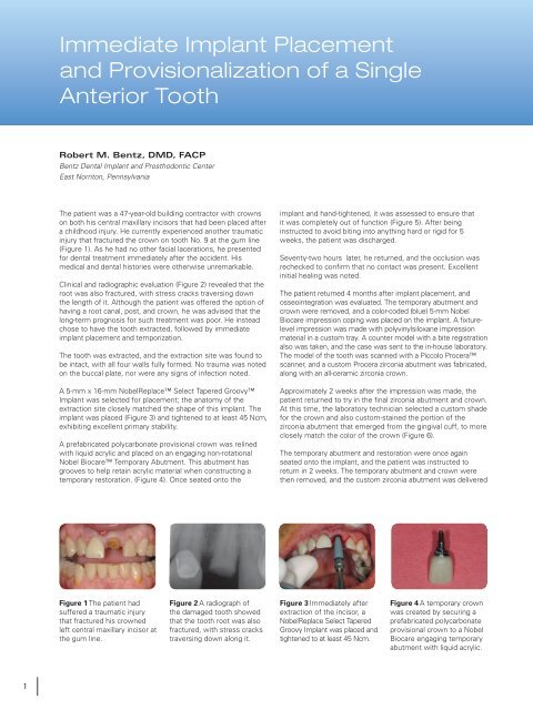
Figure 1 The patient had<br />
suffered a traumatic injury<br />
that fractured his crowned<br />
left central maxillary incisor at<br />
the gum line.<br />
Figure 2 A radiograph of<br />
the damaged tooth showed<br />
that the tooth root was also<br />
fractured, with stress cracks<br />
traversing down along it.<br />
Figure 3 <strong>Immediate</strong>ly after<br />
extraction of the incisor, a<br />
<strong>Nobel</strong>Replace Select Tapered<br />
Groovy <strong>Implant</strong> was placed <strong>and</strong><br />
tightened to at least 45 Ncm.<br />
Figure 4 A temporary crown<br />
was created by securing a<br />
prefabricated polycarbonate<br />
provisional crown to a <strong>Nobel</strong><br />
<strong>Biocare</strong> engaging temporary<br />
abutment with liquid acrylic.<br />
1
Case Report<br />
onto the implant. The final zirconia crown was secured to<br />
the abutment with temporary cement. After another month,<br />
the patient returned, the crown was removed, <strong>and</strong> excellent<br />
healing of the soft tissue around the abutment was noted,<br />
demonstrating the biocompatibility of the zirconia (Figure 7).<br />
The abutment was then secured using a torque (350 Nm) driver<br />
<strong>and</strong> ImProv ® long-term implant temporary cement (Figure 8).<br />
Conclusion<br />
The asymptomatic status of this patient’s fractured tooth <strong>and</strong><br />
the excellent status of the extraction site made this an ideal<br />
situation for immediate implant placement <strong>and</strong> temporization.<br />
Using the Procera zirconia custom CAD/CAM abutment <strong>and</strong><br />
crown enabled achievement of a highly esthetic <strong>and</strong> natural<br />
anterior restoration. This technique provided a complete<br />
solution for the patient with an exceptional result.<br />
Figure 5 The temporary<br />
abutment <strong>and</strong> crown were<br />
seated on the implant.<br />
Figure 6 Approximately 2<br />
weeks after a fixture level<br />
impression was made, the<br />
zirconia Procera abutment<br />
was tried in, <strong>and</strong> the color<br />
was adjusted.<br />
Figure 7 After wearing the<br />
definitive abutment <strong>and</strong> crown<br />
(secured with temporary<br />
cement) for a month, the patient<br />
returned. The crown was<br />
removed <strong>and</strong> vibrant, healthy<br />
gingival tissue was noted to<br />
have healed in close proximity<br />
to the zirconia abutment.<br />
Figure 8 The completed<br />
abutment <strong>and</strong> crown, 1<br />
month after placement <strong>and</strong> 6<br />
months after implant surgery.<br />
<strong>Immediate</strong> <strong>Implant</strong> <strong>Placement</strong> <strong>and</strong> Provisionalization of a Single Anterior Tooth 2