BD™ CompBeads Anti-Mouse Ig, κ/Negative Control (FBS ...
BD™ CompBeads Anti-Mouse Ig, κ/Negative Control (FBS ...
BD™ CompBeads Anti-Mouse Ig, κ/Negative Control (FBS ...

You also want an ePaper? Increase the reach of your titles
YUMPU automatically turns print PDFs into web optimized ePapers that Google loves.
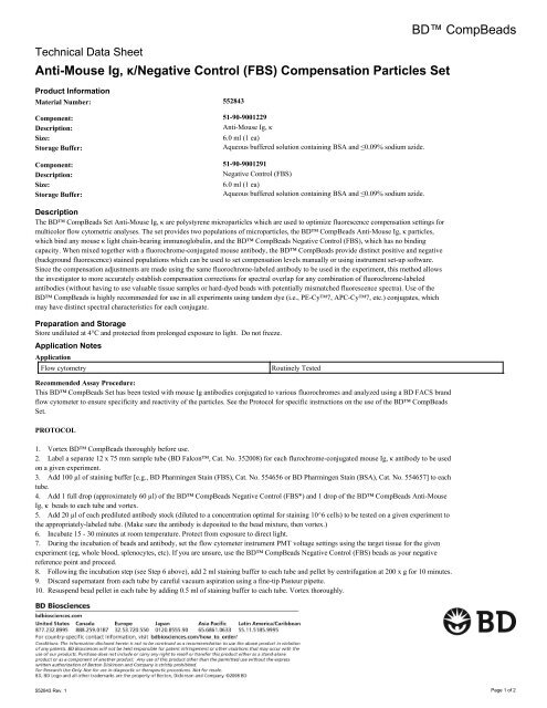
Technical Data Sheet<br />
<strong>Anti</strong>-<strong>Mouse</strong> <strong>Ig</strong>, <strong>κ</strong>/<strong>Negative</strong> <strong>Control</strong> (<strong>FBS</strong>) Compensation Particles Set<br />
Product Information<br />
Material Number: 552843<br />
Component: 51-90-9001229<br />
Description:<br />
<strong>Anti</strong>-<strong>Mouse</strong> <strong>Ig</strong>, <strong>κ</strong><br />
Size:<br />
6.0 ml (1 ea)<br />
Storage Buffer:<br />
Aqueous buffered solution containing BSA and ≤0.09% sodium azide.<br />
Component: 51-90-9001291<br />
Description:<br />
<strong>Negative</strong> <strong>Control</strong> (<strong>FBS</strong>)<br />
Size:<br />
6.0 ml (1 ea)<br />
Storage Buffer:<br />
Aqueous buffered solution containing BSA and ≤0.09% sodium azide.<br />
Description<br />
The BD <strong>CompBeads</strong> Set <strong>Anti</strong>-<strong>Mouse</strong> <strong>Ig</strong>, <strong>κ</strong> are polystyrene microparticles which are used to optimize fluorescence compensation settings for<br />
multicolor flow cytometric analyses. The set provides two populations of microparticles, the BD <strong>CompBeads</strong> <strong>Anti</strong>-<strong>Mouse</strong> <strong>Ig</strong>, <strong>κ</strong> particles,<br />
which bind any mouse <strong>κ</strong> light chain-bearing immunoglobulin, and the BD <strong>CompBeads</strong> <strong>Negative</strong> <strong>Control</strong> (<strong>FBS</strong>), which has no binding<br />
capacity. When mixed together with a fluorochrome-conjugated mouse antibody, the BD <strong>CompBeads</strong> provide distinct positive and negative<br />
(background fluorescence) stained populations which can be used to set compensation levels manually or using instrument set-up software.<br />
Since the compensation adjustments are made using the same fluorochrome-labeled antibody to be used in the experiment, this method allows<br />
the investigator to more accurately establish compensation corrections for spectral overlap for any combination of fluorochrome-labeled<br />
antibodies (without having to use valuable tissue samples or hard-dyed beads with potentially mismatched fluorescence spectra). Use of the<br />
BD <strong>CompBeads</strong> is highly recommended for use in all experiments using tandem dye (i.e., PE-Cy7, APC-Cy7, etc.) conjugates, which<br />
may have distinct spectral characteristics for each conjugate.<br />
Preparation and Storage<br />
Store undiluted at 4°C and protected from prolonged exposure to light. Do not freeze.<br />
Application Notes<br />
Application<br />
Flow cytometry<br />
Routinely Tested<br />
Recommended Assay Procedure:<br />
This BD <strong>CompBeads</strong> Set has been tested with mouse <strong>Ig</strong> antibodies conjugated to various fluorochromes and analyzed using a BD FACS brand<br />
flow cytometer to ensure specificity and reactivity of the particles. See the Protocol for specific instructions on the use of the BD <strong>CompBeads</strong><br />
Set.<br />
PROTOCOL<br />
1. Vortex BD <strong>CompBeads</strong> thoroughly before use.<br />
2. Label a separate 12 x 75 mm sample tube (BD Falcon, Cat. No. 352008) for each flurochrome-conjugated mouse <strong>Ig</strong>, <strong>κ</strong> antibody to be used<br />
on a given experiment.<br />
3. Add 100 µl of staining buffer [e.g., BD Pharmingen Stain (<strong>FBS</strong>), Cat. No. 554656 or BD Pharmingen Stain (BSA), Cat. No. 554657] to each<br />
tube.<br />
4. Add 1 full drop (approximately 60 µl) of the BD <strong>CompBeads</strong> <strong>Negative</strong> <strong>Control</strong> (<strong>FBS</strong>*) and 1 drop of the BD <strong>CompBeads</strong> <strong>Anti</strong>-<strong>Mouse</strong><br />
<strong>Ig</strong>, <strong>κ</strong> beads to each tube and vortex.<br />
5. Add 20 µl of each prediluted antibody stock (diluted to a concentration optimal for staining 10^6 cells) to be tested on a given experiment to<br />
the appropriately-labeled tube. (Make sure the antibody is deposited to the bead mixture, then vortex.)<br />
6. Incubate 15 - 30 minutes at room temperature. Protect from exposure to direct light.<br />
7. During the incubation of beads and antibody, set the flow cytometer instrument PMT voltage settings using the target tissue for the given<br />
experiment (eg, whole blood, splenocytes, etc). If you are unsure, use the BD <strong>CompBeads</strong> <strong>Negative</strong> <strong>Control</strong> (<strong>FBS</strong>) beads as your negative<br />
reference point and proceed.<br />
8. Following the incubation step (see Step 6 above), add 2 ml staining buffer to each tube and pellet by centrifugation at 200 x g for 10 minutes.<br />
9. Discard supernatant from each tube by careful vacuum aspiration using a fine-tip Pasteur pipette.<br />
10. Resuspend bead pellet in each tube by adding 0.5 ml of staining buffer to each tube. Vortex thoroughly.<br />
BD <strong>CompBeads</strong><br />
552843 Rev. 1 Page 1 of 2
11. Run each tube separately on the flow cytometer. Gate on the singlet bead population based on FSC (forward-light scatter) and SSC (side-light<br />
scatter) characteristics.<br />
12. Adjust flow rate to 200 - 300 events per second if possible.<br />
13. Create a dot plot for the given fluorochrome-conjugated antibody as appropriate [i.e., to set compensation for a fluorescein (FITC)-conjugated<br />
antibody, use an FL1 vs. FL2 dot plot].<br />
14. Place a quadrant gate such that the negative bead population is in the lower left quadrant and the positive bead population is in the upper or<br />
lower right quadrant, and adjust the compensation values until the median fluorescence intensity (MFI) of each population (as shown in the<br />
quadrant stats window) is approximately equal (i.e., for FL2 -%FL1, the FL2 MFI of both bead populations should be approximately equal when<br />
properly compensated).<br />
15. Repeat Steps 13 and 14 for other tubes, as necessary.<br />
16. Proceed to acquiring the actual staining experiment.<br />
Product Notices<br />
1. Since applications vary, each investigator should titrate the reagent to obtain optimal results.<br />
2. Please refer to www.bdbiosciences.com/pharmingen/protocols for technical protocols.<br />
3. Cy is a trademark of Amersham Biosciences Limited.<br />
4. Caution: Sodium azide yields highly toxic hydrazoic acid under acidic conditions. Dilute azide compounds in running water before<br />
discarding to avoid accumulation of potentially explosive deposits in plumbing.<br />
5. Source of all serum proteins is from USDA inspected abattoirs located in the United States.<br />
552843 Rev. 1 Page 2 of 2