Apoptosis, DNA Damage and Cell Proliferation by Becton Dickinson ...
Apoptosis, DNA Damage and Cell Proliferation by Becton Dickinson ...
Apoptosis, DNA Damage and Cell Proliferation by Becton Dickinson ...
Create successful ePaper yourself
Turn your PDF publications into a flip-book with our unique Google optimized e-Paper software.
<strong>Apoptosis</strong>, <strong>DNA</strong> <strong>Damage</strong>,<br />
<strong>and</strong> <strong>Cell</strong> <strong>Proliferation</strong> Kit<br />
Features<br />
Useful for determining the effects of small molecules,<br />
radiation, <strong>and</strong> other environmental stressors on cell<br />
cycle, proliferation, apoptosis, <strong>and</strong> <strong>DNA</strong> damage<br />
Conserve precious sample with the measurement of<br />
multiple parameters in a single tube<br />
Tested on both human <strong>and</strong> mouse samples<br />
Compatible with most flow cytometers with two<br />
or more lasers such as the BD Accuri® C6 <strong>and</strong> the<br />
BD FACSCalibur*<br />
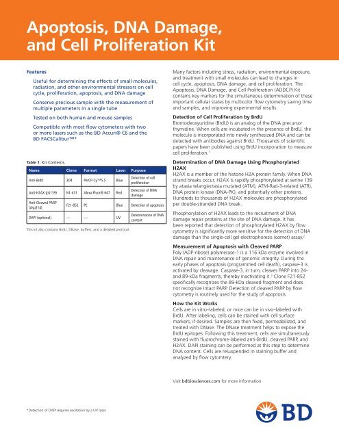
Table 1. Kit Contents.<br />
Name Clone Format Laser Purpose<br />
Anti-BrdU 3D4 PerCP-Cy5.5 Blue<br />
Anti-H2AX (pS139) N1-431 Alexa Fluor® 647 Red<br />
Anti-Cleaved PARP<br />
(Asp214)<br />
This kit also contains BrdU, DNase, buffers, <strong>and</strong> a detailed protocol.<br />
Detection of cell<br />
proliferation<br />
Detection of <strong>DNA</strong><br />
damage<br />
F21-852 PE Blue Detection of apoptosis<br />
DAPI (optional) — — UV<br />
Determination of <strong>DNA</strong><br />
content<br />
Many factors including stress, radiation, environmental exposure,<br />
<strong>and</strong> treatment with small molecules can lead to changes in<br />
cell cycle, apoptosis, <strong>DNA</strong> damage, <strong>and</strong> cell proliferation. The<br />
<strong>Apoptosis</strong>, <strong>DNA</strong> <strong>Damage</strong>, <strong>and</strong> <strong>Cell</strong> <strong>Proliferation</strong> (ADDCP) Kit<br />
contains key markers for the simultaneous determination of these<br />
important cellular states <strong>by</strong> multicolor flow cytometry saving time<br />
<strong>and</strong> samples, <strong>and</strong> improving experimental results.<br />
Detection of <strong>Cell</strong> <strong>Proliferation</strong> <strong>by</strong> BrdU<br />
Bromodeoxyuridine (BrdU) is an analog of the <strong>DNA</strong> precursor<br />
thymidine. When cells are incubated in the presence of BrdU, the<br />
molecule is incorporated into newly synthesized <strong>DNA</strong> <strong>and</strong> can be<br />
detected with antibodies against BrdU. Thous<strong>and</strong>s of scientific<br />
papers have been published using BrdU incorporation to measure<br />
cell proliferation. 1<br />
Determination of <strong>DNA</strong> <strong>Damage</strong> Using Phosphorylated<br />
H2AX<br />
H2AX is a member of the histone H2A protein family. When <strong>DNA</strong><br />
str<strong>and</strong> breaks occur, H2AX is rapidly phosphorylated at serine 139<br />
<strong>by</strong> ataxia telangiectasia mutated (ATM), ATM-Rad-3-related (ATR),<br />
<strong>DNA</strong> protein kinase (<strong>DNA</strong>-PK), <strong>and</strong> potentially other proteins.<br />
Hundreds to thous<strong>and</strong>s of H2AX molecules are phosphorylated<br />
per double-str<strong>and</strong>ed <strong>DNA</strong> break.<br />
Phosphorylation of H2AX leads to the recruitment of <strong>DNA</strong><br />
damage repair proteins at the site of <strong>DNA</strong> damage. It has<br />
been reported that detection of phosphorylated H2AX <strong>by</strong> flow<br />
cytometry is significantly more sensitive for the detection of <strong>DNA</strong><br />
damage than the single-cell gel electrophoresis (comet) assay. 2<br />
Measurement of <strong>Apoptosis</strong> with Cleaved PARP<br />
Poly (ADP-ribose) polymerase-1 is a 116 kDa enzyme involved in<br />
<strong>DNA</strong> repair <strong>and</strong> maintenance of genomic integrity. During the<br />
early phases of apoptosis (programmed cell death), caspase-3 is<br />
activated <strong>by</strong> cleavage. Caspase-3, in turn, cleaves PARP into 24-<br />
<strong>and</strong> 89-kDa fragments, there<strong>by</strong> inactivating it. 3 Clone F21-852<br />
specifically recognizes the 89-kDa cleaved fragment <strong>and</strong> does<br />
not recognize intact PARP. Detection of cleaved PARP <strong>by</strong> flow<br />
cytometry is routinely used for the study of apoptosis.<br />
How the Kit Works<br />
<strong>Cell</strong>s are in vitro–labeled, or mice can be in vivo–labeled with<br />
BrdU. After labeling, cells can be stained with cell surface<br />
markers, if desired. Samples are then fixed, permeabilized, <strong>and</strong><br />
treated with DNase. The DNase treatment helps to expose the<br />
BrdU epitopes. Following this treatment, cells are simultaneously<br />
stained with fluorochrome-labeled anti-BrdU, cleaved PARP, <strong>and</strong><br />
H2AX. DAPI staining can be performed at this step to determine<br />
<strong>DNA</strong> content. <strong>Cell</strong>s are resuspended in staining buffer <strong>and</strong><br />
analyzed <strong>by</strong> flow cytomtery.<br />
Visit bdbiosciences.com for more information.<br />
*Detection of DAPI requires excitation <strong>by</strong> a UV laser.
<strong>Apoptosis</strong>, <strong>DNA</strong> <strong>Damage</strong>, <strong>and</strong> <strong>Cell</strong> <strong>Proliferation</strong> Kit<br />
References<br />
1. Li C. Specific cell cycle synchronization with butyrate <strong>and</strong> cell cycle<br />
analysis. Methods Mol Biol. 2011;761:125-136.<br />
2. Kuo LJ, Yang LX. Gamma-H2AX - a novel biomarker for <strong>DNA</strong> doublestr<strong>and</strong><br />
breaks. In Vivo. 2008;22:305-309.<br />
PBMCs activated<br />
with CD3/CD28<br />
PBMCs activated<br />
with CD3/CD28 +<br />
Camptothecin 3 hr<br />
Figure 1. PBMCs were stimulated with Anti-CD3/CD28–coated Dynabeads® for 3<br />
days, then harvested <strong>and</strong> washed, <strong>and</strong> then replated with either 5 mM of camptothecin<br />
or left untreated as controls. The treated cell group was cultured for 3 hours<br />
with camptothecin, then washed <strong>and</strong> replated for an additional 2 hours allowing<br />
cells to recover. The untreated group was cultured for 5 hours. Both groups (control<br />
<strong>and</strong> treated) were pulsed with 50 mM of BrdU during the final 1 hour of culture.<br />
<strong>Cell</strong>s were harvested, washed with staining buffer, then fixed then analyzed using<br />
the <strong>Apoptosis</strong>, <strong>DNA</strong> <strong>Damage</strong>, <strong>and</strong> <strong>Cell</strong> Proliferaiton Kit (Cat. No. 562253).<br />
Figures A–C are the untreated control group <strong>and</strong> figures D–F are the camptothecintreated<br />
group. From the BrdU vs DAPI profile (figures A <strong>and</strong> D), it is apparent that<br />
camptothecin disrupted the cells’ ability to incorporate BrdU or decreased cell<br />
proliferation. The untreated group shows a characteristic horseshoe pattern with<br />
43% of the cells incorporating BrdU during the 1-hour pulse, compared with 21%<br />
Ordering Information<br />
Description<br />
Cat.No.<br />
<strong>Apoptosis</strong>, <strong>DNA</strong> <strong>Damage</strong>, <strong>and</strong> <strong>Cell</strong> <strong>Proliferation</strong> Kit (50 Tests) 562253<br />
Contents<br />
A<br />
BrdU PerCP-Cy5.5<br />
10 2 10 3 10 4 10 5<br />
BrdU PerCP-Cy5.5<br />
10 2 10 3 10 4 10 5<br />
PBMC-untreated<br />
50<br />
P2<br />
43%<br />
100 150 200 250<br />
DAPI (x 1,000)<br />
PBMC-3 hr Campto<br />
50<br />
P2<br />
100 150 200 250<br />
DAPI (x 1,000)<br />
H2AX Alexa Fluor® 647<br />
10 2 10 3 10 4 10 5<br />
B<br />
H2AX Alexa Fluor® 647<br />
10 2 10 3 10 4 10 5<br />
PBMC-untreated<br />
Q1<br />
Q3<br />
Q2<br />
Q4<br />
10 2 10 3 10 4 10 5<br />
BrdU PerCP-Cy5.5<br />
H2AX Alexa Fluor® 647<br />
10 2 10 3 10 4 10 5<br />
3. Krishnakumar R, Kraus WL. The PARP side of the nucleus: molecular<br />
actions, physiological outcomes, <strong>and</strong> clinical targets. Mol <strong>Cell</strong>.<br />
2010;39:8-24.<br />
PBMC-untreated<br />
Q1-2 Q2-2<br />
14.9% 32.6% 45.7% 4.3%<br />
12.2%<br />
D E F<br />
21%<br />
PBMC-3 hr Campto<br />
Q1<br />
51%<br />
Monoclonal Antibodies<br />
BrdU PerCP-Cy5.5<br />
H2AX (pSer139) Alexa Fluor® 647<br />
Cleaved PARP (Asp214) PE<br />
Other Reagents<br />
BD Cytofix/Cytoperm Fixation/Permeabilization Solution<br />
BD Perm/Wash Buffer<br />
BD Cytofix/Cytoperm Plus Permeabilization Buffer<br />
DAPI<br />
BrdU<br />
DNase<br />
Q3<br />
Q2<br />
21.7%<br />
1.1%<br />
Q4<br />
10 2 10 3 10 4 10 5<br />
BrdU PerCP-Cy5.5<br />
C<br />
H2AX Alexa Fluor® 647<br />
10 2 10 3 10 4 10 5<br />
Q3-2<br />
10 2 10 3 10 4 10 5<br />
Cleaved PARP PE<br />
PBMC-3 hr Campto<br />
Q1-2<br />
50.9%<br />
10 2 10 3 10 4<br />
Cleaved PARP PE<br />
Q2-2<br />
23.2%<br />
Q3-2 Q4-2<br />
10 5<br />
of the cells incorporating BrdU in the treated group. The H2AX signal (figures B<br />
<strong>and</strong> E) also show differences. The untreated control has a low signal intensity of<br />
H2AX with approximately 47% of the cells staining positive. The majority of the<br />
H2AX cells are on the BrdU + (proliferating) population. The camptothecin-treated<br />
group shows both a higher signal intensity <strong>and</strong> a higher percentage of positive<br />
cells (72%), with the majority of the staining on the BrdU – population (non-proliferating).<br />
The cleaved PARP (apoptosis) signal is shown in figures C <strong>and</strong> F. Untreated<br />
cells show low levels of cleaved PARP (4.3%). The camptothecin-treated group has<br />
a much higher percentage of cleaved PARP positive cells (23.2%). The percentage<br />
of H2AX + /PARP – cells is not that different when comparing the untreated cells<br />
(45.7%) <strong>and</strong> the camptothecin-treated cells (50.9%). The difference is in the signal<br />
intensity. Nearly all of the cells in this experiment that are positive for cleaved PARP<br />
are also positive for H2AX.<br />
For Research Use Only. Not for use in diagnostic or therapeutic procedures.<br />
Alexa Fluor® is a registered trademark of Molecular Probes, Inc.<br />
Cy is a trademark of Amersham Biosciences Corp. Cy dyes are subject to proprietary rights of Amersham Biosciences Corp<br />
<strong>and</strong> Carnegie Mellon University <strong>and</strong> are made <strong>and</strong> sold under license from Amersham Biosciences Corp only for research <strong>and</strong><br />
in vitro diagnostic use. Any other use requires a commercial sublicense from Amersham Biosciences Corp, 800 Centennial<br />
Avenue, Piscataway, NJ 08855-1327, USA.<br />
Dynabeads is a registered trademark of Life Technologies.<br />
BD, BD Logo <strong>and</strong> all other trademarks are property of <strong>Becton</strong>, <strong>Dickinson</strong> <strong>and</strong> Company. © 2011 BD<br />
23-13576-00<br />
BD Biosciences<br />
2350 Qume Drive<br />
San Jose, CA 95131<br />
US Orders: 855.236.2772<br />
Technical Service: 877.232.8995<br />
answers@bd.com<br />
bdbiosciences.com


