BD™ Phosflow Alexa Fluor® 647 Rat anti-Histone H3 (pS28)
BD™ Phosflow Alexa Fluor® 647 Rat anti-Histone H3 (pS28)
BD™ Phosflow Alexa Fluor® 647 Rat anti-Histone H3 (pS28)

You also want an ePaper? Increase the reach of your titles
YUMPU automatically turns print PDFs into web optimized ePapers that Google loves.
BD <strong>Phosflow</strong><br />
Technical Data Sheet<br />
<strong>Alexa</strong> <strong>Fluor®</strong> <strong>647</strong> <strong>Rat</strong> <strong>anti</strong>-<strong>Histone</strong> <strong>H3</strong> (<strong>pS28</strong>)<br />
Product Information<br />
Material Number: 558217<br />
Size:<br />
50 tests<br />
Vol. per Test: 20 µl<br />
Clone:<br />
HTA28<br />
Immunogen:<br />
Phosphorylated Human <strong>Histone</strong> <strong>H3</strong> Peptide<br />
Isotype:<br />
<strong>Rat</strong> IgG2a, κ<br />
Reactivity:<br />
Tested: Human, Mouse,<br />
Reported: Cow, Drosophila, Hamster, <strong>Rat</strong><br />
Storage Buffer:<br />
Aqueous buffered solution containing BSA and ≤0.09% sodium azide.<br />
Description<br />
<strong>Histone</strong>s are highly basic proteins that complex with DNA to form chromatin. <strong>Histone</strong> <strong>H3</strong> is a ~15-kDa protein that is phosphorylated at<br />
serine 28 (S28), S10, and/or threonine 11 during mammalian cell mitosis and meiosis. The phosphorylation sites are located in the N-terminal<br />
tail, a region that is outside of the chromatin fiber and is thus accessible for interactions with agents that may regulate chromatin or specific<br />
gene activities. The phosphorylation states of the two serine sites during the cell cycle are highly regulated by Aurora B kinase and a PP1<br />
phosphatase: S10 is in the phosphorylated state from late G2 phase to anaphase, while S28 is phosphorylated from prophase to anaphase.<br />
Furthermore, phosphorylation of histone <strong>H3</strong> S28 may be mediated by other kinases in response to external stimuli. Evidence suggests that<br />
histone phosphorylation is involved in the regulation of chromosome condensation, cell division, and gene transcription.<br />
The HTA28 monoclonal <strong>anti</strong>body reacts with histone <strong>H3</strong> phosphorylated at S28 in its N-terminal tail. It does not recognize the<br />
unphosphorylated protein.<br />
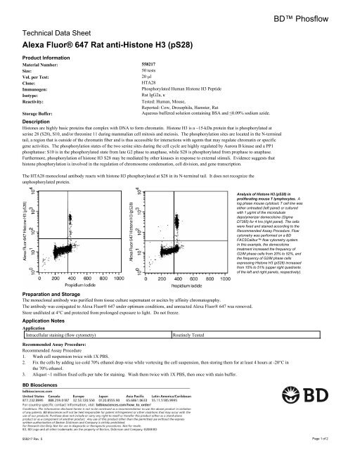
Analysis of <strong>Histone</strong> <strong>H3</strong> (<strong>pS28</strong>) in<br />
proliferating mouse T lymphocytes. A<br />
log phase mouse cytotoxic T cell line was<br />
either untreated (left panel) or cultured<br />
with 1 μg/ml of the microtubule<br />
depolymerizer demecolcine (Sigma<br />
D7385) for 4 hrs (right panel). The cells<br />
were fixed and stained according to the<br />
Recommended Assay Procedure. Flow<br />
cytometry was performed on a BD<br />
FACSCalibur flow cytometry system.<br />
In this example, the demecolcine<br />
treatment increased the frequency of<br />
G2/M phase cells from 20% to 52%, and<br />
the frequency of G2/M phase cells<br />
expressing <strong>Histone</strong> <strong>H3</strong> (<strong>pS28</strong>) increased<br />
from 15% to 51% (upper right quadrants<br />
of the left and right panels, respectively).<br />
Preparation and Storage<br />
The monoclonal <strong>anti</strong>body was purified from tissue culture supernatant or ascites by affinity chromatography.<br />
The <strong>anti</strong>body was conjugated to <strong>Alexa</strong> <strong>Fluor®</strong> <strong>647</strong> under optimum conditions, and unreacted <strong>Alexa</strong> <strong>Fluor®</strong> <strong>647</strong> was removed.<br />
Store undiluted at 4°C and protected from prolonged exposure to light. Do not freeze.<br />
Application Notes<br />
Application<br />
Intracellular staining (flow cytometry)<br />
Routinely Tested<br />
Recommended Assay Procedure:<br />
Recommended Assay Procedure<br />
1. Wash cell suspension twice with 1X PBS.<br />
2. Fix the cells by adding ice-cold 70% ethanol drop-wise while vortexing the cell suspension, then storing them for at least 4 hours at -20°C in<br />
the 70% ethanol.<br />
3. Aliquot ~1 million fixed cells per tube for staining. Wash them twice with 1X PBS, then once with stain buffer.<br />
558217 Rev. 5 Page 1 of 2
4. Stain the cells with 20 μl <strong>Alexa</strong> <strong>Fluor®</strong> <strong>647</strong> <strong>Rat</strong> <strong>anti</strong>-<strong>Histone</strong> <strong>H3</strong> (<strong>pS28</strong>) in 80 μl stain buffer for 20 minutes at room temperature, then wash<br />
them with stain buffer.<br />
5. For optimum cell cycle analysis, the cells should be treated with RNAse before staining with propidium iodide:<br />
• Treat the stained cells with 50 μg RNAse A (Sigma R5500) in 50 μl 1X PBS for 30 minutes at 37°C. Without washing, stain DNA by<br />
adding 5 μg Propidium Iodide (Sigma P4170) in 450 μl staining buffer for at least 10 minutes at room temperature.<br />
Or<br />
• Stain the cells with 0.5 ml PI/RNase Staining Buffer (Cat. No. 550825) for 15 minutes at room temperature.<br />
6. The cells are now ready for flow cytometric analysis.<br />
Suggested Companion Products<br />
Catalog Number Name Size Clone<br />
554656 Stain Buffer (FBS) 500 ml (none)<br />
550825 PI/RNase Staining Buffer 100 ml (none)<br />
Product Notices<br />
1. This reagent has been pre-diluted for use at the recommended Volume per Test. We typically use 1 × 10^6 cells in a 100-µl experimental<br />
sample (a test).<br />
2. Please refer to www.bdbiosciences.com/pharmingen/protocols for technical protocols.<br />
3. Since applications vary, each investigator should titrate the reagent to obtain optimal results.<br />
4. This product is sold under license from Shigei Medical Research Institute, Okayama, Japan.<br />
5. <strong>Alexa</strong> <strong>Fluor®</strong> <strong>647</strong> fluorochrome emission is collected at the same instrument settings as for allophycocyanin (APC).<br />
6. For fluorochrome spectra and suitable instrument settings, please refer to our Fluorochrome Web Page at www.bdbiosciences.com/colors.<br />
7. Caution: Sodium azide yields highly toxic hydrazoic acid under acidic conditions. Dilute azide compounds in running water before<br />
discarding to avoid accumulation of potentially explosive deposits in plumbing.<br />
8. Source of all serum proteins is from USDA inspected abattoirs located in the United States.<br />
9. <strong>Alexa</strong> Fluor is a registered trademark of Molecular Probes, Inc., Eugene, OR.<br />
10. The <strong>Alexa</strong> <strong>Fluor®</strong>, Pacific Blue, and Cascade Blue® dye <strong>anti</strong>body conjugates in this product are sold under license from Molecular<br />
Probes, Inc. for research use only, excluding use in combination with microarrays, or as analyte specific reagents. The <strong>Alexa</strong> <strong>Fluor®</strong> dyes<br />
(except for <strong>Alexa</strong> <strong>Fluor®</strong> 430), Pacific Blue dye, and Cascade Blue® dye are covered by pending and issued patents.<br />
References<br />
Carmena M, Earnshaw WC. The cellular geography of aurora kinases. Nat Rev Mol Cell Biol. 2003; 4:842-854.(Biology)<br />
Cheung P, Allis CD, Sassone-Corsi P. Signaling to chromatin through histone modifications. Cell. 2000; 103:263-271.(Biology)<br />
Choi HS, Choi BY, Cho Y-Y, Zhu F, Bode AM, Dong Z. Phosphorylation of Ser28 in histone <strong>H3</strong> mediated by mixed lineage kinase-like mitogen-activated protein<br />
triple kinase α. J Biol Chem. 2005; 280(14):13545-13553.(Biology)<br />
Dyson MH, Thomson S, Inagaki M, et al. MAP kinase-mediated phosphorylation of distinct pools of histone <strong>H3</strong> at S10 or S28 via mitogen- and stress-activated<br />
kinase 1/2. J Cell Sci. 2005; 118(10):2247-2259.(Clone-specific)<br />
Furukawa K, Sugiyama S, Osouda S, et al. Barrier-to-autointegration factor plays crucial roles in cell cycle progression and nuclear organization in Drosophila. J<br />
Cell Sci. 2003; 116(18):3811-3823.(Clone-specific)<br />
Goto H, Tomono Y, Ajiro K, et al. Identification of a novel phosphorylation site on histone <strong>H3</strong> coupled with mitotic chromosome condensation. J Biol Chem. 1999;<br />
274(36):25543-25549.(Immunogen)<br />
Hirata A, Inada K, Tsukamoto T, et al. Characterization of a monoclonal <strong>anti</strong>body, HTA28, recognizing a histone <strong>H3</strong> phosphorylation site as a useful marker of<br />
M-phase cells. J Histochem Cytochem. 2004; 52(11):1503-1509.(Clone-specific)<br />
Nowak SJ, Corces VG. Phosphorylation of histone <strong>H3</strong>: a balancing act between chromosome condensation and transcriptional activation. Trends Genet. 2004;<br />
20(4):214-220.(Biology)<br />
Pascreau G, Arlot-Bonnemains Y, Prigent C. Phosphorylation of histone and histone-like proteins by aurora kinases during mitosis. Prog Cell Cycle Res. 2003;<br />
5:369-374.(Biology)<br />
558217 Rev. 5 Page 2 of 2