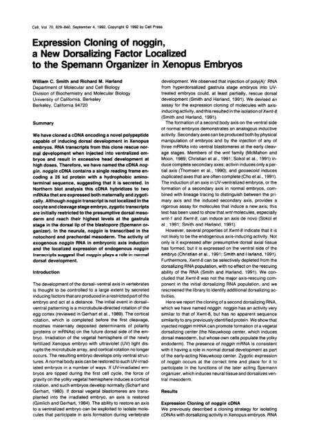
Expression Cloning of noggin, a New Dorsalizing Factor Localized ...
Expression Cloning of noggin, a New Dorsalizing Factor Localized ...
Expression Cloning of noggin, a New Dorsalizing Factor Localized ...
You also want an ePaper? Increase the reach of your titles
YUMPU automatically turns print PDFs into web optimized ePapers that Google loves.
Cell, Vol. 70, 829-840. September 4, 1992, Copyright 0 1992 by Cell Press<br />
<strong>Expression</strong> <strong>Cloning</strong> <strong>of</strong> <strong>noggin</strong>,<br />
a <strong>New</strong> <strong>Dorsalizing</strong> <strong>Factor</strong> <strong>Localized</strong><br />
to the Spemann Organizer in Xenopus Embryos<br />
William C. Smith and Richard M. Harland<br />
Department <strong>of</strong> Molecular and Cell Biology<br />
Division <strong>of</strong> Biochemistry and Molecular Biology<br />
University <strong>of</strong> California, Berkeley<br />
Berkeley, California 94720<br />
Summary<br />
We have cloned a cDNA encoding a novel polypeptide<br />
capable <strong>of</strong> inducing dorsal development in Xenopus<br />
embryos. RNA transcripts from this clone rescue normal<br />
development when injected into ventralized embryos<br />
and result in excessive head development at<br />
high doses. Therefore, we have named the cDNA <strong>noggin</strong>.<br />
<strong>noggin</strong> cDNA contains a single reading frame encoding<br />
a 26 kd protein with a hydrophobic aminoterminal<br />
sequence, suggesting that it is secreted. In<br />
Northern blot analysis this cDNA hybridizes to two<br />
mRNAs that are expressed both maternally and zygotically.<br />
Although <strong>noggin</strong> transcript is not localized in the<br />
oocyte and cleavage stage embryo, zygotic transcripts<br />
are initially restricted to the presumptive dorsal mesoderm<br />
and reach their highest levels at the gastrula<br />
stage in the dorsal lip <strong>of</strong> the blastopore (Spemann organizer).<br />
In the neurula, <strong>noggin</strong> is transcribed in the<br />
notochord and prechordal mesoderm. The activity <strong>of</strong><br />
exogenous <strong>noggin</strong> RNA in embryonic axis induction<br />
and the localized expression <strong>of</strong> endogenous <strong>noggin</strong><br />
transcripts suggest that <strong>noggin</strong> plays a role in normal<br />
dorsal development.<br />
Introduction<br />
The development <strong>of</strong> the dorsal-ventral axis in vertebrates<br />
is thought to be controlled to a large extent by secreted<br />
inducing factors that are produced in a restricted part <strong>of</strong> the<br />
embryo and act at a distance. The initial event in dorsalventral<br />
patterning is a microtubule-directed rotation <strong>of</strong> the<br />
egg cortex (reviewed in Gerhart et al., 1989). The cortical<br />
rotation, which is completed before the first cleavage,<br />
modifies maternally deposited determinants <strong>of</strong> polarity<br />
(proteins or mRNAs) on the future dorsal side <strong>of</strong> the embryo.<br />
Irradiation <strong>of</strong> the vegetal hemisphere <strong>of</strong> the newly<br />
fertilized Xenopus embryo with ultraviolet (UV) light disrupts<br />
the microtubule array, and cortical rotation no longer<br />
occurs. The resulting embryo develops only ventral structures.<br />
A normal bodyaxiscan be restored tosuch UV-irradiated<br />
embryos in a number <strong>of</strong> ways. If UV-irradiated embryos<br />
are tipped during the first cell cycle, the force <strong>of</strong><br />
gravity on the yolky vegetal hemisphere induces a cortical<br />
rotation, and such embryos develop normally (Scharf and<br />
Gerhart, 1980). If dorsal vegetal blastomeres are transplanted<br />
into the irradiated embryo, an axis is restored<br />
(Gimlich and Gerhart, 1984). The ability to restore an axis<br />
to a ventralized embryo can be exploited to isolate molecules<br />
that participate in axis formation during vertebrate<br />
development. We observed that injection <strong>of</strong> poly(A)i RNA<br />
from hyperdorsalized gastrula stage embryos into UVtreated<br />
embryos could, at least partially, rescue dorsal<br />
development (Smith and Harland, 1991). We devised an<br />
assay for the expression cloning <strong>of</strong> molecules with axisinducing<br />
activity, and this resulted in the isolation <strong>of</strong> Xwnt-8<br />
(Smith and Harland, 1991).<br />
The formation <strong>of</strong> a second body axis on the ventral side<br />
<strong>of</strong> normal embryos demonstrates an analogous inductive<br />
activity. Secondary axes can be produced both by physical<br />
manipulation <strong>of</strong> embryos and by the injection <strong>of</strong> any <strong>of</strong><br />
three mRNAs into ventral blastomeres at the early cleavage<br />
stages. Members <strong>of</strong> the wnt family (McMahon and<br />
Moon, 1989; Christian et al., 1991; Sokol et al., 1991) induce<br />
complete secondary axes; activin induces only a partial<br />
axis (Thomsen et al., 1990); and goosecoid induces<br />
duplicated axes that are <strong>of</strong>ten complete (Cho et al., 1991).<br />
The induction <strong>of</strong> an axis in UV-ventralized embryos, or the<br />
formation <strong>of</strong> a secondary axis in normal embryos, combined<br />
with lineage tracing to distinguish between the primary<br />
axis and the induced secondary axis, provides a<br />
rigorous assay for molecules that induce a new axis; this<br />
test has been used to show that wnt molecules, especially<br />
writ-7 and Xwnt-8, can induce an axis de novo (Sokol et<br />
al., 1991; Smith and Harland, 1991).<br />
However, several properties <strong>of</strong> Xwnf-8 indicate that it is<br />
not likely to be the endogenous axis-inducing activity. Not<br />
only is it expressed after presumptive dorsal axial tissue<br />
has formed, but it is expressed on the ventral side <strong>of</strong> the<br />
embryo (Christian et al., 1991; Smith and Harland, 1991).<br />
Furthermore, Xwnr-8 can be selectively depleted from the<br />
dorsalizing RNA population, with no effect on the rescuing<br />
ability <strong>of</strong> the RNA (Smith and Harland, 1991). We concluded<br />
that Xwnt-8 was not the major axis-rescuing component<br />
in the initial dorsalizing RNA population, and we<br />
rescreened the library to identify additional dorsalizing activities.<br />
Here we report the cloning <strong>of</strong> a second dorsalizing RNA,<br />
which we have named <strong>noggin</strong>. <strong>noggin</strong> has an activity very<br />
similar to that <strong>of</strong> Xwnt8, but has no apparent sequence<br />
similarity to any previously identified protein. We show that<br />
injected <strong>noggin</strong> mRNA can promote formation <strong>of</strong> a vegetal<br />
dorsalizing center (the Nieuwkoop center, which induces<br />
dorsal mesoderm, but whose own cells populate the yolky<br />
endoderm). The presence <strong>of</strong> <strong>noggin</strong> mRNA is consistent<br />
with it having a role in normal dorsal development as part<br />
<strong>of</strong> the early-acting Nieuwkoop center. Zygotic expression<br />
<strong>of</strong> <strong>noggin</strong> occurs at the correct time and place for it to<br />
participate in the functions <strong>of</strong> the later acting Spemann<br />
organizer, which induces neural tissue and dorsalizes ventral<br />
mesoderm.<br />
Results<br />
<strong>Expression</strong> <strong>Cloning</strong> <strong>of</strong> <strong>noggin</strong> cDNA<br />
We previously described a cloning strategy for isolating<br />
cDNAs with dorsalizing activity in Xenopus embryos. RNA
from dorsalized gastrulae (treated with LiCl during blastula<br />
stages) was size fractionated and the active fraction used<br />
to construct a plasmid cDNA library. RNAs were synthesized<br />
from pools <strong>of</strong> plasmids and injected into ventralized<br />
embryos produced by UV treatment (Smith and Harland,<br />
1991). Pools <strong>of</strong> plasmid DNA that directed the synthesis<br />
<strong>of</strong> dorsalizing RNA were sib selected until single active<br />
clones were isolated. In the first screen we isolated Xwnf-8,<br />
which was surprising since Xwnt-8 is down-regulated by<br />
LiCl treatment. We therefore reexamined the initial pools<br />
<strong>of</strong> 10,000 clones to ask whether Xwnt-8 was the only dorsalizing<br />
activity present.<br />
The 10 pools <strong>of</strong> 10,000 clones were analyzed by filter<br />
hybridization for the presence <strong>of</strong> Xwnt-8. The result is<br />
shown in Figure 1A. Xwnt-8 clones were present in the<br />
two active pools (8 and 9), as well as in five others. The<br />
retrospective analysis demonstrates that although Xwnr-8<br />
is less abundant in RNA from dorsalized gastrulae, it is<br />
still an abundant mRNA that is highly represented in the<br />
library.<br />
In the isolation <strong>of</strong> Xwnt-8 (Smith and Harland, 1991)<br />
pool 9 was selected for subsequent sib selections. The<br />
Xwnt-8 hybridization signal was weaker in pool 8 than in<br />
pool 9 and not much different from the inactive pools that<br />
contained Xwnf-8. To test whether pool 8 contained dorsalizing<br />
activities other than Xwnt-8, it was subdivided into<br />
12 pools <strong>of</strong> 1000 clones. Five <strong>of</strong> the pools had activity in<br />
the rescue assay and three <strong>of</strong> these did not contain Xwnt-8.<br />
The Xwnt-b-negative pool with the strongest dorsal axisrescuing<br />
activity, pool 8.12, was further sib selected until<br />
a single active clone was isolated (clone A3 from pool<br />
8.12.12.A). The activity <strong>of</strong> the pools (i.e., the degree <strong>of</strong><br />
dorsal axis rescued) increased as progressively smaller<br />
pools <strong>of</strong> clones were assayed. At the second sib selection<br />
<strong>of</strong> the library (pools <strong>of</strong> 1000 clones), A3 hybridizing clones<br />
could account for the activity <strong>of</strong> all dorsalizing pools that<br />
A. Pool: 1 2 3 4 5 6 7 8 9 10<br />
Activity: - - - - - - - + _ + _ -<br />
Xwnt-6-b<br />
Figure 1. Detection <strong>of</strong> Xwnf-8 and <strong>noggin</strong> Clones in Sib Selection<br />
Pools<br />
Five microgram samples <strong>of</strong> library plasmid DNA from the first (A) and<br />
second (6) sib selections (10,000 and 1,006 clones per pool, respectively)<br />
were digested with EcoRl and EcoRV, separated on 1% agarose<br />
gels, and transferred to nylon membranes. The membranes were hybridized<br />
with an Xwnt-8 probe alone (A) or first with Xwnt-8 and then<br />
with <strong>noggin</strong> probes (6). The activities <strong>of</strong> the various pools in the dorsal<br />
axis rescue assay are indicated (plus or minus).<br />
did not contain Xwnt-8 (Figure 16). One pool <strong>of</strong> 1000<br />
clones (8.2) hybridized with the A3 probe but did not have<br />
activity in the assay (Figure 16). The reason for this is<br />
unknown, although it is possible that this particular A3<br />
hybridizing clone was not functional. In addition, at this<br />
stage in the sib selection the active pools only conferred<br />
partial rescue <strong>of</strong> dorsal development, and pools with dorsalizing<br />
RNAs could have been missed.<br />
Finally, preliminary results indicate that at least one additional<br />
dorsalizing activity may be present in our library.<br />
RNA transcribed from Ncol linearized library plasmid DNA<br />
(rather than Notl linearized) retained axis-rescuing activity.<br />
Ncol cleaves within the 5’ untranslated region <strong>of</strong> A3 and<br />
within the coding region <strong>of</strong> Xwnt-8 and yields truncated,<br />
nonfunctional transcripts. The completeness <strong>of</strong> Ncol<br />
cleavage <strong>of</strong> A3 and Xwnt-8 plasmids in the pool was confirmed<br />
by blotting (data not shown).<br />
<strong>noggin</strong> cDNA Encodes a Novel Polypeptide<br />
The 1834 nt sequence <strong>of</strong> the A3 clone is shown in Figure<br />
2A. The sequence contains a single long open reading<br />
frame encoding a 222 aa polypeptide with a predicted molecular<br />
size <strong>of</strong> 26 kd. At the amino terminus, a hydrophobic<br />
stretch <strong>of</strong> amino acids suggests that the polypeptide enters<br />
the secretory pathway (Figure 26). There is a single<br />
potential site for N-linked glycosylation (see the asterisk in<br />
Figure 2A). Extensive untranslated regions are located<br />
both 5’and 3’<strong>of</strong> the reading frame (594 and 573 bp, respectively).<br />
The 3’ untranslated region is particularly rich in<br />
repeated dA and dT nucleotides, and contains, in addition<br />
to a polyadenylation signal sequence located 24 bp upstream<br />
<strong>of</strong> the start <strong>of</strong> the poly(A) tail, a second potential<br />
polyadenylation sequence 147 bp further upstream (both<br />
are underlined in Figure 2A).<br />
In vitro translation <strong>of</strong> RNA synthesized from the A3 clone<br />
resulted in a protein product with the approximate molecular<br />
weight predicted by the open reading frame (data not<br />
shown). Comparison <strong>of</strong> the amino acid sequence <strong>of</strong> the<br />
predicted polypeptide to the National Center for Biotechnology<br />
Information BLAST network (nonredundant data<br />
base) did not identify any similar sequence. Clone A3 thus<br />
appears to encode a new type <strong>of</strong> protein that may be secreted<br />
and that has dorsal-inducing activity in Xenopus.<br />
<strong>noggin</strong> mRNA Can Rescue a Complete<br />
Dorsal-Ventral Axis<br />
The new sequence, and its putative protein product, were<br />
named <strong>noggin</strong> based upon the phenotype resulting from<br />
mRNA injection into ventralized embryos (see below). Injection<br />
<strong>of</strong> <strong>noggin</strong> RNA into a single blastomere <strong>of</strong> a 4-cell<br />
stage UV-ventralized embryo can restore the complete<br />
spectrum <strong>of</strong> dorsal structures. The degree <strong>of</strong> axis rescue<br />
was dependent upon the amount <strong>of</strong> RNA injected: the embryos<br />
that received low doses had only posterior dorsal<br />
structures, while embryos that received higher doses had<br />
excess dorsal-anterior tissue. RNA transcripts from two<br />
<strong>noggin</strong> plasmids were tested. The first (A3) contained the<br />
full cDNA. The second @NogginAY) had a deletion removing<br />
the first 513 nt <strong>of</strong> the 5’ untranslated region up to the<br />
EcoRl site (see Figure 2A). The results <strong>of</strong> injection <strong>of</strong> RNA
l<br />
<strong>noggin</strong> Rescues<br />
831<br />
Dorsal Development<br />
A<br />
-359:CGCTGGCTGATTGCGACTGTTGCTTTCCACAGCTCCCTTCTTCC~AGTTTCTTCTAGGA~AGATCGAGTCTCTGGTTA ‘a GATCGAGCTGAAAGTGAAGAATATTTAAGAGAG<br />
NC0 I<br />
-239:ffiGAGGCTGGAGCCAGCAGGCAGACAAAGTGGTGCCACCACC~GGACTGT~GT~GffiTGAGCGCATT~AGACAGACAG~GCTCTGCTG~CTTCCACTTGACTGCGATGAGA~~G<br />
-119:GAATCCCCAATTCGCTAGGTGCCCCTGAACCCCCCA L2AAim TCCTCTGATGCATTATTTATGATCTCTGGCAAGAAATCGGAGC<br />
EC0 RI<br />
41:ProLeuValA~pLe"I1eGluHlsProAspProIleTy~A~pP~~Ly~Gl"Ly~A~pL~"A~"Gl"Th~Le"Le~A~gTh~L~~~e~V~lGly~~~PheA~pP~~A~"Ph~~~~Al~T~~<br />
121:CCACTGGTGGACCTTATTGAGCACCCQ$.XCQC ATCTATGATCCCRAGGAGRAGGATCTTRACGAGACCTTGCCTTTATGGCCACC<br />
Ban HI<br />
81:IleLeuProGl"GluArgLeuGlyValGlyValGluAspLeuGlyGluLeuAspLeuLeuLeuArgGlnLysProSerGlyAlaMetProAlaGluIleLysGlyLeuGluPheTyrGluGlyLeu<br />
241:ATCCTGCCAGAGGAGAGACTTGGAGTGGAGGACCTT~GGAGTTGGATCTCCTTCTTAGGCAG~GCCCTCGGGGGC~TGCCAGCGGA~TC~GGGACT~AGTTTTACGAGGGGCTT<br />
‘r<br />
I I I I<br />
0 I IO 100<br />
Pg RNA<br />
Figure 3. Dorsal Axis Rescue <strong>of</strong> Ventraked Embryos by <strong>noggin</strong> and<br />
Xwnt-8 RNAs<br />
Xenopus embryos were ventralized by exposure to UV light approximately<br />
0.5 hr after fertilization. The embryos were then injected into<br />
one blastomere at the 4-cell stage with <strong>noggin</strong> RNA transcribed either<br />
from the full cDNA (A3) or from a plasmid containing a truncation in<br />
the 5’ untranslated region (<strong>noggin</strong>A5’). Other embryos were injected<br />
with Xwnt-8 RNA. The RNAs were injected at 1 to 100 pg in 10 nl <strong>of</strong><br />
water. Control embryos were injected with water only. Embryos were<br />
grown until untreated embryos<strong>of</strong> the same age reached approximately<br />
stage 41. The degree <strong>of</strong> dorsoanterior development was scored according<br />
to the scale <strong>of</strong> Kao and Elinson (1988). The mean DAIS for 12 to<br />
34 embryos at each RNA dose are plotted. Bars indicate the standard<br />
errors <strong>of</strong> the mean.<br />
<strong>noggin</strong>-Injected Blastomeres Act as a<br />
Nieuwkoop Center<br />
<strong>noggin</strong>, Xwnt-8, and writ-7 mRNAs all have the ability to<br />
restore dorsal axial development when injected into ventralized<br />
embryos (Smith and Harfand, 1991; Sokol et al.,<br />
1991; this study). We have shown previously that when<br />
Xwnf-8 mRNA is injected into one vegetal blastomere <strong>of</strong><br />
UV-treated embryos at the 32cell stage, dorsal structures<br />
are rescued (Smith and Harland, 1991). The descendants<br />
<strong>of</strong> the injected vegetal cells do not fate map to the rescued<br />
dorsal tissues, but rather to the endoderm. This result is<br />
consistent with earlier blastomere transplantation experiments<br />
in which the strongest source <strong>of</strong> the axis-inducing<br />
activity was found to be localized in dorsal vegetal cells<br />
(Gimlich and Gerhart, 1984; Gimlich, 1986; Kageura, 1990).<br />
Xwnt-8 mRNAcould also rescue dorsal development when<br />
injected into marginal zone cells (in which case they did<br />
contribute progeny to rescued dorsal tissues), but not<br />
when injected into animal pole cells.<br />
The effect <strong>of</strong> varying the site <strong>of</strong> <strong>noggin</strong> mRNA injection<br />
was investigated in a similar manner, and the results were<br />
similar to those observed for Xwnt-8. UV-treated embryos<br />
at the 32-cell stage were injected with either 0.5 ng <strong>of</strong><br />
p-galactosidase mRNA alone or 0.5 ng <strong>of</strong> f3-galactosidase<br />
mixed with 25 pg <strong>of</strong> <strong>noggin</strong>A5’ mRNA, as described previously<br />
(Smith and Harland, 1991). Injection <strong>of</strong> <strong>noggin</strong><br />
mRNA into blastomeres <strong>of</strong> the vegetal pole (tier 4 blastomeres)<br />
gave the most strongly dorsoanteriorized embryos<br />
(Figure 5). Representative embryos, stained with X-gal to<br />
indicate the fates <strong>of</strong> the injected cells, are shown in Figure<br />
6. In both <strong>of</strong> the vegetally injected embryos the nuclear<br />
X-gal staining was found almost exclusively in the endoderm<br />
(the mRNA encodes a 6-galactosidase that translocates<br />
to the nucleus, allowing distinction from the diffuse<br />
background stain). One <strong>of</strong> the embryos shown was<br />
strongly hyperdorsalized (DAI = -7) as a result <strong>of</strong> the<br />
<strong>noggin</strong> mRNA injection and has a severely truncated tail<br />
and enlarged head structures. Embryos were also rescued<br />
by <strong>noggin</strong> mRNA injections into the marginal zone (blastomeres<br />
from tiers 2 and 3) (Figure 5). In these embryos<br />
6-galactosidase staining was observed primarily in the<br />
axial and head mesoderm (Figure 6). As was observed<br />
previously with Xwnt-8, injection <strong>of</strong> <strong>noggin</strong> mRNA into the<br />
animal pole (tier 1 blastomeres) had very little effect on<br />
axis formation (Figures 5 and 6). Likewise, 6-galactosidase<br />
mRNA alone was without effect (Figure 5).<br />
<strong>noggin</strong> mRNA Is Expressed Both Maternally<br />
and Zygotically<br />
In Northern blot analysis <strong>of</strong> RNA from Xenopus embryos,<br />
two <strong>noggin</strong> mRNA species <strong>of</strong> approximate sizes 1.8 and<br />
1.4 kb were observed (Figure 7). Figure 7A shows the<br />
results <strong>of</strong> probing blots containing approximately 2 ug <strong>of</strong><br />
poly(A)’ RNA from the indicated stages with both <strong>noggin</strong><br />
and c-src probes (c-src serves as a control for RNA loading;<br />
Hemmati-Brivanlou et al., 1991). A relatively low level<br />
<strong>of</strong> <strong>noggin</strong> mRNA was detected in oocytes. By stage 11 the<br />
level <strong>of</strong> <strong>noggin</strong> mRNA was significantly higher, reflecting<br />
zygotic transcription (as opposed to the maternally<br />
deposited transcripts seen in oocytes). <strong>noggin</strong> mRNA remained<br />
at the elevated level up to the latest stage examined<br />
(stage 45).<br />
Based on previous results we expect the level <strong>of</strong> primary<br />
dorsalizing RNA in our library to be elevated in LiCI-treated<br />
embryos relative to normal or UV-treated embryos (Smith<br />
and Harland, 1991). Figure 78 shows the relative amount<br />
<strong>of</strong> <strong>noggin</strong> mRNA in total RNA samples from stage 8<br />
through 10 embryos that were either untreated, UV treated<br />
30 min after fertilization, or treated with LiCl at the 32-cell<br />
stage. Lithium ion treatment resulted in a large increase<br />
in the amount <strong>of</strong> <strong>noggin</strong> mRNA expressed, relative to untreated<br />
embryos. UV treatment had the opposite effect.<br />
<strong>noggin</strong> mRNA expression was essentially undetectable in<br />
total RNA samples from these embryos. Thus, the abundance<br />
<strong>of</strong> <strong>noggin</strong> mRNA in manipulated embryos parallels<br />
the rescuing activity.<br />
A simple model would predict that cytoplasm rotation<br />
results in localization <strong>of</strong> dorsalizing RNA on the prospective<br />
dorsal side <strong>of</strong> the embryo. We therefore analyzed the<br />
distribution <strong>of</strong> <strong>noggin</strong> mRNA in oocytes and cleavage<br />
stage embryos. Since the amount <strong>of</strong> maternally deposited<br />
<strong>noggin</strong> RNA is too low for in situ hybridization to detect<br />
above background, we used an RNAase protection assay.<br />
Oocytes were dissected into animal and vegetal halves.<br />
No enrichment <strong>of</strong> <strong>noggin</strong> mRNA was seen in either hemisphere<br />
relative to total oocyte RNA (Figure 8). Four-cell<br />
stage embryos were dissected into dorsal and ventral
<strong>noggin</strong> Rescues Dorsal Development<br />
833<br />
Control<br />
UV/no injection<br />
Xwnt-8<br />
<strong>noggin</strong>As’<br />
1<br />
100<br />
Figure 4. Ve ntralized Embryos Injected with <strong>noggin</strong> and Xwnr-8 RNAs<br />
Figure shows i representative ventralized embryos injected with <strong>noggin</strong>A or Xwnf-8 R NAs. Xenopus embryos ventralized by UV irradiation before<br />
the first cleah rage were injected into one blastomere at the 4-cell stage with 1, 10, or 100 pg <strong>of</strong> either <strong>noggin</strong>A5’ or Xwnr-8 RNAs in a volume <strong>of</strong> IO<br />
nl. All embryr JS shown are the same age as the control embryos (approximately stage 41) which were not UV treated or injected. Also shown are<br />
noninjected I JV-treated embryos.
Cell<br />
634<br />
4<br />
4<br />
LacZ<br />
F<br />
+ <strong>noggin</strong><br />
obtained with embryos that were tilted 90° immediately<br />
following fertilization and then marked with a vital dye on<br />
their uppermost side to indicate the future dorsal side<br />
(Peng, 1991). Older (32-cell stage) blastula embryos were<br />
also dissected into dorsal-ventral and animal-vegetal<br />
halves. No enrichment <strong>of</strong> <strong>noggin</strong> mRNA in any <strong>of</strong> the hemispheres<br />
was seen relative to the total embryo (data not<br />
shown). In addition, UV treatment did not alter the abundance<br />
<strong>of</strong> maternally deposited <strong>noggin</strong> RNA, indicating no<br />
preferential degradation in ventral tissues (Figure 8). Samples<br />
with known amounts <strong>of</strong> in vitro synthesized <strong>noggin</strong><br />
mRNA were included in the RNAase protection assay (Figure<br />
8). From these and other data we estimate that there<br />
is approximately0.1 pg <strong>of</strong> <strong>noggin</strong> mRNA per blastula stage<br />
embryo and 1 pg per gastrula stage embryo.<br />
Figure 5. Dorsal Rescue <strong>of</strong> 32-Cell Embryos with <strong>noggin</strong> RNA<br />
Ventralized 32-cell stage embryos were coinjected with 25 pg <strong>of</strong> <strong>noggin</strong><br />
and 0.5 ng <strong>of</strong> 6-galactosidase RNAs into a single blastomere in either<br />
the animal (tier l), marginal (tiers 2 and 3) or vegetal (tier 4) regions.<br />
Embryos were grown to about stage 25 and scored for dorsal axis<br />
rescue.<br />
halves, as well as animal and vegetal halves. <strong>noggin</strong> transcripts<br />
were found to be distributed evenly between dorsal<br />
and ventral hemispheres as well as animal and vegetal<br />
hemispheres (Figure 8). The same result (not shown) was<br />
In Situ Hybridization; Zygotic <strong>Expression</strong> <strong>of</strong> <strong>noggin</strong><br />
in the Spemann Organizer<br />
The localization <strong>of</strong> <strong>noggin</strong> transcripts in developing embryos<br />
was examined in greater detail using whole-mount<br />
in situ hybridization (Harland, 1991). Whole fixed embryos<br />
were hybridized with digoxigenin-containing RNA probes.<br />
Hybridized RNA probe was then visualized with an alkaline<br />
phosphatase-conjugated anti-digoxigenin antibody. The<br />
specificity <strong>of</strong> hybridization seen with antisense <strong>noggin</strong><br />
probes was tested both by hybridizing embryos with sense<br />
Figure 6. Lineage Tracing <strong>of</strong> <strong>noggin</strong>-Injected Blastomeres<br />
Ventralized embryos (32~cell stage) were coinjected with 25 pg <strong>of</strong> <strong>noggin</strong> and 0.5 ng <strong>of</strong> 5-galactosidase RNAs. The embryos were then stained with<br />
X-gal at about stage 25. Diffuse background staining can be seen in the endoderm. Specific staining from injected f3-galactosidase RNA can be<br />
seen as darker and more discrete points <strong>of</strong> staining. Arrows indicate cement glands. Arrowheads indicate staining <strong>of</strong> lineage tracer.
<strong>noggin</strong> Rescues<br />
835<br />
Dorsal Development<br />
al<br />
5 Stage<br />
A. 8 9 11 12 13 14 20 35<br />
6.<br />
<strong>noggin</strong><br />
c-src<br />
<strong>noggin</strong><br />
c-src<br />
Control LiCl uv<br />
8 9 10 8 9 10 8 9 10<br />
Figure 7. <strong>Expression</strong> <strong>of</strong> <strong>noggin</strong> RNA in Development<br />
(A) A Northern blot <strong>of</strong> poly(A)’ RNA from embryos <strong>of</strong> the indicated<br />
stages hybridized with both <strong>noggin</strong> and c-src probes. The amount<br />
<strong>of</strong> c-src hybridization controls for RNA loading differences between<br />
samples.<br />
(B) A Northern blot <strong>of</strong> total RNA from late blastula and early gastrula<br />
(stage E-10) embryos hybridized with <strong>noggin</strong> and C-WC probes to assess<br />
the effects <strong>of</strong> UV (ventralization) and LiCl (dorsalization) treatment<br />
on <strong>noggin</strong> RNA expression.<br />
<strong>noggin</strong> probes and by using two nonoverlapping antisense<br />
probes (data not shown). Owing to both the low level <strong>of</strong><br />
expression and background staining, <strong>noggin</strong> mRNA could<br />
not be detected unequivocally before the late blastula<br />
stage. The increased level <strong>of</strong> <strong>noggin</strong> mRNA that was detected<br />
by Northern blot following activation <strong>of</strong> zygotic transcription<br />
(Figure 7) was apparent in in situ hybridization at<br />
stage 9 as a patch <strong>of</strong> staining cells on the dorsal side <strong>of</strong><br />
the embryo. Viewed from the vegetal pole (Figure 9A), this<br />
patch <strong>of</strong> cells was restricted to a sector <strong>of</strong> about 60°. A<br />
side view <strong>of</strong> the same embryo (Figure 96) shows that the<br />
staining cells were located within the marginal zone (i.e.,<br />
between the animal and vegetal poles and within the presumptive<br />
dorsal mesoderm-forming region). As with other<br />
newly activated genes, transcripts are largely restricted to<br />
the nucleus at this stage (Smith and Harland, 1991; Frank<br />
and Harland, 1991).<br />
A side view <strong>of</strong> an early gastrula stage embryo (approximately<br />
stage 10.5) shows specific hybridization primarily<br />
<strong>noggin</strong>+ ‘ -<br />
EFla+<br />
Figure 8. Distribution <strong>of</strong> Maternal <strong>noggin</strong> RNA in Oocytes and 4-Cell<br />
Stage Xenopus Embryos<br />
RNAase protection assay for <strong>noggin</strong> and EFlc in total RNA samples<br />
from dissected oocytes and 4-cell stage embryos. Also included in the<br />
assay is in vitro synthesized <strong>noggin</strong> RNA at the amounts indicated.<br />
c<br />
K<br />
in the involuting mesoderm at the dorsal lip (Figure 9C).<br />
A vegetal view <strong>of</strong> the same embryo (blastopore lip indicated<br />
with an arrowhead) shows that <strong>noggin</strong> mRNA is most<br />
abundant on the dorsal side, but expression extends at a<br />
lower level to the ventral side <strong>of</strong> the embryo. For reasons<br />
that we do not understand, this method <strong>of</strong> in situ hybridization<br />
does not detect transcripts in the most yolky endodermal<br />
region <strong>of</strong> embryos (Frank and Harland, 1992; M. E.<br />
Bolce and R. M. H., unpublished data); therefore, we cannot<br />
rule out expression in more vegetal regions than those<br />
seen in the figure. Treatments that are known to affect the<br />
size <strong>of</strong> the dorsal lip (LiCI treatment, UV irradiation) had a<br />
pr<strong>of</strong>ound effect on the pattern <strong>of</strong> <strong>noggin</strong> in situ hybridization.<br />
Figures 9D, 9E, and 9F show vegetal pole views <strong>of</strong><br />
three stage 10.5 embryos either untreated, LiCl treated at<br />
stage 6, or UV treated at stage 1, respectively. In LiCItreated<br />
embryos the staining is intense throughout the<br />
marginal zone. UV treatment reduced the hybridization<br />
signal to undetectable levels. This result is consistent with<br />
amounts <strong>of</strong> <strong>noggin</strong> mRNA seen by Northern blot analysis<br />
(see Figure 76). The UV-treated embryo also is a negative<br />
control for specificity <strong>of</strong> hybridization.<br />
Asgastrulation proceeds, <strong>noggin</strong> mRNAstaining follows<br />
the involuting dorsal mesoderm and is highest in the presumptive<br />
notochord. By the late neurula stage (approximately<br />
18) <strong>noggin</strong> mRNA-expressing cells are clearest in<br />
the most dorsal mesoderm, primarily in the notochord, but<br />
also extend more anteriorly into the prechordal mesoderm<br />
(Figures 9G and 9H). The anterior tip <strong>of</strong> the notochord is<br />
indicated by an arrow. During tailbud stages, expression<br />
<strong>of</strong> <strong>noggin</strong> in the dorsal mesoderm declines, though expression<br />
in the notochord persists in the growing tailbud. <strong>Expression</strong><br />
<strong>of</strong> <strong>noggin</strong> initiates at several new sites, which<br />
become progressively clearer as the tadpole matures. A<br />
discontinuous line <strong>of</strong> stained cells runs the length <strong>of</strong> the<br />
ro<strong>of</strong> plate <strong>of</strong> the neural tube (Figures 9J and 91). Staining<br />
is also apparent in the head mesoderm, primarily in the<br />
mandibular and gill arches. We suspect that this expression<br />
corresponds to skeletogenic neural crest cells, since<br />
the same sites also express twist (Hopwood et al., 1989;<br />
R. M. H., unpublished data); furthermore, subsets<strong>of</strong> these<br />
cells express homeobox genes that mark different anterior-posterior<br />
levels <strong>of</strong> the head neural crest; for example,<br />
En-2 in the mandibular arch is seen by antibody staining<br />
(Hemmati-Brivanlou et al., 1991) and in situ hybridization<br />
(R. M. H., unpublished data). Cells with stellate morphology<br />
stained for <strong>noggin</strong> mRNA in the tail fin. These stellate<br />
cells are also likely to be derived from the neural crest.<br />
None <strong>of</strong> these patterns were seen with the sense probe or<br />
with a number <strong>of</strong> other probes (e.g., Xwnt-8).<br />
Discussion<br />
<strong>noggin</strong>, a Novel Polypeptide with <strong>Dorsalizing</strong><br />
Activity in Xenopus Embryos<br />
Dorsal structures can be rescued in ventralized embryos<br />
by injection <strong>of</strong> mRNA from dorsalized embryos. Using this<br />
assay we have isolated cDNAs that, when transcribed into<br />
mRNA and injected into the embryo, can rescue adorsally<br />
complete anterior-posterior axis. Previously, we isolated
Figure 9. <strong>noggin</strong> In Situ Hybridization<br />
Whole embryos were hybridized with antisense <strong>noggin</strong> RNA probes. Hybridization was visualized by an alkaline phosphatase chromogenic reaction.<br />
Arrowheads in (C) and (D) indicate dorsal lip <strong>of</strong> the blastopore.<br />
(A) Stage 9, vegetal pole view. Staining restricted to wedge on dorsal side <strong>of</strong> embryo.<br />
(6) Stage 9, side view. Staining exclusively in dorsal marginal zone.<br />
(C) Stage 10.5, side view. Hybridizing cells found in dorsal lip <strong>of</strong> the blastopore.
<strong>noggin</strong> Rescues Dorsal Development<br />
837<br />
Xwnt-8 (Smith and Harland, 1991); here we have described<br />
a novel gene that is equally effective in dorsal<br />
rescue. Members <strong>of</strong> the wnt family and <strong>noggin</strong> are the<br />
only two types <strong>of</strong> molecules known that have the ability to<br />
rescue complete dorsal development in ventralized embryos.<br />
Preliminary results indicate that at least one additional<br />
dorsalizing activity may be present in our library.<br />
However, the identities <strong>of</strong> any additional dorsalizing activities<br />
remain to be determined. Our preliminary experiments<br />
eliminated only the contributions from <strong>noggin</strong> and Xwnt-8<br />
to the dorsalizing RNA transcribed from the library and did<br />
not exclude possible activities from other members <strong>of</strong> the<br />
wnt family or from <strong>noggin</strong>-related clones (if they exist).<br />
Although the results from the screening <strong>of</strong> our library<br />
clearly indicate that axis-rescuing activity is not restricted<br />
to only one type <strong>of</strong> molecule, the time and location <strong>of</strong> <strong>noggin</strong><br />
expression suggest that it participates in the normal<br />
process <strong>of</strong> axis formation at blastula and gastrula stages.<br />
In addition, as would be expected for a molecule that is<br />
involved in cell-cell signaling, the <strong>noggin</strong> sequence prediets<br />
a secreted polypeptide. In contrast to Xwnt-8, <strong>noggin</strong><br />
mRNA is present both maternally and zygotically, and is<br />
present in tissues that are known to be sources <strong>of</strong> dorsalinducing<br />
factors. Although maternal <strong>noggin</strong> transcripts do<br />
not appear to be localized within the cleaving embryo,<br />
zygotic transcription is localized to the dorsal mesoderm.<br />
Zygotic transcripts are detected in the dorsal marginal<br />
zone; this tissue involutes as part <strong>of</strong> the dorsal lip <strong>of</strong> the<br />
blastopore and becomes the notochord and head mesoderm<br />
(prechordal plate). Thus, <strong>noggin</strong> is expressed in the<br />
Spemann organizer and its descendant cells. A spatially<br />
separate and later phase <strong>of</strong> <strong>noggin</strong> expression initiates<br />
after the pattern <strong>of</strong> the tadpole is established; in the swimming<br />
tadpole, <strong>noggin</strong> transcripts are found in the ro<strong>of</strong> plate<br />
<strong>of</strong> the neural tube and in a variety <strong>of</strong> probable neural crest<br />
derivatives.<br />
(reviewed by Gerhart et al., 1989; Smith and Harland,<br />
1991). Thus, <strong>noggin</strong> mRNA is most effective at rescue<br />
when injected into vegetal blastomeres <strong>of</strong> the cleaving<br />
embryo, and lineage tracing confirms that injected vegetal<br />
blastomeres populate the endoderm <strong>of</strong> the rescued embryo.<br />
Because the injection can be shown to confer an<br />
early-acting rescue, these experiments do not address the<br />
possibility that <strong>noggin</strong> protein may also act later at the<br />
gastrula and neurula stages.<br />
The location <strong>of</strong> zygotic <strong>noggin</strong> expression suggests that<br />
it participates in activities <strong>of</strong> the Spemann organizer,<br />
namely, neural induction and dorsalization <strong>of</strong> ventral<br />
mesoderm. The development <strong>of</strong> the organizer is dependent<br />
upon zygotic transcription initiating after the midblastula<br />
transition. A number <strong>of</strong> other genes have recently<br />
been characterized that are specifically turned on in the<br />
organizer following the start <strong>of</strong> zygotic transcription, ineluding<br />
goosecoid (Blumberg et al., 1991; Cho et al.,<br />
1991), Xlim-7 (Taira et al., 1992), and XFK/-/l (Dirksen and<br />
Jamrich, 1992; Ruiz i Altaba and Jessel, 1992). In contrast<br />
to these genes, which all encode nuclear transcription factors,<br />
<strong>noggin</strong> encodes a secreted protein and could potentially<br />
mediate the inductive activities<strong>of</strong> the Spemann organizer.<br />
The later localization in the notochord suggests that<br />
<strong>noggin</strong> could be involved in induction <strong>of</strong> the floor plate<br />
<strong>of</strong> the neural tube (Yamada et al., 1991). In the tadpole,<br />
expression in the neural ro<strong>of</strong> plate and neural crest suggests<br />
yet other trophic or differentiating activities. Production<br />
<strong>of</strong> recombinant <strong>noggin</strong> protein will allow us to address<br />
the question <strong>of</strong> whether responsiveness to <strong>noggin</strong> persists<br />
to the gastrula (or later) stages, and if so, how these older<br />
tissues respond to <strong>noggin</strong> treatment.<br />
Is <strong>noggin</strong> an Endogenous Inducer <strong>of</strong><br />
Dorsal Development<br />
<strong>noggin</strong> mRNA is present throughout dorsal development<br />
and is expressed in tissues that have been identified by<br />
The Role <strong>of</strong> Zygotic <strong>noggin</strong> Transcripts dissection assources<strong>of</strong> inducing activity (i.e., dorsal vege-<br />
Because <strong>noggin</strong> was isolated from dorsalized gastrula tal cells in the blastula, dorsal lip in the gastrula, and noto-<br />
RNA (from LiCI-treated embryos), and because these em- chord in the neurula). We have demonstrated that exogebryos<br />
contain increased amounts <strong>of</strong> <strong>noggin</strong> transcript rela- nous <strong>noggin</strong> mRNA has dorsalizing activity when injected<br />
tive to controls, it is not surprising that gastrula transcripts into cleavage stage embryos. Injected <strong>noggin</strong> mRNA can<br />
are dorsally localized. However, injection assays into blas- substitute for the formation <strong>of</strong> the Nieuwkoop center in a<br />
tula embryos revealed dorsalizing activity <strong>of</strong> exogenous ventralized embryo, but it is not clear if endogenous<strong>noggin</strong><br />
<strong>noggin</strong> via the Nieuwkoop center, the early-acting source transcript performs the same role in normal development.<br />
<strong>of</strong> dorsal signals that resides in the vegetal hemisphere The amount <strong>of</strong> maternal <strong>noggin</strong> mRNA is significantly<br />
(D) Stage 10.5, vegetal pole view. Staining is restricted primarily to the dorsal side <strong>of</strong> the embryo. Faint staining can also be seen extending around<br />
the lateral and ventral sides <strong>of</strong> the embryo.<br />
(E) Stage 10.5, LiCI-treated, vegetal pole view. Strong <strong>noggin</strong> hybridization encircles the embryo as a result <strong>of</strong> dorsalizing treatment (LiCI).<br />
(F) Stage 10.5, W-treated, vegetal pole view. Only background staining was detected in the ventralized embryo. The sharp circle is the outline <strong>of</strong><br />
the blastocoel.<br />
(G) Stage 18, dorsal view. Detectable staining along dorsal midline in notochord and prechordal plate. The anterior end <strong>of</strong> the embryo is to the left.<br />
Arrow indicates the anterior limit <strong>of</strong> the notochord.<br />
(H) Stage 18, side view. Note the anterior extent <strong>of</strong> <strong>noggin</strong> expression (to the left). Staining cells extend beyond the anterior limit <strong>of</strong> the notochord<br />
into the presumptive head mesoderm.<br />
(I) stage 40, dorsal view. A line running along dorsal midline is stained as well as the mandibular and gill arches in the head, These appear as<br />
bilateral patches <strong>of</strong> stain anterior lo the eye (mandibular arch) and “fingers” <strong>of</strong> stain behind the eye. Staining in the lens and in the pharynx (between<br />
the eyes) is not specific.<br />
(J) Stage 40, side view. Broken line <strong>of</strong> staining dorsal cells extending along the anterior-posterior axis corresponds to the ro<strong>of</strong> plate <strong>of</strong> neural tube.<br />
Staining can still be detected in the posterior tip <strong>of</strong> the notochord. The speckled appearance <strong>of</strong> the tailfin is due to stained stellate cells.
Cell<br />
838<br />
lower than the quantity needed for axis rescue <strong>of</strong> ventralized<br />
embryos by injection. However, translational or<br />
posttranslational control might normally enhance the activity<br />
<strong>of</strong> <strong>noggin</strong> protein on the dorsal side, so large amounts<br />
<strong>of</strong> transcript may need to be injected into ventralized embryos<br />
to overcome reduced activity.<br />
To test whether <strong>noggin</strong> is required for normal development,<br />
its expression or activity must be eliminated experimentally.<br />
We have demonstrated a correlation between<br />
ventralization <strong>of</strong> the embryo and elimination <strong>of</strong> zygotic <strong>noggin</strong><br />
expression, but the amount <strong>of</strong> maternal mRNA is unaffected<br />
in ventralized embryos, and <strong>noggin</strong> mRNA is present<br />
in the ventral half <strong>of</strong> the cleaving embryo. Whether<br />
protein expression or activity is affected in ventral cells is<br />
not yet known. In principle, maternal <strong>noggin</strong> mRNA can be<br />
eliminated by oligonucleotide-mediated RNAase H digestion<br />
and the effects on development monitored (Kloc et al.,<br />
1989). Our recent cloning <strong>of</strong> the mouse homolog <strong>of</strong> <strong>noggin</strong><br />
(A. Huang, W. C. S., and R. M. H., unpublished data) will<br />
also facilitate genetic studies.<br />
Dorsal Mesodermal Patterning in Xenopus<br />
To date, mesoderm induction assays and axis-rescuing<br />
assays have identified different molecules. The mesoderm<br />
inducers include activin and fibroblast growth factor (FGF).<br />
Injection <strong>of</strong> FGF RNA has little effect on normal embryos<br />
(Kimelman and Maas, 1992) and activin mRNA injection<br />
produces only partial axes (Thomsen et al., 1990; Sokol et<br />
al., 1991). Although a concentration gradient <strong>of</strong> mesoderm<br />
inducer could in principle account for dorsal-ventral differences<br />
(Green and Smith, 1990), the discovery <strong>of</strong> distinct<br />
axis-rescuing (dorsalizing) molecules suggests a different<br />
mechanism for patterning the mesoderm. While molecules<br />
like Xwnt-8 have no inherent mesoderm-inducing<br />
activity (Christian et al., 1992) they sensitize cells to mesoderm<br />
inducers; thus, the combination <strong>of</strong> a ventral mesoderm<br />
inducer, FGF, with cells expressing Xwnf-8 results<br />
synergistically in differentiation <strong>of</strong> dorsal mesoderm (Christian<br />
et al., 1992). In preliminary experiments, we observed<br />
that animal caps exposed to <strong>noggin</strong> formed very little, or<br />
no, mesoderm on their own. However, when combined<br />
with a low level <strong>of</strong> activin, the animal caps differentiated<br />
a large amount <strong>of</strong> dorsal mesoderm (A. Knecht and<br />
W. C. S., unpublished data). Therefore, wnt mRNAs and<br />
<strong>noggin</strong> appear to have similar properties as dorsalizing<br />
factors that sensitize cells to mesoderm inducers.<br />
A requirement for the dorsalizing class <strong>of</strong> molecules in<br />
the embryo is also suggested by the observation that ventral<br />
animal cap tissue exposed to high concentrations <strong>of</strong><br />
activin cannot form notochord or anterior neural tissues<br />
(Sokol and Melton, 1991; Bolce et al., 1992); in contrast,<br />
dorsally derived cells can respond to activin to form these<br />
structures. In the embryo the pattern <strong>of</strong> the mesoderm<br />
could be generated by a uniform distribution <strong>of</strong> mesoderm<br />
inducers in the marginal zone that is acted on by a local<br />
source <strong>of</strong> dorsalizing factor such as a wnt or <strong>noggin</strong> (reviewed<br />
by Kimelman et al., 1992).<br />
Experimental<br />
Procedures<br />
Production <strong>of</strong> Xenopus Embryos<br />
Xenopus embryos were prepared as described previously (Condie and<br />
Harland, 1967). Embryos were staged according to the table <strong>of</strong> Nieuwkoop<br />
and Faber (1967). Ventralized embryos were produced by UV<br />
irradiation with a Stratalinker (Stratagene), and dorsalized embryos<br />
were produced by treatment with LiCl (Smith and Harland, 1991).<br />
Isolation and Sequencing <strong>of</strong> <strong>noggin</strong> cDNA<br />
The construction <strong>of</strong> the size-selected plasmid cDNA library from stage<br />
11 LiCI-treated embryos has been described previously (Smith and<br />
Harland, 1991). In vitro RNA synthesis, injection assay for dorsal axis<br />
rescue, and sib selections were also done as described previously<br />
(Smith and Harland, 1991). A slightly different protocol was used in<br />
plating bacteria for preparation <strong>of</strong> template plasmid DNAs from the<br />
sib-selected pools. As before, agarose plate cultures were used to<br />
prepare plasmid DNAs from pools <strong>of</strong> 100,000 to 1,000 clones per pool.<br />
This was done in order to ensure that particular clones did not become<br />
greatly overrepresented, as would be more likely in liquid cultures.<br />
However, for pools <strong>of</strong> 100 clones agarose plates with 100 colonies<br />
were first grown overnight. Replica plates with ten impressions from<br />
the original plates were then made, grown overnight, and used to<br />
prepare template DNA. Pools <strong>of</strong> ten were produced first by picking and<br />
growing small liquid cultures <strong>of</strong> the 100 individual colonies from the<br />
original plate <strong>of</strong> the active pool <strong>of</strong> 100 clones. These cultures were then<br />
pooled into ten pools <strong>of</strong> ten clones, while reserving some <strong>of</strong> each single<br />
clone for later expansion and assay.<br />
To test for the presence <strong>of</strong> Xwnf-8 or <strong>noggin</strong> sequences in library<br />
pools, 5 Kg <strong>of</strong> DNA from each pool was digested with EcoRl and<br />
EcoRV, which released the cDNA inserts from the plasmid vector.<br />
Blots made from the samples after electrophoresis on a 1% agarose<br />
gel were hybridized with Xwnt-8 and <strong>noggin</strong> probes.<br />
The nucleotide sequence <strong>of</strong> both strands <strong>of</strong> the isolated <strong>noggin</strong><br />
cDNA clone was determined by the dideoxy termination method<br />
(Sanger et al., 1977) using modifiedT7 DNApolymerase(US Biochemical<br />
Company). Deletions were prepared in sequencing templates by<br />
both restriction enzyme and exonuclease Ill digestion (Henik<strong>of</strong>f, 1967).<br />
RNA Isolation and Analysis<br />
Total RNA was isolated from embryos and oocytes by a small-scale<br />
protocol described previously (Condie and Harland, 1967). Oocytes<br />
and blastula stage embryos were fixed in 96% ethanol plus 2% acetic<br />
acid prior to dissection. The dissected tissues were then rinsed twice<br />
in 100% ethanol before homogenization.<br />
Samples containing either the total RNA equivalent <strong>of</strong> 2.5 embryos<br />
or approximately 2 ug <strong>of</strong> poly(A)’ RNA were analyzed by Northern<br />
blotting as described previously (Smith and Harland, 1991; Condie and<br />
Harland, 1967). Random primed DNA probes were prepared from a<br />
1323 bp fragment <strong>of</strong> <strong>noggin</strong> cDNA from the EcoRl site at nucleotide<br />
-63 to an EcoRV site that lies in the vector immediately 3’ to the end<br />
<strong>of</strong> the cDNA. Other probes used in the present study (Xenopus c-sfc<br />
and EFla) have been described previously (Hemmati-Brivanlou et al.,<br />
1991; Krieg et al., 1969).<br />
RNAase protection assays were done using a protocol detailed previously<br />
(Melton et al., 1964) with minor modifications (C. Kintner, Salk<br />
Institute, La Jolla, CA). A <strong>noggin</strong> cDNA exonuclease Ill deletion clone<br />
(clone 5.5, see Figure 2A) having a deletion from the S’end to nucle<strong>of</strong>ide<br />
363 was used as a template for synthesizing RNA probes. The<br />
template DNA was linearized by EcoRl restriction enzyme digestion,<br />
and a 463 base antisense RNA incorporating =P was synthesized with<br />
T7 RNA polymerase. A 367 base antisense EFia RNA probe was used<br />
as a control for amount <strong>of</strong> RNA per sample (Krieg et al., 1969). Probes<br />
were gel purified prior to use.<br />
In Situ Hybridization<br />
The procedure described by Frank and Harland (1991) and detailed in<br />
Harland (1991) was used with minor modifications. After fixation and<br />
storage, the embryos were checked to ensure that the blastocoel and<br />
archenteron were punctured. Care was taken to puncture the residual
<strong>noggin</strong> Rescues Dorsal Development<br />
839<br />
blastocoel <strong>of</strong> neurulae and tadpoles as well as the archenteron. Embryos<br />
were rewashed at room temperature in 100% ethanol for 2 hr<br />
to remove residual lipid. After hybridization, staining was allowed to<br />
develop overnight and the embryos were then fixed in Bouin’s, which<br />
stabilizes the stain better than MEMFA. <strong>New</strong>ly stained embryos have<br />
a high background <strong>of</strong> pink stain but most <strong>of</strong> this washes out, leaving<br />
the specific stain. Following overnight fixation, the embryos were<br />
washed well with 70% ethanol, 70% ethanol buffered with phosphatebuffered<br />
saline, and methanol. Embryos were cleared in Murray’s mix<br />
and photographed with Kodak Ektar 25 film, using a Zeiss axioplan<br />
microscope (2.5 x or 5 x objective with 3 x 128 telescope to assist<br />
with focusing).<br />
Lineage Tracing<br />
Lineage tracing with mRNA that encodes nuclear-localized &galactosidase<br />
has been described previously (Smith and Harland, 1991). Ventralized<br />
embryos were coinjected at the 32-cell stage with 0.5 ng <strong>of</strong><br />
j3-galactosidase and 25 pg <strong>of</strong> <strong>noggin</strong>A5’ mRNAs. Embryos were fixed<br />
and stained with X-gal at approximately stage 22.<br />
Acknowledgments<br />
We thank J. Gerhart, M. Dunaway, K. Anderson, and members <strong>of</strong> our<br />
lab fortheir critical reading <strong>of</strong> our manuscript. This work was supported<br />
by grants from the National Institutes <strong>of</strong> Health to R. M. H. and by a<br />
postdoctoral fellowship from the American Cancer Society to W. C. S.<br />
The costs <strong>of</strong> publication <strong>of</strong> this article were defrayed in part by<br />
the payment <strong>of</strong> page charges. This article must therefore be hereby<br />
marked “advertisement” in accordance with 18 USC Section 1734<br />
solely to indicate this fact.<br />
Received May 16, 1992; revised June 25, 1992<br />
Blumberg, B., Wright, C. V. E., De Robertis, E. M., and Cho, K. W. Y.<br />
(1991). Organizer-specific homeobox genes in Xenopus laevis embryos.<br />
Nature 253, 194-196.<br />
Bolce, M. E., Hemmati-Brivanlou, A., Kushner, P. D.. and Harland,<br />
R. M. (1992). Ventral ectoderm <strong>of</strong> Xenopusforms neural tissue, including<br />
hindbrain, in response to activin. Development 775. 681-688.<br />
Christian, J. L., McMahon, J. A., McMahon, A. P., and Moon, R. T.<br />
(1991). Xwnt-8, a Xenopus Writ-iAnt- related gene responsive to<br />
mesoderm inducing growth factors, may play a role in ventral mesodermal<br />
patterning during embryogenesis. Development 777, 1045-1055.<br />
Christian, J. L., Olson, D. J.. and Moon, R. T. (1992). Xwnt-8 modifies<br />
the character <strong>of</strong> mesoderm induced by bFGF in isolated Xenopus ectoderm.<br />
EMBO J. 77, 33-41.<br />
Cho, K. W. Y., Blumberg. B., Steinbeisser, H., and De Robertis,<br />
E. M. (1991). Molecular nature <strong>of</strong> Spemann’s organizer: the role <strong>of</strong> the<br />
Xenopus homeobox gene goosecoid. Cell 67, 111 l-l 120.<br />
Condie, B. G., and Harland, R. M. (1987). Posterior expression <strong>of</strong> a<br />
homeobox gene in early Xenopus embryos. Development 707, 93-<br />
105.<br />
Dirksen, M. L., and Jamrich, M. (1992). A novel, activininducible,<br />
blastopore lip-specific gene <strong>of</strong> Xenopus laevis contains a fork head<br />
DNA-binding domain. Genes Dev. 6, 599-608.<br />
Frank, D., and Harland, R. M. (1991). Transient expression <strong>of</strong> XMyoD<br />
in non-somitic mesoderm <strong>of</strong> Xenopus gastrulae. Development 773,<br />
1387-1393.<br />
Frank, D., and Harland, R. M. (1992). <strong>Localized</strong> expression <strong>of</strong> aXenopus<br />
POU gene depends on cell-autonomous transcriptional activation<br />
and induction-dependent inactivation. Development 7 75, 439-448.<br />
Gerhart, J. C., Danilchik, M., Doniach, T., Roberts, S., Browning, B.,<br />
and Stewart, R. (1989). Cortical rotation <strong>of</strong> the Xenopus egg: consequences<br />
for the anteroposterior pattern <strong>of</strong> embryonic dorsal development.<br />
Development (Suppl.) 707, 37-51.<br />
Gimlich, R. L. (1986). Acquisition <strong>of</strong> developmental autonomy in the<br />
equatorial region <strong>of</strong> the XenopuS embryo. Dev. Biol. 775, 340-352.<br />
Gimlich, R. L., and Gerhart, J. C. (1984). Early cellular interactions<br />
promote embryonic axis formation in Xenopus laevis. Dev. Biol. 704,<br />
117-130.<br />
Green, J. B. A., and Smith, J. C. (1990). Graded changes in dose <strong>of</strong><br />
aXenopus activin A homologue elicit stepwise transitions in embryonic<br />
cell fate. Nature 347, 391-394.<br />
Harland, R. M. (1991). In situ hybridization: an improved whole-mount<br />
method for Xenopus embryos. Meth. Cell Biol. 36, 685-695.<br />
Hemmati-Brivanlou, A., de la Terre, J. R., Holt, C., and Harland, R. M.<br />
(1991). Cephalic expression and molecular characterization <strong>of</strong> Xenopus<br />
En-2. Development 7 77, 715-724.<br />
Henik<strong>of</strong>f, S. (1987). Unidirectional digestion with exonuclease Ill in<br />
sequence analysis. Meth. Enzymol. 755, 156-165.<br />
Hopwood, N. D., Pluck,A., andGurdon, J. B. (1989).AXenopus mRNA<br />
related to Drosophila twist is expressed in response to induction in the<br />
mesoderm and the neural crest. Cell 59, 893-903.<br />
Kageura, H. (1990). Spatial distribution <strong>of</strong> the capacity to initiate a<br />
secondary embryo in the 32-cell embryo <strong>of</strong> Xenopus laevis. Dev. Biol.<br />
742, 432-438.<br />
Kao, K. R., and Elinson, R. P. (1988). The entire mesodermal mantle<br />
behaves as Spemann’s organizer in dorsoanterior enhanced Xenopus<br />
laevis embryos. Dev. Biol. 727, 64-77.<br />
Kimelman, D., and Maas, A. (1992). Induction <strong>of</strong> dorsal and ventral<br />
mesoderm by ectopically expressed Xenopus basic fibroblast growth<br />
factor. Development 774, 261-289.<br />
Kimelman, D., Christian, J., and Moon, R. T. (1992). Synergistic principles<br />
<strong>of</strong> development: overlapping patterning systems in Xenopus<br />
mesoderm induction. Development, in press.<br />
Kloc, M., Miller, M., Carrasco, A. E., Eastman, E., and Etkin, L. (1989).<br />
The maternal store <strong>of</strong> the xlgv7 mRNA in full-grown oocytes is not<br />
required for normal development in Xenopus. Development 707,899-<br />
907.<br />
Krieg. P. A., Varnum, S. M., Wormington, W. M., and Melton, D. A.<br />
(1989). The mRNA encoding elongation factor l-alpha (EF-1 alpha) is<br />
a major transcript at the midblastula transition in Xenopus. Dev. Biol.<br />
733, 93-100.<br />
Kyte, J., and Doolittle, R. F. (1982). A simple method for displaying the<br />
hydropathic character <strong>of</strong> a protein. J. Mol. Biol. 757, 105-132.<br />
McMahon, A. P., and Moon, R. T. (1989). Ectopic expression <strong>of</strong> the<br />
proto-oncogene in&l in Xenopus embryos leads to duplication <strong>of</strong> the<br />
embryonic axis. Cell 58, 1075-1084.<br />
Melton, D.A., Kreig, P. A., Rebagliati, M. R., Maniatis, T., Zinn, K., and<br />
Green, M. R. (1984). Efficient in vitro synthesis <strong>of</strong> biologically active<br />
RNA and RNA hybridization probes from plasmids containing a bacteriophage<br />
SP6 promoter. Nucl. Acids Res. 72, 7035-7056.<br />
Nieuwkoop, P. D., and Faber, J. (1967). Normal Table <strong>of</strong> Xenopus<br />
laevis (Daudin) (Amsterdam: North Holland).<br />
Peng, H. B. (1991). Appendix A: solutions and protocols. Meth. Cell<br />
Biol. 36, 661.<br />
Ruiz i Altaba, A., and Jessel, T. (1992). Pintallavis, a gene expressed<br />
in the organizer region and midline cells <strong>of</strong> frog embryos: involvement<br />
in the development <strong>of</strong> the neural axis. Development, in press.<br />
Sanger, F., Nicklen, S., and Coulson, A. (1977). DNA sequencing with<br />
chain-terminating inhibitors. Proc. Natl. Acad. Sci. USA 74, 5463-<br />
5467.<br />
Scharf, S. R., and Gerhart, J. C. (1980). Determination <strong>of</strong> the dorsalventral<br />
axis in eggs <strong>of</strong> Xenopus laevis: complete rescue <strong>of</strong> uv-impaired<br />
eggs by oblique orientation before first cleavage. Dev. Biol. 79, 181-<br />
198.<br />
Smith, W. C., and Harland, R. M. (1991). Injected Xwnt-8 RNA acts<br />
early in Xenopus embryos to promote formation <strong>of</strong> avegetal dorsalizing<br />
center. Cell 67, 753-765.<br />
Sokol, S., and Melton, D. A. (1991). Pre-existent pattern in Xenopus<br />
animal pole cells revealed by induction with activin. Nature 357, 409-<br />
411.<br />
Sokol, S., Christian, J. L., Moon, R. T., and Melton, D. A. (1991).
Cdl<br />
840<br />
Injected Wnt RNA induces a complete body axis in Xenopus embryos.<br />
Cell 67, 741-752.<br />
Taira, M., Jamrich, M., Good, P. J., and Dawid, I. B. (1992). The LIM<br />
domain-containing homeo box gene Xlim-1 is expressed specifically<br />
in the organizer region <strong>of</strong> Xenopus gastrula embryos. Genes Dev. 6,<br />
356-366.<br />
Thomsen, G., Woolf, T., Whitman, M., Sokol, S., Vaughan, J., Vale,<br />
W., and Melton, D. A. (1990). Activins are expressed early in Xenopus<br />
embryogenesis and can induce axial mesoderm and anterior structures.<br />
Cell 63, 485-493.<br />
Yamada, T., Placzek, M., Tanaka, H., Dodd, J., and Jessell, T. M.<br />
(1991). Control <strong>of</strong> cell pattern in thedeveloping nervoussystem: polarizing<br />
activity <strong>of</strong> the floor plate and notochord. Cell 64, 635-647.<br />
GenBank Accession Number<br />
The accession number for the sequence reported in this paper is<br />
M96607.<br />
Note Added<br />
in Pro<strong>of</strong><br />
In Figure 2A the numbering <strong>of</strong> the 5’ untranslated region should be<br />
adjusted to read -120, -240, etc.


