New Contribution to the Morphology and Taxonomy of Four Marine ...
New Contribution to the Morphology and Taxonomy of Four Marine ...
New Contribution to the Morphology and Taxonomy of Four Marine ...

You also want an ePaper? Increase the reach of your titles
YUMPU automatically turns print PDFs into web optimized ePapers that Google loves.
156 W. Song et al.<br />
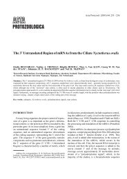
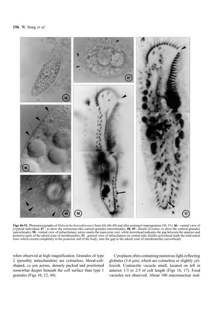
Figs 46-51. Pho<strong>to</strong>micrographs <strong>of</strong> Holosticha heter<strong>of</strong>oissneri from life (46-49) <strong>and</strong> after protargol impregnation (50, 51). 46 - ventral view <strong>of</strong><br />
a typical individual; 47 - <strong>to</strong> show <strong>the</strong> extrusome-like cortical granules (arrowheads); 48, 49 - details <strong>of</strong> cortex, <strong>to</strong> show <strong>the</strong> cortical granules<br />
(arrowheads); 50 - ventral view <strong>of</strong> infraciliature; arrow marks <strong>the</strong> transverse cirri, while arrowhead indicates <strong>the</strong> gap between <strong>the</strong> anterior <strong>and</strong><br />
posterior parts <strong>of</strong> <strong>the</strong> adoral zone <strong>of</strong> membranelles; 51 - general view <strong>of</strong> infraciliature on ventral side; double-arrowhead mark <strong>the</strong> midventral<br />
rows which extend completely <strong>to</strong> <strong>the</strong> posterior end <strong>of</strong> <strong>the</strong> body; note <strong>the</strong> gap in <strong>the</strong> adoral zone <strong>of</strong> membranelles (arrowhead)<br />
when observed at high magnification. Granules <strong>of</strong> type<br />
2 (possibly mi<strong>to</strong>chondria) are colourless, blood-cellshaped,<br />
ca µm across, densely packed <strong>and</strong> positioned<br />
somewhat deeper beneath <strong>the</strong> cell surface than type 1<br />
granules (Figs 10, 12, 44).<br />
Cy<strong>to</strong>plasm <strong>of</strong>ten containing numerous light-reflecting<br />
globules (3-6 µm), which are colourless or slightly yellowish.<br />
Contractile vacuole small, located on left in<br />
anterior 1/3 <strong>to</strong> 2/5 <strong>of</strong> cell length (Figs 16, 17). Food<br />
vacuoles not observed. About 100 macronuclear nod-