New Contribution to the Morphology and Taxonomy of Four Marine ...
New Contribution to the Morphology and Taxonomy of Four Marine ...
New Contribution to the Morphology and Taxonomy of Four Marine ...

Create successful ePaper yourself
Turn your PDF publications into a flip-book with our unique Google optimized e-Paper software.
146 W. Song et al.<br />
Petz et al. 1995, Wilbert 1995, Song <strong>and</strong> Packr<strong>of</strong>f 1997,<br />
Berger 1999, Song <strong>and</strong> Hu 1999).<br />
The current work forms part <strong>of</strong> a faunistic study <strong>of</strong><br />
ciliates in <strong>the</strong> coastal waters <strong>of</strong> <strong>the</strong> north China seas,<br />
which has been carried out for a period <strong>of</strong> over 10 years.<br />
In this paper, 4 previously known but poorly described<br />
species <strong>of</strong> hypotrichs, collected from <strong>of</strong>fshore mariculture<br />
waters near Qingdao, China, are redescribed following<br />
examination using modern techniques.<br />
MATERIALS AND METHODS<br />
Sample sites. All samples were collected from mariculture waters<br />
near <strong>the</strong> coast <strong>of</strong> Qingdao (Tsingtao, 36 o 08’N; 120 o 43’E), China.<br />
Bakuella agamalievi <strong>and</strong> Cyr<strong>to</strong>hymena marina were isolated (6 May<br />
1997) from a semi-closed pond used for mollusc culture (salinity<br />
10-15 ‰). Pseudokeronopsis flavicans was isolated on two occasions<br />
(6 <strong>and</strong> 12 May 1997) from <strong>the</strong> same sampling site as above.<br />
Two populations <strong>of</strong> Holosticha heter<strong>of</strong>oissneri were collected on<br />
separate occasions (31 March 1995 <strong>and</strong> 28 Oc<strong>to</strong>ber 1997) from an<br />
open scallop (Chlamys farreri) farming pond (salinity 31-32 ‰).<br />
General methods. After collection <strong>and</strong> isolation, specimens were<br />
maintained in <strong>the</strong> labora<strong>to</strong>ry for several weeks, ei<strong>the</strong>r as pure or raw<br />
cultures in Petri dishes, with rice grains as food source for bacteria.<br />
Observations on living morphology were undertaken using Nomarski<br />
optics. Protargol staining (Wilbert 1975) was performed for revealing<br />
<strong>the</strong> infraciliature <strong>and</strong> nuclear apparatus. Counts <strong>and</strong> measurements<br />
were made at a magnification <strong>of</strong> x1250. Drawings were made with <strong>the</strong><br />
help <strong>of</strong> a camera Lucida. Terminology is mainly according <strong>to</strong> Foissner<br />
(1982) <strong>and</strong> Hemberger (1985).<br />
<strong>New</strong> type specimens. Since no type specimens <strong>of</strong> Bakuella<br />
agamalievi, Cyr<strong>to</strong>hymena marina or Pseudokeronopsis flavicans are<br />
known <strong>to</strong> exist, one neotype slide <strong>of</strong> protargol impregnated cells <strong>of</strong><br />
each species has been deposited in <strong>the</strong> collection <strong>of</strong> <strong>the</strong> Natural<br />
His<strong>to</strong>ry Museum, London, UK with <strong>the</strong> following registration numbers:<br />
Bakuella agamalievi, 2001:1z:z8:01; Cyr<strong>to</strong>hymena<br />
marina, 2001:1z:z8:02; Pseudokeronopsis flavicans, 2001:1z:z8:03.<br />
In addition one paraneotype <strong>of</strong> each has been deposited in <strong>the</strong><br />
Labora<strong>to</strong>ry <strong>of</strong> Pro<strong>to</strong>zoology, Ocean University <strong>of</strong> Qingdao, People’s<br />
Republic China.<br />
RESULTS<br />
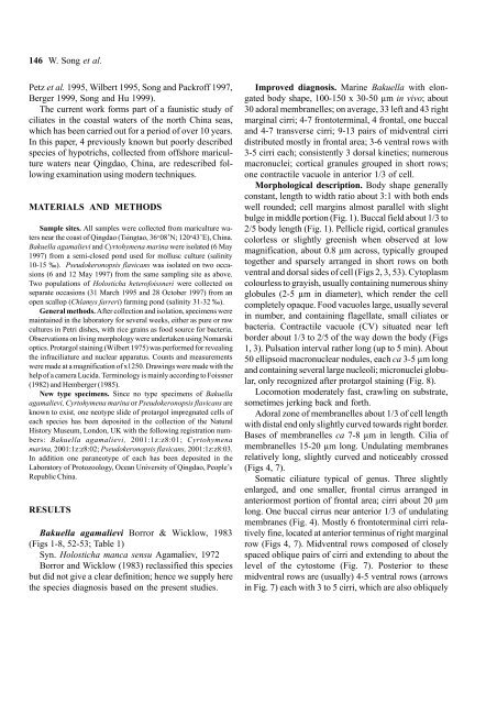
Bakuella agamalievi Borror & Wicklow, 1983<br />
(Figs 1-8, 52-53; Table 1)<br />
Syn. Holosticha manca sensu Agamaliev, 1972<br />
Borror <strong>and</strong> Wicklow (1983) reclassified this species<br />
but did not give a clear definition; hence we supply here<br />
<strong>the</strong> species diagnosis based on <strong>the</strong> present studies.<br />
Improved diagnosis. <strong>Marine</strong> Bakuella with elongated<br />
body shape, 100-150 x 30-50 µm in vivo; about<br />
30 adoral membranelles; on average, 33 left <strong>and</strong> 43 right<br />
marginal cirri; 4-7 fron<strong>to</strong>terminal, 4 frontal, one buccal<br />
<strong>and</strong> 4-7 transverse cirri; 9-13 pairs <strong>of</strong> midventral cirri<br />
distributed mostly in frontal area; 3-6 ventral rows with<br />
3-5 cirri each; consistently 3 dorsal kineties; numerous<br />
macronuclei; cortical granules grouped in short rows;<br />
one contractile vacuole in anterior 1/3 <strong>of</strong> cell.<br />
Morphological description. Body shape generally<br />
constant, length <strong>to</strong> width ratio about 3:1 with both ends<br />
well rounded; cell margins almost parallel with slight<br />
bulge in middle portion (Fig. 1). Buccal field about 1/3 <strong>to</strong><br />
2/5 body length (Fig. 1). Pellicle rigid, cortical granules<br />
colorless or slightly greenish when observed at low<br />
magnification, about 0.8 µm across, typically grouped<br />
<strong>to</strong>ge<strong>the</strong>r <strong>and</strong> sparsely arranged in short rows on both<br />
ventral <strong>and</strong> dorsal sides <strong>of</strong> cell (Figs 2, 3, 53). Cy<strong>to</strong>plasm<br />
colourless <strong>to</strong> grayish, usually containing numerous shiny<br />
globules (2-5 µm in diameter), which render <strong>the</strong> cell<br />
completely opaque. Food vacuoles large, usually several<br />
in number, <strong>and</strong> containing flagellate, small ciliates or<br />
bacteria. Contractile vacuole (CV) situated near left<br />
border about 1/3 <strong>to</strong> 2/5 <strong>of</strong> <strong>the</strong> way down <strong>the</strong> body (Figs<br />
1, 3). Pulsation interval ra<strong>the</strong>r long (up <strong>to</strong> 5 min). About<br />
50 ellipsoid macronuclear nodules, each ca 3-5 µm long<br />
<strong>and</strong> containing several large nucleoli; micronuclei globular,<br />
only recognized after protargol staining (Fig. 8).<br />
Locomotion moderately fast, crawling on substrate,<br />
sometimes jerking back <strong>and</strong> forth.<br />
Adoral zone <strong>of</strong> membranelles about 1/3 <strong>of</strong> cell length<br />
with distal end only slightly curved <strong>to</strong>wards right border.<br />
Bases <strong>of</strong> membranelles ca 7-8 µm in length. Cilia <strong>of</strong><br />
membranelles 15-20 µm long. Undulating membranes<br />
relatively long, slightly curved <strong>and</strong> noticeably crossed<br />
(Figs 4, 7).<br />
Somatic ciliature typical <strong>of</strong> genus. Three slightly<br />
enlarged, <strong>and</strong> one smaller, frontal cirrus arranged in<br />
anteriormost portion <strong>of</strong> frontal area; cirri about 20 µm<br />
long. One buccal cirrus near anterior 1/3 <strong>of</strong> undulating<br />
membranes (Fig. 4). Mostly 6 fron<strong>to</strong>terminal cirri relatively<br />
fine, located at anterior terminus <strong>of</strong> right marginal<br />
row (Figs 4, 7). Midventral rows composed <strong>of</strong> closely<br />
spaced oblique pairs <strong>of</strong> cirri <strong>and</strong> extending <strong>to</strong> about <strong>the</strong><br />
level <strong>of</strong> <strong>the</strong> cy<strong>to</strong>s<strong>to</strong>me (Fig. 7). Posterior <strong>to</strong> <strong>the</strong>se<br />
midventral rows are (usually) 4-5 ventral rows (arrows<br />
in Fig. 7) each with 3 <strong>to</strong> 5 cirri, which are also obliquely