New Contribution to the Morphology and Taxonomy of Four Marine ...
New Contribution to the Morphology and Taxonomy of Four Marine ...
New Contribution to the Morphology and Taxonomy of Four Marine ...

Create successful ePaper yourself
Turn your PDF publications into a flip-book with our unique Google optimized e-Paper software.
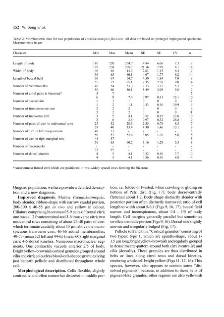
152 W. Song et al.<br />
Table 2. Morphometric data for two populations <strong>of</strong> Pseudokeronopsis flavicans. All data are based on protargol impregnated specimens.<br />
Measurements in µm<br />
Character Min Max Mean SD SE CV n<br />
Length <strong>of</strong> body 180 220 204.7 14.84 6.06 7.3 9<br />
193 238 209.1 21.16 7.99 8.1 14<br />
Width <strong>of</strong> body 40 48 44.0 2.83 1.15 6.4 9<br />
56 65 60.1 4.67 1.77 6.2 14<br />
Length <strong>of</strong> buccal field 60 67 64.7 4.50 1.84 7.0 9<br />
55 72 65.1 7.35 2.78 9.0 14<br />
Number <strong>of</strong> membranelles 46 54 51.3 2.73 1.12 5.3 9<br />
50 66 56.1 5.49 2.08 9.8 7<br />
Number <strong>of</strong> cirral pairs in bicorona* 5 7 - - - - 5<br />
6 9 7.4 0.97 0.31 13.1 10<br />
Number <strong>of</strong> buccal cirri 1 1 1 0 0 0 12<br />
1 2 1.1 0.33 0.10 30.9 9<br />
Number <strong>of</strong> fron<strong>to</strong>terminal cirri 2 2 2 0 0 0 9<br />
2 2 2 0 0 0 11<br />
Number <strong>of</strong> transverse cirri 3 5 4.1 0.52 0.15 12.6 10<br />
3 6 3.6 0.97 0.32 26.8 9<br />
Number <strong>of</strong> pairs <strong>of</strong> cirri in midventral rows 25 32 28.3 2.35 0.78 8.3 7<br />
29 40 33.4 4.39 1.46 13.1 9<br />
Number <strong>of</strong> cirri in left marginal row 40 51 - - - - 5<br />
50 57 52.4 3.05 1.36 5.8 8<br />
Number <strong>of</strong> cirri in right marginal row 44 57 - - - - 5<br />
56 65 60.2 3.16 1.29 5.3 8<br />
Number <strong>of</strong> macronuclei - - - - - - -<br />
72 97 - - - - 2<br />
Number <strong>of</strong> dorsal kineties 4 5 4.1 0.32 0.10 7.7 10<br />
4 5 4.1 0.36 0.10 8.8 14<br />
*Anteriormost frontal cirri which are positioned in two widely spaced rows forming <strong>the</strong> bicorona<br />
Qingdao population, we here provide a detailed description<br />
<strong>and</strong> a new diagnosis.<br />
Improved diagnosis. <strong>Marine</strong> Pseudokeronopsis,<br />
body slender, ribbon-shape with narrow caudal portion,<br />
200-300 x 40-55 µm in vivo <strong>and</strong> yellow in colour.<br />
Ciliature comprising bicorona <strong>of</strong> 5-9 pairs <strong>of</strong> frontal cirri;<br />
one buccal, 2 fron<strong>to</strong>terminal <strong>and</strong> 3-6 transverse cirri; two<br />
midventral rows consisting <strong>of</strong> about 25-40 pairs <strong>of</strong> cirri<br />
which terminate caudally about 15 µm above <strong>the</strong> inconspicuous<br />
transverse cirri; 46-66 adoral membranelles;<br />
40-57 (mean 52) left <strong>and</strong> 44-65 (mean 60) right marginal<br />
cirri; 4-5 dorsal kineties. Numerous macronuclear segments.<br />
One contractile vacuole anterior 2/5 <strong>of</strong> body.<br />
Bright yellow-brownish cortical granules grouped around<br />
cilia <strong>and</strong> cirri; colourless blood-cell-shaped granules lying<br />
just beneath pellicle <strong>and</strong> distributed throughout whole<br />
cell.<br />
Morphological description. Cells flexible, slightly<br />
contractile <strong>and</strong> <strong>of</strong>ten somewhat dis<strong>to</strong>rted in middle portion,<br />
i.e. folded or twisted, when crawling or gliding on<br />
bot<strong>to</strong>m <strong>of</strong> Petri dish (Fig. 17); body dorsoventrally<br />
flattened about 1:2. Body shape distinctly slender with<br />
posterior portion <strong>of</strong>ten distinctly narrowed; ratio <strong>of</strong> cell<br />
length <strong>to</strong> width about 5-6:1 (Figs 9, 16, 17); buccal field<br />
narrow <strong>and</strong> inconspicuous, about 1/4 - 1/5 <strong>of</strong> body<br />
length. Cell margins generally parallel but sometimes<br />
swollen in middle portion (Figs 9, 16). Dorsal side slightly<br />
uneven <strong>and</strong> irregularly bulged (Fig. 17).<br />
Pellicle s<strong>of</strong>t <strong>and</strong> thin. “Cortical granules” consisting <strong>of</strong><br />
two types: type 1, which are spindle-shape, about 1-<br />
1.5 µm long, bright yellow-brownish <strong>and</strong> regularly grouped<br />
in dense rosette-pattern around both cirri (ventrally) <strong>and</strong><br />
cilia (dorsally). These granules are thus distributed in<br />
belts or lines along cirral rows <strong>and</strong> dorsal kineties,<br />
rendering whole cell bright yellow (Figs 11, 12, 16). This<br />
species, however, also appears <strong>to</strong> contain some “dissolved<br />
pigments” because, in addition <strong>to</strong> <strong>the</strong>se belts <strong>of</strong><br />
pigment-like granules, o<strong>the</strong>r regions are also yellowish