Issue 19, 2013 - Balliol College - University of Oxford
Issue 19, 2013 - Balliol College - University of Oxford
Issue 19, 2013 - Balliol College - University of Oxford

You also want an ePaper? Increase the reach of your titles
YUMPU automatically turns print PDFs into web optimized ePapers that Google loves.
speeds and behave as relativistic particles.<br />
Combining this with the near transparent<br />
nature <strong>of</strong> a one-atom-thick sheet means<br />
there are a vast number <strong>of</strong> applications for<br />
graphene in electronics and opto-electronics,<br />
such as touch-screen displays and solar cells.<br />
Critical to the implementation <strong>of</strong> graphene<br />
in technology applications is the ability to<br />
synthesise a high-quality material, and the key<br />
to this is being able to determine the structure<br />
<strong>of</strong> graphene at the atomic level.<br />
My team have been working on growing<br />
synthetic graphene, atom by atom, by flowing<br />
carbon containing gas precursors across metal<br />
catalyst substrates inside high-temperature<br />
furnaces (1000°C). this high-quality synthetic<br />
material now provides a basis for studying<br />
fundamental atomic structure in graphene<br />
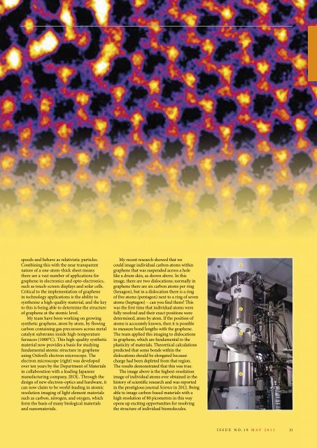
using oxford’s electron microscope. the<br />
electron microscope (right) was developed<br />
over ten years by the Department <strong>of</strong> Materials<br />
in collaboration with a leading Japanese<br />
manufacturing company, JeoL. through the<br />
design <strong>of</strong> new electron-optics and hardware, it<br />
can now claim to be world-leading in atomic<br />
resolution imaging <strong>of</strong> light element materials<br />
such as carbon, nitrogen, and oxyge n, which<br />
form the basis <strong>of</strong> many biological materials<br />
and nanomaterials.<br />
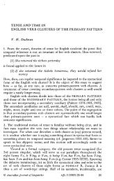
My recent research showed that we<br />
could image individual carbon atoms within<br />
graphene that was suspended across a hole<br />
like a drum skin, as shown above. in this<br />
image, there are two dislocations; normally in<br />
graphene there are six carbon atoms per ring<br />
(hexagon), but in a dislocation there is a ring<br />
<strong>of</strong> five atoms (pentagon) next to a ring <strong>of</strong> seven<br />
atoms (heptagon) – can you find them this<br />
was the first time that individual atoms were<br />
fully resolved and their exact positions were<br />
determined, atom by atom. if the position <strong>of</strong><br />
atoms is accurately known, then it is possible<br />
to measure bond lengths with the graphene.<br />
the team applied this imaging to dislocations<br />
in graphene, which are fundamental to the<br />
plasticity <strong>of</strong> materials. theoretical calculations<br />
predicted that some bonds within the<br />
dislocations should be elongated because<br />
charge had been depleted from that region.<br />
the results demonstrated that this was true.<br />
the image above is the highest-resolution<br />
image <strong>of</strong> individual atoms ever obtained in the<br />
history <strong>of</strong> scientific research and was reported<br />
in the prestigious journal Science in 2012. Being<br />
able to image carbon-based materials with a<br />
high resolution <strong>of</strong> 80 picometres in this way<br />
opens up exciting opportunities for resolving<br />
the structure <strong>of</strong> individual biomolecules.<br />
1<br />
2<br />
3<br />
4<br />
issue no.<strong>19</strong> MAY <strong>2013</strong><br />
21