
Create successful ePaper yourself
Turn your PDF publications into a flip-book with our unique Google optimized e-Paper software.
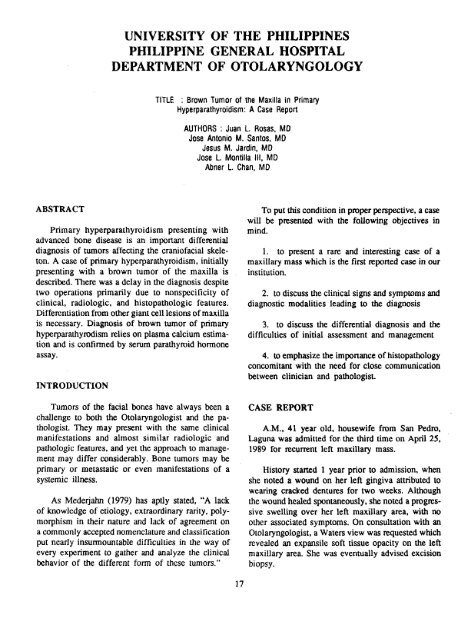
UNIVERSITY OF THE PHILIPPINES<br />
PHILIPPINE GENERAL HOSPITAL<br />
DEPARTMENT OF OTOLARYNGOLOGY<br />
TITLE : BrownTumorof the Maxillain Primary<br />
Hyperparathyroidism: A CaseReport<br />
AUTHORS: Juank. Rosas,MD<br />
JoseAntonioM. Santos,MD<br />
JesusM. Jardin,MD<br />
JoseL. MontillaIII, MD<br />
AbnerL. Chart,MD<br />
ABSTRACT<br />
To put this condition in proper perspective, a case<br />
will be presented with the following objectives in<br />
Primary hyperparathyroidism presenting with mind.<br />
advanced bone disease is an important differential<br />
diagnosis of tumors affecting the craniofacial skele- I. to present a rare and interesting case of a<br />
ton. A case of primary hyperparathymidism, initially maxillary mass which is the first reported case in our<br />
presenting with a brown tumor of the maxilla is institution.<br />
described. There was a delay in the diagnosis despite<br />
two operations primarily due to nonspccificity of 2. to discuss the clinical signs and symptoms and<br />
clinical, radiologic, and histopathologic features, diagnostic modalities leading to the diagnosis<br />
Differentiation from other giant c¢11lesions of maxilla<br />
is necessary. Diagnosis of brown tumor of primary 3. to discuss the differential diagnosis and the<br />
hyperparathyrodism relies on plasma calcium estima- difficulties of initial assessment and management<br />
tion and is confirmed by serum parathyroid hormone<br />
assay.<br />
INTRODUCTION<br />
4. to emphasize the importance of histopathology<br />
concomitant with the need for close communication<br />
between clinician and pathologist.<br />
Tumors of the facial bones have always been a CASE REPORT<br />
challenge to both the Otolaryngologist and the pathologist.<br />
They may present with the same clinical A.M., 41 year old, housewife from San Pedro,<br />
manifestations and almost similar radiologic and Laguna was admitted for. the third time on April 25,<br />
pathologic features, and yet the approach to manage- 1989 for recurrent left maxillary mass.<br />
merit may differ considerably. Bone tumors may be<br />
primary or metastatic or even manifestations of a History started 1 year prior to admission, when<br />
systemic illness, she noted a wound on her left gingiva attributed to<br />
wearing cracked dentures for two weeks. Although<br />
As Mederjahn (1979) has aptly stated, "A lack the wound he',tied spontaneously, she noted a progresof<br />
knowledge of etiology, extraordinary rarity, poly- sive swelling over her left maxillary area, with no<br />
morphism in their nature and lack of agreement on other associated symptoms. On consultation with an<br />
a commonly accepted nomenclature and classification Otolaryngologist, a Waters view was requested which<br />
put nearly insurmountable difficulties in the way of revealed an expansile soft tissue opacity on the left<br />
every experiment to gather and analyze the clinical maxillary area. She was eventually advised excision<br />
behavior of the different form of these tumors." biopsy.<br />
17