Doxorubicin and b-Lapachone Release and ... - UT Southwestern
Doxorubicin and b-Lapachone Release and ... - UT Southwestern
Doxorubicin and b-Lapachone Release and ... - UT Southwestern

You also want an ePaper? Increase the reach of your titles
YUMPU automatically turns print PDFs into web optimized ePapers that Google loves.
1092 S<strong>UT</strong>TON ET AL<br />
Copolymer. PEG-b-PLA was synthesized by ring-opening<br />
polymerization of D,L-lactide under dry argon at 1108C.<br />
Monomethyl ether hydroxyl (HO-PEG-OCH 3 ; number<br />
average molecular weight M n ¼ 5000 Da) was used as a<br />
macroinitiator. D,L-lactide was added as a monomer <strong>and</strong><br />
stannous (II) octoate (Sn(Oct) 2 ) was added as a catalyst.<br />
After reacting for 4 hrs at 1108C, the mixture was allowed to<br />
cool to room temperature. PEG-b-PLA was purified by<br />
redissolving in tetrahydrofuran (THF) <strong>and</strong> precipitating in<br />
hexane three times. The overall yield was 95%. The degree<br />
of polymerization of the PLA was calculated by comparing<br />
the integral intensity of the characteristic resonance of the<br />
PLA at 5.2 ppm ( C(¼O)-CH( CH 3 )) <strong>and</strong> PEG resonance<br />
at 3.64 ppm ( OCH 2 CH 2 ) in the 1 H nuclear magnetic<br />
resonance (NMR) spectrum in chloroform (CDCl 3 ). The<br />
molecular weight <strong>and</strong> polydispersity index (PDI) of PEG-b-<br />
PLA were also characterized by gel permeation chromatography<br />
(THF as eluent), <strong>and</strong> the results were found to be<br />
consistent with 1 H NMR data. PEG-b-PLA (M n ¼ 10.0 kD;<br />
PDI ¼ 1.2) was used in this study.<br />
Synthesis of PEG-b-PCL Copolymer. The PEGb-PCL<br />
copolymer was synthesized (with yields .95%) by<br />
ring-opening polymerization of e-caprolactone under dry<br />
argon at 1158C for 24 hrs using PEG as a macroinitiator <strong>and</strong><br />
Sn(Oct) 2 as a catalyst. The product was purified by<br />
precipitating twice into cold methanol from CH 2 Cl 2<br />
solution, <strong>and</strong> was then vacuum dried at 408C. The block<br />
copolymer was characterized by 1 H NMR in CDCl 3 at room<br />
temperature. The degree of polymerization of the PCL block<br />
was calculated by comparing the integrals of the 1 HNMR<br />
characteristic peaks of the PCL block at 2.31 ppm (triplet,<br />
C(¼O)-CH 2 ) <strong>and</strong> PEG block at 3.39 ppm (singlet,<br />
OCH 2 CH 2 ). The molecular weight <strong>and</strong> polydispersity of<br />
PEG-b-PCL were also characterized by gel permeation<br />
chromatography (THF as eluent), <strong>and</strong> the results were found<br />
to be consistent with 1 H NMR data. PEG-b-PCL (M n ¼ 10.0<br />
kD; PDI ¼ 1.3) was used in this study.<br />
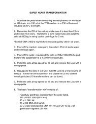
Preparation of Drug-Loaded Micelles. Drug-containing<br />
polymer micelles were prepared as follows: 18 mg<br />
of PEG-b-PLA or PEG-b-PCL copolymer <strong>and</strong> 2 mg of drug<br />
(hydrophobic DOX was predissolved in 0.12 ml dimethylsulfoxide<br />
[DMSO]) were added to 1.08 ml THF in a glass<br />
vial. Next, the mixture was slowly added to 13 ml of water<br />
under sonication (60 Sonic Dismembrator; Fisher Scientific;<br />
Pittsburgh, PA). The mixture was vigorously stirred overnight<br />
to remove THF followed by filtration through a<br />
syringe filter (pore size 0.45 lm; Millipore, Billerica, MA)<br />
to remove large drug aggregates. Micelles were characterized<br />
by dynamic light scattering (see below). 1 H NMR was<br />
used to confirm the formation of core-shell structure. The<br />
strong resonance of methylene proton in PEG was detected,<br />
whereas all of the D,L-lactide or caprolactone proton<br />
resonances were hardly observed, demonstrating the coreshell<br />
structure of these micelles.<br />
Drug-Loading Content Determination. The drugloading<br />
content, defined as the weight percentage of DOX<br />
or b-lap based on the total micellar weight (i.e., weight of<br />
copolymer <strong>and</strong> drug) was quantified by UV-Vis analysis<br />
using a Lambda 20 spectrophotometer (Perkin-Elmer,<br />
Boston, MA). First, micelle solutions were frozen <strong>and</strong><br />
lyophilized to yield the solid micelle samples. Then, the<br />
dried samples were weighed <strong>and</strong> redissolved in CDCl 3 for<br />
the b-lap micelles or a mixture of CDCl 3 <strong>and</strong> DMSO (1:1, v/<br />
v) for the DOX micelles followed by ultraviolet-visible<br />
spectroscopy (UV-Vis) analysis. The amount of loaded drug<br />
was determined based on the absorbance at 480 nm for<br />
DOX <strong>and</strong> at 257 nm for b-lap.<br />
Dynamic Light Scattering (DLS). DLS was performed<br />
on a DLS Model 802 (Viscotek, Houston, TX).<br />
Scattered light was detected at an angle of 908 at room<br />
temperature <strong>and</strong> analyzed on an autocorrelator. Sample<br />
concentration during measurement was 1.4 mg/ml. The data<br />
for each sample was obtained in five independent measurements.<br />
The average hydrodynamic diameters <strong>and</strong> their<br />
st<strong>and</strong>ard deviations are provided in Table 1.<br />
In Vitro <strong>Release</strong> of Drugs from Polymer Micelles.<br />
The drug-loaded micelles were purified using<br />
Millipore centrifugal filters with a molecular weight cutoff<br />
of 100 kD to remove the free drug <strong>and</strong> to concentrate the<br />
samples in preparation for release studies. Approximately<br />
15 mg of DOX-loaded polymeric micelles were placed into<br />
a total of 2 ml of water inside dialysis tubing, resulting in a<br />
micelle concentration of 7.5 mg/ml. The tubing was placed<br />
into 13 ml phosphate-buffered saline (PBS; pH 7.4) or<br />
acetate-buffered saline (pH 5.0) solutions. For b-lap release,<br />
10 mg of drug-loaded micelles in water (2 ml), resulting in a<br />
micelle concentration of 5 mg/ml, were transferred into<br />
dialysis tubing (MW cutoff, 100 kDa). The tubing was<br />
placed into 8 ml PBS (pH 7.4) or acetate-buffered saline (pH<br />
5.0) solutions. <strong>Release</strong> studies were performed at 378C ina<br />
C24 Incubator Shaker (New Brunswick Scientific, Edison,<br />
NJ). At selected time intervals, all of the buffered solution<br />
outside the dialysis bag was removed for UV-Vis analysis<br />
<strong>and</strong> replaced with fresh buffer solution. The DOX <strong>and</strong> b-lap<br />
concentrations were calculated based on the absorbance<br />
intensity at 480 <strong>and</strong> 257 nm, respectively. Free drug<br />
transport from the dialysis tubing was studied under the<br />
same conditions as drug release from micelles, except the<br />
amount of the free drug was different: 1 ml solution of 2 mg/<br />
ml DOX-HCl at pH 5.0 <strong>and</strong> 2 ml of saturated deionized<br />
water solution (with 0.04 mg/ml of b-lap) were placed<br />
inside the dialysis tubing, respectively.<br />
Mathematical Modeling. To simulate drug release<br />
kinetics from PEG-b-PCL <strong>and</strong> PEG-b-PLA micelles, we<br />
applied Higuchi’s model (34). The advantage of this model<br />
over the Fickian diffusion model (35, 36) is that it accounts<br />
for the difference in solubility of the drugs in the buffer<br />
solution. As discussed below, this feature is especially<br />
important in underst<strong>and</strong>ing DOX release at different pH<br />
values.<br />
The cumulative amount of drug released (SdQ) per unit<br />
time (dt) is given by the following equation: