Doxorubicin and b-Lapachone Release and ... - UT Southwestern
Doxorubicin and b-Lapachone Release and ... - UT Southwestern
Doxorubicin and b-Lapachone Release and ... - UT Southwestern

You also want an ePaper? Increase the reach of your titles
YUMPU automatically turns print PDFs into web optimized ePapers that Google loves.
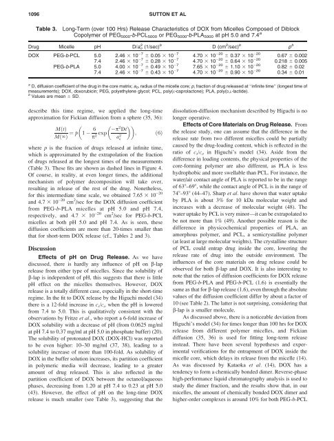
1096 S<strong>UT</strong>TON ET AL<br />
Table 3.<br />
Long-Term (over 100 Hrs) <strong>Release</strong> Characteristics of DOX from Micelles Composed of Diblock<br />
Copolymer of PEG 5000 -b-PCL 5000 or PEG 5000 -b-PLA 5000 at pH 5.0 <strong>and</strong> 7.4 a<br />
Drug Micelle pH D/a o 2 (1/sec) b D (cm 2 /sec) b p b<br />
DOX PEG-b-PCL 5.0 2.46 3 10 7 6 0.05 3 10 7 4.70 3 10 20 6 0.37 3 10 20 0.67 6 0.002<br />
7.4 2.46 3 10 7 6 0.28 3 10 7 4.70 3 10 20 6 0.64 3 10 20 0.218 6 0.005<br />
PEG-b-PLA 5.0 4.00 3 10 7 6 0.49 3 10 7 7.65 3 10 20 6 1.10 3 10 20 0.82 6 0.02<br />
7.4 2.46 3 10 7 6 0.43 3 10 7 4.70 3 10 20 6 0.90 3 10 20 0.34 6 0.01<br />
a D, diffusion coefficient of the drug in the core matrix; a 0 , radius of the micelle core; p, fraction of drug released at ‘‘infinite time’’ (longest time of<br />
measurements); DOX, doxorubicin; PEG, polyethylene glycol; PCL, poly(e-caprolactone); PLA, poly(D,L-lactide).<br />
b Values are mean 6 SD.<br />
describe this time regime, we applied the long-time<br />
approximation for Fickian diffusion from a sphere (35, 36):<br />
<br />
MðtÞ<br />
Mð‘Þ ¼ p 1 6<br />
p 2 exp p 2 <br />
Dt<br />
a 2 ; ð6Þ<br />
o<br />
where p is the fraction of drugs released at infinite time,<br />
which is approximated by the extrapolation of the fraction<br />
of drugs released at the longest times of the measurements<br />
(Table 3). These fits are shown as dashed lines in Figure 4.<br />
Of course, in reality, at even longer times, the additional<br />
mechanism of polymer decomposition will take over,<br />
resulting in release of the rest of the drug. Nonetheless,<br />
for this intermediate time scale, we obtained 7.65 3 10 20<br />
<strong>and</strong> 4.7 3 10 20 cm 2 /sec for the DOX diffusion coefficient<br />
from PEG-b-PLA micelles at pH 5.0 <strong>and</strong> pH 7.4,<br />
respectively, <strong>and</strong> 4.7 3 10 20 cm 2 /sec for PEG-b-PCL<br />
micelles at both pH 5.0 <strong>and</strong> pH 7.4. As is seen, these<br />
diffusion coefficients are more than 20-times smaller than<br />
that for short-term DOX release (cf., Tables 2 <strong>and</strong> 3).<br />
Discussion<br />
Effects of pH on Drug <strong>Release</strong>. As we have<br />
discussed, there is hardly any influence of pH on b-lap<br />
release from either type of micelles. Since the solubility of<br />
b-lap is independent of pH, this suggests that there is little<br />
pH effect on the micelles themselves. However, DOX<br />
release is a totally different case, especially in the short-time<br />
regime. In the fit to DOX release by the Higuchi model (34)<br />
there is a 12-fold increase in c s /c o when the pH is lowered<br />
from 7.4 to 5.0. This is qualitatively consistent with the<br />
observations by Fritze et al., who report a 6-fold increase of<br />
DOX solubility with a decrease of pH (from 0.0625 mg/ml<br />
at pH 7.4 to 0.37 mg/ml at pH 5.0 in phosphate buffer) (20).<br />
The solubility of protonated DOX (DOX-HCl) was reported<br />
to be even higher: 10–30 mg/ml (37, 38), leading to a<br />
solubility increase of more than 100-fold. As solubility of<br />
DOX in the buffer solution increases, its partition coefficient<br />
in polymeric media will decrease, leading to a greater<br />
amount of drug released. This is also reflected in the<br />
partition coefficient of DOX between the octanol/aqueous<br />
phases, decreasing from 1.20 at pH 7.4 to 0.23 at pH 5.0<br />
(43). However, the effect of pH on the long-time DOX<br />
release is much smaller (see Table 3), suggesting that the<br />
dissolution-diffusion mechanism described by Higuchi is no<br />
longer operative.<br />
Effects of Core Materials on Drug <strong>Release</strong>. From<br />
the release study, one can assume that the difference in the<br />
release rate from two different micelles could be partially<br />
caused by the drug-loading content, which is reflected in the<br />
ratio of c s /c o in Higuchi’s model (34). Aside from the<br />
difference in loading contents, the physical properties of the<br />
core-forming polymer are also different, as PLA is less<br />
hydrophobic <strong>and</strong> more swellable than PCL. For instance, the<br />
water/air contact angle of PLA is reported to be in the range<br />
of 638–698, while the contact angle of PCL is in the range of<br />
748–938 (44–47). Sharp et al. have shown that water uptake<br />
by PLA is about 3% for 10 kDa molecular weight <strong>and</strong><br />
increases with a decrease of molecular weight (48). The<br />
water uptake by PCL is very minor—it can be extrapolated to<br />
be not more than 1% (49). Another possible reason is the<br />
difference in physicochemical properties of PLA, an<br />
amorphous polymer, <strong>and</strong> PCL, a semicrystalline polymer<br />
(at least at large molecular weights). The crystalline structure<br />
of PCL could entrap drug inside the core, lowering the<br />
release rate of drug into the outside environment. The<br />
influences of the core materials on drug release could be<br />
observed for both b-lap <strong>and</strong> DOX. It is also interesting to<br />
note that the ratios of diffusion coefficients for DOX release<br />
from PEG-b-PLA <strong>and</strong> PEG-b-PCL (1.6) is essentially the<br />
same as that for b-lap release (1.6), even through the absolute<br />
values of the diffusion coefficient differ by about a factor of<br />
10 (see Table 2). The latter is not surprising, considering that<br />
b-lap is a smaller molecule.<br />
As discussed above, there is a noticeable deviation from<br />
Higuchi’s model (34) for times longer than 100 hrs for DOX<br />
release from different polymer micelles, <strong>and</strong> Fickian<br />
diffusion (35, 36) is used for fitting long-term release<br />
instead. There have been several hypotheses <strong>and</strong> experimental<br />
verifications for the entrapment of DOX inside the<br />
micelle core, which delays its release from the micelle (14).<br />
As was discussed by Kataoka et al. (14), DOX has a<br />
tendency to form a chemically bonded dimer. Reverse-phase<br />
high-performance liquid chromatography analysis is used to<br />
study the dimer fraction, <strong>and</strong> the results show that, in our<br />
micelles, the amount of chemically bonded DOX dimer <strong>and</strong><br />
higher-order complexes is around 10% for both PEG-b-PCL