

Volume 6 Issue 3 - Australasian Society for Ultrasound in Medicine
Volume 6 Issue 3 - Australasian Society for Ultrasound in Medicine
Volume 6 Issue 3 - Australasian Society for Ultrasound in Medicine
- No tags were found...

You also want an ePaper? Increase the reach of your titles
YUMPU automatically turns print PDFs into web optimized ePapers that Google loves.
<strong>Ultrasound</strong> of salivary glands<br />
<strong>Ultrasound</strong> of salivary glands<br />
Wong KT; ; Ahuja AT; ; Yuen HY; ; K<strong>in</strong>g AD<br />
Department of Diagnostic Radiology and Organ Imag<strong>in</strong>g, The Ch<strong>in</strong>ese University of Hong Kong, Pr<strong>in</strong>ce of Wales<br />
Hospital, Shat<strong>in</strong>, New Territories, Hong Kong SAR<br />
AR, , Ch<strong>in</strong>a<br />
INTRODUCTION<br />
Salivary gland masses are commonly encountered by<br />
surgeons and radiologists <strong>in</strong> daily practice. Cl<strong>in</strong>ical<br />
exam<strong>in</strong>ation alone is often <strong>in</strong>sufficient to identify the orig<strong>in</strong><br />
and nature of the mass. Imag<strong>in</strong>g is required <strong>in</strong> the vast<br />
majority of cases. Be<strong>in</strong>g paired superficial structures, the<br />
parotid and submandibular glands are suitable <strong>for</strong> highresolution<br />
ultrasound exam<strong>in</strong>ation and its role is well<br />
established 1,2 . The ultrasound exam<strong>in</strong>ation can be easily<br />
comb<strong>in</strong>ed with f<strong>in</strong>e needle aspiration cytology (FNAC)<br />
further enhanc<strong>in</strong>g its ability to differentiate between benign<br />
and malignant lesions.<br />
TECHNIQUE<br />
<strong>Ultrasound</strong> of the parotid gland is per<strong>for</strong>med us<strong>in</strong>g a highresolution<br />
(7-12MHz) l<strong>in</strong>ear array transducer. Transverse and<br />
longitud<strong>in</strong>al scans are obta<strong>in</strong>ed with the patient sup<strong>in</strong>e and<br />
the head turned away from the side be<strong>in</strong>g exam<strong>in</strong>ed.<br />
Transverse scans are per<strong>for</strong>med with the transducer<br />
perpendicular and <strong>in</strong>ferior to the ear lobe. When per<strong>for</strong>m<strong>in</strong>g<br />
longitud<strong>in</strong>al scans, particular attention should be paid to the<br />
tail of the parotid gland which may be obscured by the<br />
ramus of mandible.<br />
The submandibular gland is evaluated us<strong>in</strong>g a highresolution<br />
(7-12MHz) l<strong>in</strong>ear transducer. Transverse scans<br />
us<strong>in</strong>g a submandibular view provide most of the<br />
<strong>in</strong><strong>for</strong>mation. Oblique and coronal adjustments help to<br />
localize lesions and to trace vessels.<br />
Colour-flow imag<strong>in</strong>g is a useful adjunctive tool and should<br />
be per<strong>for</strong>med whenever a mass is seen on gray scale<br />
ultrasound.<br />
It is important to scan both sides <strong>for</strong> symmetry and to<br />
exclude further cl<strong>in</strong>ically non-palpable lesions, as there is a<br />
chance of bilateral disease (eg Warth<strong>in</strong>’s tumour). Regional<br />
nodal territories <strong>in</strong> the neck should be <strong>in</strong>cluded as part of<br />
ultrasound exam<strong>in</strong>ation of the salivary glands.<br />
ANATOMY<br />
The parotid gland is the largest of the salivary glands. The<br />
gland lies <strong>in</strong> the parotid space which is the most lateral space<br />
<strong>in</strong> the nasopharyngeal area. It extends from the external<br />
auditory canal superiorly to the level of angle of mandible<br />
<strong>in</strong>feriorly. On high-resolution ultrasound it shows uni<strong>for</strong>m,<br />
f<strong>in</strong>e bright <strong>in</strong>ternal echoes. The facial nerve creates an<br />
artificial plane divid<strong>in</strong>g the gland <strong>in</strong>to superficial and deep<br />
lobes which is important from the surgical po<strong>in</strong>t of view.<br />
<strong>Ultrasound</strong> cannot identify the facial nerve def<strong>in</strong>itely, but<br />
its course can be <strong>in</strong>ferred from the vascular plane (which is<br />
readily identified by ultrasound and consists of external<br />
carotid artery and retromandibular ve<strong>in</strong>). Stensen’s duct is<br />
visible as a f<strong>in</strong>e echogenic l<strong>in</strong>e with<strong>in</strong> the superficial lobe.<br />
Intraparotid lymph nodes are commonly seen on ultrasound.<br />
They ma<strong>in</strong>ly lie with<strong>in</strong> superficial lobe and appear as round<br />
or oval hypoechoic nodules, generally less than 5mm <strong>in</strong><br />
diameter and are usually well-def<strong>in</strong>ed. An echogenic hilum<br />
differentiates <strong>in</strong>traparotid nodes from other parotid masses.<br />
The submandibular gland is a well-encapsulated structure<br />
with homogeneous hyperechogenicity similar to that of<br />
parotid gland. On high-resolution ultrasound, multiple<br />
discrete f<strong>in</strong>e l<strong>in</strong>ear streaks represent<strong>in</strong>g <strong>in</strong>traglandular<br />
ductules are commonly seen. The free border of the<br />
mylohyoid muscle divide the gland <strong>in</strong>to imag<strong>in</strong>ary<br />
superficial and deep lobes. Wharton’s duct is clearly seen<br />
when it is abnormally dilated, but can be seen <strong>in</strong> normal<br />
cases <strong>in</strong> the oblique scans.<br />
PATHOLOGY<br />
Sialolithiasis<br />
Submandibular calculi are more common than parotid<br />
calculi. The greater number (80%) of calculi that occur <strong>in</strong><br />
the submandibular gland is attributed to the greater mucous<br />
content of the saliva produced by the submandibular gland.<br />
90% of submandibular calculi are radio-opaque whereas<br />
only 10% of parotid ductal calculi are opaque. For the<br />
detection of salivary calculi, ultrasound is the <strong>in</strong>vestigation<br />
of choice, with a sensitivity of 94%, specificity of 100% and<br />
an accuracy of 96% 3 . Intra-glandular ductal dilatation and<br />
an <strong>in</strong>tra-ductal echogenic fill<strong>in</strong>g defect cast<strong>in</strong>g posterior<br />
acoustic shadow<strong>in</strong>g are the hallmark ultrasound features of<br />
sialolithiasis (Figure 1). <strong>Ultrasound</strong> can accurately localize<br />
whether the calculus is <strong>in</strong>traglandular or with<strong>in</strong> the ma<strong>in</strong><br />
salivary duct. This affects patient management, particularly<br />
<strong>for</strong> submandibular calculi. For <strong>in</strong>traglandular calculi, the<br />
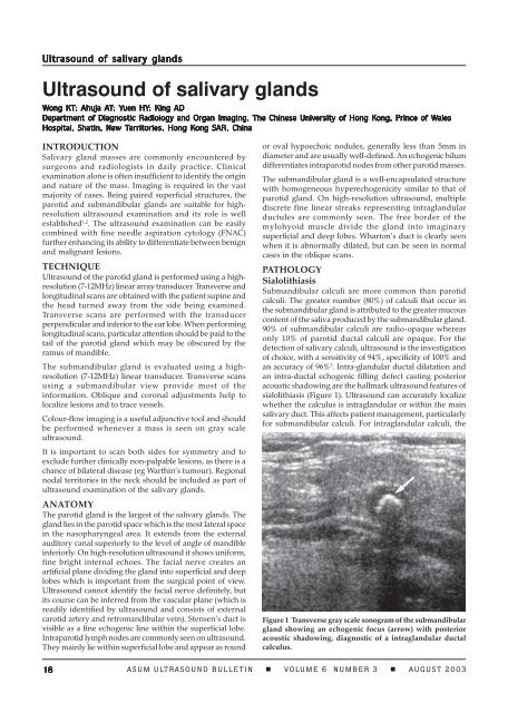
Figure 1 Transverse gray scale sonogram of the submandibular<br />
gland show<strong>in</strong>g an echogenic focus (arrow) with posterior<br />
acoustic shadow<strong>in</strong>g, diagnostic of a <strong>in</strong>traglandular ductal<br />
calculus.<br />
18 ASUM ULTRASOUND BULLETIN VOLUME 6 NUMBER 3 AUGUST 2003


