Varshni parameters for InAs/GaSb strained layer superlattice ...
Varshni parameters for InAs/GaSb strained layer superlattice ...
Varshni parameters for InAs/GaSb strained layer superlattice ...

You also want an ePaper? Increase the reach of your titles
YUMPU automatically turns print PDFs into web optimized ePapers that Google loves.
Home Search Collections Journals About Contact us My IOPscience<br />
<strong>Varshni</strong> <strong>parameters</strong> <strong>for</strong> <strong>InAs</strong>/<strong>GaSb</strong> <strong>strained</strong> <strong>layer</strong> <strong>superlattice</strong> infrared photodetectors<br />
This article has been downloaded from IOPscience. Please scroll down to see the full text article.<br />
2011 J. Phys. D: Appl. Phys. 44 075102<br />
(http://iopscience.iop.org/0022-3727/44/7/075102)<br />
View the table of contents <strong>for</strong> this issue, or go to the journal homepage <strong>for</strong> more<br />
Download details:<br />
IP Address: 64.106.37.191<br />
The article was downloaded on 09/02/2011 at 16:50<br />
Please note that terms and conditions apply.
IOP PUBLISHING<br />
JOURNAL OF PHYSICS D: APPLIED PHYSICS<br />
J. Phys. D: Appl. Phys. 44 (2011) 075102 (5pp) doi:10.1088/0022-3727/44/7/075102<br />
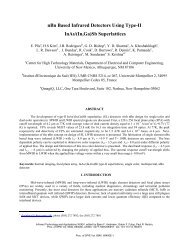
<strong>Varshni</strong> <strong>parameters</strong> <strong>for</strong> <strong>InAs</strong>/<strong>GaSb</strong><br />
<strong>strained</strong> <strong>layer</strong> <strong>superlattice</strong> infrared<br />
photodetectors<br />
B Klein, E Plis, M N Kutty, N Gautam, A Albrecht, S Myers and S Krishna<br />
Center <strong>for</strong> High Technology Materials, Department of Electrical and Computer Engineering,<br />
University of New Mexico, Albuquerque, NM 87106, USA<br />
Received 4 October 2010, in final <strong>for</strong>m 29 November 2010<br />
Published 28 January 2011<br />
Online at stacks.iop.org/JPhysD/44/075102<br />
Abstract<br />
The temperature-dependent behaviour of the bandgap of mid- and long-wavelength as well as<br />
dual-colour (mid-/long-wavelength) infrared detectors based on <strong>InAs</strong>/<strong>GaSb</strong> <strong>strained</strong> <strong>layer</strong><br />
<strong>superlattice</strong>s (SLSs) with p-i-n and nBn designs has been investigated with<br />
temperature-dependent absorption, photoluminescence and spectral response techniques.<br />
Values of <strong>Varshni</strong> <strong>parameters</strong>, zero temperature bandgap E 0 and empirical coefficient α, were<br />
extracted and tabulated. The MWIR and LWIR <strong>superlattice</strong> detectors showed a temperature<br />
change of 0.325 meV K −1 and 0.282 meV K −1 , respectively. These values are a factor of two<br />
lower than that of HgCdTe and InSb, making them attractive <strong>for</strong> higher operating temperatures.<br />
(Some figures in this article are in colour only in the electronic version)<br />
1. Introduction<br />
The development of third generation infrared imagers has<br />
led to a concerted research ef<strong>for</strong>t on the development of<br />
higher operating temperature (HOT) detectors in the midwave<br />
infrared (MWIR, 3–5 µm) and the long-wave infrared<br />
(LWIR, 8–12 µm) range [1, 2]. The present dominant IR<br />
technologies in the MWIR and LWIR wavelength ranges are<br />
indium antimonide (InSb) and mercury–cadmium–telluride<br />
(MCT) detectors, respectively. <strong>InAs</strong>/<strong>GaSb</strong> type II strain<strong>layer</strong><br />
<strong>superlattice</strong>s (SLS), first proposed <strong>for</strong> IR detection in the<br />
1980s [3, 4], are good candidates <strong>for</strong> the development of HOT<br />
detectors. Most of the <strong>InAs</strong>/<strong>GaSb</strong> SLS previously reported<br />
are based on the p-i-n design [5, 6]. One of the major dark<br />
current components in these devices is Shockley–Read–Hall<br />
(SRH) generation-recombination (GR) current associated with<br />
the depletion region of the p-i-n diode. The recently proposed<br />
nBn heterostructure design [7] eliminates the GR component<br />
of dark current as the nBn structure is intended to operate with<br />
n-type <strong>layer</strong>s in the flatband with little depletion. Klipstein<br />
showed that the diffusion contribution to dark current is directly<br />
proportional to the zero temperature semiconductor bandgap<br />
E 0 <strong>for</strong> <strong>InAs</strong>Sb-based nBn devices [8]. In order to design HOT<br />
detectors based on <strong>superlattice</strong>s, the variation of the bandgap<br />
needs to be investigated. This is especially interesting <strong>for</strong><br />
the <strong>superlattice</strong> since the band structure of the <strong>superlattice</strong><br />
consists of minibands with a 2D density of states, unlike bulk<br />
semiconductors with 3D density of states. In this paper, we<br />
report on a systematic study of the temperature dependence<br />
of the bandgap of both MWIR and LWIR <strong>superlattice</strong>s using<br />
optical absorption, photoluminescence and spectral response<br />
measurements. The most common binary <strong>superlattice</strong>s in the<br />
literature <strong>for</strong> the MWIR and LWIR detector consists of 10<br />
mono<strong>layer</strong>s (ML) <strong>InAs</strong>/10 ML <strong>GaSb</strong> and 14 ML <strong>InAs</strong>/7 ML<br />
<strong>GaSb</strong> [9–11], respectively. Hence, these particular thicknesses<br />
were chosen <strong>for</strong> the study. In order to study the effect of<br />
transport in the heterostructure, both p-i-n and nBn detectors<br />
were included in the study. Finally, reference samples of<br />
<strong>InAs</strong>Sb were included, to serve as a quantitative comparison<br />
with the literature. The <strong>Varshni</strong> <strong>parameters</strong> <strong>for</strong> all the samples<br />
were obtained using these three techniques. It was found<br />
that the bandgap in the <strong>superlattice</strong> changed on average by<br />
0.33 meV K −1 <strong>for</strong> MWIR and 0.282 meV K −1 <strong>for</strong> LWIR. This<br />
is about a factor of two lower than InSb (0.6 meV K −1 ) [12]<br />
and MCT (0.5 meV K −1 ) [13].<br />
The variation of the bandgap of a III–V semiconductor<br />
with temperature is described by the linear-quadratic relation<br />
proposed by <strong>Varshni</strong> [14]<br />
E(T ) = E 0 − αT 2 /(T + β), (1)<br />
0022-3727/11/075102+05$33.00 1 © 2011 IOP Publishing Ltd Printed in the UK & the USA
J. Phys. D: Appl. Phys. 44 (2011) 075102 B Klein et al<br />
Table 1. Summary of investigated detector structures and extracted <strong>Varshni</strong> <strong>parameters</strong>.<br />
Wavelength Heterostructure α E 0<br />
Structure range design Material (eV K −1 ) (eV) Method<br />
A MWIR nBn undoped barrier Bulk <strong>InAs</strong>Sb 3.8 × 10 −4 0.344 PL<br />
3.8 × 10 −4 0.352 Absorption<br />
B MWIR nBn doped barrier Bulk <strong>InAs</strong>Sb 3.8 × 10 −4 0.340 Absorption<br />
C MWIR nBn doped barrier Bulk <strong>InAs</strong>Sb 2.5 × 10 −4 0.350 PL<br />
4.6 × 10 −4 0.341 Absorption<br />
D MWIR pin 10 ML/10 ML SLS 3.1 × 10 −4 0.234 Absorption<br />
3.0 × 10 −4 0.228 Spectral response<br />
E LWIR pin 14 ML/7 ML SLS 3.2 × 10 −4 0.138 Absorption<br />
F LWIR nBn 14 ML/7 ML SLS 2.5 × 10 −4 0.133 Absorption<br />
2.8 × 10 −4 0.144 Spectral response<br />
G MWIR nBn 10 ML/10 ML SLS 3.7 × 10 −4 0.233 Absorption<br />
3.2 × 10 −4 0.241 Spectral response<br />
LWIR nBn 14 ML/7 ML SLS 2.0 × 10 −4 0.118 Absorption<br />
3.6 × 10 −4 0.159 Spectral response<br />
Figure 1. Schematics of heterostructures used in this study. Samples A, B, C and F used the single-colour nBn architecture, samples D and<br />
E were p-i-n, and sample G was a dual colour nBn structure.<br />
where β is a constant (K), E 0 is the width of the semiconductor<br />
bandgap at 0 K (eV), α is a fitting parameter (eV K −1 ) and T is<br />
the temperature (K). The literature indicates a β of 266 K <strong>for</strong><br />
bulk <strong>GaSb</strong> [15], and a similar value of 270 K <strong>for</strong> <strong>InAs</strong>/<strong>GaSb</strong><br />
SLS [16]. Using experimentally determined temperaturedependent<br />
bandgaps and β allows <strong>for</strong> the other two <strong>parameters</strong><br />
in the <strong>Varshni</strong> equation, α and E 0 , to be extracted. The value<br />
of β chosen <strong>for</strong> this study was 270 K.<br />
Previously, values of α equal to 2.5 × 10 −4 eV K −1 and<br />
2.76×10 −4 eV K −1 <strong>for</strong> MWIR [17] and LWIR [16] <strong>InAs</strong>/<strong>GaSb</strong><br />
SLS detectors, found through photoresponse experiments,<br />
have been reported. However, there was no correlation<br />
between the photocurrent measurements, which represent<br />
the convolution between the absorption and photocarrier<br />
collection process and the absorption or photoluminescence<br />
measurements that are insensitive to the photocarrier collection<br />
process.<br />
2. Experiment<br />
The seven detector structures used in this study are summarized<br />
in table 1; generic schematics of these structures are shown<br />
in figure 1. All detectors were grown by solid source<br />
molecular beam epitaxy (MBE) on n-type (Te-doped) epiready<br />
(1 0 0) <strong>GaSb</strong> substrates. Structures A–C are nBn detectors<br />
consisting of a 3 µm n-type (1 × 10 17 cm −3 ) absorbing <strong>layer</strong><br />
<strong>for</strong>med by <strong>InAs</strong> 0.91 Sb 0.09 , grown on top of a 1 µm n-type<br />
(1 × 10 18 cm −3 ) contact <strong>layer</strong>. A 100 nm thick AlAs x Sb 1−x<br />
barrier <strong>layer</strong> was deposited on top of an absorber <strong>layer</strong>,followed<br />
by a 0.3 µm thick n-type <strong>InAs</strong> 0.91 Sb 0.09 (1 × 10 17 cm −3 ) top<br />
contact. The barrier <strong>layer</strong> with an arsenic mole fraction<br />
in AlAsSb of 15% was nominally undoped (in structure A)<br />
and n-type with a carrier concentration of 1 × 10 17 cm −3 (in<br />
structure B). In structure C, the composition of the barrier<br />
<strong>layer</strong> was AlAs 0.1 Sb 0.9 with an n-type carrier concentration<br />
of 1 × 10 17 cm −3 . The structure was terminated by a 10 nm<br />
thick n-type (1 × 10 18 cm −3 ) <strong>InAs</strong> <strong>layer</strong>. It is to be noted that<br />
the purpose of using the nBn-based <strong>InAs</strong>Sb samples was to<br />
validate the study by comparing the <strong>Varshni</strong> <strong>parameters</strong> with<br />
previously reported bulk <strong>InAs</strong>Sb films grown by MBE [18].<br />
Structures D and E are p-i-n detectors designed to operate<br />
in the MWIR and LWIR spectral regions, respectively. The<br />
non-intentionally doped absorbing regions of these detector<br />
structures were ∼2.5 µm thick, <strong>for</strong>med by 10 ML <strong>InAs</strong>/10 ML<br />
<strong>GaSb</strong> SLS (D) and 14 ML <strong>InAs</strong>/7 ML <strong>GaSb</strong> SLS (E). N-type<br />
(4 × 10 18 cm −3 ) bottom contacts with thicknesses of ∼0.6 µm<br />
2
J. Phys. D: Appl. Phys. 44 (2011) 075102 B Klein et al<br />
holder. A schematic of the setup is illustrated in figure 2.<br />
Then, absorption was calculated by<br />
Absorption(%) = I sam /I tsam − I subs /I tsub .<br />
Figure 2. Configuration of samples in cryostat <strong>for</strong> absorption<br />
measurements. The incident and transmitted light are (respectively)<br />
made and detected with an FTIR system.<br />
were <strong>for</strong>med by 10 ML <strong>InAs</strong>/10 ML <strong>GaSb</strong> SLS (D) and 8 ML<br />
<strong>InAs</strong>/8 ML <strong>GaSb</strong> SLS (E). The top contact consisted of a p-type<br />
(2 × 10 18 cm −3 ) <strong>GaSb</strong> <strong>layer</strong> with thicknesses of 160 nm (D)<br />
and 100 nm (E). In order to improve carrier transport in the<br />
detector structure, the 0.25 µm top and bottom components of<br />
SLS absorber were doped. In addition, 10 periods of graded<br />
<strong>layer</strong>s were added between the absorber and the top contact<br />
<strong>layer</strong>.<br />
Structure F is an nBn detector intended to operate in the<br />
LWIR wavelength range. The device structure consists of a<br />
∼2 µm 14 ML <strong>InAs</strong>/7 MLs <strong>GaSb</strong> SLS absorber grown on top<br />
of a ∼0.6 µm thick n-type contact <strong>layer</strong> (made of SLS with<br />
the same composition and thickness but with Te-doped <strong>InAs</strong><br />
<strong>layer</strong>s). This was followed by a 100 nm Al 0.2 Ga 0.8 Sb n-type<br />
(1×10 16 cm −3 ) doped barrier <strong>layer</strong>. The structure was capped<br />
by ∼0.13 µm thick top contact <strong>layer</strong> with the same <strong>superlattice</strong><br />
composition, thickness and doping concentration as the bottom<br />
contact <strong>layer</strong>.<br />
Structure G is a dual-colour detector with an nBn design.<br />
It is composed of ∼2 µm thick MWIR and LWIR absorbing<br />
<strong>layer</strong>s <strong>for</strong>med by 10 ML <strong>InAs</strong>/10 ML <strong>GaSb</strong> SLS and 14 ML<br />
<strong>InAs</strong>/7 ML <strong>GaSb</strong> SLS, respectively, separated by 100 nm<br />
thick n-type (1 × 10 17 cm −3 ) Al 0.2 Ga 0.8 Sb barrier. N-type<br />
(4 × 10 18 cm −3 ) top (120 nm thick) and bottom (0.5 µm<br />
thick) contact <strong>layer</strong>s were <strong>for</strong>med with SLSs with the same<br />
composition as the MWIR and LWIR absorbing <strong>layer</strong>s,<br />
respectively. This sample was included to verify the validity of<br />
the <strong>Varshni</strong> <strong>parameters</strong> since the MWIR and LWIR absorbers<br />
were similar to those in samples D–F.<br />
The temperature dependent changes in the bandgap of the<br />
detector were determined by absorption, photoluminescence<br />
(PL) and spectral response measurements. Absorption<br />
measurements were per<strong>for</strong>med within the 77–270 K range with<br />
10 K increments using a Thermo Nicolet Nexus 870 Fourier<br />
Trans<strong>for</strong>m Infrared (FT-IR) spectrometer, equipped with a<br />
liquid nitrogen-cooled HgCdTe detector. First, single beam<br />
transmission measurements were taken through a sample (I sam )<br />
and a reference <strong>GaSb</strong> n-type substrate (I subs ), both mounted at<br />
Brewster’s angle; transmission measurements were also taken<br />
through the holes on the sample (I tsam ) and substrate (I tsub )<br />
For PL measurements, the sample was placed in a closecycle<br />
liquid helium cryostat and pumped with a 514 nm argon<br />
laser, with a power of 1 W and chopping frequency of 207 Hz.<br />
PL collection optics consisted of calcium fluoride lenses,<br />
aluminium mirrors and a germanium filter. A monochromator<br />
was equipped with a grating optimized <strong>for</strong> wavelengths from<br />
2.5 to 6 µm. PL was measured from 10 to 160 K, with<br />
increments of 10 K. It is to be noted that the cutoff wavelength<br />
of the PL setup was 5.3 µm. There<strong>for</strong>e, the PL was not suitable<br />
<strong>for</strong> the LWIR samples.<br />
The detector material was processed into single element<br />
detectors with 410 × 410 µm 2 mesas having apertures ranging<br />
from 25 to 300 µm. Details of p-i-n, nBn and dualcolour<br />
detector fabrication are reported elsewhere [11, 19, 20].<br />
Spectral response measurements were per<strong>for</strong>med within<br />
20–230 K temperature range with 10 K increments, using<br />
a glow-bar source within the FTIR spectrometer. The<br />
cutoff wavelength <strong>for</strong> PL measurements was defined as the<br />
wavelength where the PL signal was at a maximum. For<br />
absorption and spectral response measurements, the cutoff was<br />
defined at the wavelength where the signal was reduced to 50%<br />
of its peak value. The characterization methods utilized <strong>for</strong><br />
each investigated structure are summarized in table 1.<br />
3. Results and discussion<br />
Figure 3 shows a typical plot <strong>for</strong> the temperaturedependent<br />
absorption, photoluminescence and spectral<br />
response measurements. Measurements were made using<br />
small temperature steps to obtain a meaningful fit. <strong>Varshni</strong><br />
fits <strong>for</strong> MWIR (A, B and C), LWIR (F) and dual-colour (G)<br />
nBn detector structures using data extracted from absorption<br />
(solid lines), PL (dash lines) and spectral response (dashed–<br />
dotted line) measurements are shown in figure 4. The symbols<br />
correspond to the values of the energy bandgap measured at<br />
each given temperature. <strong>Varshni</strong> fits <strong>for</strong> MWIR (D) and LWIR<br />
(E) p-i-n detectors, based on data extracted from absorption<br />
and spectral response measurements are shown in figure 5. The<br />
50% cutoff wavelength of the absorption spectrum corresponds<br />
to the maximum of the PL curve, as illustrated by the inset in<br />
figure 5.<br />
The slight difference between values of energy bandgaps<br />
extracted from temperature-dependent PL and absorption<br />
measurements is possibly due to band filling effects, since<br />
the number of carriers created by the laser is different<br />
from the number of carriers created by the glow-bar source.<br />
Presumably, the laser excitation promotes many carriers to<br />
the conduction band, which fills the low-energy states within<br />
the band so that the bandgap appears widened. This would<br />
manifest as a blue-shift in the peak of the photoluminescence.<br />
The samples in the absorption and spectral response do not<br />
experience this phenomenon nearly as much, as the incident<br />
radiation from the glow-bar is of low power compared with<br />
that of the laser.<br />
3
J. Phys. D: Appl. Phys. 44 (2011) 075102 B Klein et al<br />
Figure 3. Representative plots of (a) absorption (77–270 K), (b) spectral response (20–200 K) and (c) photoluminescence (20–160 K) at<br />
multiple temperatures. Sharp peaks and troughs in the plots are due to atmospheric absorption. For all three plots, the cutoff wavelength<br />
increases with increasing temperature. The magnitude of spectral response and photoluminescence decreases with increasing temperature,<br />
as indicated by the arrows.<br />
Figure 4. <strong>Varshni</strong> fits <strong>for</strong> MWIR (A, B and C), LWIR (F) and<br />
dual-colour (G) nBn detector structures extracted from absorption<br />
(solid lines), PL (dash lines) and spectral response (dashed–dotted<br />
lines) measurements. The symbols correspond to the values of<br />
energy bandgap measured at each given temperature.<br />
The zero temperature bandgap of <strong>InAs</strong>Sb-based nBn<br />
detectors with n-type barriers (structures B and C) is slightly<br />
lower than one of the <strong>InAs</strong>Sb-based detector with n.i.d. barrier.<br />
We attribute this to variations in the quality of the samples.<br />
For samples C (MWIR) and G (LWIR), α varies<br />
significantly by the measurement used to determine it. We<br />
believe that spectral response has the least error, as it can<br />
be normalized with a background transmission measurement<br />
to minimize effects from atmospheric distortion. Similarly,<br />
a more complex normalization scheme is per<strong>for</strong>med <strong>for</strong> the<br />
absorption measurements; the only measurement that does<br />
not have normalization is photoluminescence. However,<br />
Figure 5. <strong>Varshni</strong> fits <strong>for</strong> MWIR (D) and LWIR (E) p-i-n detectors<br />
extracted from absorption (solid lines) and spectral response<br />
(dashed–dotted line) measurements. The inset shows representative<br />
absorption and PL spectra. The λ 50%cutoff of the absorption spectrum<br />
corresponds to the maximum on the PL curve.<br />
the greatest advantage of photoluminescence is that the<br />
cutoff wavelength is easily identified by the peak of the<br />
spectra, rather than the 50% point, which is sometimes<br />
difficult to define clearly. There<strong>for</strong>e, photoluminescence<br />
and absorption measurements have about an equal level of<br />
accuracy.<br />
Table 1 summarizes the <strong>Varshni</strong> <strong>parameters</strong> obtained <strong>for</strong><br />
each sample. All three characterization methods produced<br />
results that are in good agreement with each other. Moreover,<br />
our findings <strong>for</strong> <strong>InAs</strong>/<strong>GaSb</strong> SLS MWIR and LWIR material<br />
compare closely with previously published data [16, 17]. A<br />
comparison of the resulting measurements of the <strong>InAs</strong>Sb<br />
samples with already well-documented <strong>Varshni</strong> equations<br />
shows good agreement. For a composition of <strong>InAs</strong> 0.91 Sb 0.09 ,a<br />
0 K bandgap of 0.338 eV and α parameter of 3.4×10 −4 eV K −1<br />
4
J. Phys. D: Appl. Phys. 44 (2011) 075102 B Klein et al<br />
have been reported [18]. Our results are quite similar, with<br />
average values of 0.345 eV <strong>for</strong> the 0 K bandgap and 3.7 ×<br />
10 −4 eV K −1 <strong>for</strong> the α parameter. This similarity validates the<br />
methods used to determine cutoff energies.<br />
4. Conclusions<br />
In conclusion, the temperature-dependent behaviour of<br />
MWIR, LWIR and dual-colour (MWIR/LWIR) detectors<br />
based on bulk <strong>InAs</strong>Sb and <strong>InAs</strong>/<strong>GaSb</strong> SLS with p-i-n and nBn<br />
designs have been investigated with conventional absorption,<br />
PL and spectral response techniques. Values of <strong>Varshni</strong><br />
<strong>parameters</strong>, zero temperature bandgap E 0 and empirical<br />
coefficient α, were extracted. All three characterization<br />
methods produced results that were in good agreement.<br />
To the best of our knowledge this is the first systematic<br />
study of <strong>Varshni</strong> <strong>parameters</strong> per<strong>for</strong>med on single and dual<br />
colour <strong>InAs</strong>/<strong>GaSb</strong> SLS detectors operating in different spectral<br />
regions.<br />
Acknowledgment<br />
Support from AFOSR grant FA9550-10-1-0113 is acknowledged.<br />
References<br />
[1] Rogalski A 2005 Rep. Prog. Phys. 68 2267<br />
[2] Rogalski A, Antoszewski J and Faraone L 2009 J. Appl. Phys.<br />
105 091101<br />
[3] Tsu R, Esaki L and Sai-Halasz B A 1977 Appl. Phys. Lett.<br />
30 651<br />
[4] Smith D L and Mailhiot C 1987 J. Appl. Phys. 62 2545<br />
[5] Rhiger D R, Kvaas R E, Harris S F and Hill C J 2009 Infrared<br />
Phys. Technol. 52 304<br />
[6] Sharabani Y, Paltiel Y, Sher A, Raizman A and Zussman A<br />
2007 Appl. Phys. Lett. 90 232106<br />
[7] Maimon S and Wicks G 2006 Appl Phys. Lett. 89 151109<br />
[8] Klipstein P 2008 Proc. SPIE 6940 69402U<br />
[9] Gautam N, Kim H S, Kutty M N, Plis E, Dawson LRand<br />
Krishna S 2010 Appl. Phys. Lett. 96 231107<br />
[10] Myers S, Plis E, Khoshakhlagh A, Kim H S, Sharma Y,<br />
Dawson R, Krishna S and Gin A 2009 Appl. Phys. Lett.<br />
95 121110<br />
[11] Plis E, Myers S, Khoshakhlagh A, Kim H S, Sharma Y,<br />
Gautam N, Dawson R and Krishna S 2009 Infrared Phys.<br />
Technol. 52 335–9<br />
[12] Levinstein M, Rumyantsev S and Shur M 1999 Handbook<br />
Series on Semiconductor Parameters vols 1 and 2 (London:<br />
World Scientific)<br />
[13] Ksendzov A, Pollak F H, Wilson J A and Cotton V A 1989 J.<br />
Appl. Phys. 66 5528<br />
[14] <strong>Varshni</strong> Y P 1967 Physica (Amsterdam) 34 149<br />
[15] Passler R 1999 Phys. Status Solidi b 216 975<br />
[16] Hoffman D, Hood A, Fuchs F and Razeghi M 2006 J. Appl.<br />
Phys. 99 043503<br />
[17] Lee S J, Noh S K, Plis E, Krishna S and Lee K-S 2009 Appl.<br />
Phys. Lett. 95 102106<br />
[18] Marciniak M A, Hengehold R L, Yeo Y K and Turner G W<br />
1998 J. Appl. Phys. 84 480<br />
[19] Khoshakhlagh A, Rodriguez J B, Plis E, Bishop G D,<br />
Sharma Y D, Kim H S, Dawson L R and Krishna S 2007<br />
Appl. Phys. Lett. 91 263504<br />
[20] Khoshakhlagh A, Myers S, Kim H S, Plis E, Gautam N,<br />
Lee S J, Noh S K, Dawson L R and Krishna S 2010 IEEE J.<br />
Quantum Electron. 46 959<br />
5