Flow Generic - Lab-JOT
Flow Generic - Lab-JOT
Flow Generic - Lab-JOT

You also want an ePaper? Increase the reach of your titles
YUMPU automatically turns print PDFs into web optimized ePapers that Google loves.
ANTIBODIES, KITS, AND REAGENTS FOR<br />
FLOW CYTOMETRY<br />
Unparalleled Product Quality, Validation, and Technical Support<br />
Events<br />
Androgen Receptor<br />
(Alexa Fluor ® 488 Conjugate)
XP ®<br />
XP® monoclonal antibodies are a line of high<br />
quality rabbit monoclonal antibodies exclusively<br />
available from Cell Signaling Technology (CST).<br />
Any product labeled with XP has been carefully<br />
selected based on superior performance in the<br />
most relevant research applications.<br />
These antibodies are generated using XMT®<br />
technology, a proprietary monoclonal<br />
method developed at CST. The technology<br />
provides access to a broad range of antibodies<br />
unattainable with traditional monoclonal<br />
technologies, allowing more comprehensive<br />
screening and the identification of XP<br />
monoclonal antibodies with exceptional<br />
specificity, sensitivity, and performance.<br />
kDa<br />
200<br />
140<br />
100<br />
80<br />
60<br />
50<br />
40<br />
30<br />
Monoclonal Antibodies<br />
one antibody, multiple applications <br />
A<br />
HT-29<br />
SK-BR-3<br />
T-47D<br />
Pro Met<br />
Met<br />
B<br />
eXceptional specificity<br />
As with all of our antibodies, the antibody is specific to your<br />
target of interest, saving you valuable time and resources.<br />
+ eXceptional sensitivity<br />
The antibody will provide a stronger signal for your target<br />
protein in cells and tissues, allowing you to monitor<br />
expression of low levels of endogenous proteins, saving<br />
you valuable materials.<br />
+ eXceptional stability and reproducibility<br />
XMT technology combined with our stringent quality<br />
control ensures maximum lot-to-lot consistency and the<br />
most reproducible results.<br />
= eXceptional Performance<br />
XMT technology coupled with our extensive antibody<br />
validation and stringent quality control delivers<br />
XP monoclonal antibodies with eXceptional Performance<br />
in the widest range of applications.<br />
C<br />
WINNER!<br />
Life Science<br />
Industry Awards ®<br />
:: Best Antibodies<br />
Please see page 4<br />
for full details about<br />
antibody validation for<br />
flow cytometry.<br />
:: Best Breakthrough Products<br />
for Cancer Research<br />
Antibodies and Reagents for<br />
<strong>Flow</strong> Cytometry<br />
Cell Signaling Technology (CST) provides the highest quality<br />
activation state and total protein antibodies available for use in<br />
<strong>Flow</strong> Cytometry research. Our in-house <strong>Flow</strong> Cytometry Group<br />
has validated each <strong>Flow</strong> Cytometry recommended CST antibody<br />
using multiple approaches. Technical support is provided by the<br />
same scientists who validate the products.<br />
4<br />
5<br />
6<br />
9<br />
11<br />
12<br />
14<br />
16<br />
18<br />
20<br />
Antibody Validation for <strong>Flow</strong> Cytometry<br />
Complementary Reagents<br />
Conjugated Antibodies<br />
Immunology and Inflammation<br />
Jak/Stat Signaling Table<br />
Cytokines and Growth Factors<br />
Cell Cycle, Checkpoint Control, and DNA Damage<br />
Stem Cell Markers, Development, and Differentiation<br />
MAP Kinase Signaling<br />
PI3K/Akt Signaling<br />
20<br />
200<br />
140<br />
100<br />
80<br />
60<br />
50<br />
40<br />
30<br />
Events<br />
D<br />
E<br />
21<br />
22<br />
23<br />
Translational Control<br />
Tyrosine Kinases<br />
Apoptosis and Autophagy<br />
20<br />
Met<br />
Met (D1C2) XP ® Rabbit mAb #8198: Western blot analysis (A) of extracts from HT-29 (Met+), SK-BR-3 (Met-), and T-47D (Met-)<br />
cells using #8198 (upper) or β-Actin Antibody #4967 (lower). Confocal IF analysis of HT-29 (B) and T-47D (C) cells using #8198<br />
(green). Actin filaments were labeled with DY-554 phalloidin (red). Blue pseudocolor = DRAQ5 ® #4084 (fluorescent DNA dye).<br />
<strong>Flow</strong> cytometric analysis (D) of T-47D (blue) and HT-29 (green) cells using #8198. IHC analysis (E) of paraffin-embedded human<br />
hepatocellular carcinoma using #8198.<br />
Visit our website for more experimental details, additional information,<br />
and a complete list of available XP ® monoclonal antibodies.<br />
Application Key:<br />
W Western / IP Immunoprecipitation / IHC Immunohistochemistry<br />
IF Immunofluorescence / F <strong>Flow</strong> Cytometry / ChIP Chromatin<br />
Immunoprecipitation / (-IC Immunocytochemistry, -P Paraffin,<br />
-F Frozen) / E-P Peptide ELISA<br />
Reactivity Key:<br />
H human / M mouse / R rat / Hm hamster / Mk monkey<br />
C chicken / Mi mink / Dm D. melanogaster / X Xenopus<br />
Z zebra fish / B bovine / Dg dog / Pg pig / Sc S. cerevisiae<br />
All all species expected / ( ) 100% sequence homology<br />
For Research Use Only. Not For Use In Diagnostic Procedures.<br />
24 Metabolism<br />
25<br />
27<br />
Other Antibodies<br />
<strong>Flow</strong> Cytometry Protocols
Antibody Validation for <strong>Flow</strong> Cytometry<br />
Cell Signaling Technology (CST) antibodies have undergone rigorous validation by our in-house <strong>Flow</strong> Cytometry<br />
Group. Our goal is to provide our customers with the most specific antibodies that yield the brightest signal<br />
with the lowest background possible. All of our 550+ antibodies validated for flow cytometry research have been<br />
screened to determine optimal dilution and to verify specificity.<br />
Titration:<br />
Serial dilution is used to determine optimal dilution.<br />
Mean Channel Fluorescence<br />
7.00<br />
6.00<br />
5.00<br />
4.00<br />
3.00<br />
2.00<br />
1.00<br />
0.00<br />
0.00<br />
0 0.2 0.4 0.6 0.8 1.0 1.2 1.4 1.6 1.8 2.0<br />
Isotype Control:<br />
Comparison of signal to isotype control is<br />
used to estimate the nonspecific binding<br />
of primary antibodies.<br />
Events<br />
CREB<br />
Rabbit (DA1E) mAb IgG XP ® Isotype Control<br />
#3900: <strong>Flow</strong> cytometric analysis of SH-SY5Y<br />
cells using CREB (D76D11) Rabbit mAb Antibody<br />
#4820 (blue) compared to concentration matched<br />
#3900 (red).<br />
Antibody Dilution (µg/ml)<br />
Positive and Negative Cell Lines:<br />
Use of known positive and negative cell lines verifies target specificity.<br />
Events<br />
A<br />
Zap-70 (Alexa Fluor ® 647 Conjugate)<br />
BrdU incorporated<br />
Unincorporated<br />
Fold Induction<br />
Activation-state Specificity:<br />
Comparison of the signal on treated and<br />
untreated cells helps to verify activationstate<br />
specificity.<br />
SSC-H<br />
1000<br />
800<br />
600<br />
400<br />
200<br />
0<br />
10 0 10 1 10 2 10 3 10 4<br />
Phospho-Stat3 (Tyr705)<br />
(Alexa Fluor ® 647 Conjugate)<br />
Phospho-Stat3 (Tyr705) (D3A7) XP ® Rabbit<br />
mAb (Alexa Fluor ® 647 Conjugate) #4324:<br />
<strong>Flow</strong> cytometric analysis of human whole blood,<br />
untreated (red) or treated with Human Granulocyte<br />
Colony Stimulating Factor (hG-CSF) #8930 (blue),<br />
using #4324.<br />
Ramos<br />
Jurkat<br />
Jurkat<br />
10 0 10 1 10 2 10 3<br />
CD3-PE<br />
Zap-70 (136F12) Rabbit mAb (Alexa Fluor ® 647 Conjugate) #2707: <strong>Flow</strong> cytometric analysis (A) of Ramos<br />
B (blue) and Jurkat T (green) cells using #2707. Two-color flow cytometric analysis (B) of a mixed population<br />
of T and B cells (Jurkat and Ramos, respectively) using #2702 and a CD3 antibody. CD3-negative B cells have<br />
little or no Zap-70 staining, while CD3-positive T cells stain brightly for Zap-70 protein.<br />
Zap70 (Alexa Fluor ® 647)<br />
10 3<br />
10 2<br />
10 1<br />
10 0<br />
B<br />
3.50<br />
3.00<br />
2.50<br />
2.00<br />
1.50<br />
1.00<br />
0.50<br />
Fold Induction<br />
BrdU (Bu20a) Mouse mAb #5292:<br />
<strong>Flow</strong> cytometric analysis of Jurkat<br />
cells, unincorporated (red) or after<br />
30 min of BrdU incorporation (blue),<br />
using serial dilutions of #5292. The<br />
fold-induction ratio is shown in green.<br />
Optimal concentration of #5292 was<br />
determined to be 0.044 µg/ml.<br />
Inhibitor Treatment:<br />
Treatment of cell lines with pathway-specific<br />
inhibitors helps to verify target specificity.<br />
Events<br />
Phospho-p44/42 MAPK (Thr202/Tyr204)<br />
(Alexa Fluor ® 488 Conjugate)<br />
Phospho-p44/42 MAPK (Erk1/2) (Thr202/<br />
Tyr204) (D13.14.4E) XP ® Rabbit mAb (Alexa<br />
Fluor ® 488 Conjugate) #4344: <strong>Flow</strong> cytometric<br />
analysis of Jurkat cells treated with U0126 #9903<br />
(blue) or TPA #4174 (green), using #4344.<br />
Other Validation Steps<br />
Include:<br />
:: The use of blocking peptides,<br />
siRNA, and expression vectors<br />
verifies specificity of staining.<br />
:: Phosphatase treatment confirms<br />
phospho-specificity of the antibody.<br />
:: Extensive quality control testing<br />
guarantees stability over time and<br />
eliminates lot-to-lot variability.<br />
:: Optimized protocols are provided<br />
and dilutions are predetermined.<br />
:: Concurrent testing on other<br />
relevant applications.<br />
Complementary Reagents<br />
CST offers a wide selection of conjugated secondary antibodies and cellular dyes, as well as isotype controls.<br />
These same reagents are also used in-house for antibody validation in flow cytometry analysis and therefore<br />
work optimally with our primary antibodies. Our research protocols (included in the back of this brochure)<br />
also recommend the use of these reagents. Technical support is provided by the product scientists who make<br />
the reagents and know them best.<br />
Isotype Controls<br />
#5415 Mouse (G3A1) mAb IgG1 Isotype Control<br />
New #6899 Mouse (G3A1) mAb IgG1 Isotype Control (PE Conjugate)<br />
#4878 Mouse (MOPC-21) mAb IgG1 Isotype Control (Alexa Fluor ® 488 Conjugate)<br />
#4843 Mouse (MOPC-21) mAb IgG1 Isotype Control (Alexa Fluor ® 647 Conjugate)<br />
#4097 Mouse (MOPC-21) mAb IgG1 Isotype Control (Biotinylated)<br />
#3900 Rabbit (DA1E) mAb IgG XP ® Isotype Control<br />
#2975 Rabbit (DA1E) mAb IgG XP ® Isotype Control (Alexa Fluor ® 488 Conjugate)<br />
#2985 Rabbit (DA1E) mAb IgG XP ® Isotype Control (Alexa Fluor ® 647 Conjugate)<br />
#4096 Rabbit (DA1E) mAb IgG XP ® Isotype Control (Biotinylated)<br />
#5742 Rabbit (DA1E) mAb IgG XP ® Isotype Control (PE Conjugate)<br />
#4340 Rabbit IgG Isotype Control (Alexa Fluor ® 488 Conjugate)<br />
#3452 Rabbit IgG Isotype Control (Alexa Fluor ® 647 Conjugate)<br />
Product References:<br />
Rabbit IgG Isotype Control (Alexa Fluor ® 488 Conjugate) #4340: Anand, S. et al. (2011)<br />
Increased basal intracellular signaling patterns do not correlate with JAK2 genotype in human<br />
myeloproliferative neoplasms. Blood. 118, 1610-1621.<br />
Dyes and Secondary Antibodies<br />
#4408 Anti-mouse IgG (H+L), F(ab’) 2 Fragment (Alexa Fluor ® 488 Conjugate)<br />
#4410 Anti-mouse IgG (H+L), F(ab’) 2 Fragment (Alexa Fluor ® 647 Conjugate)<br />
#4412 Anti-rabbit IgG (H+L), F(ab’) 2 Fragment (Alexa Fluor ® 488 Conjugate)<br />
#4414 Anti-rabbit IgG (H+L), F(ab’) 2 Fragment (Alexa Fluor ® 647 Conjugate)<br />
#4416 Anti-rat IgG (H+L), (Alexa Fluor ® 488 Conjugate)<br />
#4418 Anti-rat IgG (H+L), (Alexa Fluor ® 647 Conjugate)<br />
#4084 DRAQ5 ®<br />
New #7406 DRAQ7<br />
New #4087 Propidium Iodide (PI)/ RNase Staining Solution<br />
Chemical Activators and Inhibitors<br />
#9944 AICAR<br />
#9841 Bisindolylmaleimide I,<br />
Hydrochloride<br />
#9972 Brefeldin A<br />
#9902 Calyculin A (Serine/Threonine<br />
Phosphatase Inhibitor)<br />
#9973 Cyclosporin A<br />
New #9052 Dasatinib<br />
#9886 Docetaxel<br />
#9974 FK-506<br />
#3828 Forskolin<br />
#9843 Geldanamycin<br />
#9844 H-89, Dihydrochloride<br />
New #9084 Imatinib<br />
#9995 Ionomycin, Calcium Salt<br />
New #9676 Leptomycin B<br />
#9901 LY294002 (PI3 Kinase Inhibitor)<br />
New #8158 SB202190<br />
#5633 SB203580 (p38 Inhibitor)<br />
New #8177 SP600125<br />
#9903 U0126 (MEK1/2 Inhibitor)<br />
#9900 PD98059 (MEK1 Inhibitor)<br />
#9996 Oligomycin<br />
#9807 Paclitaxel<br />
New #9493 PKC412<br />
#9904 Rapamycin (FRAP/mTOR<br />
Inhibitor)<br />
#9885 Roscovitine<br />
#9953 Staurosporine<br />
#4174 TPA (12-O-Tetradecanoylphorbol-<br />
13-Acetate)<br />
#9950 Trichostatin A (TSA)<br />
#9842 Tyrphostin AG 1478<br />
#9951 Wortmannin<br />
DRAQ7 <br />
Phospho-Histone H3 (Ser10)<br />
(Alexa Fluor ® 488 Conjugate)<br />
Events<br />
10 3<br />
10 2<br />
10 1<br />
Phospho-Stat5 (Tyr694)<br />
10 0 0<br />
1023<br />
DNA (PI)<br />
10 3<br />
10 2<br />
10 1<br />
10 0 1023<br />
Events<br />
0<br />
DNA (PI)<br />
Phospho-S6 Ribosomal Protein (Ser235/236)<br />
(Alexa Fluor ® 488 Conjugate)<br />
Rabbit (DA1E) mAb<br />
IgG XP ® Isotype<br />
Control #3900: <strong>Flow</strong><br />
cytometric analysis of<br />
K-562 cells, untreated<br />
(green) or treated with<br />
imatinib #9084 (blue),<br />
using Phospho-Stat5<br />
(Tyr694) (C71E5) Rabbit<br />
mAb #9314 compared to<br />
concentration-matched<br />
#3900 (red).<br />
DRAQ7 #7406:<br />
<strong>Flow</strong> cytometric<br />
analysis of live<br />
(unpermeabilized)<br />
Jurkat cells treated<br />
with Staurosporine<br />
#9953. Gated<br />
population represents<br />
DRAQ7-positive<br />
apoptotic cells.<br />
Propidium Iodide<br />
(PI)/RNase Staining<br />
Solution #4087: <strong>Flow</strong><br />
cytometric analysis<br />
of Jurkat cells using<br />
Phospho-Histone H3<br />
(Ser10) Antibody (Alexa<br />
Fluor ® 488 Conjugate)<br />
#9708 and #4087.<br />
LY294002 #9901,<br />
Wortmannin #9951,<br />
and U0126 #9903: <strong>Flow</strong><br />
cytometric analysis of<br />
Jurkat cells, untreated<br />
(green) or treated with<br />
#9901, #9951, and<br />
#9903 (blue), using<br />
Phospho-S6 Ribosomal<br />
Protein (Ser235/236)<br />
(D57.2.2E) XP ® Rabbit<br />
mAb (Alexa Fluor ® 488<br />
Conjugate) #4803.<br />
4 Application and Reactivity Keys, pg 3. Monoclonal Antibody. Please see page 2 for details.<br />
Unparalleled Product Quality, Validation, and Technical Support www.cellsignal.com<br />
5
Conjugated Antibodies<br />
Cell Signaling Technology (CST) offers Alexa Fluor® and PE-conjugated antibodies that are optimized for<br />
flow cytometry research. The combination of Alexa Fluor® and PE dyes, which exhibit superior brightness<br />
and photostability, with the highest quality CST antibodies results in conjugates with bright signal and<br />
low background. All conjugated antibodies are validated in-house. Price-competitive and high-quality<br />
custom antibody conjugation of CST antibodies is offered from our Custom Conjugation Group, please<br />
see page 24 for additional information. Contact your sales representative with any inquiries.<br />
emission strength<br />
100<br />
80<br />
60<br />
40<br />
20<br />
0<br />
300<br />
SS Lin: SS Lin<br />
SS Lin: SS Lin<br />
1000<br />
800<br />
600<br />
400<br />
200<br />
Untreated +TPA +LPS<br />
1000<br />
1000<br />
800<br />
600<br />
400<br />
200<br />
0<br />
0<br />
0<br />
10 0 10 1 10 2 10 3 10 4 10 0 10 1 10 2 10 3 10 4 10 0 10 1 10 2 10 3 10 4<br />
Phospho-S6 Ribosomal Protein (Ser235/236) (D57.2.2E) XP ® Rabbit mAb (Alexa Fluor ® 488 Conjugate) #4803<br />
1000<br />
800<br />
600<br />
400<br />
200<br />
1000<br />
800<br />
600<br />
400<br />
200<br />
Emission Spectra<br />
400 500 600 700<br />
wavelength (nm)<br />
Untreated +TPA +LPS<br />
0<br />
0<br />
0<br />
10 0 10 1 10 2 10 3 10 4 10 0 10 1 10 2 10 3 10 4 10 0 10 1 10 2 10 3 10 4<br />
Phospho-p44/42 MAPK (Erk1/2) (Thr202/Tyr204) (D13.14.4E) XP ® Rabbit mAb (Alexa Fluor ® 647 Conjugate) #4284<br />
<strong>Flow</strong> cytometric analysis of whole blood, either untreated, treated with TPA #4174, or LPS-treated, using either Phospho-S6 Ribosomal Protein<br />
(Ser235/236) (D57.2.2E) XP ® Rabbit mAb (Alexa Fluor ® 488 Conjugate) #4803 (top) or Phospho-p44/42 MAPK (Erk1/2) (Thr202/Tyr204)<br />
(D13.14.4E) XP ® Rabbit mAb (Alexa Fluor ® 647 Conjugate) #4284 (bottom).<br />
800<br />
600<br />
400<br />
200<br />
1000<br />
800<br />
600<br />
400<br />
200<br />
Alexa Fluor ® 488<br />
R-Phycoerythrin (PE)<br />
Propidium Iodide (PI)<br />
Alexa Fluor ® 647<br />
Isotype Controls<br />
Reactivity<br />
Conjugates<br />
488 PE 647<br />
Mouse IgG1 Isotype Control (MOPC-21) – #4878 – #4843<br />
#3900 Rabbit (DA1E) mAb IgG XP ® Isotype Control – #2975 #5742 #2985<br />
Rabbit IgG (Polyclonal) Isotype Control – #4340 – #3452<br />
#5415 Mouse (G3A1) mAb IgG1 Isotype Control – – #6899 –<br />
Epitope Tag Antibody Reactivity 488 PE 647<br />
#2368 DYKDDDDK Tag Antibody (Binds to same<br />
epitope as Sigma’s Anti-FLAG ® M2 Antibody)<br />
All #5407 – #3916<br />
#2624 GST (26H1) Mouse mAb All #3368 – #3445<br />
#2367 HA-Tag (6E2) Mouse mAb All #2350 – #3444<br />
#2276 Myc-Tag (9B11) Mouse mAb All #2279 #3739 #2233<br />
Unconjugated Antibody Reactivity 488 PE 647<br />
#2855 Phospho-4E-BP1 (Thr37/46) (236B4)<br />
Rabbit mAb<br />
H, M, R, Mk, Dm #2846 #7547 #5123<br />
#4970 β-Actin (13E5) Rabbit mAb H, M, R, Mk, B, Pg, (C, Dg) #8844 – #8584<br />
#2965 Phospho-Akt (Thr308) (C31E5E) Rabbit mAb H, M, R, Hm, Mk #2918 #9088 #3375<br />
#4060 Phospho-Akt (Ser473) (D9E) XP ® Rabbit mAb H, M, R, Hm, Mk, Dm, Z, B,<br />
(C, X, Dg, Pg)<br />
#4071 #5315 #4075<br />
#4058 Phospho-Akt (Ser473) (193H12) Rabbit mAb H, M, R #2336 – #2337<br />
#4691 Akt (pan) (C67E7) Rabbit mAb H, M, R, Mk, Dm #5084 – #5186<br />
#2966 Akt (5G3) Mouse mAb H, M, R, Hm #2917 – #2944<br />
#5153 Androgen Receptor (D6F11) XP ® Rabbit mAb H #7395 #8428 #7397<br />
#2914 Phospho-Aurora A (Thr288)/Aurora B<br />
(Thr232)/Aurora C (Thr198) (D13A11) XP ®<br />
Rabbit mAb<br />
H, M, R #8525 – –<br />
#2827 Phospho-Bcl-2 (Ser70) (5H2) Rabbit mAb H #2834 – –<br />
#2764 Bcl-xL (54H6) Rabbit mAb H, M, R, Mk #2767 – –<br />
#3195 E-Cadherin (24E10) Rabbit mAb H, M, (Dg, Pg) #3199 #7559 #9835<br />
#9664 Cleaved Caspase-3 (Asp175) (5A1E)<br />
Rabbit mAb<br />
H, M, R, Mk, (Dg) – #9978 –<br />
#9661 Cleaved Caspase-3 (Asp175) Antibody H, M, R, Mk, (B, Dg, Pg) #9669 – –<br />
#2677 β-Catenin (L54E2) Mouse mAb (IF Preferred) H, (M, R, Mk, Pg) #2849 #6898 #4627<br />
#3570 CD44 (156-3C11) Mouse mAb H #3516 #8724 –<br />
#4850 COX IV (3E11) Rabbit mAb H, R, Mk, Z, B, Pg #4853 – #7561<br />
#9198 Phospho-CREB (Ser133) (87G3) Rabbit mAb H, M, R #9187 – –<br />
#4135 Cyclin B1 (V152) Mouse mAb H, M, (Hm) #4112 – #4118<br />
#4267 EGF Receptor (D38B1) XP ® Rabbit mAb H, M, Mk #5616 #8839 #5588<br />
#2929 EpCAM (VU1D9) Mouse mAb H #5198 #8995 #5447<br />
#2251 FosB (5G4) Rabbit mAb H, M, R #2023 – –<br />
#2118 GAPDH (14C10) Rabbit mAb H, M, R, Mk #3906 – #3907<br />
#2024 Hexokinase I (C35C4) Rabbit mAb H, M #3689 – #3540<br />
#9718 Phospho-Histone H2A.X (Ser139) (20E3)<br />
Rabbit mAb<br />
#3377 Phospho-Histone H3 (Ser10) (D2C8) XP ®<br />
Rabbit mAb<br />
#9701 Phospho-Histone H3 (Ser10) Antibody H, M, R, Mk, C, Dm, Z,<br />
Sc, (X)<br />
H, M, R, Mk #9719 #5763 #9720<br />
H, M, R, Mk, Z #3465 #5764 #3458<br />
#9708 – #9716<br />
#9649 Acetyl-Histone H3 (Lys9) (C5B11) Rabbit mAb H, M, R, Mk, Z, (Sc) #9683 – #4484<br />
#4876 HSP70 (D69) Antibody H, M, R, Mk #4837 – #4008<br />
#4814 IκBα (L35A5) Mouse mAb (Amino-terminal<br />
Antigen)<br />
H, M, R, Mk, B, Pg #5743 #7523 –<br />
#4545 Pan-Keratin (C11) Mouse mAb H, R, Mk #4523 #5075 #4528<br />
#3308 c-Kit (Ab81) Mouse mAb H #3310 – –<br />
#4777 Lamin A/C (4C11) Mouse mAb H, M, R, Mk #8617 – –<br />
#2230 LEF1 (C12A5) Rabbit mAb H, M, R #8490 – –<br />
continued on next page<br />
Phospho-Histone H3 (Ser10) (PE Conjugate)<br />
Events<br />
Androgen Receptor<br />
(Alexa Fluor ® 488 Conjugate)<br />
Androgen Receptor (D6F11) XP ® Rabbit<br />
mAb (Alexa Fluor ® 488 Conjugate)<br />
#7395: <strong>Flow</strong> cytometric analysis of Jurkat<br />
(blue) and LNCaP (green) cells using #7395.<br />
Events<br />
Cleaved Caspase-3 (Alexa Fluor ® 488)<br />
Cleaved Caspase-3 (Asp175) Antibody<br />
(Alexa Fluor ® 488 Conjugate) #9669: <strong>Flow</strong><br />
cytometric analysis of Jurkat cells, untreated<br />
(blue) or etoposide-treated (green), using #9669.<br />
Events<br />
EGF Receptor (Alexa Fluor ® 647 Conjugate)<br />
EGF Receptor (D38B1) XP ® Rabbit mAb<br />
(Alexa Fluor ® 647 Conjugate) #5588: <strong>Flow</strong><br />
cytometric analysis of Jurkat (blue) and HeLa<br />
(green) cells using #5588.<br />
10 3<br />
10 2<br />
10 1<br />
10 0<br />
0<br />
1023<br />
DNA (DRAQ5 ® )<br />
Phospho-Histone H3 (Ser10) (D2C8)<br />
XP ® Rabbit mAb (PE Conjugate)<br />
#5764: <strong>Flow</strong> cytometric analysis of Jurkat<br />
cells using #5764 and DRAQ5 ® #4084<br />
(DNA content). Gated population represents<br />
phospho-histone H3-positive cells.<br />
6 Application and Reactivity Keys, pg 3. Monoclonal Antibody. Please see page 2 for details.<br />
Unparalleled Product Quality, Validation, and Technical Support www.cellsignal.com<br />
7
Conjugated Antibodies Continued<br />
Events<br />
Phospho-MAPKAPK-2 (Thr334)<br />
(Alexa Fluor ® 647 Conjugate)<br />
Phospho-MAPKAPK-2 (Thr334) (27B7) Rabbit<br />
mAb (Alexa Fluor ® 647 Conjugate) #4320: <strong>Flow</strong><br />
cytometric analysis of THP-1 cells, untreated (blue)<br />
or anisomycin-treated (green), using #4320.<br />
Events<br />
Phospho-p44/42 MAPK (Thr202/Tyr204)<br />
(Alexa Fluor ® 647 Conjugate)<br />
Phospho-p44/42 MAPK (Erk1/2) (Thr202/<br />
Tyr204) (D13.14.4E) XP ® Rabbit mAb (Alexa<br />
Fluor ® 647 Conjugate) #4284: <strong>Flow</strong> cytometric<br />
analysis of Jurkat cells, treated with U0126 #9951<br />
(blue) or TPA #4174 (green), using #4284.<br />
Events<br />
Phospho-SAPK/JNK<br />
(Thr183/Tyr185) (PE Conjugate)<br />
Phospho-SAPK/JNK (Thr183/Tyr185) (G9)<br />
Mouse mAb (PE Conjugate) #5755: <strong>Flow</strong><br />
cytometric analysis of THP-1 cells, untreated<br />
(blue) or anisomycin-treated (green), using #5755.<br />
Events<br />
Phospho-Tyrosine (P-Tyr-100)<br />
(Alexa Fluor ® 488 Conjugate)<br />
Phospho-Tyrosine Mouse mAb (P-Tyr-100)<br />
(Alexa Fluor ® 488 Conjugate) #9414: <strong>Flow</strong><br />
cytometric analysis of K-562 cells, untreated<br />
(green) or treated with imatinib #9084 (blue), using<br />
#9414 compared to a concentration-matched<br />
nonspecific negative control antibody (red).<br />
Immunology and Inflammation<br />
Conjugates<br />
Unconjugated Antibody<br />
Reactivity<br />
488 PE 647<br />
Applications Reactivity<br />
one antibody, multiple applications Phospho-NF-κB p65 (Ser536) (93H1) Rabbit mAb<br />
#9777 Pirin (1E8) Rat mAb W, IP, F H, M, R, Hm, Mk, B (Alexa Fluor ® 647 Conjugate) #4887: Kalland,<br />
#3007 Phospho-MAPKAPK-2 (Thr334) (27B7) H, M, R, Mk #4338 – #4320<br />
#4327 Phospho-AML1 (Ser249) Antibody W, IP, IF-IC, F H<br />
Rabbit mAb<br />
#4336 AML1 (D33G6) XP ® Rabbit mAb W, IHC-P, IF-IC, F H, Mk<br />
#8198 Met (D1C2) XP ® Rabbit mAb H #8494 – –<br />
#4334 AML1 Antibody W, IF-IC, F H, Mk<br />
#2983 mTOR (7C10) Rabbit mAb H, M, R, Mk #5043 – #5048<br />
New #8638 BATF (D7C5) Rabbit mAb W, IP, IF-IC, F H, M<br />
#4903 Nanog (D73G4) XP ® Rabbit mAb H, (Mk) – – #5448<br />
#4443 CD3ε (CD3-12) Rat mAb W, IP, F H, M, (Pg)<br />
#5482 Phospho-NDRG1 (Thr346) (D98G11) XP ® H, M, R, Mk #6992 – #7497<br />
#3563 CD4 (Edu-2) Mouse mAb F H<br />
Rabbit mAb<br />
#3572 CD8 (RIV11) Mouse mAb F H<br />
#3033 Phospho-NF-κB p65 (Ser536) (93H1) H, M, R, Hm, Mk, Pg, (Dg) #4886 #5733 #4887<br />
Rabbit mAb<br />
#3565 CD10 (CB-CALLA) Mouse mAb F H<br />
#2840 Oct-4A (C30A3) Rabbit mAb H, M #5177 – #5263<br />
#3574 CD19 Antibody W, IP, IF-IC, F H, M<br />
#2947 p21 Waf1/Cip1 (12D1) Rabbit mAb H, Mk #5487 #8865 #8587<br />
#3568 CD31 (PECAM-1) (158-2B3) Mouse mAb F H<br />
GATA-3<br />
#3528 CD31 (PECAM-1) (89C2) Mouse mAb W, IP, IHC-P, IF-IC, F H<br />
#9215 Phospho-p38 MAPK (Thr180/Tyr182) (3D7) H, M, R, Mk, Dm, Pg, Sc, – #6908 –<br />
GATA-3 (D13C9) XP ® Rabbit mAb #5852:<br />
Rabbit mAb<br />
(Hm, Mi, Z, B)<br />
#3569 CD34 (ICO115) Mouse mAb IHC-P, F H<br />
<strong>Flow</strong> cytometric analysis of THP-1 cells (blue)<br />
#9216 Phospho-p38 MAPK (Thr180/Tyr182) (28B10) H, M, R, Mk, Sc, (Z) #4551 – #4552<br />
#3570 CD44 (156-3C11) Mouse mAb W, IP, IHC-P, IF-IC, F H<br />
and MCF7 cells (green) using #5852.<br />
Mouse mAb<br />
#4041 CD44 (156-3C11) Mouse mAb (Biotinylated) W, F H<br />
#4370 Phospho-p44/42 MAPK (Erk1/2) (Thr202/ H, M, R, Hm, Mk, Mi, Dm, #4344 #5682 #4284<br />
#5640 CD44 (8E2) Mouse mAb W, IP, IF-IC, F H, M, R<br />
Tyr204) (D13.14.4E) XP ® Rabbit mAb Z, B, Dg, Pg, Sc<br />
#5173 Phospho-CD79A (Tyr182) Antibody W, IP, IF-IC, F H, (M, R)<br />
#9106 Phospho-p44/42 MAPK (Erk1/2) (Thr202/ H, M, R, Hm, Mk, Mi, Dm, #4374 – #4375<br />
Tyr204) (E10) Mouse mAb<br />
Z, B, Pg<br />
#4841 Cox1 Antibody W, IP, F H, M, R, Mk<br />
#4695 p44/42 MAPK (Erk1/2) (137F5) Rabbit mAb<br />
#2294 DRAK2 (33D7) Rabbit mAb W, IP, IHC-P, F M<br />
H, M, R, Hm, Mk, Mi, Dm, #4780 – #5376<br />
Z, B, Dg, Pg, (C)<br />
#2593 Evi-1 (C50E12) Rabbit mAb W, IP, IF-IC, F H<br />
#9286 Phospho-p53 (Ser15) (16G8) Mouse mAb H #9235 – #8695<br />
#4589 GATA-1 (D24E4) XP ® Rabbit mAb W, IP, IF-IC, F H<br />
#2527 p53 (7F5) Rabbit mAb H, Mk #5429 – –<br />
#3535 GATA-1 (D52H6) XP ® Rabbit mAb W, IP, IHC-P, IF-IC, F H, M, R<br />
#2524 p53 (1C12) Mouse mAb H, M, R, Mk #2015 – #2533<br />
New #5852 GATA-3 (D13C9) XP ® Rabbit mAb W, IF-IC, F H, (Mk)<br />
#5625 Cleaved PARP (Asp214) (D64E10) XP ® H, Mk #9148 #8978 #6087<br />
Rabbit mAb<br />
New #8714 Phospho-HS1 (Tyr397) (D12C1) XP ® Rabbit mAb W, IF-IC, F H, (M, R)<br />
#2258 PU.1 (9G7) Rabbit mAb H, M, (Mk, Pg) #2216 – #2240<br />
#3892 HS1 (D5A9) XP ® Rabbit mAb (Rodent Specific) W, IP, IHC-P, F M, R<br />
#4858 Phospho-S6 Ribosomal Protein (Ser235/236) H, M, R, Mk, Sc, (C) #4803 #5316 #4851<br />
Phospho-Jak2 (Tyr1008)<br />
#3890 HS1 (D83A8) XP<br />
(D57.2.2E) XP ® Rabbit mAb<br />
Rabbit mAb (Human Specific) W, IP, IHC-P, IF-IC, F H<br />
Phospho-Jak2 (Tyr1008) (D4A8) Rabbit mAb<br />
#4856 Phospho-S6 Ribosomal Protein (Ser235/236) H, M, R, Mk #4854 – –<br />
#4503 HS1 Antibody (Human Specific) W, IP, F H<br />
#8082: <strong>Flow</strong> cytometric analysis of BaF3 cells,<br />
(2F9) Rabbit mAb<br />
#4814 IκBα (L35A5) Mouse mAb (Amino-terminal<br />
W, IP, IHC-P, IF-IC, F H, M, R, Mk, B, Pg untreated (blue) or treated with mIL-3 #8923<br />
Antigen)<br />
#5364 Phospho-S6 Ribosomal Protein (Ser240/244) H, M, R, Mk #5018 – #5044<br />
(green), using #8082.<br />
(D68F8) XP ® Rabbit mAb<br />
#3416 IKKε (D61F9) XP ® Rabbit mAb W, IP, IF-IC, F M, R<br />
#2317 S6 Ribosomal Protein (54D2) Mouse mAb H, M, R, Mk, Dm #5317 – #5548<br />
New #8478 IRF-1 (D5E4) XP ® Rabbit mAb W, IP, IF-IC, F H, M, R, (Mk)<br />
#9255 Phospho-SAPK/JNK (Thr183/Tyr185) (G9) H, M, R, Hm, Sc – #5755 #9257<br />
Mouse mAb<br />
#4964 IRF-4 Antibody W, IP, IF-IC, F, ChIP H<br />
® #3579 Sox2 (D6D9) XP Rabbit mAb B, Dg) H, (Mk, #5049 – #5067<br />
New #8082 Phospho-Jak2 (Tyr1008) (D4A8) Rabbit mAb W, F H, M, (R, Mk, X,<br />
B, Pg)<br />
#4755 SSEA4 (MC813) Mouse mAb H – – #5836<br />
#3230 Jak2 (D2E12) XP ® Rabbit mAb W, IP, IHC-P, IF-IC, F H, M, R, (Hm, Mk, C,<br />
#9167 Phospho-Stat1 (Tyr701) (58D6) Rabbit mAb H, M #9174 #8062 #8009<br />
X, B, Dg, Pg)<br />
Product References:<br />
GATA-3 (D13C9) XP ® Rabbit mAb #5852: Malu, D.T.<br />
#9145 Phospho-Stat3 (Tyr705) (D3A7) XP ® H, M, R, Mk, (Hm, B, Pg) #4323 #8119 #4324<br />
#4040 Jak2 (D2E12) XP ® Rabbit mAb (Biotinylated) W, F H, M, R<br />
et al. (2011) Interferon-regulatory factor 4 is essential<br />
Rabbit mAb<br />
for the developmental program of T helper 9 cells. J.<br />
#3329 MNDA (3C1) Rat mAb W, IF-IC, F H<br />
Immunol. 186, 6271–6279.<br />
#9314 Phospho-Stat5 (Tyr694) (C71E5) Rabbit mAb H, M, (R, Mk, B) #3939 – #9365<br />
#9166 LAT Antibody W, IP, IHC-P, F H, M, (R)<br />
IκBα (L35A5) Mouse mAb (Amino-terminal<br />
#2808 Survivin (71G4B7) Rabbit mAb H, M, R #2810 – #2866<br />
Antigen) (Alexa Fluor<br />
#2787 Lck (73A5) Rabbit mAb W, IF-IC, F H<br />
488 Conjugate) #5743:<br />
#2125 α-Tubulin (11H10) Rabbit mAb H, M, R, Mk, Dm, B, (Dg) #5063 – #5046<br />
Katzman, S.D. et al. (2010) Duration of antigen receptor<br />
#2088 LEDGF (C57G11) Rabbit mAb W, IHC-P, IF-IC, F H, M, R, (Mk)<br />
signaling determines T-cell tolerance or activation. Proc.<br />
#2128 β-Tubulin (9F3) Rabbit mAb H, M, R, Mk, Z, B, (C) #3623 #5867 #3624<br />
#3576 CD56 (NCAM) (123C3) Mouse mAb W, IHC-P, IF-IC, F H<br />
Natl. Acad. Sci. U.S.A. 107, 18085–18090.<br />
#9411 Phospho-Tyrosine Mouse mAb (P-Tyr-100) All #9414 – #9415<br />
New #5861 NFAT1 (D43B1) XP ® IRF-4 Antibody #4964: Staudt, V. et al. (2010)<br />
Rabbit mAb W, IP, IHC-P, IF-IC, F H, M<br />
Interferon-regulatory factor 4 is essential for the<br />
#5741 Vimentin (D21H3) XP ® Rabbit mAb H, M, R, Mk #9854 – #9856<br />
developmental program of T helper 9 cells. Immunity<br />
#3165 Zap-70 (D1C10E) XP ® Rabbit mAb H, M #9473 – –<br />
#3017 NF-κB2 p100/p52 (18D10) Rabbit mAb<br />
W, IHC-P, F H, Mk<br />
33, 192–202.<br />
(Human Specific)<br />
MNDA (3C1) Rat mAb #3329: McClintock-Treep, S.A.<br />
Zap-70 (136F12) Rabbit mAb H – – #2707<br />
#3033 Phospho-NF-κB p65 (Ser536) (93H1)<br />
W, IP, IF-IC, F H, M, R, Hm, Mk,<br />
et al. (2011) Quantitative assessment of myeloid nuclear<br />
Rabbit mAb<br />
Pg, (Dg)<br />
differentiation antigen distinguishes myelodysplastic<br />
New #8242 NF-κB p65 (D14E12) XP ® Rabbit mAb<br />
syndrome from normal bone marrow. Am. J. Clin.<br />
W, IP, IHC-P, IF-IC, H, M, R, Hm, Mk, Dg Pathol. 135, 380–385.<br />
F, ChIP<br />
Phospho-NF-κB p65 (Ser536) (93H1) Rabbit<br />
#4764 NF-κB p65 (C22B4) Rabbit mAb W, IHC-P, IF-IC, F H, M, R, Mk, B, (Dg) mAb #3033: Xie, S. et al. (2010) IL-17 activates the<br />
canonical NF-kappaB signaling pathway in autoimmune<br />
New #6956 NF-κB p65 (L8F6) Mouse mAb<br />
W, IP, IHC-P, IF-IC, H, M, R, Hm, Mk, Mi,<br />
B cells of BXD2 mice to upregulate the expression of<br />
F, ChIP<br />
B, Dg, Pg<br />
regulators of G-protein signaling 16. J. Immunol. 184,<br />
XP ®<br />
New #8970 PAX5 (D19F8) XP ® Rabbit mAb W, IP, IF-IC, F H, M, (X)<br />
2289–2296. / Dental, C. et al. (2011) Hepatitis C<br />
virus fails to activate NF-κB signaling in plasmacytoid<br />
Monoclonal Antibodies<br />
#3550 PIAS1 (D33A7) XP ® dendritic cells. J. Virol. 86, 1090–1096.<br />
Rabbit mAb W, IF-IC, F H, M, R, Mk<br />
#2258 PU.1 (9G7) Rabbit mAb W, IP, IHC-P, IF-IC, H, M, (Mk, Pg)<br />
M.E. et al. (2011) T cell-signaling network analysis<br />
F, ChIP<br />
reveals distinct differences between CD28 and CD2<br />
#2266 PU.1 Antibody<br />
costimulation responses in various subsets and in the<br />
W, IP, IHC-P, IF-IC, H, M, (Mk, Pg)<br />
MAPK pathway between resting and activated regulatory<br />
F, ChIP<br />
T cells. J. Immunol. 187, 5233–5245.<br />
continued on next page<br />
Events<br />
Events<br />
8 Application and Reactivity Keys, pg 3. Monoclonal Antibody. Please see page 2 for details.<br />
Also available as a Conjugated Antibody. See page 8 for details.<br />
Unparalleled Product Quality, Validation, and Technical Support www.cellsignal.com<br />
9
Immunology and Inflammation Continued<br />
Jak/Stat Signaling<br />
Cytokines<br />
Events<br />
Events<br />
Phospho-Stat1 (Tyr701)<br />
Phospho-Stat1 (Tyr701) (D4A7) Rabbit<br />
mAb #7649: <strong>Flow</strong> cytometric analysis of Jurkat<br />
cells, untreated (blue) or treated with hIFN-α1<br />
#8927 (green), using #7649.<br />
Phospho-Stat3 (Tyr705) (PE Conjugate)<br />
Phospho-Stat3 (Tyr705) (D3A7) XP ® Rabbit<br />
mAb (PE Conjugate) #8119: <strong>Flow</strong> cytometric<br />
analysis of Jurkat cells, untreated (blue) or treated<br />
with hIFN-α1 #8927 (green), using #8119.<br />
Zap-70<br />
Events<br />
Zap-70 (Alexa Fluor ® 488 Conjugate)<br />
Zap-70 (D1C10E) XP ® Rabbit mAb (Alexa<br />
Fluor ® 488 Conjugate) #9473: <strong>Flow</strong> cytometric<br />
analysis of Ramos (blue) and Jurkat (green) cells<br />
using #9473.<br />
Applications<br />
Reactivity<br />
#4727 c-Rel Antibody W, IP, IHC-P, IF-IC, F H, Mk<br />
#5025 Phospho-RelB (Ser552) (D41B9) XP ® Rabbit mAb W, IP, IF-IC, F H, M, (R, Mk, B, Dg)<br />
#4999 Phospho-RelB (Ser552) Antibody W, IP, IF-IC, F H, M, (R, Mk, B, Dg)<br />
#2805 SH2D1A (XLP 1D12) Rat mAb W, F H<br />
#9167 Phospho-Stat1 (Tyr701) (58D6) Rabbit mAb W, IP, IHC-P, IHC-F,<br />
IF-IC, F, ChIP<br />
New #7649 Phospho-Stat1 (Tyr701) (D4A7) Rabbit mAb W, IP, IF-IC, F, ChIP H, M, R, (Mk)<br />
#9177 Phospho-Stat1 (Ser727) Antibody W, IF-IC, F, ChIP H, M, R, (B)<br />
#9145 Phospho-Stat3 (Tyr705) (D3A7) XP ® Rabbit mAb W, IP, IHC-P, IHC-F,<br />
IF-IC, F, ChIP<br />
H, M<br />
H, M, R, Mk,<br />
(Hm, B, Pg)<br />
#9138 Phospho-Stat3 (Tyr705) (3E2) Mouse mAb W, IP, F H, M, (R, B)<br />
#4113 Phospho-Stat3 (Tyr705) (M9C6) Mouse mAb W, IP, IHC-P, IF-IC, F H, M, R, Mk<br />
#9139 Stat3 (124H6) Mouse mAb W, IP, IHC-P, IF-IC,<br />
F, ChIP<br />
H, M, R, Mk<br />
New #4134 Phospho-Stat4 (Tyr693) (D2E4) Rabbit mAb W, IP, IF-IC, F, ChIP H, (M, R, Mk, Pg)<br />
#4322 Phospho-Stat5 (Tyr694) (D47E7) XP ® Rabbit mAb W, IP, IF-IC, F H, M, (R, Mk, B)<br />
#9359 Phospho-Stat5 (Tyr694) (C11C5) Rabbit mAb W, IP, IHC-P, F H, M, (R, Mk, B)<br />
#9314 Phospho-Stat5 (Tyr694) (C71E5) Rabbit mAb W, IHC-P, IF-IC, F H, M, (R, Mk, B)<br />
#9351 Phospho-Stat5 (Tyr694) Antibody W, F, ChIP H, M, (R, B)<br />
#4807 Stat5a (4H1) Mouse mAb W, IP, F H<br />
#9361 Phospho-Stat6 (Tyr641) Antibody W, IP, IF-IC, F H, (B)<br />
#5483 Phospho-TBK1/NAK (Ser172) (D52C2)<br />
XP ® Rabbit mAb<br />
W, IP, IF-IC, F H, (M, R,<br />
Mk, X, B, Dg)<br />
#5251 Phospho-TCTP (Ser46) Antibody W, IHC-P, IF-IC, F H, M, R, Mk<br />
#2701 Phospho-Zap-70 (Tyr319)/Syk (Tyr352) Antibody W, IP, IF-IC, F H, M<br />
#2704 Phospho-Zap-70 (Tyr493) Antibody W, IP, F H, (M, R)<br />
#3165 Zap-70 (D1C10E) XP ® Rabbit mAb W, IP, IF-F, F H, M<br />
#2705 Zap-70 (99F2) Rabbit mAb W, IP, IHC-P, F H, M<br />
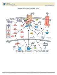
The Syk family protein tyrosine kinase Zap-70 is expressed in T and NK cells and plays a critical role in mediating T cell<br />
activation in response to T cell receptor (TCR) engagement. Following TCR engagement, Zap-70 is rapidly phosphorylated<br />
on several tyrosine residues through autophosphorylation and transphosphorylation by the Src family tyrosine<br />
kinase Lck. Tyrosine phosphorylation correlates with increased Zap-70 kinase activity and downstream signaling<br />
events. Researchers have found that expression of Zap-70 is correlated with disease progression and survival in patients<br />
with chronic lymphocytic leukemia. 1,2<br />
1 Wiestner, A. et al. (2003) Blood 101, 4944–4951.<br />
2 Crespo, M. et al. (2003) N. Engl. J. Med. 348, 1764–1775.<br />
Product References:<br />
Phospho-Stat3 (Tyr705) (D3A7) XP ® Rabbit mAb (Alexa Fluor ® 488 Conjugate) #4323: Anand, S. et al. (2011)<br />
Increased basal intracellular signaling patterns do not correlate with JAK2 genotype in human myeloproliferative neoplasms.<br />
Blood 118, 1610–1621.<br />
P<br />
Tyr394 Lck<br />
P<br />
Tyr505<br />
TCR<br />
P<br />
Zap-70<br />
P P<br />
Tyr319 Tyr493<br />
IP 3<br />
PIP 2<br />
Ca 2+<br />
Calcineurin<br />
DAG<br />
CD4<br />
membrane<br />
Tyr113<br />
P<br />
Tyr128<br />
P<br />
SLP76<br />
Tyr171 P<br />
P<br />
Tyr783<br />
P PLCγ1<br />
PKC<br />
Tyr142<br />
LAT<br />
P Tyr191<br />
Grb2<br />
Ras-Erk<br />
Vav<br />
P<br />
P<br />
Rho/Rac<br />
pathways<br />
Cytokine signaling is integral to an efficient immune response. A key pathway<br />
involved in cytokine signaling is the Janus kinase-signal transducer and<br />
activator of transcription (Jak/Stat) pathway. Jaks and Stats regulate growth,<br />
survival, differentiation, and pathogen resistance. Cytokine binding induces<br />
receptor dimerization, thereby activating the associated Jaks, which undergo<br />
autophosphorylation and subsequently phosphorylate the receptor. These<br />
phosphorylated sites serve as docking sites for the SH2 domain-containing<br />
Stats, such as Stat3, and for SH2-containing proteins and adaptors that<br />
link the receptor to MAP kinase, PI3K/Akt, and other cellular pathways.<br />
Receptor-bound Stats phosphorylated by Jaks dimerize and translocate to<br />
the nucleus where they regulate target gene transcription.<br />
Jak/Stat Utilization Table<br />
Combinatorial use of Tyrosine Kinases and Stat Proteins in Cytokine/Growth Factor Signaling<br />
Ligand Receptor Jak-kinase Other Tyrosine Kinases Stat Family Members<br />
IL-6 IL-6Rα+gp130 Jak1, Jak2, Tyk2 Hck Stat1, Stat3<br />
IL-11 IL-11R+gp130 Jak1, Jak2, Tyk2 Src, Yes Stat3<br />
CNTF, CT-1, LIF, OSM<br />
CNTFR+gp130, CT-1R+gp130, LIFR+gp130,<br />
OSMR+gp130<br />
Jak1, Jak2, Tyk2 Src family Predominant: Stat3<br />
Secondary: Stat1, Stat5<br />
G-CSF G-CSFR Jak2, Tyk2 Lyn Stat3<br />
IL-12 (p40+p35) IL-12Rβ1+IL-12Rβ2 Jak2, Tyk2 Lck Stat4<br />
Leptin LeptinR Jak2 not determined Stat3, Stat5, Stat6<br />
IL-3 IL-3Rα+βc Jak2 Fyn, Hck, Lyn Stat3, Stat5, Stat6<br />
IL-5 IL-5R+βc Jak2 Btk Stat3, Stat5, Stat6<br />
GM-CSF GM-CSFR+βc Jak2 Hck, Lyn Stat3, Stat5<br />
Angiotensin GPCR Jak2, Tyk2 Stat1, Stat2, Stat3<br />
Serotonin GPCR Jak2 Stat3<br />
α-Thrombin GPCR Jak2 Stat1, Stat3<br />
Chemokines CXCR4 Jak2, Jak3<br />
Cytoplasm<br />
Nucleus<br />
Transcription<br />
Factors<br />
P Tyk2<br />
P<br />
ISRE/GAS<br />
IL-2 IL-2Rα+IL-2Rb+γc Jak1, Jak2, Jak3 Fyn, Hck, Lck, Syk, Tec Stat3, Stat5<br />
IL-4 IL-4Rα+γc or IL-4Rα+IL-13Rα1 Jak1, Jak3 Lck, Tec Stat6<br />
IL-7 IL-7R+γc Jak1, Jak3 Lyn Stat3, Stat5<br />
IL-9 IL-9R+γc Jak1, Jak3 not determined Stat1, Stat3, Stat5<br />
IL-13 IL-13Rα1+IL-4Rα Jak1, Jak2, Tyk2 Ctk Stat6<br />
IL-15 IL-15Rα+IL-2Rβ+γc Jak1, Jak3 Lck Stat3, Stat5<br />
IL-19 IL-20Rα+IL-20Rβ Jak1, ? Stat3<br />
IL-20 IL-20Rα, IL-22R+IL-20Rβ Jak1, ? Stat3<br />
IL-21 IL-21R+γc Jak1, Jak3 Stat1, Stat3, Stat5<br />
IL-22 IL-22R+IL-10Rβ Jak1, Tyk2 Stat1, Stat3, Stat5<br />
IL-23 (p40+p19) IL-12Rβ1+IL-23R Jak2, Tyk2 Stat4<br />
IL-24 same as IL-20 Jak1, ? Stat3<br />
IL-26 IL-20Rα+IL-10Rβ Jak1, Tyk2 Stat3<br />
IL-27 (EBI3+p28) gp130+WSX1 Jak1, Jak2, Tyk2 Stat1, Stat2, Stat3, Stat4, Stat5<br />
IL-28A, IL-28B, IL-29 IL-28R+IL-10Rβ Jak1, Tyk2 Stat1, Stat2, Stat3, Stat4, Stat5<br />
IL-31 IL-31Rα+OSMR Jak1, Jak2, Tyk2 Stat1, Stat3, Stat5<br />
IL-35 (p35+EBI3) gp130+WSX1 Jak1, Jak2, Tyk2 Stat1, Stat3, Stat5<br />
GH GHR Jak2 Src family Stat3, Stat5 (mainly Stat5a)<br />
Tpo TpoR (c-Mpl) Tyk2, Jak2 Lyn Stat1, Stat3, Stat5<br />
Epo, Pro EpoR, ProlactinR Jak2 Src Family Stat5 (mainly Stat5a)<br />
Interferon (IFNα/β) IFNAR1+IFNAR2 Jak1, Tyk2 Lck Predominant: Stat1, Stat2<br />
Secondary: Stat3, Stat4, Stat5<br />
IFN-γ IFN-γR1+IFN-γR2 Jak1, Jak2 Hck, Lyn Stat1<br />
IL-10 IL-10Rα+IL-10Rβ Jak1, Tyk2 not determined Stat3, Stat1, Stat5<br />
TLSP TLSPR and IL7R Jak1, possibly Jak2 not determined Stat3, Stat5<br />
EGF EGFR Jak1 EGFR, Src Stat1, Stat3, Stat5<br />
PDGF PDGFR Jak1, Jak2 PDGFR, Src Stat1, Stat3, Stat5<br />
Stats<br />
P<br />
P<br />
Stats<br />
P<br />
Jaks<br />
P P<br />
Stats<br />
P<br />
P<br />
P<br />
P<br />
Stats<br />
PI3K<br />
Akt<br />
mTOR<br />
Membrane<br />
Transcription<br />
Raf<br />
Erk<br />
10 Application and Reactivity Keys, pg 3. Monoclonal Antibody. Please see page 2 for details. Also available as a Conjugated Antibody. See page 8 for details.<br />
Unparalleled Product Quality, Validation, and Technical Support www.cellsignal.com<br />
11
Bioactive Cytokines and Growth Factors<br />
The world’s highest quality research antibody provider has extended<br />
its expertise to Cytokine and Growth Factor production.<br />
Quality<br />
:: Most are greater than 98% pure as demonstrated<br />
by SDS-PAGE.<br />
:: Endotoxin levels are tested by the LAL assay<br />
and are less than 0.01 ng/µg cytokine.<br />
:: Reduced and non-reduced protein is run<br />
on SDS-PAGE.<br />
:: ED 50 or maximum 50% response is determined<br />
by a standard cell based assay for every lot.<br />
:: Several lots are tested side-by-side to ensure<br />
consistent bioactivity.<br />
:: Bioactivity and purity data is shown on each<br />
product webpage and datasheet.<br />
Consistency<br />
:: Strict specifications are set and enforced.<br />
:: Each lot is compared to previous lots for<br />
consistency in purity and bioactivity.<br />
:: Lyophilized lots are quality assured for<br />
sterility and bioactivity.<br />
Dependability<br />
:: Products are produced in-house and<br />
ready to ship.<br />
:: Products are available in multi-milligram sizes.<br />
:: Most customers receive overnight delivery.<br />
:: CST sales and technical support ensure the<br />
highest quality customer service support.<br />
New #8445 Human C-C Motif Chemokine 3 (hCCL3/MIP-1-α)<br />
#3583 Human CD40 Ligand (hCD40L)<br />
#5717 Human Cystatin C (hCystatin C)<br />
#8916 Human Epidermal Growth Factor (hEGF)<br />
New #5331 Mouse Epidermal Growth Factor (mEGF)<br />
#5493 Human Epigen<br />
New #6980 Human Erythropoietin (hEPO)<br />
#5494 Human Epiregulin<br />
#5452 Human His6 Fas Ligand/TNFSF6 (h His6 FasL)<br />
New #8910 Human Basic Fibroblast Growth Factor (hFGF basic/FGF2)<br />
#5234 Human FGF acidic (hFGF acidic)<br />
#5414 Mouse Basic Fibroblast Growth Factor (mFGF basic/FGF2)<br />
#8924 Human Fms-related Tyrosine Kinase 3 Ligand (hFLT3L)<br />
#8930 Human Granulocyte Colony Stimulating Factor (hG-CSF)<br />
#8922 Human Granulocyte Macrophage Colony Stimulating Factor<br />
(hGM-CSF)<br />
#5191 Mouse Granulocyte Macrophage Colony Stimulating Factor<br />
(mGM-CSF)<br />
#8927 Human Interferon-α1 (hIFN-α1)<br />
#8901 Human Interferon-γ (hIFN-γ)<br />
New #5222 Mouse Interferon-γ (mIFN-γ)<br />
#8917 Human Insulin-like Growth Factor I (hIGF-I)<br />
New #9897 Mouse Insulin-like Growth Factor I (mIGF-I)<br />
New #5238 Human Insulin-like Growth Factor II (hIGF-II)<br />
#5236 Human Interleukin-1α (hIL-1α)<br />
#5273 Mouse Interleukin-1α (mIL-1α)<br />
#8900 Human Interleukin-1β (hIL-1β)<br />
#5204 Mouse Interleukin-1β (mIL-1β)<br />
#8907 Human Interleukin-2 (hIL-2)<br />
#5454 Human Interleukin-2 (hIL-2) (mammalian derived)<br />
#5201 Mouse Interleukin-2 (mIL-2)<br />
#8918 Human Interleukin-3 (hIL-3)<br />
#8923 Mouse Interleukin-3 (mIL-3)<br />
#8919 Human Interleukin-4 (hIL-4)<br />
#5208 Mouse Interleukin-4 (mIL-4)<br />
#8904 Human Interleukin-6 (hIL-6)<br />
New #5216 Mouse Interleukin-6 (mIL-6)<br />
New #8170 Human Interleukin-7 (hIL-7)<br />
#5217 Mouse Interleukin-7 (mIL-7)<br />
New #8921 Human Interleukin-8 (hIL-8)<br />
#8903 Human Interleukin-10 (hIL-10)<br />
#5358 Human Interleukin-10 (hIL-10) (mammalian derived)<br />
New #5261 Mouse Interleukin-10 (mIL-10)<br />
#8905 Human Interleukin-13 (hIL-13)<br />
#5242 Mouse Interleukin-13 (mIL-13)<br />
#8928 Human Interleukin-17A (hIL-17A)<br />
#5227 Mouse Interleukin-17A (mIL-17A)<br />
New #9584 Mouse Interleukin-17B (mIL-17B)<br />
#8906 Human Interleukin-17F (hIL-17F)<br />
#8920 Human Interleukin-21 (hIL-21)<br />
#8931 Human Interleukin-22 (hIL-22)<br />
#5224 Mouse Interleukin-22 (mIL-22)<br />
New #5725 Mouse His6 Interleukin-27 (m His6 IL-27)<br />
#5164 Human Interleukin-28A (hIL-28A/IFN-λ2)<br />
New #8796 Human Interleukin-28B (hIL-28B/IFN-λ3)<br />
#5183 Human Interleukin-29 (hIL-29)<br />
New #5719 Human Leptin/OB (hLeptin)<br />
#8929 Human Macrophage Colony Stimulating Factor (hM-CSF)<br />
New #5228 Mouse Macrophage Colony Stimulating Factor (mM-CSF)<br />
#5221 Human β-Nerve Growth Factor (hβ-NGF)<br />
#5218 Human Neuregulin-1 (hNRG-1)<br />
New #5237 Human Neurotrophin-3 (hNT-3)<br />
New #5592 Human Neurotrophin-4 (hNT-4)<br />
New #5367 Human Oncostatin M (hOSM)<br />
#5371 Mouse Oncostatin M (mOSM)<br />
#8912 Human Platelet-Derived Growth Factor BB (hPDGF-BB)<br />
#8913 Human Platelet-Derived Growth Factor AA (hPDGF-AA)<br />
#8925 Human Stem Cell Factor (hSCF)<br />
New #5223 Mouse Stem Cell Factor (mSCF)<br />
New #5712 Human Stromal Cell-derived Factor 1β/CXCL12 (hSDF1β)<br />
IL-6 (pink) is a potent inducer of the acute phase response and is produced by T cells,<br />
macrophages, fibroblasts, endothelial and other cells. IL-6 induces proliferation and<br />
differentiation, and in concert with TGF-β is important for developing Th17 responses.<br />
IL-6 binds to IL-6Rα (light blue) inducing gp130 (yellow) homodimerization. gp130<br />
homodimerization triggers the Jak/STAT cascade and the SHP2/Erk MAP Kinase<br />
cascade. IL-6, through increasing expression of proangiogenic VEGF, may contribute<br />
to metastatic breast cancer.<br />
#5495 Human Transforming Growth Factor α (hTGF-α)<br />
#5154 Human Latent Transforming Growth Factor β1 (hLatent TGF-β1)<br />
#8915 Human Transforming Growth Factor β1 (hTGF-β1)<br />
#5231 Mouse Transforming Growth Factor β1 (mTGF-β1)<br />
#8406 Human Transforming Growth Factor β2 (hTGF-β2)<br />
#8425 Human Transforming Growth Factor β3 (hTGF-β3)<br />
#8902 Human Tumor Necrosis Factor-α (hTNF-α)<br />
#4698 Mouse His6 Tumor Necrosis Factor-α (m His6 TNF-α)<br />
#5178 Mouse Tumor Necrosis Factor-α (mTNF-α)<br />
New #8230 Human Lymphotoxin-α/TNF-β/TNFSF1 (hLT-α)<br />
New #8460 Human His6 41BB Ligand/TNFSF9 (h His6 4-1BBL)<br />
New #5413 Human His6 BAFF/TNFSF13B (h His6 BAFF)<br />
New #5233 Human BAFF/TNFSF13B (hBAFF)<br />
New #8012 Human His6 Thymic Stromal Lymphopoietin (h His6 TSLP)<br />
#5314 Mouse Vascular Endothelial Growth Factor-120 (mVEGF 120 )<br />
#8908 Human Vascular Endothelial Growth Factor-121 (hVEGF 121 )<br />
#5211 Mouse Vascular Endothelial Growth Factor-164 (mVEGF 164 )<br />
New #5874 Rat Vascular Endothelial Growth Factor-164 (rVEGF 164 )<br />
#8065 Human Vascular Endothelial Growth Factor-165 (hVEGF 165 )<br />
Mouse Insulin-like Growth Factor I (mIGF-I) #9897<br />
OD450-OD650<br />
1.5<br />
1.0<br />
0.5<br />
0.00<br />
Bioactivity<br />
0 0.01 0.1 1 10 100 1000<br />
mIGF-I (ng/ml)<br />
kDa<br />
200<br />
116<br />
97<br />
66<br />
55<br />
37<br />
31<br />
22<br />
14<br />
6<br />
4<br />
Purity<br />
+ – kDa<br />
200<br />
140<br />
100<br />
80<br />
60<br />
50<br />
40<br />
30<br />
mIGF-I<br />
20<br />
200<br />
140<br />
100<br />
80<br />
60<br />
50<br />
40<br />
30<br />
20<br />
Downstream Signaling<br />
0<br />
0.1<br />
1<br />
10<br />
100<br />
Phospho-Akt<br />
(Ser473)<br />
Akt<br />
mIGF-I (ng/ml)<br />
Human Interleukin-6 (hIL-6) #8904<br />
Bioactivity<br />
OD 450<br />
-OD 650<br />
0.6<br />
0.3<br />
0<br />
0 .01 .1 1 10 100<br />
hIL-6 (ng/ml)<br />
kDa<br />
200<br />
116<br />
97<br />
66<br />
55<br />
37<br />
31<br />
22<br />
14<br />
6<br />
4<br />
Purity<br />
+ – kDa<br />
hIL-6<br />
200<br />
140<br />
100<br />
80<br />
60<br />
50<br />
200<br />
140<br />
100<br />
80<br />
60<br />
50<br />
Downstream Signaling<br />
0<br />
0.1<br />
1<br />
10<br />
100<br />
Phospho-Stat1<br />
(Tyr701)<br />
Stat1<br />
hIL-6 (ng/ml)<br />
The ability of mIGF-I to induce phosphorylation of Akt<br />
was assessed. After serum starvation, NIH/3T3 cells<br />
were treated with increasing concentrations of mIGF-I<br />
for 10 min. Cells were lysed, and phospho-Akt was<br />
quantified using PathScan ® Phospho-Akt (Thr308)<br />
Sandwich ELISA Kit #7252. OD 450 is shown.<br />
The purity of recombinant mIGF-I<br />
was determined by SDS-PAGE of 6 µg<br />
reduced (+) and non-reduced (-)<br />
recombinant mIGF-I and staining<br />
overnight with Coomassie Blue.<br />
Western blot analysis of extracts from NIH/3T3 cells,<br />
untreated or treated with increasing concentrations<br />
of mIGF-I for 10 min, using Phospho-Akt (Ser473)<br />
(D9E) XP ® Rabbit mAb #4060 (upper) and Akt1<br />
(C73H10) Rabbit mAb #2938 (lower).<br />
The proliferation of TF-1 cells treated with increasing<br />
concentrations of hIL-6 was assessed. After 48<br />
hr treatment with hIL-6, cells were incubated with<br />
a tetrazolium salt and the OD 450 - OD 650 was<br />
determined.<br />
The purity of recombinant hIL-6<br />
was determined by SDS-PAGE of<br />
6 µg reduced (+) and non-reduced<br />
(-) recombinant hIL-6 and staining<br />
overnight with Coomassie Blue.<br />
Western blot analysis of extracts from<br />
TF-1 cells, untreated or treated with hIL-6<br />
for 10 min, using Phospho-Stat1 (Tyr701)<br />
Antibody #9171 (upper) and Stat1 Antibody<br />
#9172 (lower).<br />
12 Application and Reactivity Keys, pg 3. Monoclonal Antibody. Please see page 2 for details.<br />
Unparalleled Product Quality, Validation, and Technical Support www.cellsignal.com<br />
13
BrdU<br />
Phospho-Histone H3 (Ser10)<br />
Cell Cycle, Checkpoint Control,<br />
and DNA Damage<br />
10 3<br />
10 2<br />
10 1<br />
10 0 0<br />
1023<br />
DNA (PI)<br />
BrdU (Bu20a) Mouse mAb #5292:<br />
<strong>Flow</strong> cytometric analysis of Jurkat cells,<br />
incorporated with BrdU (30 min), using<br />
#5292 and Propidium Iodide (PI) #4087.<br />
10 3<br />
10 2<br />
10 1<br />
10 0<br />
0<br />
DNA (PI)<br />
1023<br />
Phospho-Histone H3 (Ser10) (D2C8)<br />
XP ® Rabbit mAb #3377: <strong>Flow</strong> cytometric<br />
analysis of Jurkat cells using #3377 and<br />
Propidium Iodide (PI) #4087. The gated<br />
population indicates Phospho-Histone H3<br />
(Ser10)-positive cells.<br />
p21 Waf1/Cip1 (Alexa Fluor ® 488 Conjugate)<br />
10 2<br />
10 1<br />
10 0 0 200 400 600<br />
DNA (PI)<br />
p21 Waf1/Cip1 (12D1) Rabbit mAb<br />
(Alexa Fluor ® 488 Conjugate) #5487:<br />
<strong>Flow</strong> cytometric analysis of COS-7 cells<br />
using #5487 and Propidium Iodide (PI)<br />
#4087. Red = positive cells.<br />
Applications Reactivity<br />
#2675 Phospho-53BP1 (Ser1778) Antibody W, IF-IC, F H, Mk<br />
#2914 Phospho-Aurora A (Thr288)/Aurora B (Thr232)/Aurora W, IF-IC, F H, M, R<br />
C (Thr198) (D13A11) XP ® Rabbit mAb<br />
#3094 Aurora B/AIM1 Antibody W, IP, F H, M, R, Mk<br />
#5292 BrdU (Bu20a) Mouse mAb IHC-P, IF-IC, F All<br />
#4539 Phospho-cdc2 (Tyr15) (10A11) Rabbit mAb W, IP, IF-IC, F H, M, R, Mk<br />
#9529 Phospho-cdc25C (Ser198) Antibody W, F H<br />
#2561 Phospho-CDK2 (Thr160) Antibody W, IP, F, E-P H, M, R<br />
#2546 CDK2 (78B2) Rabbit mAb W, IP, F H, M, R, Mk<br />
#2316 CDK9 (C12F7) Rabbit mAb W, IP, IHC-P, IHC-F,<br />
IF-IC, F<br />
H, M, R, Hm,<br />
Mk, B, Dg<br />
#2348 Phospho-Chk1 (Ser345) (133D3) Rabbit mAb W, IF-IC, F H, M, R, Mk<br />
#2197 Phospho-Chk2 (Thr68) (C13C1) Rabbit mAb W, IP, IHC-P, F H, (Mk)<br />
#2661 Phospho-Chk2 (Thr68) Antibody W, IP, IF-IC, F H, Mk<br />
#4656 Cyclin A (BF683) Mouse mAb W, F H<br />
#4135 Cyclin B1 (V152) Mouse mAb W, F H, M, (Hm)<br />
#3300 Phospho-Cyclin D1 (Thr286) (D29B3) XP ® Rabbit mAb W, IP, IF-IC, F H, (Mk)<br />
#2978 Cyclin D1 (92G2) Rabbit mAb W, IHC-P, F H, M, R<br />
#4136 Phospho-Cyclin E (Thr62) Antibody W, IP, IHC-P, F H<br />
#9718 Phospho-Histone H2A.X (Ser139) (20E3) Rabbit mAb W, IHC-P, IF-IC, F H, M, R, Mk<br />
#2577 Phospho-Histone H2A.X (Ser139) Antibody W, IHC-P, IF-IC, F H, M, R<br />
#5438 Phospho-Histone H2A.X (Ser139/Tyr142) Antibody W, IP, IF-IC, F H, M, R, Mk<br />
#3377 Phospho-Histone H3 (Ser10) (D2C8) XP ® Rabbit mAb W, IF-IC, F H, M, R, Mk, Z<br />
#3642 Phospho-Histone H3 (Ser10) (D2C8) XP ® Rabbit mAb<br />
(Biotinylated)<br />
#9701 Phospho-Histone H3 (Ser10) Antibody W, IP, IHC-P, IHC-F,<br />
IF-IC, F<br />
W, IF-F, IF-IC, F H, M, R, Mk<br />
H, M, R, Mk,<br />
C, Dm, Z, Sc, (X)<br />
#9706 Phospho-Histone H3 (Ser10) (6G3) Mouse mAb W, IF-F, IF-IC, F H, M, R<br />
#9767 Phospho-Histone H3 (Thr11) (C2A6) Rabbit mAb W, IP, F H, M, R, (X)<br />
#9764 Phospho-Histone H3 (Thr11) Antibody W, IP, IF-IC, F H, M, R, (X)<br />
#9713 Phospho-Histone H3 (Ser28) Antibody W, IP, IF-F, IF-IC, F H, M, Hm, Dm,<br />
(R, C, X, Z, B)<br />
#9649 Acetyl-Histone H3 (Lys9) (C5B11) Rabbit mAb W, IHC-P, IF-IC, H, M, R, Mk, Z, (Sc)<br />
F, ChIP<br />
New #5545 hnRNP A0 (D8A3) XP ® Rabbit mAb W, IP, IF-IC, F H, M, R, Mk<br />
New #8443 hnRNP A1 (D21H11) Rabbit mAb W, IP, IF-IC, F H, M, R, Mk<br />
#9304 hnRNP A2/B1 (2A2) Mouse mAb W, F H, Mk<br />
#2616 HP1α Antibody W, IP, IHC-P, IF-IC, F H, M, R, Mk, (B)<br />
#2619 HP1γ Antibody W, IP, IF-IC, F H, M, R, Mk<br />
#4944 HR6A/HR6B Antibody W, IF-IC, F H, M, R, Mk,<br />
(C, Dm, X, Z)<br />
#2786 INCENP (A841) Antibody W, IF-IC, F H<br />
#2807 INCENP (P240) Antibody W, IF-IC, F H<br />
#4822 p15 INK4B Antibody W, F H, M, R<br />
#2753 Ku80 Antibody W, IP, IHC-P, IF-IC, F H, Mk, (M, R)<br />
#2139 LSD1 Antibody W, IP, IHC-P, IF-IC, F H, M, R, Mk<br />
#3515 MLH1 (4C9C7) Mouse mAb W, IP, IF-IC, F H, Mk<br />
#4847 Mre11 (31H4) Rabbit mAb W, IP, IHC-P, IHC-F, F H<br />
#2017 MSH2 (D24B5) XP ® Rabbit mAb W, IP, IHC-P, IF-IC, F H, M, Mk<br />
#3520 Phospho-NPM (Ser4) (D19C1) XP ® Rabbit mAb W, F H, (M, R, Mk, X, B, Dg)<br />
#3517 Phospho-NPM (Thr95) Antibody W, IP, IF-IC, F H, (M, R, Mk)<br />
#2947 p21 Waf1/Cip1 (12D1) Rabbit mAb W, IP, IHC-P, IF-IC, F H, Mk<br />
#9286 Phospho-p53 (Ser15) (16G8) Mouse mAb W, IF-IC, F H<br />
#4030 Phospho-p53 (Ser15) (16G8) Mouse mAb (Biotinylated) W, F H<br />
#9289 Phospho-p53 (Ser37) Antibody W, IF-IC, F H, Mk<br />
#2521 Phospho-p53 (Ser46) Antibody W, IP, IF-IC, F H, Mk<br />
Product References:<br />
Phospho-Histone H3 (Ser10) (D2C8) XP ® Rabbit mAb (Alexa Fluor ® 488 Conjugate) #3465: Mathews, H.L. et al. (2011) Epigenetic<br />
patterns associated with the immune dysregulation that accompanies psychosocial distress. Brain Behav. Immun. 25, 830–839.<br />
Phospho-Histone H3 (Ser10) Antibody (Alexa Fluor ® 647 Conjugate) #9716: Kalland, M.E. et al. (2011) T cell-signaling network<br />
analysis reveals distinct differences between CD28 and CD2 costimulation responses in various subsets and in the MAPK pathway between<br />
resting and activated regulatory T cells. J. Immunol. 187, 5233–5245.<br />
Applications<br />
Reactivity<br />
#2527 p53 (7F5) Rabbit mAb W, IHC-P, IF-IC, H, Mk<br />
F, ChIP<br />
#4667 p53 (7F5) Rabbit mAb (Biotinylated) W, F H, Mk<br />
#2015 p53 (1C12) Mouse mAb (Alexa Fluor ® 488 Conjugate) IF-IC, F H, M, R, Mk<br />
#2533 p53 (1C12) Mouse mAb (Alexa Fluor ® 647 Conjugate) F H<br />
#4981 Phospho-p63 (Ser160/162) Antibody W, IHC-P, F H, (M, R, C, X)<br />
#4941 Phospho-PBK/TOPK (Thr9) Antibody W, IF-IC, F H, M<br />
#4852 PCTAIRE 1 Antibody W, IP, F M, R, (H)<br />
#9309 Rb (4H1) Mouse mAb W, IP, IHC-P, IF-IC, H, Mk, B, Pg<br />
F, ChIP<br />
#2267 RPA70 Antibody W, IP, IF-IC, F H, R, Mk<br />
#2314 Phospho-SirT1 (Ser47) Antibody W, IP, IF-IC, F H<br />
#4124 TIF1β (C42G12) Rabbit mAb W, IHC-P, IF-IC, F H, M, R, Mk<br />
#4733 Topoisomerase IIα Antibody W, IP, F H, M, R, Mk<br />
#2648 VCP Antibody W, IF-IC, F H, M, R, Mk,<br />
(X, Z, B, Pg, Sc)<br />
#4936 Wee1 Antibody W, IP, F H, R, Mk<br />
Events<br />
p53 Tumor Suppressor and<br />
Aurora Kinases<br />
p53 (Alexa Fluor ® 488 Conjugate)<br />
A<br />
Phospho-Histone H3 (Ser10)<br />
(Alexa Fluor ® 647 Conjugate)<br />
p53 (7F5) Rabbit mAb<br />
(Alexa Fluor ® 488<br />
Conjugate) #5429: <strong>Flow</strong><br />
cytometric analysis of<br />
K-562 (blue) and HT-29<br />
(green) cells using #5429.<br />
10 3<br />
10 2<br />
B<br />
p53<br />
MDM2<br />
10 0 10 1 10 2 10 3<br />
Phospho-Aurora A (Thr288)/Aurora B (Thr232)<br />
/Aurora C (Thr198) (Alexa Fluor ® 488 Conjugate)<br />
Ser315<br />
P<br />
Degradation<br />
HIPK2<br />
Aurora A<br />
53BP1<br />
p300<br />
Events<br />
TIF1β<br />
TIF1β (C42G12) Rabbit mAb #4124: <strong>Flow</strong><br />
cytometric analysis of HeLa cells using #4124<br />
(blue) compared to a concentration-matched<br />
nonspecific negative control antibody (red).<br />
DNA DAMAGE<br />
Ser9<br />
Ser15<br />
Ser20<br />
Ser6 P P P P<br />
Ser46 P Ser37<br />
P p53 P Ser33<br />
P<br />
Ser315<br />
Ser392<br />
P<br />
MDM2<br />
P<br />
P<br />
PP<br />
P P<br />
P<br />
P P<br />
P P P P P<br />
P P<br />
Bax, p53AIP, etc. Apoptosis<br />
p21Waf1/Cip1, etc. G2 arrest<br />
p53R2, etc. DNA repair<br />
Phospho-Aurora A (Thr288)/Aurora B (Thr232)/Aurora<br />
C (Thr198) (D13A11) XP ® Rabbit mAb (Alexa Fluor ® 488<br />
10 1<br />
Conjugate) #8525: Confocal IF analysis (A) of HT-1080 cells<br />
using #8525 (green) and β-Tubulin (9F3) Rabbit mAb (Alexa Fluor ®<br />
555 Conjugate) #2116 (red). Blue pseudocolor = DRAQ5 ® #4084<br />
10 0 (fluorescent DNA dye). Two-color flow cytometric analysis (B) of<br />
asynchronous Jurkat cells using #8525 and Phospho-Histone H3<br />
(Ser10) (D2C8) XP ® Rabbit mAb (Alexa Fluor ® 647 Conjugate)<br />
#3458. Gated population represents cells that are are mitotic and<br />
positive for phospho-Aurora and phospho-histone H3, while cells<br />
represented in blue are non-mitotic.<br />
A B C<br />
Events<br />
ATR<br />
CK1δ/ε<br />
Phospho-p53 (Ser15)<br />
(Alexa Fluor ® 488 Conjugate)<br />
p300<br />
p53<br />
DNA-PK<br />
p53<br />
ATM<br />
Chk2<br />
DNA-PK<br />
Ser166<br />
MDM2<br />
CAK<br />
Phospho-p53 (Ser15) (16G8)<br />
Mouse mAb (Alexa Fluor ® 488<br />
Conjugate) #9235: Confocal IF<br />
analysis of HT-29 cells, untreated<br />
(A) or UV-treated (B), using #9235<br />
(green). Blue pseudocolor = DRAQ5 ®<br />
#4084 (fluorescent DNA dye). <strong>Flow</strong><br />
cytometric analysis of HT-29 cells (C),<br />
untreated (blue) or UV-treated (green),<br />
using #9235.<br />
14 Application and Reactivity Keys, pg 3. Monoclonal Antibody. Please see page 2 for details. Also available as a Conjugated Antibody. See page 8 for details.<br />
Unparalleled Product Quality, Validation, and Technical Support www.cellsignal.com<br />
15
Stem Cell Markers, Development,<br />
and Differentiation<br />
Events<br />
β-Catenin (L54E2) Mouse mAb (PE Conjugate)<br />
#6898: <strong>Flow</strong> cytometric analysis of NCI-H28 (blue)<br />
and HeLa (green) cells using #6898.<br />
Events<br />
E-Cadherin (PE Conjugate)<br />
E-Cadherin (24E10) Rabbit mAb (PE Conjugate)<br />
#7559: <strong>Flow</strong> cytometric analysis of HeLa (blue) and<br />
MCF7 (green) cells using #7559.<br />
Events<br />
Applications<br />
Reactivity<br />
New #8225 CACYBP (D43G11) Rabbit mAb W, IHC-P, F H, M, R, Mk<br />
#3354 CACYBP Antibody W, IHC-P, F H, M, R, Mk<br />
New #8480 β-Catenin (D10A8) XP ® Rabbit mAb<br />
W, IP, IHC-P, IHC-F,<br />
IF-F, IF-IC, F, ChIP<br />
#3195 E-Cadherin (24E10) Rabbit mAb W, IHC-P, IHC-F,<br />
IF-IC, F<br />
H, M, R, Mk,<br />
(Z, B, Pg)<br />
H, M, (Dg, Pg)<br />
#2677 β-Catenin (L54E2) Mouse mAb (IF Preferred) IP, IF-IC, F H, (M, R,<br />
Mk, Pg)<br />
#5265 β-Catenin (L54E2) Mouse mAb (Biotinylated) F H<br />
#2929 EpCAM (VU1D9) Mouse mAb W, IHC-P, IF-IC, F H<br />
#4402 FoxP1 (D35D10) XP ® Rabbit mAb W, IP, IHC-P, F H, M, R, Mk<br />
#2005 FoxP1 Antibody W, IP, IHC-P, IF-IC, F H, M, (R)<br />
#2230 LEF1 (C12A5) Rabbit mAb W, IP, IF-IC, F H, M, R<br />
#2286 LEF1 (C18A7) Rabbit mAb W, IP, F H, M<br />
New #8641 LIN28A (D1A1A) XP ® Rabbit mAb W, IF-IC, F H, M, (R, Mk)<br />
#3695 LIN28A (D84C11) XP ® Rabbit mAb W, IF-IC, F H, (R, Mk)<br />
#3978 LIN28A (A177) Antibody W, IP, IHC-P, IF-IC, F H, M, (Mk)<br />
#4903 Nanog (D73G4) XP ® Rabbit mAb W, IHC-P, IF-IC, F H, (Mk)<br />
#3580 Nanog Antibody W, IF-IC, F, ChIP H<br />
#4893 Nanog (1E6C4) Mouse mAb W, IHC-P, IF-IC, F H<br />
#4380 Notch1 (D6F11) XP ® Rabbit mAb W, IF-IC, F H, M, R<br />
#5732 Notch2 (D76A6) XP ® Rabbit mAb W, IP, IF-IC, F H, M, R<br />
#2756 Numb (C29G11) Rabbit mAb W, IP, IF-IC, F H, M, R, Mk<br />
#2761 Numb (C44B4) Rabbit mAb W, IP, IF-IC, F H<br />
#2840 Oct-4A (C30A3) Rabbit mAb W, IF-IC, F H, M<br />
#2890 Oct-4A (C52G3) Rabbit mAb W, IHC-P, IF-IC, F,<br />
ChIP<br />
#2750 Oct-4 Antibody W, IHC-P, IF-IC, F,<br />
ChIP<br />
#9516 Phospho-Smad1/5 (Ser463/465) (41D10)<br />
Rabbit mAb<br />
H<br />
H, (Mk)<br />
W, IF-IC, F H, M, R<br />
New #6944 Smad1 (D59D7) XP ® Rabbit mAb W, IP, IF-IC, F, ChIP H, M, (Mk, X, B)<br />
#9510 Phospho-Smad2 (Ser465/467)/ Smad3<br />
(Ser423/425) (D6G10) XP ® Rabbit mAb<br />
IF-IC, F<br />
#5339 Smad2 (D43B4) XP ® Rabbit mAb W, IP, IF-IC, F, ChIP H, M, R, Mk<br />
New #8685 Smad2/3 (D7G7) XP ® Rabbit mAb W, IP, IF-IC, F, ChIP H, M, R, Mk<br />
H<br />
Pluripotency in Human<br />
Embryonic Stem Cells<br />
The predominant signaling pathways involved in human embryonic stem<br />
cell (ESC) pluripotency and self renewal are TGF-β, which signals through<br />
Smad2/3, and FGFR, which activates the MAPK and Akt pathways. The Wnt<br />
pathway also promotes pluripotency, although this may occur through a<br />
non-canonical mechanism involving a balance between the transcriptional<br />
activator TCF1 and the repressor TCF3. Signaling through these pathways<br />
results in the expression and activation of three key transcription factors:<br />
Oct-4, Sox2, and Nanog. These transcription factors activate gene expression<br />
of ESC-specific genes, regulate their own expression, and also serve as<br />
human ESC markers.<br />
membrane<br />
cytoplasm<br />
nucleus<br />
PI3K<br />
Akt<br />
FGF2<br />
IGF<br />
Erk1/2<br />
Oct-4<br />
LRP<br />
Sox2<br />
Wnt<br />
Oct-4 Sox2 FoxD3<br />
Oct-4 Sox2 Nanog<br />
TGF-β/<br />
Activin/Nodal<br />
β-catenin Smad2/3 Smad1/5/8<br />
Oct-4,<br />
Sox2,<br />
FoxD3,<br />
FGF4<br />
Nanog<br />
gene<br />
expression<br />
BMP4<br />
Pluripotency<br />
and Self Renewal<br />
hESC Markers:<br />
Oct-4<br />
Nanog<br />
Sox2<br />
SSEA 3/4<br />
TRA-1-60<br />
TRA-1-81<br />
Events<br />
Oct-4A (Alexa Fluor ® 488 Conjugate)<br />
Oct-4A (C30A3) Rabbit mAb (Alexa Fluor ®<br />
488 Conjugate) #5177: <strong>Flow</strong> cytometric<br />
analysis of HeLa (blue) and NTERA-2 (green)<br />
cells using #5177.<br />
Events<br />
Sox2 (Alexa Fluor ® 647 Conjugate)<br />
Sox2 (D6D9) XP ® Rabbit mAb (Alexa Fluor ®<br />
647 Conjugate) #5067: <strong>Flow</strong> cytometric<br />
analysis of HeLa (blue) and NTERA-2 (green)<br />
cells using #5067.<br />
Events<br />
EpCAM (PE Conjugate)<br />
EpCAM (VU1D9) Mouse mAb (PE Conjugate)<br />
#8995: <strong>Flow</strong> cytometric analysis of unpermeablized<br />
HT-29 cells using #8995 (green) compared to<br />
concentration-matched Mouse (G3A1) mAb IgG1<br />
Isotype Control (PE Conjugate) #6899 (red).<br />
LIN28A<br />
LIN28A (D1A1A) XP ® Rabbit mAb #8641:<br />
<strong>Flow</strong> cytometric analysis of HeLa (red) and<br />
NTERA-2 (blue) cells using #8641.<br />
#5678 Smad2/3 Antibody W, IP, IF-IC, F, ChIP H, M, R, Mk, (X)<br />
#9523 Smad3 (C67H9) Rabbit mAb W, IP, IF-IC, F, ChIP H, M, R, Mk,<br />
(X, Z, B)<br />
#3579 Sox2 (D6D9) XP ® Rabbit mAb W, IHC-P, IF-IC, F H, (Mk, B, Dg)<br />
#4900 Sox2 (L1D6A2) Mouse mAb W, IF-IC, F H, M, (R, B, Dg)<br />
#4744 SSEA1 (MC480) Mouse mAb IHC-P, IF-IC, F M<br />
#2203 TCF1 (C63D9) Rabbit mAb W, IP, IHC-P, IF-IC, F H, M<br />
#4746 TRA-1-60(S) (TRA-1-60(S)) Mouse mAb W, IHC-P, IF-IC, F H<br />
#4745 TRA-1-81 (TRA-1-81) Mouse mAb IHC-P, IF-IC, F H<br />
#5741 Vimentin (D21H3) XP ® Rabbit mAb W, IHC-P, IF-IC, F H, M, R, Mk<br />
XP ®<br />
Monoclonal Antibodies<br />
one antibody, multiple applications <br />
Events<br />
Nanog (Alexa Fluor ® 647 Conjugate)<br />
Nanog (D73G4) XP ® Rabbit mAb (Alexa<br />
Fluor ® 647 Conjugate) #5448: <strong>Flow</strong><br />
cytometric analysis of HeLa (blue) and NTERA-2<br />
(green) cells using #5448.<br />
16 Application and Reactivity Keys, pg 3. Monoclonal Antibody. Please see page 2 for details.<br />
Also available as a Conjugated Antibody. See page 8 for details.<br />
Unparalleled Product Quality, Validation, and Technical Support www.cellsignal.com<br />
17
MAP Kinase Signaling<br />
Events<br />
Events<br />
Phospho-p38 MAPK (Thr180/Tyr182)<br />
(PE Conjugate)<br />
Phospho-p38 MAPK (Thr180/Tyr182) (3D7)<br />
Rabbit mAb (PE Conjugate) #6908: <strong>Flow</strong><br />
cytometric analysis of THP-1 cells, untreated (blue)<br />
or anisomycin-treated (green), using #6908.<br />
Events<br />
Phospho-c-Fos (Ser32)<br />
Phospho-c-Fos (Ser32) (D82C12) XP ® Rabbit<br />
mAb #5348: <strong>Flow</strong> cytometric analysis of HeLa<br />
cells, untreated (blue) or treated with TPA #4174<br />
(green), using #5348.<br />
p38 MAPK<br />
p38 MAPK (D13E1) XP ® Rabbit mAb #8690:<br />
<strong>Flow</strong> cytometric analysis of HeLa cells using #8690<br />
(blue) compared to concentration-matched Rabbit<br />
(DA1E) mAb IgG XP ® Isotype Control #3900 (red).<br />
Applications<br />
Reactivity<br />
#5112 Phospho-ATF-2 (Thr71) (11G2) Rabbit mAb W, F H, M, R, Mk<br />
#9221 Phospho-ATF-2 (Thr71) Antibody W, IP, IHC-P, IHC-F,<br />
IF-IC, F<br />
H, M, R, Mk<br />
#4067 Erk3 Antibody W, IP, F H, M, R, Mk<br />
#5122 FAM129B Antibody W, IP, IF-IC, F H, Mk<br />
#5348 Phospho-c-Fos (Ser32) (D82C12) XP ®<br />
Rabbit mAb<br />
W, IP, IF-IC, F H, M, R, (Hm, Mk, B, Pg)<br />
#2250 c-Fos (9F6) Rabbit mAb W, IF-IC, F H, M, R, (Hm, B, Pg)<br />
#2251 FosB (5G4) Rabbit mAb W, IP, IHC-P, IF-IC, F H, M, R<br />
#2023 FosB (5G4) Rabbit mAb (Alexa Fluor ® 488<br />
Conjugate)<br />
F<br />
H, M, Mk<br />
New #5519 JIP4/SPAG9 (D72F4) XP ® Rabbit mAb W, IP, IHC-P, IF-IC, F H, M, R, Mk<br />
#9261 Phospho-c-Jun (Ser63) II Antibody W, IP, IF-IC, F H, M, R, Mk, Pg<br />
#3270 Phospho-c-Jun (Ser73) (D47G9) XP ®<br />
Rabbit mAb<br />
W, IP, IHC-P, IF-IC, F H, M, R, Mk<br />
New #5000 JunD (D17G2) Rabbit mAb W, IP, IF-IC, F H, Mk, B, Pg<br />
#3007 Phospho-MAPKAPK-2 (Thr334) (27B7)<br />
Rabbit mAb<br />
W, IHC-P, IF-IC, F H, M, R, Mk<br />
#3042 MAPKAPK-2 Antibody W, IP, F H, M, R, Mk, (Hm)<br />
#4660 Phospho-MEK1/2 (Ser217/221) (41G9)<br />
Rabbit mAb (Alexa Fluor ® 488 Conjugate)<br />
#5068 Phospho-MEK1/2 (Ser217/221) (41G9)<br />
Rabbit mAb (Alexa Fluor ® 647 Conjugate)<br />
#2338 Phospho-MEK1/2 (Ser221) (166F8)<br />
Rabbit mAb<br />
Product References:<br />
Phospho-p38 MAPK (Thr180/Tyr182) (28B10) Mouse mAb (Alexa Fluor ® 647 Conjugate)<br />
#4552: Kalland, M.E. et al. (2011) T cell-signaling network analysis reveals distinct differences<br />
between CD28 and CD2 costimulation responses in various subsets and in the MAPK pathway<br />
between resting and activated regulatory T cells. J. Immunol. 187, 5233–5245.<br />
Phospho-p44/42 MAPK (Erk1/2) (Thr202/Tyr204) (D13.14.4E) XP ® Rabbit mAb #4370:<br />
Koenigsmann, J. et al. (2009) Nf1 haploinsufficiency and Icsbp deficiency synergize in the<br />
development of leukemias. Blood 113, 4690–4701.<br />
Phospho-p44/42 MAPK (Erk1/2) (Thr202/Tyr204) (197G2) Rabbit mAb #4377: Gross, A.J.<br />
et al. (2009) Developmental acquisition of the Lyn-CD22-SHP-1 inhibitory pathway promotes B cell<br />
tolerance. J. Immunol. 182, 5382–5392. / Messal, N. et al. (2011) Differential role for CD277 as a<br />
co-regulator of the immune signal in T and NK cells. Eur. J. Immunol. 41, 3443–3454.<br />
F<br />
F<br />
H, M, R, Mk, Dm, (C)<br />
H, M, R, Mk, Dm, (C)<br />
W, IHC-P, F H, M, R, Mk, (Dg)<br />
#9127 Phospho-MEK1 (Thr286) Antibody W, IP, IF-IC, F H, R, Mk, (M)<br />
New #8727 MEK1/2 (D1A5) Rabbit mAb W, IF-IC, F H, M, R, Mk, (Hm, Dm,<br />
X, Z, B, Dg, Pg, Ce)<br />
#4694 MEK1/2 (L38C12) Mouse mAb W, IHC-P, IF-IC, F H, M, R, Mk<br />
#4511 Phospho-p38 MAPK (Thr180/Tyr182) (D3F9)<br />
XP ® Rabbit mAb<br />
#9215 Phospho-p38 MAPK (Thr180/Tyr182) (3D7)<br />
Rabbit mAb<br />
#4092 Phospho-p38 MAPK (Thr180/Tyr182) (3D7)<br />
Rabbit mAb (Biotinylated)<br />
#9211 Phospho-p38 MAPK (Thr180/Tyr182)<br />
Antibody<br />
#9216 Phospho-p38 MAPK (Thr180/Tyr182)<br />
(28B10) Mouse mAb<br />
W, IP, IHC-P, IF-IC, F H, M, R, Mk, Sc,<br />
(Hm, C, Z, B, Pg)<br />
W, IF-IC, F H, M, R, Mk, Dm, Pg, Sc,<br />
(Hm, Z, B)<br />
W, F H, M, R, Mk, Dm, Pg, Sc<br />
W, IP, IF-IC, F H, M, R, Mk, Dm, Pg, Sc,<br />
(Hm, Z, B)<br />
W, IP, IF-IC, F H, M, R, Mk, Sc, (Z)<br />
New #8690 p38 MAPK (D13E1) XP ® Rabbit mAb W, IHC-P, IF-IC, F H, M, R, Hm, Mk, Pg<br />
#9212 p38 MAPK Antibody W, IHC-P, IF-IC, F H, M, R, Mk, (C)<br />
#9228 p38α MAPK (L53F8) Mouse mAb W, IF-IC, F H, M, R, Mk, Sc<br />
#4370 Phospho-p44/42 MAPK (Erk1/2) (Thr202/<br />
Tyr204) (D13.14.4E) XP ® Rabbit mAb<br />
#4094 Phospho-p44/42 MAPK (Erk1/2) (Thr202/<br />
Tyr204) (D13.14.4E) XP ® Rabbit mAb<br />
(Biotinylated)<br />
#4377 Phospho-p44/42 MAPK (Erk1/2) (Thr202/<br />
Tyr204) (197G2) Rabbit mAb<br />
#9101 Phospho-p44/42 MAPK (Erk1/2) (Thr202/<br />
Tyr204) Antibody<br />
#9106 Phospho-p44/42 MAPK (Erk1/2) (Thr202/<br />
Tyr204) (E10) Mouse mAb<br />
W, IP, IHC-P, IF-IC, F H, M, R, Hm, Mk, Mi, Dm,<br />
Z, B, Dg, Pg, Sc, (Ce)<br />
W, F H, M, R, Hm, Mk, Mi, Dm,<br />
Z, B, Pg, Sc<br />
W, IF-IC, F H, M, R, Mk, Mi, Dm,<br />
Z, Pg<br />
W, IP, IF-IC, F H, M, R, Hm, Mk, Mi, Dm,<br />
Z, B, Pg, Ce, (C)<br />
W, IP, F H, M, R, Hm, Mk, Mi, Dm,<br />
Z, B, Pg<br />
#4695 p44/42 MAPK (Erk1/2) (137F5) Rabbit mAb W, IP, IHC-P, IF-IC, F H, M, R, Hm, Mk, Mi, Dm,<br />
Z, B, Dg, Pg, Ce, (C)<br />
Phospho-p44/42 MAPK (Erk1/2) (Thr202/Tyr204) (E10) Mouse mAb (Alexa Fluor ® 647<br />
Conjugate) #4375: Soond, D.R. et al. (2010) PI3K p110delta regulates T-cell cytokine production<br />
during primary and secondary immune responses in mice and humans. Blood 115, 2203–2213.<br />
/ Kalland, M.E. et al. (2011) T cell-signaling network analysis reveals distinct differences between<br />
CD28 and CD2 costimulation responses in various subsets and in the MAPK pathway between<br />
resting and activated regulatory T cells. J. Immunol. 187, 5233–5245.<br />
Phospho-MAPKAPK-2 (Thr334) (27B7) Rabbit mAb (Alexa Fluor ® 647 Conjugate) #4320:<br />
Kalland, M.E. et al. (2011) T cell-signaling network analysis reveals distinct differences between<br />
CD28 and CD2 costimulation responses in various subsets and in the MAPK pathway between<br />
resting and activated regulatory T cells. J. Immunol. 187, 5233–5245.<br />
SS Lin: SS Lin<br />
20<br />
15<br />
10<br />
#5013 p44/42 MAPK (Erk1/2) (137F5) Rabbit mAb<br />
(Biotinylated)<br />
5<br />
0<br />
Untreated<br />
20<br />
15<br />
10<br />
5<br />
Applications Reactivity<br />
W, IP, F H, M, R, Mk, Mi, Dm,<br />
Z, B, Pg<br />
#9102 p44/42 MAPK (Erk1/2) Antibody W, IP, IHC-P, IF-IC, F H, M, R, Hm, Mk, Mi, Z,<br />
B, Pg, Sc<br />
#4696 p44/42 MAPK (Erk1/2) (L34F12) Mouse mAb W, IHC-P, IF-IC, F H, M, R, Mk, Mi, Z, Pg<br />
#9434 B-Raf (L12G7) Mouse mAb W, IP, F H, M, R, Mk, Dm<br />
New #5528 RSK2 (D21B2) XP ® Rabbit mAb W, IF-IC, F H, M, R, Mk, B, Pg, (Dg)<br />
#9255 Phospho-SAPK/JNK (Thr183/Tyr185) (G9) W, IP, IF-IC, F H, M, R, Hm, Sc<br />
Mouse mAb<br />
#4514 Phospho-SEK1/MKK4 (Ser257) (C36C11) W, F H, M, R, Mk<br />
Rabbit mAb<br />
#3225 TAB1 Antibody W, IF-IC, F H, M, R, Mk<br />
#4505 TAK1 Antibody W, F H, M, R, Mk, B, (C, X)<br />
p44/42 MAPK<br />
Mitogen-activated protein kinases (MAPKs) are a<br />
widely conserved family of serine/threonine protein<br />
kinases involved in many cellular programs such as<br />
cell proliferation, differentiation, motility, and death.<br />
The p44/42 MAPK (Erk1/2) signaling pathway can be<br />
activated in response to a diverse range of extracellular<br />
stimuli including mitogens, growth factors, and<br />
cytokines and is an important target in the diagnosis<br />
and treatment of cancer.<br />
Upon stimulation, a sequential three-part protein<br />
kinase cascade is initiated, consisting of a MAP kinase<br />
kinase kinase (MAPKKK or MAP3K), a MAP kinase<br />
kinase (MAPKK or MAP2K), and a MAP kinase<br />
(MAPK). Multiple p44/42 MAP3Ks have been identified,<br />
including members of the Raf family, as well as<br />
Mos and Tpl2/Cot. MEK1 and MEK2 are the primary<br />
MAPKKs in this pathway. MEK1 and MEK2 activate<br />
p44 and p42 through phosphorylation of activation<br />
loop residues Thr202/Tyr204 and Thr185/Tyr187,<br />
respectively.<br />
Several downstream targets of p44/42 have been<br />
identified, including p90RSK and the transcription<br />
factor Elk-1. p44/42 are negatively regulated by a family<br />
of dual-specificity (Thr/Tyr) MAPK phosphatases,<br />
known as DUSPs or MKPs, along with MEK inhibitors<br />
such as U0126 and PD98059.<br />
TPA<br />
Events<br />
Phospho-p44/42 MAPK (Erk 1/2)<br />
(Thr202/Tyr204)<br />
0<br />
0<br />
10 0 10 1 10 2 10 3<br />
10 0 10 1 10 2 10 3<br />
10 0 10 1 10 2 10 3<br />
Phospho-p44/42 MAPK (Erk1/2) (Thr202/Tyr204) (Alexa Fluor ® 488 Conjugate)<br />
20<br />
15<br />
10<br />
5<br />
Growth/Neurotrophic Factors<br />
EGF<br />
Overlay<br />
Membrane<br />
RAS<br />
GRB SOS<br />
P Tyr1068<br />
Ser338 Akt<br />
P<br />
P Ser261<br />
PP2A<br />
c-Raf P Ser259 PKC<br />
14-3-3<br />
P Ser296 AMPK<br />
Ser298 P<br />
P<br />
Ser217<br />
14-3-3<br />
Ser297 Ser392 MEK1/2<br />
Thr286<br />
P Ser221<br />
P P<br />
P<br />
KSR<br />
KSR P<br />
U0126<br />
IMP<br />
Ser298<br />
Thr202<br />
P<br />
PD98059<br />
MEK1/2 Tyr204 P MAPK Erk1/2<br />
Cytoplasm<br />
EGFR<br />
Ser363<br />
Thr359 P P<br />
Transcription<br />
Events<br />
Thr58<br />
Ser380 P<br />
P<br />
Ser62<br />
P<br />
p90RSK c-Myc Elk1<br />
Nucleus<br />
Ser383<br />
P<br />
SRE<br />
RSK2<br />
RSK2 (D21B2) XP ® Rabbit mAb #5528: <strong>Flow</strong><br />
cytometric analysis of HeLa cells using #5528<br />
(blue) compared to concentration-matched Rabbit<br />
(DA1E) mAb IgG XP ® Isotype Control #3900 (red).<br />
Transcription<br />
Phospho-p44/42 MAPK<br />
(Erk1/2) (Thr202/Tyr204)<br />
(D13.14.4E) XP ® Rabbit mAb<br />
#4370: <strong>Flow</strong> cytometric analysis<br />
of Jurkat cells, treated with<br />
U0126 #9951 (blue) or TPA<br />
#4174 (green), using #4370.<br />
Phospho-p44/42 MAPK<br />
(Erk1/2) (Thr202/<br />
Tyr204) (E10) Mouse<br />
mAb (Alexa Fluor ® 488<br />
Conjugate) #4374: <strong>Flow</strong><br />
cytometric analysis of<br />
whole blood, untreated or<br />
treated with TPA #4174,<br />
using #4374.<br />
18 Application and Reactivity Keys, pg 3. Monoclonal Antibody. Please see page 2 for details. Also available as a Conjugated Antibody. See page 8 for details.<br />
Unparalleled Product Quality, Validation, and Technical Support www.cellsignal.com<br />
19
PI3K/Akt Signaling<br />
Events<br />
Phospho-Akt (Thr308) (PE Conjugate)<br />
Phospho-Akt (Thr308) (C31E5E) Rabbit<br />
mAb (PE Conjugate) #9088: <strong>Flow</strong><br />
cytometric analysis of NIH/3T3 cells, treated<br />
with hPDGF-BB #8912 (green) or with<br />
LY294002 #9901, Wortmannin #9951, and<br />
U0126 #9903 (blue), using #9088.<br />
Events<br />
Phospho-Akt (Ser473) (PE Conjugate)<br />
Phospho-Akt (Ser473) (D9E) XP ® Rabbit<br />
mAb (PE Conjugate) #5315: <strong>Flow</strong> cytometric<br />
analysis of Jurkat cells, untreated (green) or<br />
treated with LY294002 #9901, Wortmannin<br />
#9551, and U0126 #9903 (blue), using #5315.<br />
Events<br />
Phospho-GSK-3β (Ser9)<br />
Amino<br />
Acids<br />
Growth factors,<br />
insulin, etc.<br />
PI3K<br />
Thr308<br />
P P Ser473<br />
Akt<br />
Thr246 P<br />
PRAS40<br />
Wnt<br />
Signaling<br />
LKB1<br />
Ser1254 Ser1462<br />
Ser939 P P<br />
P P Tyr1571<br />
TSC2<br />
TSC1<br />
AMP:<br />
ATP<br />
Rheb<br />
Rag A/B<br />
Ser2448 Ser2481<br />
Rapamycin<br />
Rag C/D<br />
P P<br />
mTORC1<br />
Thr389<br />
Thr37<br />
P<br />
P<br />
Ser371<br />
P Thr46<br />
P p70 S6K<br />
4E-BP1<br />
Ser65 P P Thr70<br />
Ser236 Ser240<br />
P P<br />
PDCD4<br />
P P P Ser244<br />
Ser67<br />
Ser235 S6<br />
Translation and<br />
Cell Growth<br />
mTORC2<br />
GSK-3<br />
AMPK<br />
mTORC2:<br />
mTOR<br />
GβL (mLST8)<br />
rictor<br />
Sin1<br />
DEPTOR<br />
PRR5<br />
mTORC1:<br />
mTOR<br />
GβL (mLST8)<br />
raptor<br />
DEPTOR<br />
Applications Reactivity<br />
#2965 Phospho-Akt (Thr308) (C31E5E) Rabbit mAb W, IF-IC, F H, M, R, Hm, Mk<br />
#9275 Phospho-Akt (Thr308) Antibody W, IP, F H, M, R, Hm<br />
#4060 Phospho-Akt (Ser473) (D9E) XP ® Rabbit mAb W, IP, IHC-P, IHC-F,<br />
IF-IC, F<br />
#5012 Phospho-Akt (Ser473) (D9E) XP ® Rabbit mAb<br />
(Biotinylated)<br />
H, M, R, Hm, Mk,<br />
Dm, Z, B, (C, X,<br />
Dg, Pg)<br />
W, F H, M, R, Hm, Dm,<br />
Z, B<br />
#4058 Phospho-Akt (Ser473) (193H12) Rabbit mAb W, IP, IF-IC, F H, M, R<br />
#9271 Phospho-Akt (Ser473) Antibody W, IP, IF-IC, F H, M, R, Hm, Dm, B,<br />
Dg, Pg, (Mk, C, X)<br />
#9272 Akt Antibody W, IP, IF-IC, F H, M, R, Hm, Mk, C,<br />
Dm, B, Pg, (Dg)<br />
#2966 Akt (5G3) Mouse mAb IP, IF-IC, F H, M, R, Hm<br />
#4685 Akt (pan) (11E7) Rabbit mAb W, IP, IHC-P, IF-IC, F H, M, R, Mk<br />
#4691 Akt (pan) (C67E7) Rabbit mAb W, IP, IHC-P, IF-IC, F H, M, R, Mk, Dm<br />
#2920 Akt (pan) (40D4) Mouse mAb W, IP, IHC-P, IF-IC, F H, M, R, Mk<br />
#4821 Akt (pan) (40D4) Mouse mAb (Biotinylated) W, IP, F H, M, R, Mk<br />
#4612 CTMP Antibody W, IP, F H, M, R<br />
#2134 DJ-1 Antibody W, F H, M, R<br />
#4818 GSK-3α (D80D1) XP ® Rabbit mAb IF-IC, F H, M, (R)<br />
#5558 Phospho-GSK-3β (Ser9) (D85E12) XP ® Rabbit mAb W, IP, IF-IC, F H, M, R, Hm<br />
New #9832 GSK-3β (3D10) Mouse mAb W, IP, IF-IC, F H, M, R, Hm, Mk<br />
#5482 Phospho-NDRG1 (Thr346) (D98G11) XP ®<br />
W, IHC-P, IF-IC, F H, M, R, Mk<br />
Rabbit mAb<br />
#4912 YAP Antibody W, IP, IHC-P, IF-IC, F H, M, R, Mk<br />
Translational Control<br />
Applications<br />
Reactivity<br />
#2855 Phospho-4E-BP1 (Thr37/46) (236B4) Rabbit mAb W, IHC-P, IF-IC, F H, M, R, Mk, Dm<br />
#3929 Phospho-4E-BP1 (Thr37/46) (236B4) Rabbit mAb W, F H, M, R, Mk, Dm<br />
(Biotinylated)<br />
#4923 Non-phospho-4E-BP1 (Thr46) (87D12) Rabbit mAb W, F H, M, R, Mk<br />
#9644 4E-BP1 (53H11) Rabbit mAb W, IP, IHC-P, IF-IC, F H, M, R, Mk<br />
#2845 4E-BP2 Antibody W, IP, IHC-P, F H, M, R, Mk, B<br />
#2119 BRF1/2 Antibody W, IF-IC, F H, M, R, Mk, (C, B)<br />
#9407 DDX6/RCK Antibody W, IF-IC, F H, M, R, Mk<br />
#2469 eIF4G (C45A4) Rabbit mAb W, IHC-P, IF-IC, F H, M, R, Mk<br />
#2498 eIF4G Antibody W, IHC-P, IF-IC, F H, M, R, Mk<br />
#5169 eIF4G2/p97 (D88B6) XP ® Rabbit mAb W, IP, IF-IC, F H, M, R, Mk<br />
#5033 ERp72 (D70D12) XP ® Rabbit mAb W, IF-IC, F H, M, R, Mk<br />
#4675 hnRNP K (R332) Antibody W, IP, IF-IC, F H, M, R, Mk<br />
#2983 mTOR (7C10) Rabbit mAb W, IHC-P, IF-IC, F H, M, R, Mk<br />
#9480 RagC (D8H5) Rabbit mAb W, IP, IF-IC, F H, M, R, Mk<br />
New #6960 RCAS1 Antibody W, IP, IF-IC, F H, M, R, Mk<br />
#4935 Rheb Antibody W, IHC-P, F H, M, R, Sc, (B)<br />
#4858 Phospho-S6 Ribosomal Protein (Ser235/236)<br />
W, IHC-P, IHC-F, H, M, R, Mk, Sc, (C)<br />
(D57.2.2E) XP ® Rabbit mAb<br />
IF-IC, F<br />
#3945 Phospho-S6 Ribosomal Protein (Ser235/236)<br />
W, F H, M, R, Mk, Sc<br />
(D57.2.2E) XP ® Rabbit mAb (Biotinylated)<br />
#4856 Phospho-S6 Ribosomal Protein (Ser235/236)<br />
W, IF-IC, F H, M, R, Mk<br />
(2F9) Rabbit mAb<br />
#2211 Phospho-S6 Ribosomal Protein (Ser235/236)<br />
Antibody<br />
W, IP, IHC-P, IHC-F,<br />
IF-IC, F<br />
H, M, R, Mk, Sc,<br />
(C, X)<br />
#5364 Phospho-S6 Ribosomal Protein (Ser240/244)<br />
W, IP, IHC-P, IF-IC, F H, M, R, Mk<br />
(D68F8) XP ® Rabbit mAb<br />
#4278 Phospho-S6 Ribosomal Protein (Ser240/244)<br />
W, F H, M, R, Mk<br />
(61H9) Rabbit mAb (Biotinylated)<br />
#2317 S6 Ribosomal Protein (54D2) Mouse mAb W, IP, IHC-P, IF-IC, F H, M, R, Mk, Dm<br />
#3944 S6 Ribosomal Protein (54D2) Mouse mAb<br />
W, F H, M, R, Mk<br />
(Biotinylated)<br />
New #8509 TIAR (D32D3) XP ® Rabbit mAb W, IP, IF-IC, F H, M, R, Mk<br />
Product References:<br />
Phospho-S6 Ribosomal Protein (Ser235/236) (2F9) Rabbit mAb #4856: Kotecha, N. et al. (2008) Single-cell profiling identifies<br />
aberrant STAT5 activation in myeloid malignancies with specific clinical and biologic correlates. Cancer Cell 14, 335–343.<br />
Phospho-S6 Ribosomal Protein (Ser235/236) (D57.2.2E) XP ® Rabbit mAb (Alexa Fluor ® 647 Conjugate) #4851: Soond,<br />
D.R. et al. (2010) PI3K p110delta regulates T-cell cytokine production during primary and secondary immune responses in mice and<br />
humans. Blood 115, 2203–2213. / Kalland, M.E. et al. (2011) T cell-signaling network analysis reveals distinct differences between<br />
CD28 and CD2 costimulation responses in various subsets and in the MAPK pathway between resting and activated regulatory T cells.<br />
J. Immunol. 187, 5233–5245.<br />
S6 Ribosomal Protein<br />
One way that growth factors and mitogens effectively<br />
promote sustained cell growth and proliferation is by<br />
upregulating mRNA translation. Growth factors and<br />
mitogens induce the activation of p70 S6 kinase and the<br />
subsequent phosphorylation of the S6 ribosomal protein.<br />
Phosphorylation of S6 ribosomal protein correlates with an<br />
increase in translation of mRNA transcripts that contain<br />
an oligopyrimidine tract in their 5’ untranslated regions.<br />
Events<br />
Phospho-4E-BP1 (Thr37/46) (PE Conjugate)<br />
Phospho-4E-BP1 (Thr37/46) (236B4) Rabbit<br />
mAb (PE Conjugate) #7547: <strong>Flow</strong> cytometric<br />
analysis of Jurkat cells, untreated (green) or<br />
treated with LY294002 #9901, Wortmannin<br />
#9951, and U0126 #9903 (blue), using #7547.<br />
Events<br />
mTOR (Alexa Fluor ® 647 Conjugate)<br />
mTOR (7C10) Rabbit mAb (Alexa Fluor ®<br />
647 Conjugate) #5048: <strong>Flow</strong> cytometric<br />
analysis of HeLa cells using #5048 (blue)<br />
compared to concentration-matched Rabbit<br />
(DA1E) mAb IgG XP ® Isotype Control (Alexa<br />
Fluor ® 647 Conjugate) #2985 (red).<br />
These particular mRNA transcripts (5’TOP) encode proteins<br />
involved in cell cycle progression as well as ribosomal<br />
proteins and elongation factors necessary for translation.<br />
Important S6 ribosomal protein phosphorylation sites<br />
include several residues (Ser235, Ser236, Ser240, and<br />
Ser244) located within a small, carboxy-terminal region of<br />
the S6 protein.<br />
Phospho-GSK-3β (Ser9) (D85E12) XP ®<br />
Rabbit mAb #5558: <strong>Flow</strong> cytometric analysis<br />
of NIH/3T3 cells, untreated (blue) or treated with<br />
hPDGF-BB #8912 (green), using #5558.<br />
Unparalleled Product<br />
Quality, Validation,<br />
and Technical Support<br />
Product References:<br />
Phospho-Akt (Thr308) (C31E5E) Rabbit mAb #2965: Sykes, S.M. et al. (2011) AKT/FOXO signaling enforces reversible<br />
differentiation blockade in myeloid leukemias. Cell 146, 697–708.<br />
Phospho-Akt (Ser473) (D9E) XP ® Rabbit mAb #4060: Sykes, S.M. et al. (2011) AKT/FOXO signaling enforces reversible<br />
differentiation blockade in myeloid leukemias. Cell 146, 697–708.<br />
Phospho-Akt (Ser473) (D9E) XP ® Rabbit mAb (Alexa Fluor ® 647 Conjugate) #4075: Soond, D.R. et al. (2010) PI3K<br />
p110delta regulates T-cell cytokine production during primary and secondary immune responses in mice and humans.<br />
Blood 115, 2203–2213. / Kalland, M.E. et al. (2011) T cell-signaling network analysis reveals distinct differences between<br />
CD28 and CD2 costimulation responses in various subsets and in the MAPK pathway between resting and activated<br />
regulatory T cells. J. Immunol. 187, 5233–5245.<br />
Phospho-Akt (Ser473) (193H12) Rabbit mAb #4058: Firaguay, G. and Nunès, J.A. (2009) Analysis of signaling events<br />
by dynamic phosphoflow cytometry. Sci Signal 2, pl3. / Messal, N. et al. (2011) Differential role for CD277 as a co-regulator<br />
of the immune signal in T and NK cells. Eur. J. Immunol. 41, 3443–3454.<br />
Phospho-Akt (Ser473) (193H12) Rabbit mAb (Alexa Fluor ® 488 Conjugate) #2336: Anand, S. et al. (2011) Increased<br />
basal intracellular signaling patterns do not correlate with JAK2 genotype in human myeloproliferative neoplasms. Blood.<br />
118, 1610–1621.<br />
Phospho-Akt (Ser473) (193H12) Rabbit mAb (Alexa Fluor ® 647 Conjugate) #2337: Mahajan, K. et al. (2010) Ack1<br />
mediated AKT/PKB tyrosine 176 phosphorylation regulates its activation. PLoS One 5, e9646.<br />
Events<br />
Phospho-S6 Ribosomal Protein (Ser240/244)<br />
(Alexa Fluor ® 647 Conjugate)<br />
Phospho-S6 Ribosomal Protein (Ser240/244)<br />
(D68F8) XP ® Rabbit mAb (Alexa Fluor ® 647<br />
Conjugate) #5044: <strong>Flow</strong> cytometric analysis of Jurkat<br />
cells, untreated (green) or treated with LY294002<br />
#9901, Wortmannin #9951 and U0126 #9903 (blue),<br />
using #5044.<br />
Events<br />
Phospho-S6 Ribosomal Protein (Ser235/236)<br />
(Alexa Fluor ® 647 Conjugate)<br />
Phospho-S6 Ribosomal Protein (Ser235/236)<br />
(D57.2.2E) XP ® Rabbit mAb (Alexa Fluor ®<br />
647 Conjugate) #4851: <strong>Flow</strong> cytometric analysis<br />
of K-562 cells, untreated (green) or treated with<br />
imatinib #9084 (blue), using #4851.<br />
Events<br />
S6 Ribosomal Protein<br />
(Alexa Fluor ® 488 Conjugate)<br />
S6 Ribosomal Protein (54D2) Mouse mAb<br />
(Alexa Fluor ® 488 Conjugate) #5317: <strong>Flow</strong><br />
cytometric analysis of Jurkat cells using #5317<br />
(blue) compared to concentration-matched<br />
Mouse (MOPC-21) mAb IgG1 Isotype Control<br />
(Alexa Fluor ® 488 Conjugate) #4878 (red).<br />
20 Application and Reactivity Keys, pg 3. Monoclonal Antibody. Please see page 2 for details. Also available as a Conjugated Antibody. See page 8 for details.<br />
Unparalleled Product Quality, Validation, and Technical Support www.cellsignal.com<br />
21
Tyrosine Kinases<br />
Applications<br />
Events<br />
Phospho-CrkL (Tyr207)<br />
Phospho-CrkL (Tyr207) Antibody #3181: <strong>Flow</strong><br />
cytometric analysis of K-562 cells, untreated (blue)<br />
or treated with imatinib #9084 (green), using<br />
#3181 compared to a concentration-matched<br />
nonspecific negative control antibody (red).<br />
Events<br />
Phospho-PZR (Tyr241)<br />
Phospho-PZR (Tyr241) (D6F9) Rabbit mAb #8131:<br />
<strong>Flow</strong> cytometric analysis of BAE cells, untreated (blue)<br />
or H 2<br />
O 2<br />
-treated (green), using #8131.<br />
ALK<br />
Anaplastic lymphoma kinase (ALK) is a tyrosine<br />
kinase receptor for pleiotrophin (PTN), a growth factor<br />
involved in embryonic brain development 1-3 . In ALKexpressing<br />
cells, PTN induces phosphorylation of both<br />
ALK and the downstream effectors IRS-1, Shc, PLCγ,<br />
and PI3 kinase 1 . ALK was originally discovered as a<br />
nucleophosmin (NPM)-ALK fusion protein produced<br />
by a translocation 4 . Investigators have found that the<br />
NPM-ALK fusion protein is a constitutively active,<br />
oncogenic tyrosine kinase associated with anaplastic<br />
lymphoma 4 . Research literature suggests that activation<br />
of PLCγ by NPM-ALK may be a crucial step for<br />
its mitogenic activity and involved in the pathogenesis<br />
of anaplastic lymphomas 5 . A distinct ALK oncogenic<br />
fusion protein involving ALK and echinoderm<br />
microtubule-associated protein like 4 (EML4) has been<br />
described in the research literature from a non-small<br />
cell lung cancer (NSCLC) cell line, with corresponding<br />
fusion transcripts present in some cases of lung adenocarcinoma.<br />
The EML4-ALK fusion protein has been<br />
found in 3-7% of NSCLC samples 6-14 .<br />
Events<br />
ALK<br />
Reactivity<br />
New #3633 ALK (D5F3) XP ® Rabbit mAb W, IP, IHC-P, IF-IC, F H<br />
#4566 Axl (C44G1) Rabbit mAb W, IP, F H, Mk<br />
#3901 Phospho-Bcr (Tyr177) Antibody W, IHC-P, F H, M<br />
#3902 Bcr Antibody W, IF-IC, F H, M, R, Pg<br />
#3181 Phospho-CrkL (Tyr207) Antibody W, IP, IHC-P, F H, M, R, Mk<br />
#3155 Phospho-M-CSF Receptor (Tyr723) (49C10)<br />
Rabbit mAb<br />
#3777 Phospho-EGF Receptor (Tyr1068) (D7A5)<br />
XP ® Rabbit mAb<br />
#2085 EGF Receptor (E746-A750del Specific)<br />
(6B6) XP ® Rabbit mAb<br />
#3197 EGF Receptor (L858R Mutant Specific)<br />
(43B2) Rabbit mAb<br />
W, IP, IHC-P, F H, M<br />
W, IHC-P, IF-IC, F H, M, R, Mk<br />
W, IP, IHC-P, IF-IC,<br />
IF-P, F<br />
W, IP, IHC-P, IF-IC,<br />
IF-P, F<br />
#4267 EGF Receptor (D38B1) XP ® Rabbit mAb W, IP, IHC-P, IF-IC, F H, M, Mk<br />
#2736 Fes Antibody W, IP, F H<br />
#2755 Fgr Antibody W, IP, F H<br />
#2165 HER2/ErbB2 (29D8) Rabbit mAb W, IP, IHC-P, IHC-F,<br />
IF-IC, F<br />
#3308 c-Kit (Ab81) Mouse mAb W, IP, IF-IC, F H<br />
New #8198 Met (D1C2) XP ® Rabbit mAb<br />
W, IP, IHC-P, IHC-F,<br />
IF-IC, F<br />
New #5631 Met (11C4) Mouse mAb (<strong>Flow</strong> Specific) F H<br />
#5241 PDGF Receptor α (D13C6) XP ® Rabbit mAb W, IHC-P, IF-IC, F H<br />
#3174 PDGF Receptor α (D1E1E) XP ® Rabbit mAb W, IP, IHC-P, IF-IC, F H, M<br />
ALK (D5F3) XP ® Rabbit<br />
mAb #3633: <strong>Flow</strong> cytometric<br />
analysis of DU 145 (blue) and<br />
KARPAS-299 (green) cells<br />
using #3633.<br />
22 Application and Reactivity Keys, pg 3. Monoclonal Antibody. Please see page 2 for details. Also available as a Conjugated Antibody. See page 8 for details.<br />
H<br />
H<br />
H, (M, R)<br />
#3164 PDGF Receptor α Antibody W, IP, IHC-P, F H, M, R<br />
#3161 Phospho-PDGF Receptor β (Tyr751) Antibody W, F H, M, R<br />
#4564 PDGF Receptor β (C82A3) Rabbit mAb W, IHC-P, F H, M, R<br />
New #8131 Phospho-PZR (Tyr241) (D6F9) Rabbit mAb W, IP, IF-IC, F H, M, R, B<br />
New #8088 Phospho-PZR (Tyr263) (D6A5) Rabbit mAb W, IP, IF-IC, F H, M, R, B<br />
#3223 Ret (C31B4) Rabbit mAb W, IP, IF-IC, F H<br />
#2093 SCF (C19H6) Rabbit mAb W, IHC-P, F H<br />
#2109 Src (36D10) Rabbit mAb W, IP, IHC-P, IHC-F,<br />
IF-F, IF-IC, F<br />
H<br />
H, M, R, Hm, Mk,<br />
B, Pg, (C)<br />
#2108 Src Antibody W, IP, IF-IC, F H, M, R, Mk, (C)<br />
1 Stoica, G.E. et al. (2001) J. Biol.<br />
Chem. 276, 16772–16779.<br />
2 Iwahara, T. et al. (1997)<br />
Oncogene 14, 439–449.<br />
3 Morris, S.W. et al. (1997)<br />
Oncogene 14, 2175–2188.<br />
4 Morris, S.W. et al. (1994)<br />
Science 263, 1281–1284.<br />
5 Bai, R.Y. et al. (1998) Mol.<br />
Cell. Biol. 18, 6951–6961.<br />
6 Rikova, K. et al. (2007)<br />
Cell 131, 1190–1203.<br />
7 Takeuchi, K. et al. (2008) Clin.<br />
Cancer Res. 14, 6618–6624.<br />
8 Soda, M. et al. (2007) Nature<br />
448, 561–566.<br />
9 Takeuchi, K. et al. (2009)<br />
Clin. Cancer Res. 15, 3143–3149.<br />
10 Palmer, R.H. et al. (2009)<br />
Biochem. J. 420, 345–361.<br />
11 Horn, L. and Pao, W. (2009)<br />
J. Clin. Oncol. 27, 4232–4235.<br />
12 Rodig, S.J. et al. (2009) Clin.<br />
Cancer Res. 15, 5216–5223.<br />
13 Mino-Kenudson, M. et al. (2010)<br />
Clin. Cancer Res. 16, 1561–1571.<br />
14 Kwak, E.L. et al. (2010) N. Engl.<br />
J. Med. 363, 1693–1703.<br />
Apoptosis and Autophagy<br />
Ser104<br />
P<br />
Ser116 P PEA-15<br />
ER Stress<br />
[Ca 2+ ]<br />
Applications<br />
Reactivity<br />
#2300 Aven Antibody W, IF-IC, F H, M, R, Mk<br />
#5284 Phospho-Bad (Ser112) (40A9) Rabbit mAb W, IHC-P, F H, M, R, Mk<br />
#9291 Phospho-Bad (Ser112) Antibody W, IP, F, E-P H, M, R, Mk<br />
#2827 Phospho-Bcl-2 (Ser70) (5H2) Rabbit mAb W, IF-IC, F H<br />
#2764 Bcl-xL (54H6) Rabbit mAb W, IP, IHC-P, IHC-F,<br />
IF-IC, F<br />
H, M, R, Mk<br />
#2933 Bim (C34C5) Rabbit mAb W, IP, IHC-P, IF-IC, F H, M, R, (Mk, B, Dg)<br />
#2819 Bim Antibody W, IP, IF-IC, F H, M, R, (Mk)<br />
New #9579 Cleaved Caspase-3 (Asp175) (D3E9) Rabbit mAb IHC-P, IF-IC, F H, (M, R, Mk, B, Pg)<br />
#9664 Cleaved Caspase-3 (Asp175) (5A1E) Rabbit mAb W, IP, IHC-P, IHC-F,<br />
IF-IC, F<br />
#9661 Cleaved Caspase-3 (Asp175) Antibody W, IP, IHC-P, IHC-F,<br />
IF-IC, F<br />
H, M, R, Mk, (Dg)<br />
H, M, R, Mk,<br />
(B, Dg, Pg)<br />
New #8438 Cleaved Caspase-7 (Asp198) (D6H1) Rabbit mAb W, IF-IC, F H, M, R, (Mk)<br />
New #8592 Cleaved Caspase-8 (Asp387) (D5B2) XP ® Rabbit<br />
mAb (Mouse Specific)<br />
W, IP, IF-IC, F M<br />
#9496 Cleaved Caspase-8 (Asp391) (18C8) Rabbit mAb W, IHC-P, IF-IC, F H<br />
#9502 Caspase-9 Antibody (Human Specific) W, F H<br />
New #8023 Fas (4C3) Mouse mAb W, IP, IF-IC, F H<br />
#4777 Lamin A/C (4C11) Mouse mAb W, IP, IHC-P, IF-F,<br />
IF-IC, F<br />
H, M, R, Mk<br />
#4599 LC3A (D50G8) XP ® Rabbit mAb W, IP, IHC-P, IF-IC, F H, M, R, (Mk, Dg)<br />
#4108 LC3A/B Antibody W, IF-IC, F H, M, R, (Mk, C, X,<br />
Z, Dg)<br />
#3868 LC3B (D11) XP ® Rabbit mAb W, IP, IHC-P, IF-IC, F H, M, R, (Mk, B, Pg)<br />
#2775 LC3B Antibody W, IF-IC, F H, M, R, (Mk, B, Pg)<br />
New #9891 NBR1 (D2E6) Rabbit mAb W, IP, IF-IC, F H, M<br />
#5605 c-Myc (D84C12) XP ® Rabbit mAb W, IP, IF-IC, F H, M, R, (Mk, Dg, Pg)<br />
#2328 PAR-4 Antibody W, IP, IF-IC, F H, M, R, Mk<br />
#5625 Cleaved PARP (Asp214) (D64E10) XP ® Rabbit mAb W, IP, IHC-P, IF-IC, F H, Mk<br />
#9541 Cleaved PARP (Asp214) Antibody<br />
(Human Specific)<br />
W, IHC-P, IF-IC, F H<br />
#3693 Perforin Antibody (Mouse Specific) W, IF-IC, F M<br />
#2808 Survivin (71G4B7) Rabbit mAb W, IP, IHC-P, IHC-F,<br />
IF-IC, F<br />
H, M, R<br />
#4037 Survivin (71G4B7) Rabbit mAb (Biotinylated) W, F H, M, R<br />
#3694 TRADD Antibody W, IP, F H, M, R, Mk<br />
#4715 TRAF1 (45D3) Rabbit mAb W, IP, IHC-P, IF-IC, F H, (Mk)<br />
#3219 TRAIL (C92B9) Rabbit mAb W, IP, IHC-P, IF-IC, F H<br />
Product References:<br />
TNF-α<br />
Caspase-12<br />
Caspase-6<br />
Lamin A<br />
TRADD<br />
P Ser191<br />
FADD<br />
P Ser194<br />
Caspase-8/10<br />
FLIP<br />
Apaf-1<br />
Cyto c<br />
Caspase-9<br />
α-Fodrin<br />
Caspase-3<br />
Apoptosis<br />
IAPs<br />
DFF<br />
FasL<br />
FADD<br />
Caspase-2<br />
RIP<br />
CRADD<br />
Smac/<br />
HtrA2<br />
Death stimuli<br />
Caspase-7<br />
Bim Antibody #2819: Kurtulus, S. et al. (2011) Bcl-2 allows effector and memory CD8+ T cells to tolerate higher expression of<br />
Bim. J. Immunol. 186, 5729-5737.<br />
Aven<br />
PARP<br />
Phospho-Bcl-2 (Ser70) (Alexa Fluor ® 488 Conjugate)<br />
Unparalleled Product Quality, Validation, and Technical Support www.cellsignal.com<br />
10 3<br />
10 2<br />
10 1<br />
10 0 0<br />
DNA (PI)<br />
1023<br />
10 3<br />
10 2<br />
10 1<br />
63<br />
63<br />
FL3 Lin<br />
10 0 0<br />
1023<br />
DNA (PI)<br />
0<br />
0<br />
FL3 Lin<br />
Phospho-Bcl-2 (Ser70) (5H2) Rabbit mAb<br />
(Alexa Fluor ® 488 Conjugate) #2834: <strong>Flow</strong><br />
cytometric analysis of Jurkat cells, untreated<br />
(upper) or treated with paclitaxel #9807 (lower),<br />
using #2834 and Propidium Iodide (PI) #4087.<br />
The blue inserts represent propidium iodide<br />
staining alone, showing cells in mitotic arrest in<br />
the paclitaxel-treated sample.<br />
Events<br />
Cleaved Caspase-3<br />
Cleaved Caspase-3 (Asp175) (5A1E)<br />
Rabbit mAb #9664: <strong>Flow</strong> cytometric analysis<br />
of Jurkat cells, untreated (blue) or etoposidetreated<br />
(green), using #9664 compared to a<br />
concentration-matched nonspecific negative<br />
control antibody (red).<br />
Events<br />
Cleaved PARP (Asp214)<br />
(Alexa Fluor ® 488 Conjugate)<br />
Cleaved PARP (Asp214) (D64E10) XP ® Rabbit<br />
mAb (Alexa Fluor ® 488 Conjugate) #9148:<br />
<strong>Flow</strong> cytometric analysis of Jurkat cells, untreated<br />
(blue) or etoposide-treated (green), using #9148.<br />
1023<br />
1023<br />
23
Metabolism<br />
Events<br />
ASCT2<br />
ASCT2 (D7C12) Rabbit mAb #8057: <strong>Flow</strong><br />
cytometric analysis of HT-29 cells using #8057<br />
(blue) compared to concentration-matched Rabbit<br />
(DA1E) mAb IgG XP ® Isotype Control #3900 (red).<br />
Events<br />
Applications<br />
Reactivity<br />
#3676 Acetyl-CoA Carboxylase (C83B10) Rabbit mAb W, IHC-P, IF-IC, F H, M, R, Hm<br />
#3662 Acetyl-CoA Carboxylase Antibody W, IP, IHC-P, IF-IC, F H, M, R, Mk, B,<br />
(C, Dm)<br />
#4150 AMPKβ1/2 (57C12) Rabbit mAb W, IHC-P, IF-IC, F H, M, R, Hm, Mk<br />
New #8057 ASCT2 (D7C12) Rabbit mAb W, IP, IF-IC, F H, M, Mk<br />
#5345 ASCT2 (V501) Antibody W, IP, IF-IC, F H, M, R<br />
New #5864 CA12 (D75C6) XP ® Rabbit mAb W, IP, F H<br />
#4850 COX IV (3E11) Rabbit mAb W, IP, IHC-P, IHC-F,<br />
IF-IC, F<br />
H, R, Mk, Z, B, Pg<br />
#4844 COX IV Antibody W, IP, IHC-P, IF-IC, F H, M, R, Mk, B<br />
#2118 GAPDH (14C10) Rabbit mAb W, IHC-P, IF-IC, F H, M, R, Mk<br />
#5014 GAPDH (14C10) Rabbit mAb (Biotinylated) W, IF-IC, F H, M, R, Mk<br />
#3893 Glycogen Synthase Antibody W, IP, F H, M, R<br />
#3689 Hexokinase I (C35C4) Rabbit mAb (Alexa Fluor ®<br />
488 Conjugate)<br />
#3540 Hexokinase I (C35C4) Rabbit mAb (Alexa Fluor ®<br />
647 Conjugate)<br />
IF-IC, F<br />
IF-IC, F<br />
New #8137 IDH1 (D2H1) Rabbit mAb W, IP, IF-IC, F H, M, R, Mk<br />
New #9750 IGF-I Receptor β (D23H3) XP ® Rabbit mAb W, IP, IF-IC, F H, M, R, Mk<br />
#3014 Insulin (C27C9) Rabbit mAb IHC-P, IF-F, IF-IC, F H, M, R<br />
#4590 Insulin Antibody IHC-P, IF-F, IF-IC, F H, M, R<br />
H, M<br />
H, M<br />
Other Antibodies<br />
Calcium, cAMP, and<br />
Lipid Signaling Applications Reactivity<br />
New #8235 Annexin A2 (D11G2) Rabbit mAb W, IHC-P, IF-IC, F H, M, R, Mk, B,<br />
Pg, (Dg)<br />
#9198 Phospho-CREB (Ser133) (87G3) Rabbit mAb W, IHC-P, IHC-F, IF-F,<br />
IF-IC, F, ChIP<br />
H, M, R<br />
#5607 MARCKS (D88D11) XP ® Rabbit mAb W, IHC-P, IF-IC, F H, Mk<br />
#9694 PIP5K2B Antibody W, F H, M, R, Mk<br />
#4782 PKA C-α Antibody W, IP, IF-IC, F H, M, R<br />
#2056 PKCα Antibody W, IP, IF-IC, F H, M, R, Mk, (Dg)<br />
#2998 PKCι (C83H11) Rabbit mAb W, F H, M, R, Mk<br />
#2052 PKD/PKCμ Antibody W, F H, M, R, Mk<br />
#2821 Phospho-PLCγ1 (Tyr783) Antibody W, F H, M, R<br />
New #8713 Phospho-PLCγ1 (Ser1248) (D25A9) Rabbit mAb W, IP, IHC-P, IF-IC, F H, M, Mk, (R)<br />
New #5529 S100A10 (4E7E10) Mouse mAb W, IHC-P, IF-IC, F H, Mk<br />
Nuclear Receptor Signaling<br />
#5153 Androgen Receptor (D6F11) XP ® Rabbit mAb W, IHC-P, IF-IC, F H<br />
#3960 Nur77 (D63C5) XP ® Rabbit mAb W, IP, IF-IC, F H, (Mk)<br />
#3157 Progesterone Receptor B (C1A2) Rabbit mAb W, IHC-P, IF-IC, F H<br />
#2115 SRC-3 (11B1) Mouse mAb W, IF-IC, F H<br />
Events<br />
Phospho-CREB (Ser133)<br />
(Alexa Fluor ® 488 Conjugate)<br />
Phospho-CREB (Ser133) (87G3) Rabbit mAb<br />
(Alexa Fluor ® 488 Conjugate) #9187: <strong>Flow</strong><br />
cytometric analysis of NIH/3T3 cells, untreated<br />
(blue) or treated with hPDGF-BB #8912 (green),<br />
using #9187.<br />
Events<br />
COX IV (Alexa Fluor ® 647 Conjugate)<br />
COX IV (3E11) Rabbit mAb (Alexa Fluor ® 647<br />
Conjugate) #7561: <strong>Flow</strong> cytometric analysis of HeLa<br />
cells using #7561 (green) compared to concentrationmatched<br />
Rabbit (DA1E) mAb IgG XP ® Isotype Control<br />
(Alexa Fluor ® 647 Conjugate) #2985 (red).<br />
Events<br />
Hexokinase I (Alexa Fluor ® 647 Conjugate)<br />
Hexokinase I (C35C4) Rabbit mAb (Alexa<br />
Fluor ® 647 Conjugate) #3540: <strong>Flow</strong> cytometric<br />
analysis of MCF7 cells using #3540 (green)<br />
compared to concentration-matched Rabbit (DA1E)<br />
mAb IgG XP ® Isotype Control (Alexa Fluor ® 647<br />
Conjugate) #2985 (red).<br />
Unparalleled Product<br />
Quality, Validation,<br />
and Technical Support<br />
Custom Conjugation<br />
Cell Signaling Technology (CST) offers custom conjugation of our<br />
highest quality antibodies to Alexa Fluor® dyes, R-Phycoerythrin (PE),<br />
sepharose or magnetic beads, as well as biotin or HRP. This service<br />
includes research application consultation and extensive technical<br />
support from our Custom Conjugation Group. Contact your local<br />
sales representative for more information and multiple sizing options.<br />
Custom conjugations are optimized by degree of labeling<br />
(DOL) testing to identify the optimal antibody: dye molecule<br />
ratio, resulting in conjugates with maximum signal intensity.<br />
S/N Ratio<br />
45<br />
40<br />
35<br />
30<br />
25<br />
20<br />
DOL 2.34<br />
15<br />
DOL 4.28<br />
10<br />
DOL 5.51<br />
5<br />
DOL 7.27<br />
0<br />
0 1 2 3 4 5<br />
Concentration (µg/ml)<br />
:: All CST custom conjugates are validated in the intended application.<br />
:: CST antibodies can be conjugated to Alexa Fluor® 488, 555, 594, or<br />
647, PE, sepharose or magnetic beads, biotin, or HRP.<br />
:: CST monoclonal antibodies, including XP® monoclonal antibodies,<br />
yield highly reproducible results, minimizing lot-to-lot variation.<br />
Phosphatases<br />
#2614 Pan-Calcineurin A Antibody W, IP, IF-IC, F H, M, R, Mk, Dm,<br />
(C, X, B, Pg)<br />
#2039 PP2A A Subunit Antibody W, IF-IC, F H, M, R, Mk, Dm<br />
#4953 PP2A B Subunit Antibody W, IP, IHC-P, IF-IC, F H, M, R, Mk, (C)<br />
#2259 PP2A C Subunit (52F8) Rabbit mAb W, IP, IHC-P, F H, M, R, Mk, Dm,<br />
(C, Pg)<br />
#2038 PP2A C Subunit Antibody W, IP, IHC-P, IF-IC, F H, M, R, Mk, Dm,<br />
(C, Pg)<br />
#3549 PP2C-α (D18C10) XP ® Rabbit mAb W, IP, IHC-P, IF-IC, F H, Mk<br />
#2289 PP5 Antibody W, F H, M, R, Mk<br />
#2725 SHIP1 (C15C9) Rabbit mAb W, IP, F H<br />
#2727 SHIP1 (C40G9) Rabbit mAb W, IP, IF-IC, F H<br />
#2726 SHIP1 (P290) Antibody W, IP, IF-IC, F H<br />
#5445 Phospho-SHIP2 (Tyr1135) (D33C6) XP ® Rabbit<br />
mAb<br />
W, IP, F H<br />
#2839 SHIP2 (C76A7) Rabbit mAb W, IP, IF-IC, F H<br />
#5431 Phospho-SHP-2 (Tyr580) (D66F10) Rabbit mAb W, IP, F M, R, (H)<br />
Phospho-Ser/Thr/Tyr Motif Antibodies<br />
#9411 Phospho-Tyrosine Mouse mAb (P-Tyr-100) W, IP, IHC-P, IF-F, IF-IC,<br />
IF-P, F, E-P<br />
#9416 Phospho-Tyrosine Mouse mAb (P-Tyr-102) W, IP, IHC-P, F, E-P All<br />
Product References:<br />
Phospho-Tyrosine Mouse mAb (P-Tyr-100) (Alexa Fluor ® 647 Conjugate) #9415: Kalland, M.E. et al. (2011) T cellsignaling<br />
network analysis reveals distinct differences between CD28 and CD2 costimulation responses in various subsets<br />
and in the MAPK pathway between resting and activated regulatory T cells. J. Immunol. 187, 5233–5245.<br />
All<br />
Phospho-PLCγ1 (Ser1248) (D25A9) Rabbit<br />
mAb #8713: <strong>Flow</strong> cytometric analysis of Jurkat<br />
cells, treated with U0126 #9903 (blue) or TPA<br />
#4174 (green), using #8713.<br />
Events<br />
Androgen Receptor (PE Conjugate)<br />
Androgen Receptor (D6F11) XP ® Rabbit<br />
mAb (PE Conjugate) #8428: <strong>Flow</strong> cytometric<br />
analysis of Jurkat (blue) and LNCaP (green) cells<br />
using #8428.<br />
Events<br />
Phospho-SHIP2 (Tyr1135)<br />
Phospho-SHIP2 (Tyr1135) (D33C6) XP ®<br />
Rabbit mAb #5445: <strong>Flow</strong> cytometric analysis of<br />
K-562 cells, untreated (green) or λ phosphatasetreated<br />
(blue), using #5445 compared to<br />
concentration-matched Rabbit (DA1E) mAb IgG<br />
XP ® Isotype Control #3900 (red).<br />
24 Application and Reactivity Keys, pg 3. Monoclonal Antibody. Please see page 2 for details. Also available as a Conjugated Antibody. See page 8 for details.<br />
Unparalleled Product Quality, Validation, and Technical Support www.cellsignal.com<br />
25
Other Antibodies Continued<br />
Events<br />
DYKDDDDK Tag<br />
DYKDDDDK Tag (9A3) Mouse mAb (Binds to<br />
same epitope as Sigma’s Anti-FLAG ® M2<br />
Antibody) #8146: <strong>Flow</strong> cytometric analysis of<br />
COS-7 cells, untreated (blue) or transfected with<br />
a construct expressing DYKDDDDK-tagged<br />
IRF3 (green), using #8146.<br />
Events<br />
HOP<br />
HOP (D10E2) XP ® Rabbit mAb #5670: <strong>Flow</strong><br />
cytometric analysis of HeLa cells using #5670<br />
(blue) compared to concentration-matched Rabbit<br />
(DA1E) mAb IgG XP ® Isotype Control #3900 (red).<br />
Events<br />
RCAS1<br />
RCAS1 Antibody #6960: <strong>Flow</strong> cytometric<br />
analysis of Jurkat cells using #6960 (blue)<br />
compared to concentration-matched Rabbit<br />
(DA1E) mAb IgG XP ® Isotype Control #3900 (red).<br />
Epitope Tag Antibodies Applications<br />
New #8146 DYKDDDDK Tag (9A3) Mouse mAb (Binds to same<br />
epitope as Sigma’s Anti-FLAG ® M2 Antibody)<br />
W, IP, IHC-P, IF-IC, F All<br />
#2372 β-Gal (14B7) Mouse mAb W, IP, IF-IC, F All<br />
#2956 GFP (D5.1) XP ® Rabbit mAb W, IHC-P, IF-IC, F All<br />
#2955 GFP (4B10) Mouse mAb W, IHC-P, F All<br />
#3724 HA-Tag (C29F4) Rabbit mAb W, IP, IHC-P, IF-IC, F All<br />
#5017 HA-Tag (C29F4) Rabbit mAb (Biotinylated) W, F All<br />
#2367 HA-Tag (6E2) Mouse mAb W, IHC-P, IF-IC, F All<br />
#3368 GST (26H1) Mouse mAb (Alexa Fluor ® 488<br />
Conjugate)<br />
#3445 GST (26H1) Mouse mAb (Alexa Fluor ® 647<br />
Conjugate)<br />
Cell Signaling Technology ® , CST, eXceptional Performance, one antibody, multiple applications, XMT ® , and XP ® are trademarks of Cell Signaling Technology, Inc. / Motif antibodies are covered by<br />
U.S. Patent Nos.: 6,441,140; 6,982,318; 7,259,022; 7,344,714; U.S.S.N. 11,484,485; and all foreign equivalents. Use of Motif antibodies from Cell Signaling Technology (CST) within certain methods (e.g.,<br />
U.S. Patent No.’s 7,198,896 & 7,300,753) may require a license from CST. For information regarding academic licensing terms please have your technology transfer office contact CST Legal Department<br />
at CST_ip@cellsignal.com. For information regarding commercial licensing terms please contact CST Pharma Services at ptmscan@cellsignal.com. Jak antibodies are sold under license from Chemicon<br />
International, Inc. relating to U.S. Patent No. 5,658,791. Selected Rabbit Monoclonals are produced under license (granting certain rights, including those under U.S. Patent No. 5,675,063 and in some<br />
instances 7,429,487) from Epitomics, Inc. / Anti-FLAG ® is a trademark of Sigma-Aldrich. / DRAQ5 ® and DRAQ7 are trademarks of Biostatus Ltd. / Alexa Fluor ® and SYBR ® are trademarks of Molecular<br />
Probes, Inc. / The Alexa Fluor ® dye conjugated secondary antibodies are sold under license from Life Technologies, Inc., for research use only in immunocytochemistry, immunohistochemistry, high<br />
content screening (HCS) analysis, or flow cytometry applications. / Life Science Industry Awards ® is a jointly-owned registered trademark of BioInformatics, LLC and The Scientist LLC. / Milli-Q ® is a<br />
trademark of Millipore Corporation.<br />
All content of this Brochure and Technical Reference is protected by U.S. and foreign intellectual property laws. You may not copy, modify, upload, download, post, transmit, republish or distribute any<br />
of the content without prior written permission from CST except for your own personal and non-commercial purposes. Except as provided in the preceding sentence, nothing contained in this Brochure<br />
and Technical Reference shall be construed as granting a license or other rights under any patent, trademark, copyright or other intellectual property of Cell Signaling Technology, Inc. or any third party.<br />
Unauthorized use of any CST trademark, service mark or logo may be a violation of federal and state trademark laws.<br />
IF-IC, F<br />
#2278 Myc-Tag (71D10) Rabbit mAb W, IP, IF-IC, F All<br />
#3946 Myc-Tag (71D10) Rabbit mAb (Biotinylated) W, F All<br />
#2276 Myc-Tag (9B11) Mouse mAb W, IP, IHC-P, IF-IC, F All<br />
#2272 Myc-Tag Antibody W, IF-IC, F All<br />
Reactivity<br />
Protein Folding and Stability<br />
#2539 Calpain 2 Large Subunit (M-type) Antibody W, IP, IF-F, F H, M, R<br />
#2284 Cathepsin D Antibody W, IHC-P, F H, Mk<br />
New #5670 HOP (D10E2) XP ® Rabbit mAb W, IF-IC, F H, Mk, B<br />
#4356 HSF1 Antibody W, IP, IHC-P, IF-IC,<br />
F, ChIP<br />
#2405 Phospho-HSP27 (Ser78) Antibody W, IHC-P, F H, Mk<br />
F<br />
All<br />
All<br />
H, M, R, Mk<br />
#2406 Phospho-HSP27 (Ser82) Antibody II W, IHC-P, IHC-F, IF-IC, F H, M, R, Mk<br />
#2442 HSP27 Antibody (Rodent Preferred) W, IF-IC, F M, R<br />
#4868 HSP40 Antibody W, IP, IF-IC, F H, M, R, Mk<br />
#4870 HSP60 (D307) Antibody W, IF-IC, F H, M, R, Mk, Dm<br />
#4869 HSP60 (D85) Antibody W, IF-IC, F H, M, R, Mk, Dm<br />
#4876 HSP70 (D69) Antibody W, IHC-P, IHC-F, IF-IC, F H, M, R, Mk<br />
#4873 HSP70 (6B3) Rat mAb W, IP, IHC-P, IF-IC, F H, Mk<br />
#4877 HSP90 (C45G5) Rabbit mAb W, IHC-P, IF-IC, F H, M, R, Mk, (B)<br />
#4875 HSP90 (E289) Antibody W, IHC-P, F H, M, R, Mk, (B)<br />
#2743 ISG15 Antibody W, F, E-P H, M, Mk<br />
#2740 NEDD4 Antibody W, IF-IC, F H, M, R, Mk<br />
#2754 NEDD8 (19E3) Rabbit mAb W, IP, IHC-P, F H, M, R, Mk, (X,<br />
Z, B)<br />
#2745 NEDD8 Antibody W, IP, IHC-P, F H, M, R, Mk, (X,<br />
Z, B)<br />
New #6960 RCAS1 Antibody W, IP, IF-IC, F H, M, R, Mk<br />
#2156 Skp1 Antibody W, IF-IC, F H, M, R<br />
#4997 UBC3 Antibody W, F H, M, R<br />
#4996 UBC3B Antibody W, IHC-P, F H, M, R<br />
#4890 UBE1a Antibody W, IP, IHC-P, IF-IC, F H, M, R<br />
#2648 VCP Antibody W, IF-IC, F H, M, R, Mk, (X, Z,<br />
B, Pg, Sc)<br />
Product References:<br />
HA-Tag (6E2) Mouse mAb (Alexa Fluor ® 488 Conjugate) #2350: Mahajan, K. et al. (2010) Ack1 mediated AKT/PKB tyrosine<br />
176 phosphorylation regulates its activation. PLoS One 5, e9646.<br />
<strong>Flow</strong> Cytometry Protocols<br />
Cell Signaling Technololgy recommends using the associated optimized protocol for each of our flow cytometry validated antibodies.<br />
Please find a link to the appropriate protocol from the product webpage on our website.<br />
General <strong>Flow</strong> Cytometry Protocol<br />
A. Solutions and Reagents<br />
NOTE: Prepare solutions with Milli-Q ® or equivalently purified water.<br />
1. 1X Phosphate Buffered Saline (PBS): Dissolve 8 g NaCl, 0.2 g KCl, 1.44 g<br />
Na 2<br />
HPO 4<br />
and 0.24 g KH 2<br />
PO 4<br />
in 800 mL dH 2<br />
O. Adjust the pH to 7.4 with HCl and<br />
the volume to 1L. Store at room temperature.<br />
2. Formaldehyde (methanol free).<br />
3. Incubation Buffer: Dissolve 0.5 g bovine serum albumin (BSA) in 100mL 1X<br />
PBS. Store at 4°C.<br />
B. Fixation<br />
1. Collect cells by centrifugation and aspirate supernatant.<br />
2. Resuspend cells briefly in 0.5-1 ml PBS. Add formaldehyde to a final<br />
concentration of 2-4% formaldehyde.<br />
3. Fix for 10 minutes at 37°C.<br />
4. Chill tubes on ice for 1 minute.<br />
5. For extracellular staining with antibodies that do not require permeabilization,<br />
proceed to step D1 or store cells in PBS with 0.1% sodium azide at 4°C; for<br />
intracellular staining, proceed to permeabilization step C1.<br />
C. Permeabilization<br />
1. Permeabilize cells by adding ice-cold 100% methanol slowly to pre-chilled cells,<br />
while gently vortexing, to a final concentration of 90% methanol. Alternatively, to<br />
remove fix prior to permeabilization, pellet cells by centrifugation and resuspend<br />
in 90% methanol.<br />
2. Incubate 30 minutes on ice.<br />
3. Proceed with staining or store cells at -20°C in 90% methanol.<br />
D. Immunostaining<br />
Protocol for Combined Staining of Intracellular<br />
Proteins and Cell Surface Markers in Blood<br />
A. Solutions and Reagents<br />
NOTE: Prepare solutions with Milli-Q ® or equivalently purified water.<br />
1. 10X Phosphate Buffered Saline (PBS): To prepare 1 L add 80 g sodium<br />
chloride (NaCl), 2 g potassium chloride (KCl), 14.4 g sodium phosphate, dibasic<br />
(Na 2<br />
HPO 4<br />
) and 2.4 g potassium phosphate, monobasic (KH 2<br />
PO 4<br />
) to 1 L dH 2<br />
O.<br />
Adjust pH to 7.4. Store at room temperature.<br />
2. Formaldehyde (methanol free).<br />
3. Incubation Buffer: Dissolve 0.5 g bovine serum albumin (BSA) in 100 mL<br />
1X PBS. Store at 4°C.<br />
B. Preparation of Whole Blood (fixation, lysis,<br />
and permeabilization) for Immunostaining<br />
1. Aliquot 100 μl fresh whole blood per assay tube.<br />
2. OPTIONAL: Place tubes in rack in 37°C water bath for short-term treatments<br />
with ligands, inhibitors, drugs, etc.<br />
3. Add 65 μl of 10% formaldehyde to each tube.<br />
4. Vortex briefly and let stand for 15 minutes at room temperature.<br />
5. Add 1 ml of Triton X-100 in PBS to make 0.1% Triton final concentration.<br />
6. Vortex and let stand for 30 minutes at room temperature.<br />
7. Add 1 ml Incubation Buffer.<br />
8. Pellet cells by centrifugation and aspirate supernatant.<br />
NOTE: Account for isotype matched controls for monoclonal antibodies or species<br />
matched IgG for polyclonal antibodies. Count cells using a hemocytometer or<br />
alternative method.<br />
1. Aliquot 0.5-1x10 6 cells into each assay tube (by volume).<br />
2. Add 2-3 ml Incubation Buffer to each tube and rinse by centrifugation. Repeat.<br />
3. Resuspend cells in 100 µl Incubation Buffer per assay tube.<br />
4. Block in Incubation Buffer for 10 minutes at room temperature.<br />
5. Add the unconjugated, biotinylated, or fluorochrome-conjugated primary<br />
antibody at the appropriate dilution to the assay tubes (see individual antibody<br />
datasheet for the appropriate dilution).<br />
6. Incubate for 1 hour at room temperature.<br />
7. Rinse as before in Incubation Buffer by centrifugation.<br />
8. If using a fluorochrome-conjugated primary antibody, resuspend cells in 0.5 ml<br />
PBS and analyze on flow cytometer; for unconjugated or biotinylated primary<br />
antibodies, proceed to step D9.<br />
9. Resuspend cells in fluorochrome-conjugated secondary antibody* or<br />
fluorochrome-conjugated avidin, diluted in Incubation Buffer at the<br />
recommended dilution.<br />
10. Incubate for 30 minutes at room temperature.<br />
11. Rinse as before in Incubation Buffer by centrifugation.<br />
12. Resuspend cells in 0.5 ml PBS and analyze on flow cytometer; alternatively, for<br />
DNA staining, proceed to step E1.<br />
* Recommended secondary antibodies are listed on page 5.<br />
E. Optional DNA Stain<br />
1. Resuspend cells in 0.5 ml of DNA dye (e.g. DRAQ5 ® #4084).<br />
2. Incubate for at least 5 minutes at room temperature.<br />
3. Analyze cells in DNA stain on flow cytometer.<br />
9. Resuspend cells in cold 50% methanol in PBS (store methanol solution at –20°C<br />
until just before use).<br />
10. Incubate at least 10 minutes on ice.<br />
11. Proceed with staining or store cells at –20°C in 50% methanol.<br />
C. Staining Using Unlabeled Primary and<br />
Conjugated Secondary Antibodies<br />
1. Add 1 ml Incubation Buffer to each tube and rinse by centrifugation. Repeat.<br />
2. Add primary antibodies diluted as recommended on datasheet in Incubation<br />
Buffer.<br />
3. Incubate for 30–60 minutes at room temperature.<br />
4. Rinse as before in incubation buffer by centrifugation.<br />
5. Resuspend cells in fluorochrome-conjugated secondary antibody* diluted in<br />
Incubation Buffer according to the manufacturer’s recommendations.<br />
6. Incubate for 30 minutes at room temperature.<br />
7. Rinse as before in Incubation Buffer by centrifugation.<br />
8. Resuspend cells in 0.5 ml PBS and analyze on flow cytometer.<br />
Reference: Chow S, Hedley D, Grom P, Magari R, Jacobberger JW, Shankey<br />
TV (2005) Whole blood fixation and permeabilization protocol with red blood<br />
cell lysis for flow cytometry of intracellular phosphorylated epitopes in leukocyte<br />
subpopulations. Cytometry A 67(1), 4–17.<br />
26 Application and Reactivity Keys, pg 3. Monoclonal Antibody. Please see page 2 for details.<br />
Also available as a Conjugated Antibody. See page 8 for details.<br />
Unparalleled Product Quality, Validation, and Technical Support www.cellsignal.com<br />
27
3 Trask Lane, Danvers, MA 01923 USA<br />
PRESORTED<br />
STANDARD<br />
U.S. POSTAGE<br />
PAID<br />
RAM COMPANIES<br />
Scan the code<br />
to view our<br />
up to date<br />
product list.<br />
USA Headquarters<br />
Cell Signaling Technology, Inc.<br />
3 Trask Lane, Danvers, MA 01923<br />
Tel: 978-867-2300 / E-mail: info@cellsignal.com<br />
www.cellsignal.com<br />
International Subsidiaries<br />
Cell Signaling Technology (China) Limited<br />
Tel: (86) 21-5835-6288 / E-mail: info@cst-c.com.cn<br />
www.cellsignal.com<br />
Cell Signaling Technology Europe<br />
Tel: +31 (0)71 568 1060<br />
E-mail: info@cellsignal.eu<br />
www.cellsignal.com<br />
Cell Signaling Technology Japan, K.K.<br />
Tel: 03-3295-1630<br />
E-mail: info@cstj.co.jp<br />
www.cstj.co.jp<br />
International Distributors<br />
ARGENTINA: Migliore Laclaustra S.r.l.<br />
Tel: 5411-43729045<br />
E-mail: info@migliorelaclaustra.com.ar<br />
AUSTRALIA: Genesearch Pty. Ltd.<br />
Toll Free: 1800 074 278 / www.genesearch.com.au<br />
BELGIUM/LUXEMBOURG: BIOKÉ<br />
Tel: 0800-71640 / www.bioke.com<br />
BRAZIL: Uniscience Do Brazil<br />
Tel: (011) 3622 2320 / www.uniscience.com<br />
CANADA: New England Biolabs Ltd.<br />
Toll Free: 1-800-387-1095 / www.neb.ca<br />
CHILE: Genetica Y Technologia Ltda.<br />
Tel: 56-2-633 52 69 / www.genytec.cl<br />
COLOMBIA/PANAMA: Bio Products, Inc.<br />
dba Subiotec Ltda.<br />
Tel: 561-434-2121 / www.bioproducts.net<br />
CZECH REPUBLIC: Biotech A.s.<br />
Toll Free: +420 800124683 / www.biotech.cz<br />
DENMARK: BioNordika Denmark A/S<br />
Tel: +45 3956 2000 / www.bionordika.dk<br />
ESTONIA/LATVIA/LITHUANIA: BioNordika Baltic Oü<br />
Tel: +372 6306 520 / www.bionordika.ee<br />
FINLAND: Fisher Scientific Oy<br />
Tel: +358 9 802 76 280 / www.fishersci.fi<br />
FRANCE: Ozyme<br />
Tel: (1) 34 60 24 24 / www.ozyme.fr<br />
GERMANY/AUSTRIA: New England Biolabs GmbH<br />
Tel: +49/ (0) 69 305 23140 / www.neb-online.de<br />
GREECE: Bioline Scientific Douros Bro –<br />
E. Demagos O.e.<br />
Tel: 210-5226547 / E-mail: demagos@hol.gr<br />
Hong Kong: Gene Company Limited<br />
Tel: (852) 2896-6283 / www.genehk.com<br />
HUNGARY: Kvalitex Kft.<br />
Tel: (36) 1340-4700 / www.kvalitex.hu<br />
ICELAND: Groco ehf<br />
Tel: +354-568-8533 / www.groco.is<br />
INDIA: <strong>Lab</strong>mate (Asia) Pvt Ltd.<br />
Tel: 44 222 000 66 / www.labmateasia.com<br />
INDONESIA: P T Research Biolabs<br />
Tel: 62-21-5859365<br />
E-mail: Indonesia@researchbiolabs.com<br />
REPUBLIC OF IRELAND: Isis Ltd.<br />
Tel: (1) 286 7777 / www.isisco.ie<br />
ISRAEL: Eldan Electronic Instruments Co., Ltd<br />
Tel: (3) 9371132 / www.eldan.biz<br />
ITALY: Euroclone<br />
Toll Free: 800-315911 / www.euroclonegroup.it<br />
KOREA: Koram Biotech Corp.<br />
Tel: (02) 556-0311 / www.korambiotech.com<br />
MALAYSIA: Research Biolabs Sdn Bhd<br />
Tel: 60358829588 / www.researchbiolabs.com<br />
MEXICO: Quimica Valaner S.a. De C.v.<br />
Tel: 5525-5725 / www.valaner.com<br />
THE NETHERLANDS: BIOKÉ<br />
Tel: +31 (0)71 568 1000 / www.bioke.com<br />
NEW ZEALAND: Biolab Ltd<br />
Tel: (09) 980-6700 / www.biolabgroup.com<br />
NORWAY: BioNordika Norway AS<br />
Tel: 47 67 11 14 60 / www.bionordika.no<br />
POLAND: <strong>Lab</strong>-<strong>JOT</strong><br />
Tel: +48 22 2034155 / www.labjot.com<br />
PORTUGAL: Izasa Lisbon<br />
Tel: (21) 424 73 64 / www.izasa.es<br />
SINGAPORE: Research Biolabs Pte Ltd<br />
Tel: +65 6777 5366 / www.researchbiolabs.com<br />
SLOVAK REPUBLIC: Biotech S.r.o.<br />
Tel: (07) 54774488 / E-mail: biotech@biotech.cz<br />
SOUTH AFRICA: <strong>Lab</strong>oratory Specialist Services cc<br />
Tel: +27 (0)21 7887755 / www.lss.co.za<br />
SPAIN: Izasa, S.a.<br />
Tel: (34) 902 20 30 70 / www.izasa.es<br />
SWEDEN: BioNordika Sweden AB<br />
Tel: 46 8 30 60 10 / www.bionordika.se<br />
SWITZERLAND: Bioconcept<br />
Tel: (061) 486 80 80 / www.bioconcept.ch<br />
TAIWAN: Taigen Bioscience Corp.<br />
Tel: (02) 28802913 / www.taigen.com<br />
THAILAND: Theera Trading Co. Ltd.<br />
Tel: (02) 412-5672 / www.theetrad.com<br />
TURKEY: Sacem Hayat Teknolojileri<br />
Tel: +90 312 231 52 72 / www.sacem.com.tr<br />
UNITED KINGDOM: New England Biolabs (UK) Ltd.<br />
Toll Free: 0800 318486 / www.neb.uk.com<br />
URUGUAY: Tanirel SA<br />
Tel: 00598 24804895 / E-mail: ventas@tanirel.com.uy<br />
VENEZUELA: Bioproducts, Inc. DBA Corporacion<br />
Internacional De Tecnologia, S.a. (Corpointer)<br />
Tel: 561-434-2121 / www.bioproducts.net<br />
Printed in the USA on 55% recycled paper (30% post-consumer waste fiber) using soy inks and processed chlorine free. © 2005–04/2012 Cell Signaling Technology, Inc.<br />
For Research Use Only. Not For Use In Diagnostic Procedures.