2012 IHC generic com.. - Lab-JOT
2012 IHC generic com.. - Lab-JOT
2012 IHC generic com.. - Lab-JOT
- No tags were found...
Create successful ePaper yourself
Turn your PDF publications into a flip-book with our unique Google optimized e-Paper software.
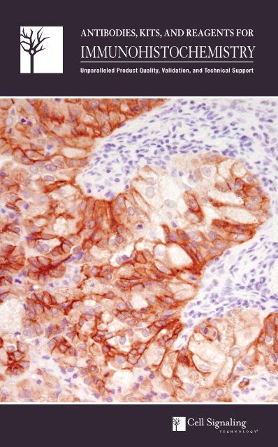
ANTIBODIES, KITS, AND REAGENTS FORIMMUNOHISTOCHEMISTRYUnparalleled Product Quality, Validation, and Technical Support
Antibody Validation for ImmunohistochemistryCell Signaling Technology (CST) antibodies have been extensively validated by ourin-house immunohistochemistry (<strong>IHC</strong>) group in tissue samples and clinically relevantmouse models. Target specificity for <strong>IHC</strong> analysis is determined throughmultiple validation steps. Our in-house <strong>IHC</strong> group uses a variety of approaches foreach antibody validated for use in <strong>IHC</strong> to demonstrate that staining achieved withthe antibody is specific and accurate.#4060UntreatedLY294002Western Blot Analysis: CST antibodies are initially tested by western blot, andonly antibodies that yield clear bands of the appropriate molecular weight with nosignificant additional cross-reacting bands are chosen for validation in applicationssuch as <strong>IHC</strong>.Paraffin-embedded Cell Pellets: Cells are subjected to treatments known toinduce signaling changes to verify modification specificity (e.g. phosphorylation,acetylation, cleavage, etc.). To determine phospho-specificity of Phospho-Akt(Ser473) (D9E) XP ® Rabbit mAb #4060, LNCaP cells were treated with the PI3Kinhibitor LY294002 #9901, which dramatically reduced signal as <strong>com</strong>pared tothe untreated cells, consistent with an expected loss of Akt phosphorylation withthe treatment (A). Cell lines known to express or lack expression of the targetof interest are used to verify specificity of antibodies recognizing total protein.Alternatively, cells can be treated to detect anticipated changes in localization ofthe target as <strong>com</strong>pared to control, or siRNA can be used to block expression ofthe target. In figure (A), treatment of LNCaP cells with LY294002 #9901 shiftedstaining from the membrane to the cytoplasm as detected using Akt (pan) (C67E7)Rabbit mAb #4691.#4691APhospho-Akt (Ser473) (D9E) XP ® Rabbit mAb #4060,Akt (pan) (C67E7) Rabbit mAb #4691: <strong>IHC</strong> analysis ofSignalSlide ® Phospho-Akt (Ser473) <strong>IHC</strong> Controls #8101: LNCaPcells, untreated (left) or treated with LY294002 #9901 (right),using #4060 (top) and #4691 (bottom).Phosphatase Treatment: Treatment with phosphatase is used as an additionaltest to verify phospho-specificity of the antibody. As shown in (B), signal obtainedusing Phospho-EGF Receptor (Tyr1068) (D7A5) XP ® Rabbit mAb #3777 was <strong>com</strong>pletelyablated after treatment of the tissue with λ phosphatase.Tissue Arrays: Having stained cells consistently and appropriately in models withknown target expression levels, antibodies can then be applied to arrays of humancancer tissues to assess performance over a broad spectrum of tissue types.Xenografts: Xenografts generated from cell lines with known target expression levelsare a valuable tool to verify target specificity of phospho-specific and total proteinantibodies. Furthermore, when <strong>com</strong>bined with a previously characterized drugtreatment, these models can be used to show target modulation upon treatment.Blocking Peptides: Peptide blocking verifies sequence specificity of the phosphospecificor total protein antibody and rules out F c receptor-mediated binding, stainingassociated with endogenous biotin, and other non-specific staining. As shownin (C), signal obtained using DDR1 (D1G6) XP ® Rabbit mAb #5583 is blocked inthe presence of antigen-specific peptide.Mouse Models of Cancer: We routinely verify antibody performance in relevantmouse models of cancer. As shown in (D), tissues from WT and PTEN (-/-) mouseprostate were used to assess staining of Phospho-Akt (Ser473) (D9E) XP ® RabbitmAb #4060. The PTEN protein/lipid phosphatase is a negative regulator of thePI3K/Akt pathway that is often mutated or absent in various cancers. <strong>IHC</strong> analysisof prostate from PTEN (-/-), but not WT, mice shows strong staining usingPhospho-Akt (Ser473) (D9E) XP ® Rabbit mAb #4060 indicating that the stainingis specific and due to the lack of PTEN activity in the PTEN (-/-) mice.Apc (Min/+) mouse intestinal adenoma was used to assess staining obtained withβ-Catenin Antibody (Carboxy-terminal Antigen) #9587 (E). This mouse strain ishighly susceptible to spontaneous intestinal adenoma formation due to a mutationin adenomatous polyposis coli (APC). In the normal intestine, β-catenin residesat the membrane at adherens junctions; APC functions as a negative regulator ofWnt signaling by contributing to the destabilization of cytosolic β-catenin. In theintestinal adenoma, the cytoplasmic pool of β-catenin is stabilized and transportedto the nucleus. Significant nuclear and cytoplasmic staining was observed in Apc(Min/+) mouse intestinal adenoma using β-Catenin Antibody (Carboxy-terminalAntigen) #9587, consistent with diminished APC.BCDEPhospho-EGF Receptor (Tyr1068) (D7A5) XP ® Rabbit mAb#3777: <strong>IHC</strong> analysis of paraffin-embedded HCC827 xenograft,untreated (left) or λ phosphatase-treated (right), using #3777.DDR1 (D1G6) XP ® Rabbit mAb #5583: <strong>IHC</strong> analysis of paraffinembeddedhuman lung carcinoma using #5583 in the presence ofcontrol peptide (left) or antigen-specific peptide (right).Phospho-Akt (Ser473) (D9E) XP ® Rabbit mAb #4060: <strong>IHC</strong> analysisof paraffin-embedded WT (left) and PTEN (-/-) (right) mouse prostateusing #4060. Tissue courtesy of Dr. David Guertin, The WhiteheadInstitute for Biomedical Research, Cambridge, MA.β-Catenin Antibody (Carboxy-terminal Antigen) #9587: <strong>IHC</strong>analysis of paraffin-embedded normal mouse intestine (left) and Apc(Min/+) mouse intestinal adenoma (right) using #9587.
Clinically Relevant AntibodiesMany <strong>IHC</strong>-validated antibodies from Cell Signaling Technology are directed against proteins that play a critical role in affectinghuman diseases including cancer, diabetes, obesity, and immune disorders. Several of these antibodies have been validated usinga shorter 2 hour incubation at 37°C, which gives the same results as the longer overnight incubation at 4°C. Please visit our websitefor optimal incubation conditions and re<strong>com</strong>mendations specific for each product.#2855 Phospho-4E-BP1 (Thr37/46) (236B4) Rabbit mAb#9644 4E-BP1 (53H11) Rabbit mAb#3289 5-Lipoxygenase (C49G1) Rabbit mAb#4970 β-Actin (13E5) Rabbit mAb#3700 β-Actin (8H10D10) Mouse mAb#4060 Phospho-Akt (Ser473) (D9E) XP ® Rabbit mAb#4691 Akt (pan) (C67E7) Rabbit mAb#2938 Akt1 (C73H10) Rabbit mAb#4336 AML1 (D33G6) XP ® Rabbit mAb#3796 α-Amylase (D55H10) XP ® Rabbit mAb#5153 Androgen Receptor (D6F11) XP ® Rabbit mAb#2087 Axin1 (C76H11) Rabbit mAb#2774 Bax Antibody (Human Specific)#2764 Bcl-xL (54H6) Rabbit mAb#2933 Bim (C34C5) Rabbit mAb#4593 C-Peptide Antibody#8225 CACYBP (D43G11) Rabbit mAb#3195 E-Cadherin (24E10) Rabbit mAb#2173 Calbindin (C26D12) Rabbit mAb#9579 Cleaved Caspase-3 (Asp175) (D3E9) Rabbit mAb#9582 β-Catenin (6B3) Rabbit mAb#9587 β-Catenin Antibody (Carboxy-terminal Antigen)#3267 Caveolin-1 (D46G3) XP ® Rabbit mAb#3528 CD31 (PECAM-1) (89C2) Mouse mAb#3569 CD34 (ICO115) Mouse mAb#3570 CD44 (156-3C11) Mouse mAb#3575 CD45 (136-4B5) Mouse mAb#4915 CD54 (ICAM-1) Antibody#3576 CD56 (NCAM) (123C3) Mouse mAb#2383 CEA/CD66e (CB30) Mouse mAb#2197 Phospho-Chk2 (Thr68) (C13C1) Rabbit mAb#4842 Cox2 Antibody#9198 Phospho-CREB (Ser133) (87G3) Rabbit mAb#9197 CREB (48H2) Rabbit mAb#2978 Cyclin D1 (92G2) Rabbit mAb#3777 Phospho-EGF Receptor (Tyr1068) (D7A5) XP ® Rabbit mAb#4407 Phospho-EGF Receptor (Tyr1173) (53A5) Rabbit mAb#4267 EGF Receptor (D38B1) XP ® Rabbit mAb#2085 EGF Receptor (E746-A750del Specific) (6B6) XP ® Rabbit mAb#3197 EGF Receptor (L858R Mutant Specific) (43B2) Rabbit mAb#2067 eIF4E (C46H6) Rabbit mAb#2929 EpCAM (VU1D9) Mouse mAb#5246 Ezh2 (D2C9) XP ® Rabbit mAb#3145 Ezrin Antibody#4233 Fas (C18C12) Rabbit mAb#3180 Fatty Acid Synthase (C20G5) Rabbit mAb#4574 FGF Receptor 3 (C51F2) Rabbit mAb#2880 FoxO1 (C29H4) Rabbit mAb#3670 GFAP (GA5) Mouse mAb#8233 Glucagon (D16G10) XP ® Rabbit mAb#9323 Phospho-GSK-3β (Ser9) (5B3) Rabbit mAb#7558 HDAC6 (D2E5) Rabbit mAb#2243 Phospho-HER2/ErbB2 (Tyr1221/1222) (6B12) Rabbit mAb#4290 HER2/ErbB2 (D8F12) XP ® Rabbit mAb#4791 Phospho-HER3/ErbB3 (Tyr1289) (21D3) Rabbit mAb#2024 Hexokinase I (C35C4) Rabbit mAb#2867 Hexokinase II (C64G5) Rabbit mAb#9718 Phospho-Histone H2A.X (Ser139) (20E3) Rabbit mAb#9701 Phospho-Histone H3 (Ser10) Antibody#2401 Phospho-HSP27 (Ser82) Antibody#2402 HSP27 (G31) Mouse mAb#4876 HSP70 (D69) Antibody#4877 HSP90 (C45G5) Rabbit mAb#3027 IGF-I Receptor β Antibody#3014 Insulin (C27C9) Rabbit mAb#3230 Jak2 (D2E12) XP ® Rabbit mAb#3753 JunB (C37F9) Rabbit mAb#4545 Pan-Keratin (C11) Mouse mAb#4546 Keratin 8/18 (C51) Mouse mAb#3984 Keratin 17/19 (D32D9) XP ® Rabbit mAb#4548 Keratin 18 (DC10) Mouse mAb#4558 Keratin 19 (BA17) Mouse mAb#2984 Lck (D88) XP ® Rabbit mAb#3582 LDHA (C4B5) Rabbit mAb#3558 LDHA/LDHC (C28H7) Rabbit mAb#3007 Phospho-MAPKAPK-2 (Thr334) (27B7) Rabbit mAb#3619 MCM2 (D7G11) XP ® Rabbit mAb#4012 MCM3 Antibody#3735 MCM7 (D10A11) XP ® Rabbit mAb#2338 Phospho-MEK1/2 (Ser221) (166F8) Rabbit mAb#8198 Met (D1C2) XP ® Rabbit mAb#3077 Phospho-Met (Tyr1234/1235) (D26) XP ® Rabbit mAb#3801 MMP-7 (D4H5) XP ® Rabbit mAb#3852 MMP-9 Antibody#2017 MSH2 (D24B5) XP ® Rabbit mAb#2976 Phospho-mTOR (Ser2448) (49F9) Rabbit mAb (<strong>IHC</strong> Specific)#4538 MUC1 (VU4H5) Mouse mAb#4903 Nanog (D73G4) XP ® Rabbit mAb#2837 Neurofilament-L (C28E10) Rabbit mAb#8242 NF-κB p65 (D14E12) XP ® Rabbit mAb#6956 NF-κB p65 (L8F6) Mouse mAb#3608 Notch1 (D1E11) XP ® Rabbit mAb#3542 NPM Antibody#3625 NUT (C52B1) Rabbit mAb#2890 Oct-4A (C52G3) Rabbit mAb#2947 p21 Waf1/Cip1 (12D1) Rabbit mAb#4511 Phospho-p38 MAPK (Thr180/Tyr182) (D3F9) XP ® Rabbit mAb#4370 Phospho-p44/42 MAPK (Erk1/2) (Thr202/Tyr204) (D13.14.4E)XP ® Rabbit mAb#2527 p53 (7F5) Rabbit mAb#5625 Cleaved-PARP (Asp214) (D64E10) XP ® Rabbit mAb#2586 PCNA (PC10) Mouse mAb#9535 PDCD4 (D29C6) XP ® Rabbit mAb#5241 PDGF Receptor α (D13C6) XP ® Rabbit mAb#4564 PDGF Receptor β (C82A3) Rabbit mAb#4249 PI3 Kinase p110α (C73F8) Rabbit mAb#4053 PKM2 (D78A4) XP ® Rabbit mAb#2435 PPARγ (C26H12) Rabbit mAb#2997 Phospho-PRAS40 (Thr246) (C77D7) Rabbit mAb#2691 PRAS40 (D23C7) XP ® Rabbit mAb#3153 Progesterone Receptor A/B (C89F7) Rabbit mAb#3157 Progesterone Receptor B (C1A2) Rabbit mAb#3409 PSD95 (D74D3) XP ® Rabbit mAb#9188 PTEN (D4.3) XP ® Rabbit mAb#3205 Pyruvate Dehydrogenase (C54G1) Rabbit mAb#9309 Rb (4H1) Mouse mAb#4858 Phospho-S6 Ribosomal Protein (Ser235/236) (D57.2.2E) XP ®Rabbit mAb#5364 Phospho-S6 Ribosomal Protein (Ser240/244) (D68F8) XP ®Rabbit mAb#2217 S6 Ribosomal Protein (5G10) Rabbit mAb#2317 S6 Ribosomal Protein (54D2) Mouse mAb#4668 Phospho-SAPK/JNK (Thr183/Tyr185) (81E11) Rabbit mAb#2093 SCF (C19H6) Rabbit mAb#9585 Slug (C19G7) Rabbit mAb#3579 Sox2 (D6D9) XP ® Rabbit mAb4 Application and Reactivity Keys, pg 3. Monoclonal Antibody. Please see page 6 for details.
#8725 SPARC (D10F10) Rabbit mAb#2109 Src (36D10) Rabbit mAb#2191 SRC-1 (128E7) Rabbit mAb#9167 Phospho-Stat1 (Tyr701) (58D6) Rabbit mAb#9145 Phospho-Stat3 (Tyr705) (D3A7) XP ® Rabbit mAb#4113 Phospho-Stat3 (Tyr705) (M9C6) Mouse mAb#9359 Phospho-Stat5 (Tyr694) (C11C5) Rabbit mAb#4191 Phospho-Stathmin (Ser38) (D19H10) Rabbit mAb#3352 Stathmin Antibody#2808 Survivin (71G4B7) Rabbit mAb#4179 α-Synuclein (D37A6) XP ® Rabbit mAb#2647 α-Synuclein (Syn204) Mouse mAb#4019 Tau (Tau46) Mouse mAb#3557 TGM2 (D11A6) XP ® Rabbit mAb#3219 TRAIL (C92B9) Rabbit mAb#2508 TrkA (14G6) Rabbit mAb#4607 TrkB (80G2) Rabbit mAb#5666 β3-Tubulin (D65A4) XP ® Rabbit mAb#9319 Tyrosinase (T311) Mouse mAb#3933 Ubiquitin Antibody#2479 VEGF Receptor 2 (55B11) Rabbit mAb#5741 Vimentin (D21H3) XP ® Rabbit mAb#8418 YAP/TAZ (D24E4) Rabbit mAb#4202 YB1 (D299) Antibody#2705 Zap-70 (99F2) Rabbit mAbMetMet, a high affinity tyrosine kinase receptorfor hepatocyte growth factor (HGF),is a disulfide-linked heterodimer made of45 kDa α- and 145 kDa β-subunits. Theα-subunit and the amino-terminal regionof the β-subunit form the extracellulardomain. The remainder of the β-chainspans the plasma membrane and containsa cytoplasmic region with tyrosine kinaseactivity. Interaction of Met with HGF resultsin autophosphorylation at multiple tyrosinesites, which results in the recruitment ofseveral downstream signaling <strong>com</strong>ponents,including Gab1, c-Cbl, and PI3 kinase.These fundamental events are important forall of the biological functions involving Metkinase activity. Altered Met levels and/ortyrosine kinase activities are found in severaltypes of tumors, including renal, colon,and breast. Thus, Met is an attractive cancertherapeutic and diagnostic target.AMet (D1C2) XP ® Rabbit mAb #8198: <strong>IHC</strong> analysis of colon carcinoma using #8198 (A) and a<strong>com</strong>petitor’s <strong>IHC</strong>-approved rabbit monoclonal Met antibody (B). Note that <strong>com</strong>petitor’s antibodylacks specific membrane staining, but prominently stains lymphocytes.ABJurkatMet negative cell linesT-47DSK-BR-3HT-29BMet positive cell linesMKN-45kDa2001401008060T-47DSK-BR-3HT-29MKN-45JurkatT-47DSK-BR-3HT-29MKN-45JurkatT-47DSK-BR-3HT-29MKN-45JurkatPro-MetMet▲ Met (D1C2) XP ® Rabbit mAb #8198 (A)was <strong>com</strong>pared to a <strong>com</strong>petitor’s <strong>IHC</strong>-approvedrabbit monoclonal Met antibody (B). <strong>IHC</strong> analysisof various cell lines expressing Met (HT-29 andMKN-45) and cell lines not expressing Met(Jurkat, T-47D, and SK-BR-3). The optimal dilutionof each antibody was individually evaluated tomaximize specific signal in cell lines expressingMet and minimize background non-specific signalin cell lines not expressing Met. The determinedoptimal dilution for each antibody was utilizedin <strong>IHC</strong> analysis of paraffin-embedded humancolon carcinoma. Note accurate staining of XP ®monoclonal antibody in MKN-45 and HT-29 cells,and absence of staining in Jurkat, T-47D, andSK-BR-3 cells <strong>com</strong>pared to aberrant staining of<strong>com</strong>petitor’s Met antibody.5040Met (D1C2) XP ®Rabbit mAb #8198CompetitorMet Abβ-Tubulin (9F3)Rabbit mAb #2128◀Met (D1C2) XP ® Rabbit mAb #8198 was<strong>com</strong>pared to the same <strong>com</strong>petitor’s <strong>IHC</strong>-approvedrabbit monoclonal Met antibody. WB analysisof various cell lines expressing Met (HT-29 andMKN-45) and cell lines not expressing Met (T-47D,SK-BR-3, and Jurkat).Unparalleled Product Quality, Validation, and Technical Support www.cellsignal.<strong>com</strong>5
SignalStain ® <strong>IHC</strong> Sampler KitsSignalStain ® <strong>IHC</strong> Sampler Kits contain antibodies that areextensively validated for use in immunohistochemical assaysusing multiple approaches. The kits contain a selection ofprimary antibodies, SignalStain ® Antibody Diluent, as well ascontrol slides that can be used to verify the performance ofeach antibody.#8107 SignalStain ® Akt Pathway <strong>IHC</strong> Sampler KitPhospho-Akt (Ser473) (D9E) XP ® Rabbit mAb #4060, Akt (pan)(C67E7) Rabbit mAb #4691, PTEN (D4.3) XP ® Rabbit mAb#9188, Phospho-S6 Ribosomal Protein (Ser235/236) (D57.2.2E)XP ® Rabbit mAb #4858, SignalStain ® Antibody Diluent #8112,SignalSlide ® Phospho-Akt (Ser473) <strong>IHC</strong> Controls #8101#8109 SignalStain ® Proliferation/Apoptosis <strong>IHC</strong> Sampler KitCleaved Caspase-3 (Asp175) Antibody #9661, Phospho-HistoneH3 (Ser10) Antibody #9701, PCNA (PC10) Mouse mAb #2586,Survivin (71G4B7) Rabbit mAb #2808, SignalStain ® AntibodyDiluent #8112, SignalSlide ® Cleaved Caspase-3 (Asp175) <strong>IHC</strong>Controls #8104Phospho-Akt (Ser473)Akt (pan)UntreatedALY294002-treatedB#8113 SignalStain ® Phospho-Stat <strong>IHC</strong> Sampler KitPhospho-Stat1 (Tyr701) (58D6) Rabbit mAb #9167, Phospho-Stat3 (Tyr705) (D3A7) XP ® Rabbit mAb #9145, Phospho-Stat5(Tyr694) (C11C5) Rabbit mAb #9359, SignalStain ® Antibody Diluent#8112, SignalSlide ® Phospho-Stat1/3/5 <strong>IHC</strong> Controls #8105PTENSignalStain ® Akt Pathway <strong>IHC</strong> Sampler Kit #8107: <strong>IHC</strong> analysisof paraffin-embedded LNCaP cell pellets (A), either untreated (left) ortreated with LY294002 (PI3 Kinase Inhibitor) #9901 (right), or NIH/3T3cell pellets (B), using Phospho-Akt (Ser473) (D9E) XP ® Rabbit mAb#4060, Akt (pan) (C67E7) Rabbit mAb #4691, PTEN (D4.3) XP ®Rabbit mAb #9188, and Phospho-S6 Ribosomal Protein (Ser235/236)(D57.2.2E) XP ® Rabbit mAb #4858.Phospho-S6 RibosomalProtein (Ser235/236)SignalSlide ® <strong>IHC</strong> ControlsFormalin-fixed, paraffin-embedded cell pellet control slidesare available for many of our <strong>IHC</strong>-approved antibodies. Eachslide contains a negative and positive pellet, and each setcontains five slides. Custom control slides are available uponrequest. Please contact technical support with any questions.NDRG1UntreatedLY294002-treated#8101 SignalSlide ® Phospho-Akt (Ser473) <strong>IHC</strong> ControlsFor use with Antibodies: 2211, 2217, 2317, 2691, 2855, 2920,2938, 2997, 3787, 4060, 4685, 4691, 4857, 4858, 5196,5364, 5482, 8107, 9323, 9644#8104 SignalSlide ® Cleaved Caspase-3 (Asp175) <strong>IHC</strong> ControlsFor use with Antibodies: 2035, 5625, 8109, 9541, 9661, 9662,9664#8102 SignalSlide ® Phospho-EGF Receptor <strong>IHC</strong> ControlsFor use with Antibodies: 2234, 2235, 2236, 2237, 3777, 4267,4404, 4407, 8713, 9411, 9416, 9417#8118 SignalSlide ® Phospho-Met (Tyr1234/1235) <strong>IHC</strong> ControlsFor use with Antibody: 3077, 8198#8103 SignalSlide ® Phospho-p44/42 MAPK (Thr202/Tyr204) <strong>IHC</strong>ControlsFor use with Antibodies: 4370, 4376, 4695, 4696, 9102#8106 SignalSlide ® PTEN <strong>IHC</strong> ControlsFor use with Antibodies: 9188, 9559#8105 SignalSlide ® Phospho-Stat1/3/5 <strong>IHC</strong> ControlsFor use with Antibodies: 4113, 4904, 8113, 9132, 9139, 9314,9359, 9145, 9167, 9175Phospho-NDRG1 (Thr346)SignalSlide ® Phospho-Akt (Ser473) <strong>IHC</strong> Controls #8101: <strong>IHC</strong> analysis on #8101[paraffin-embedded LNCaP cell pellets, untreated (left) or treated with LY294002 (PI3Kinase Inhibitor) #9901 (right)] using NDRG1 Antibody #5196 (top) or Phospho-NDRG1(Thr346) (D98G11) XP ® Rabbit mAb #5482 (bottom).Please visit the immunohistochemistry section of our website for the mostupdated information on re<strong>com</strong>mended diluents, detection reagents, and controls.Unparalleled Product Quality, Validation, and Technical Support www.cellsignal.<strong>com</strong>9
Tyrosine KinasesApplications Reactivity#3901 Phospho-Bcr (Tyr177) Antibody W, <strong>IHC</strong>-P, F H, MNew #5583 DDR1 (D1G6) XP ® Rabbit mAb W, IP, <strong>IHC</strong>-P, IF-IC H, M, R, Mk#2235 Phospho-EGF Receptor (Tyr992) Antibody W, <strong>IHC</strong>-P H, (R)#2237 Phospho-EGF Receptor (Tyr1045) Antibody W, <strong>IHC</strong>-P, IF-IC H, R#3777 Phospho-EGF Receptor (Tyr1068) (D7A5) XP ® W, <strong>IHC</strong>-P, IF-IC, F H, M, R, MkRabbit mAb#2234 Phospho-EGF Receptor (Tyr1068) Antibody W, <strong>IHC</strong>-P H, M, R#2236 Phospho-EGF Receptor (Tyr1068) (1H12) Mouse mAb W, IP, <strong>IHC</strong>-P H, R, Mk#4404 Phospho-EGF Receptor (Tyr1148) Antibody W, <strong>IHC</strong>-P H, Mk, (R)#4407 Phospho-EGF Receptor (Tyr1173) (53A5) Rabbit mAb W, IP, <strong>IHC</strong>-P H, M, R#2085 EGF Receptor (E746-A750del Specific) (6B6) XP ®Rabbit mAbW, IP, <strong>IHC</strong>-P, IF-IC,IF-P, FH#3197 EGF Receptor (L858R Mutant Specific) (43B2)Rabbit mAbW, IP, <strong>IHC</strong>-P, IF-IC,IF-P, F#4267 EGF Receptor (D38B1) XP ® Rabbit mAb W, IP, <strong>IHC</strong>-P, IF-IC, F H, M, Mk#3285 FAK Antibody W, IP, <strong>IHC</strong>-P H, M, R, Mk,B, Pg, (C)#4574 FGF Receptor 3 (C51F2) Rabbit mAb W, IP, <strong>IHC</strong>-P, IF-IC H#3462 FLT3 (8F2) Rabbit mAb W, IP, <strong>IHC</strong>-P H, M#2243 Phospho-HER2/ErbB2 (Tyr1221/1222) (6B12) W, <strong>IHC</strong>-P, <strong>IHC</strong>-F HRabbit mAb#4290 HER2/ErbB2 (D8F12) XP ® Rabbit mAb W, <strong>IHC</strong>-P H, (M, R)#2165 HER2/ErbB2 (29D8) Rabbit mAb W, IP, <strong>IHC</strong>-P, <strong>IHC</strong>-F, H, (M, R)IF-IC, F#2242 HER2/ErbB2 Antibody W, <strong>IHC</strong>-P H#4791 Phospho-HER3/ErbB3 (Tyr1289) (21D3) Rabbit mAb W, IP, <strong>IHC</strong>-P H, (M, R, Dg)#2984 Lck (D88) XP ® Rabbit mAb W, <strong>IHC</strong>-P H#3155 Phospho-M-CSF Receptor (Tyr723) (49C10)W, IP, <strong>IHC</strong>-P, F H, MRabbit mAb#3077 Phospho-Met (Tyr1234/1235) (D26) XP ® Rabbit mAb W, IP, <strong>IHC</strong>-P, <strong>IHC</strong>-F H, M, RNew #8198 Met (D1C2) XP ® Rabbit mAbW, <strong>IHC</strong>-P, <strong>IHC</strong>-F, HIF-IC, F#5241 PDGF Receptor α (D13C6) XP ® Rabbit mAb W, <strong>IHC</strong>-P, IF-IC, F H#3174 PDGF Receptor α (D1E1E) XP ® Rabbit mAb W, IP, <strong>IHC</strong>-P, IF-IC, F H, M#3164 PDGF Receptor α Antibody W, IP, <strong>IHC</strong>-P, F H, M, R#4564 PDGF Receptor β (C82A3) Rabbit mAb W, <strong>IHC</strong>-P, F H, M, R#3169 PDGF Receptor β (28E1) Rabbit mAb W, IP, <strong>IHC</strong>-P, <strong>IHC</strong>-F, IF-IC H, M, R#2093 SCF (C19H6) Rabbit mAb W, <strong>IHC</strong>-P, F H#2105 Phospho-Src (Tyr527) Antibody W, <strong>IHC</strong>-P H, M, R, (C)#2109 Src (36D10) Rabbit mAb W, IP, <strong>IHC</strong>-P, <strong>IHC</strong>-F,IF-F, IF-IC, F#4991 Phospho-VEGF Receptor 2 (Tyr951) (15D2)Rabbit mAb#2478 Phospho-VEGF Receptor 2 (Tyr1175) (19A10)Rabbit mAbHW, <strong>IHC</strong>-P H, MW, <strong>IHC</strong>-P, IF-IC H, M#2479 VEGF Receptor 2 (55B11) Rabbit mAb W, IP, <strong>IHC</strong>-P, IF-F, IF-IC, F H, M#2705 Zap-70 (99F2) Rabbit mAb W, IP, <strong>IHC</strong>-P, F H, MApplication References:Phospho-Met (Tyr1234/1235) (D26) XP ® Rabbit mAb #3077: Benedettini, E. et al. (2010) Met activationin non-small cell lung cancer is associated with de novo resistance to EGFR inhibitors and the development ofbrain metastasis. Am. J. Pathol. 177, 415–423.Unparalleled ProductQuality, Validation, andTechnical SupportH, M, R, Hm,Mk, B, Pg, (C)DDR1 (D1G6) XP ® Rabbit mAb #5583: <strong>IHC</strong> analysis ofparaffin-embedded human colon carcinoma using #5583.MET SignalingMembraneHGF β αTyr1003 P Met degradationTyr1234 PTyr1235 Pp110 AktTyr1349p85 CrkLP Gab1PaxillinSHP2 CASTyr1356P Grb2SosRasKinase domain AutophosphorylationMAPKPhospho-Met (Tyr1234/1235) (D26) XP ® RabbitmAb #3077: <strong>IHC</strong> analysis of paraffin-embeddedhuman papillary renal cell carcinoma using #3077.Met (D1C2) XP ® Rabbit mAb #8198: <strong>IHC</strong> analysisof paraffin-embedded human papillary renal cellcarcinoma using #8198.10 Application and Reactivity Keys, pg 3. Monoclonal Antibody. Please see page 6 for details.
EGFR and Mutant EGFRThe epidermal growth factor receptor (EGFR) is a 170 kDa transmembrane tyrosine kinase that belongs to the HER/ErbBprotein family. Ligand binding results in receptor dimerization, autophosphorylation, activation of downstream signaling,and internalization. The adaptor protein GRB2 binds activated EGFR phosphorylated at Tyr1068; this binding is necessaryfor the stimulation of EGFR-induced Ras/MAPK signaling.Somatic mutations in the tyrosine kinase domain of EGFR are present in a subset of lung adenocarcinomas that respond toEGFR inhibitors, such as gefinitib and erlotinib. Two types of mutations account for approximately 90% of mutated cases:a specific point mutation L858R in exon 21 and short in-frame deletions in exon 19. The most frequent deletion is exon 19E746-A750. Mutation-specific antibodies from Cell Signaling Technology (CST) allow the examination of tissues or cellswith antibodies designed to detect these two <strong>com</strong>mon mutations in EGFR. CST also offers the highest quality phospho- andtotal protein EGFR antibodies to assess EGFR activity and expression levels.EGF Receptor (D38B1) XP ® Rabbit mAb #4267: <strong>IHC</strong> analysis ofparaffin-embedded human placenta (left) and human lung carcinoma(right) using #4267.Lung Carcinoma E746-A750del+ Lung Carcinoma L858R+Phospho-EGF Receptor (Tyr1068) (D7A5) XP ® Rabbit mAb #3777:<strong>IHC</strong> analysis of paraffin-embedded HCC827 xenograft, control (left) or λphosphatase-treated (right), using #3777.A B C DEGF Receptor (L858R Mutant Specific) (43B2) Rabbit mAb #3197, EGF Receptor (E746-A750del Specific) (6B6) XP ® Rabbit mAb #2085,and EGF Receptor (D38B1) XP ® Rabbit mAb #4267: <strong>IHC</strong> analysis of paraffin-embedded human lung carcinoma of known mutational status using #3197(A), #2085 (B), and #4267 (C). EGFR L858R-positive lung (top); EGFR deletion-positive lung (bottom). WB analysis of various EGFR expressing cell lines(D), showing the specificity of detection with #4267 (top), #3197 (upper middle), and #2085 (lower middle). β-Actin (13E5) Rabbit mAb #4970 was used toconfirm equal protein loading (bottom).kDa20014010080200140100802001401008060504030H1975H3255H1650HCC827HeLaKYSE-450EGFREGFR L858REGFRE746-A750delβ-ActinApplication References:EGF Receptor (E746-A750del Specific) (6B6) XP ® Rabbit mAb #2085 and EGF Receptor (L858R Mutant Specific) (43B2) Rabbit mAb #3197:Yu, J. et al. (2009) Mutation-specific antibodies for the detection of EGFR mutations innon-small-cell lung cancer. Clin. Cancer Res. 15, 3023–3028. / Brevet, M. et al. (2010)Assessment of EGFR mutation status in lung adenocarcinoma by immunohistochemistryusing antibodies specific to the two major forms of mutant EGFR. J. Mol. Diagn. 12,169–176. / Kato, Y. et al. (2010) Novel epidermal growth factor receptor mutation-specificantibodies for non-small cell lung cancer: immunohistochemistry as a possible screeningmethod for epidermal growth factor receptor mutations. J. Thorac. Oncol. 5, 1551–1558. /Kawahara, A. et al. (2010) Molecular diagnosis of activating EGFR mutations innon-small cell lung cancer using mutation-specific antibodies for immunohistochemicalanalysis. Clin. Cancer Res. 16, 3163–3170. / Kitamura, A. et al. (2010)Immunohistochemical Detection of EGFR Mutation Using Mutation-Specific Antibodies inLung Cancer. Clin. Cancer Res. 16, 3349–3355. / Simonetti, S. et al. (2010) Detection ofEGFR mutations with mutation-specific antibodies in stage IV non-small-cell lung cancer.J. Transl. Med. 8, 135.Unparalleled Product Quality, Validation, and Technical Support www.cellsignal.<strong>com</strong>11
Immunology and InflammationApplicationsReactivity#4959 AID (EK2 5G9) Rat mAb W, <strong>IHC</strong>-P H#4336 AML1 (D33G6) XP ® Rabbit mAb W, IP, <strong>IHC</strong>-P, IF-IC, F H, MkNew #8547 Btk (D3H5) Rabbit mAb W, IP, <strong>IHC</strong>-P H, M, (R, Hm, Mk,B, Dg, Pg#3528 CD31 (PECAM-1) (89C2) Mouse mAb W, IP, <strong>IHC</strong>-P, IF-IC, F H#3569 CD34 (ICO115) Mouse mAb <strong>IHC</strong>-P, F H#3570 CD44 (156-3C11) Mouse mAb W, IP, <strong>IHC</strong>-P, IF-IC, F H#3575 CD45 (136-4B5) Mouse mAb <strong>IHC</strong>-P H#4842 Cox2 Antibody W, <strong>IHC</strong>-P H, M, (R)#4233 Fas (C18C12) Rabbit mAb W, <strong>IHC</strong>-P H#3535 GATA-1 (D52H6) XP ® Rabbit mAb W, IP, <strong>IHC</strong>-P, IF-IC, F H, M, R#3890 HS1 (D83A8) XP ® Rabbit mAb (Human Specific) W, IP, <strong>IHC</strong>-P, IF-IC, F HPhospho-IKKα/β (Ser176/180) (16A6) RabbitmAb #2697: <strong>IHC</strong> analysis of paraffin-embedded#3892 HS1 (D5A9) XP ® Rabbit mAb (Rodent Specific) W, IP, <strong>IHC</strong>-P, F M, Rhuman chronic cholecystitis using #2697.#4814 IκBα (L35A5) Mouse mAb (Amino-terminal W, IP, <strong>IHC</strong>-P, IF-IC, F H, M, R, Mk, B, PgAntigen)#5443 Ikaros Antibody W, IP, <strong>IHC</strong>-P, IF-IC, F H, M, R#2697 Phospho-IKKα/β (Ser176/180) (16A6) Rabbit mAb W, <strong>IHC</strong>-P, <strong>IHC</strong>-F H, M, R, Mk, (B)#2685 IKKγ Antibody W, <strong>IHC</strong>-P H, M, R, Mk#3344 Jak1 (6G4) Rabbit mAb W, IP, <strong>IHC</strong>-P H, M, R, (Mk,Dg, Pg)#3230 Jak2 (D2E12) XP ® Rabbit mAb W, IP, <strong>IHC</strong>-P, IF-IC, F H, M, R, (Mk, X, B,Dg, Hm, Pg)#9166 LAT Antibody W, IP, <strong>IHC</strong>-P, F H, M, (R)#2984 Lck (D88) XP ® Rabbit mAb W, <strong>IHC</strong>-P H#2796 Lyn (C13F9) Rabbit mAb W, IP, <strong>IHC</strong>-P H, M, R, MkNew #8242 NF-κB p65 (D14E12) XP ® Rabbit mAbW, IP, <strong>IHC</strong>-P, IF-IC,F, ChIPH, M, R, Hm,Mk, Dg#4764 NF-κB p65 (C22B4) Rabbit mAb W, <strong>IHC</strong>-P, IF-IC, F H, M, R, Mk,B, (Dg)New #6956 NF-κB p65 (L8F6) Mouse mAb#4808 Phospho-NF-κB p105 (Ser933) (178F3)Rabbit mAb (<strong>IHC</strong> Specific)#3017 NF-κB2 p100/p52 (18D10) Rabbit mAb(Human Specific)W, IP, <strong>IHC</strong>-P, IF-IC,F, ChIP<strong>IHC</strong>-PW, <strong>IHC</strong>-P, F H, MkH, M, R, Hm, Mk,Mi, B, Dg, PgHLck (D88) XP ® Rabbit mAb #2984: <strong>IHC</strong> analysisof paraffin-embedded human lung carcinomausing #2984.NF-κB SignalingInflammatory Cytokines, Growth Factors,CD23, CD3/CD28, LPS, Fas, etc.GFIL-1RTNFRGF-RRASSOSP GRBP P MAPK/ErkCascadeNAKRacSAPK/JNKCascadeIRAKTRAF-6PKRTRAFsMembraneTRADDBtk (D3H5) Rabbit mAb #8547: <strong>IHC</strong> analysisof paraffin-embedded human B-cell lymphomausing #8547.Ser176 Ser180CaspasePPNIKIKKsSer32 Ser36P P polyubiquitinationApoptosisp65p50 IκB IκB ProteasomeReldegradationp50P Ser536p65 Active NF-κBRel P Ser276P Ser468Nucleusp50p65κB siteTranscriptionCytokines, Chemokines,Transcription Factors,Adhesion Molecules,Acute Phase Proteins, IκB-αNF-κB p65 (D14E12) XP ® Rabbit mAb #8242:<strong>IHC</strong> analysis of paraffin-embedded human chroniccholecystitis using #8242.12 Application and Reactivity Keys, pg 3. Monoclonal Antibody. Please see page 6 for details.
Applications#3187 NQO1 (A180) Mouse mAb W, <strong>IHC</strong>-P, IF-IC H#4730 Pim-2 (D1D2) XP ® Rabbit mAb W, IP, <strong>IHC</strong>-P, IF-IC H#2258 PU.1 (9G7) Rabbit mAb W, IP, <strong>IHC</strong>-P, IF-IC,F, ChIP#2266 PU.1 Antibody W, IP, <strong>IHC</strong>-P, IF-IC,F, ChIPReactivity#4800 RAGE 1 Antibody W, <strong>IHC</strong>-P H, (Mk)#4727 c-Rel Antibody W, IP, <strong>IHC</strong>-P, IF-IC, F H, Mk#4958 SLP-76 Antibody W, IP, <strong>IHC</strong>-P H, M#8113 SignalStain ® Phospho-Stat <strong>IHC</strong> Sampler Kit <strong>IHC</strong>-P#9167 Phospho-Stat1 (Tyr701) (58D6) Rabbit mAb W, IP, <strong>IHC</strong>-P, <strong>IHC</strong>-F,IF-IC, F, ChIPH, M, (Mk, Pg)H, M, (Mk, Pg)H, M#9175 Stat1 (42H3) Rabbit mAb W, <strong>IHC</strong>-P H, Mk#9145 Phospho-Stat3 (Tyr705) (D3A7) XP ® Rabbit mAb W, IP, <strong>IHC</strong>-P, <strong>IHC</strong>-F,IF-IC, F, ChIPH, M, R, Mk, (Hm,B, Pg)#4113 Phospho-Stat3 (Tyr705) (M9C6) Mouse mAb W, IP, <strong>IHC</strong>-P, IF-IC, F H, M, R, Mk#4904 Stat3 (79D7) Rabbit mAb W, IP, <strong>IHC</strong>-P, ChIP H, M, R, Mk#9132 Stat3 Antibody W, IP, <strong>IHC</strong>-P, ChIP H, M, R, Mk, (B)#9139 Stat3 (124H6) Mouse mAb W, IP, <strong>IHC</strong>-P, IF-IC,F, ChIPH, M, R, Mk#9359 Phospho-Stat5 (Tyr694) (C11C5) Rabbit mAb W, IP, <strong>IHC</strong>-P, F H, M, (R, Mk, B)#9314 Phospho-Stat5 (Tyr694) (C71E5) Rabbit mAb W, <strong>IHC</strong>-P, IF-IC, F H, M, (R, Mk, B)#5251 Phospho-TCTP (Ser46) Antibody W, <strong>IHC</strong>-P, IF-IC, F H, M, R, Mk#2429 Thioredoxin 1 (C63C6) Rabbit mAb W, <strong>IHC</strong>-P H, M, R#4715 TRAF1 (45D3) Rabbit mAb W, IP, <strong>IHC</strong>-P, IF-IC, F H, (Mk)#4710 TRAF1 (1F3) Rat mAb W, IP, <strong>IHC</strong>-P, IF-IC H, M, RPim-2 (D1D2) XP ® Rabbit mAb #4730: <strong>IHC</strong> analysisof paraffin-embedded human testis using #4730.Application References:Phospho-IκBα (Ser32/36) (5A5) Mouse mAb #9246:Wright, T. et al. (2007) Regulation of early wave of germ cellapoptosis and spermatogenesis by deubiquitinating enzymeCYLD. Dev. Cell. 5, 705–716.NF-κB p65 (C22B4) Rabbit mAb #4764: Hideshima, T.et al. (2009) Biologic sequelae of IκB kinase (IKK) inhibitionin multiple myeloma: therapeutic implications. Blood 113,5228–5236.Stat3 Antibody #9132: Peterson, W.M. et al. (2000) Ciliaryneurotrophic factor and stress stimuli activate the Jak-STATpathway in retinal neurons and glia. J. Neurosci. 20,4081–4090.Stat3The Stat3 transcription factor is an important signaling moleculefor many cytokines and growth factor receptors and is required formurine fetal development. Stat3 is constitutively activated in a numberof human tumors and possesses oncogenic potential and antiapoptoticactivities. Stat3 is activated by phosphorylation at Tyr705,which induces dimerization, nuclear translocation, and DNA binding.Stat3 isoform expression appears to reflect biological function asthe relative expression levels of Stat3α (86 kDa) and Stat3β (79 kDa)depend on cell type, ligand exposure, or cell maturation stage.CytokinesPhosho-Stat3 (Tyr705) (D3A7) XP ® Rabbit mAb#9145: <strong>IHC</strong> analysis of paraffin-embedded Apc(Min/+) mouse intestine using #9145.P Tyk2PJaksPPMembraneCytoplasmStatsPStatsPI3KRafPAktPStatsPmTORErkNucleusTranscriptionFactorsP PStatsPISRE/GASTranscriptionStat3 (79D7) Rabbit mAb #4904: <strong>IHC</strong> analysis ofparaffin-embedded human renal clear cell carcinomausing #4904.Unparalleled Product Quality, Validation, and Technical Support www.cellsignal.<strong>com</strong>13
Cytoskeletal Regulationand AdhesionApplicationsReactivity#4968 Pan-Actin Antibody W, <strong>IHC</strong>-P H, M, R, Mk, Z,(Dm, X, B, Pg)#4970 β-Actin (13E5) Rabbit mAb W, <strong>IHC</strong>-P, <strong>IHC</strong>-F,IF-IC, FH, M, R, Mk, B, Pg,(C, Dg)#3700 β-Actin (8H10D10) Mouse mAb W, <strong>IHC</strong>-P, IF-IC, F H, M, R, Hm, Mk#4068 Pan-Cadherin Antibody W, IP, <strong>IHC</strong>-P H, M, R, Mk, Dm,Z, B#3195 E-Cadherin (24E10) Rabbit mAb W, <strong>IHC</strong>-P, <strong>IHC</strong>-F,IF-IC, FH, M, (Dg, Pg)#4065 E-Cadherin Antibody W, IP, <strong>IHC</strong>-P, IF-IC H, M, (B, Dg)#2309 γ-Catenin Antibody W, IP, <strong>IHC</strong>-P, IF-IC H, M, R, Hm, Mk#3267 Caveolin-1 (D46G3) XP ® Rabbit mAb W, IP, <strong>IHC</strong>-P, IF-IC, F H, M, R, Hm, Mk,B, Dg#3238 Caveolin-1 Antibody W, IP, <strong>IHC</strong>-P, <strong>IHC</strong>-F,IF-IC, F#4915 CD54 (ICAM-1) Antibody W, <strong>IHC</strong>-P H#3576 CD56 (NCAM) (123C3) Mouse mAb W, <strong>IHC</strong>-P, F H#2383 CEA/CD66e (CB30) Mouse mAb W, <strong>IHC</strong>-P, IF-IC, F H#3512 Connexin 43 Antibody W, <strong>IHC</strong>-P, <strong>IHC</strong>-F, IF-F,IF-ICH, M, R, Hm, Z,B, PgH, M, R, Mk, Z,(Dg, Pg)#3502 Cortactin Antibody W, <strong>IHC</strong>-P H, M, R, Mk#2929 EpCAM (VU1D9) Mouse mAb W, <strong>IHC</strong>-P, IF-IC, F H#3149 Phospho-Ezrin (Thr567)/Radixin (Thr564)/Moesin (Thr558) (41A3) Rabbit mAbW, <strong>IHC</strong>-P, IF-IC H, M, R, Mk, Dm,B, (X, Dg)#3142 Ezrin/Radixin/Moesin Antibody W, <strong>IHC</strong>-P H, M, R, Mk, B, (X)#3145 Ezrin Antibody W, IP, <strong>IHC</strong>-P, IF-IC, F H, M, R, Mk, B#3862 ILK1 Antibody W, <strong>IHC</strong>-P H, M, R, Mk, B#4545 Pan-Keratin (C11) Mouse mAb W, <strong>IHC</strong>-P, IF-IC, IF-P, F H, R, Mk#4546 Keratin 8/18 (C51) Mouse mAb W, <strong>IHC</strong>-P, F H, Mk#3984 Keratin 17/19 (D32D9) XP ® Rabbit mAb W, <strong>IHC</strong>-P H, M, R, Mk#4548 Keratin 18 (DC10) Mouse mAb W, <strong>IHC</strong>-P, IF-IC, F H#4558 Keratin 19 (BA17) Mouse mAb W, <strong>IHC</strong>-P HCaveolin-1 (D46G3) XP ® Rabbit mAb #3267: <strong>IHC</strong> analysisof paraffin-embedded human lymphoma using #3267.Unparalleled ProductQuality, Validation, andTechnical SupportApplication References:EpCAM (VU1D9) Mouse mAb #2929: Luk, J. M. et al (2011)DLK1-DIO3 genomic imprinted microRNA cluster at 14q32.2 definesa stemlike subtype of hepatocellular carcinoma associated with poorsurvival. J. Biol. Chem. 286, 30706-30713.KeratinKeratins (cytokeratins) are intermediate filament proteins that are mainly expressed in epithelial cells. Keratin heterodimers<strong>com</strong>posed of an acidic keratin (or type I keratin, keratins 9 to 23) and a basic keratin (or type II keratin, keratins 1 to8) assemble to form filaments. Keratin isoforms demonstrate tissue- and differentiation-specific profiles that make themuseful as biomarkers. Mutations in keratin genes are associated with skin disorders, liver and pancreatic diseases, andinflammatory intestinal diseases.Keratin 17/19 (D32D9) XP ® Rabbit mAb #3984: <strong>IHC</strong> analysis of paraffinembeddedhuman breast carcinoma using #3984.Keratin 18 (DC10) Mouse mAb #4548: <strong>IHC</strong> analysis of paraffinembeddedhuman benign prostate hyperplasia using #4548.14 Application and Reactivity Keys, pg 3. Monoclonal Antibody. Please see page 6 for details.
ApplicationsReactivity#3389 LPP (8B3A11) Mouse mAb W, IP, <strong>IHC</strong>-P, IF-IC H, M, Hm, Mk#4538 MUC1 (VU4H5) Mouse mAb W, IP, <strong>IHC</strong>-P, IF-IC, F H#2602 PAK1 Antibody W, IP, <strong>IHC</strong>-P H, M, R, Mk#4191 Phospho-Stathmin (Ser38) (D19H10)Rabbit mAbW, IP, <strong>IHC</strong>-P, IF-IC H, Mk#3352 Stathmin Antibody W, <strong>IHC</strong>-P H, M, R, Mk#2125 α-Tubulin (11H10) Rabbit mAb W, <strong>IHC</strong>-P, IF-IC, F H, M, R, Mk, Dm,B, (Dg)#2144 α-Tubulin Antibody W, <strong>IHC</strong>-P, IF-IC, F H, M, R, Mk, Dm,B, (X)#3873 α-Tubulin (DM1A) Mouse mAb W, IP, <strong>IHC</strong>-P, IF-IC H, M, R, Mk#2148 α/β-Tubulin Antibody W, <strong>IHC</strong>-P, IF-IC, F H, M, R, Mk, Z, B#2128 β-Tubulin (9F3) Rabbit mAb W, <strong>IHC</strong>-P, IF-IC, F H, M, R, Mk, Z,B, (C)#2146 β-Tubulin Antibody W, <strong>IHC</strong>-P, IF-IC, F H, M, R, Mk, Z,B, (X)#3111 Phospho-VASP (Ser157) Antibody W, <strong>IHC</strong>-P H, M, R, Mk#4866 VDAC Antibody W, <strong>IHC</strong>-P H, M, R, B#5741 Vimentin (D21H3) XP ® Rabbit mAb W, <strong>IHC</strong>-P, IF-IC, F H, M, R, MkPre-optimizationFrom Lot to LotEvery antibody validated for <strong>IHC</strong>undergoes thorough testing prior toany new lot release. This ensures thestability and reproducibility necessaryfor consistently accurate results.ABα-Tubulin Antibody#2144: <strong>IHC</strong> analysis ofparaffin-embedded humanbreast carcinoma, showingcytoplasmic localization,using #2144.Cβ-Tubulin (9F3) RabbitmAb #2128: <strong>IHC</strong> analysis ofparaffin-embedded human lungcarcinoma using #2128.DVimentin (D21H3) XP ® RabbitmAb #5741: <strong>IHC</strong> analysis ofparaffin-embedded humanbreast carcinoma using #5741.Phospho-p44/42 MAPK (Erk1/2) (Thr202/Tyr204) (D13.14.4E) XP ® Rabbit mAb #4370:<strong>IHC</strong> analysis of adjacent sections of paraffinembeddedhuman colon carcinoma using #4370lot 7 at a 1:400 dilution (A), #4370 lot 9 at a1:200 dilution (B), #4370 lot 9 at a 1:400 dilution(C) and #4370 lot 9 at a 1:800 dilution (D). There<strong>com</strong>mended dilution for lot 9 remained 1:400.Unparalleled Product Quality, Validation, and Technical Support www.cellsignal.<strong>com</strong>15
PI3 Kinase/Akt SignalingPI3K/Akt SignalingGrowth factors, Hormones, Cytokines,Neurotrophins, Oxidative Stress, etc.P P P P P P P PP P PPTyr376 PPDK1 PIP 3 PI3K PThr308 Tyr373 PPP PIP 3 PAkt/PKBSer241P Thr450 PTENPSHIPP Ser473Tyr315 PTyr326Thr308PSrc DEPTORAkt/PKBP Ser473mTORSIN1RictorPRR5GβLMembraneReceptorNOsynthesisNeuroprotectionProliferationCell Migrationand AdhesionApplications#8107 SignalStain ® Akt Pathway <strong>IHC</strong> Sampler Kit <strong>IHC</strong>-P#4060 Phospho-Akt (Ser473) (D9E) XP ® Rabbit mAb W, IP, <strong>IHC</strong>-P, <strong>IHC</strong>-F,IF-IC, FReactivityH, M, R, Hm, Mk,Dm, Z, B, (C, X,Dg, Pg)#3787 Phospho-Akt (Ser473) (736E11) Rabbit mAb <strong>IHC</strong>-P, <strong>IHC</strong>-F H, M, (R)#4685 Akt (pan) (11E7) Rabbit mAb W, IP, <strong>IHC</strong>-P, IF-IC, F H, M, R, Mk#4691 Akt (pan) (C67E7) Rabbit mAb W, IP, <strong>IHC</strong>-P, IF-IC, F H, M, R, Mk, Dm#2920 Akt (pan) (40D4) Mouse mAb W, IP, <strong>IHC</strong>-P, IF-IC, F H, M, R, Mk#2938 Akt1 (C73H10) Rabbit mAb W, IP, <strong>IHC</strong>-P H, M, R, Mk#5880 eNOS (6H2) Mouse mAb W, <strong>IHC</strong>-P H, B#2880 FoxO1 (C29H4) Rabbit mAb W, <strong>IHC</strong>-P, IF-IC H, M, R, Mk#9467 FoxO3a Antibody W, <strong>IHC</strong>-P H, Mk, (M, R)#9316 Phospho-GSK-3α (Ser21) (36E9) Rabbit mAb W, <strong>IHC</strong>-P H, M, R, Mk#9331 Phospho-GSK-3α/β (Ser21/9) Antibody W, <strong>IHC</strong>-P H, M, R, Mk, Z#9323 Phospho-GSK-3β (Ser9) (5B3) Rabbit mAb W, <strong>IHC</strong>-P, IF-IC H, M, R, Mk#9315 GSK-3β (27C10) Rabbit mAb W, IP, <strong>IHC</strong>-P H, M, R, Mk#5482 Phospho-NDRG1 (Thr346) (D98G11) XP ® RabbitmAbW, <strong>IHC</strong>-P, IF-IC, F H, M, R, Mk#5196 NDRG1 Antibody W, IP, <strong>IHC</strong>-P H, M, R, Mk#4249 PI3 Kinase p110α (C73F8) Rabbit mAb W, IP, <strong>IHC</strong>-P H, M, R, B#2997 Phospho-PRAS40 (Thr246) (C77D7) Rabbit mAb W, IP, <strong>IHC</strong>-P H, M, R, Mk#2691 PRAS40 (D23C7) XP ® Rabbit mAb W, IP, <strong>IHC</strong>-P H, M, R, Mk#9188 PTEN (D4.3) XP ® Rabbit mAb W, IP, <strong>IHC</strong>-P H, M, R, Mk#9559 PTEN (138G6) Rabbit mAb W, IP, <strong>IHC</strong>-P H, M, R, Mk#4042 TCL1 Antibody W, <strong>IHC</strong>-P H, (M)#4911 Phospho-YAP (Ser127) Antibody W, <strong>IHC</strong>-P H, M, R, (Mk, B)New #8418 YAP/TAZ (D24E4) Rabbit mAb W, IP, <strong>IHC</strong>-P H, M, Mk#4912 YAP Antibody W, IP, <strong>IHC</strong>-P, IF-IC, F H, M, R, Mk#4202 YB1 (D299) Antibody W, <strong>IHC</strong>-P, IF-IC H, M, R, Mk, (X, B)Application References:InsulinActionProtein SynthesisSurvivalPhospho-Akt (Ser473) (D9E) XP ® Rabbit mAb#4060: <strong>IHC</strong> analysis of paraffin-embedded humanlung carcinoma (upper) and PTEN (-/-) mouseprostate (lower) using #4060. (Mouse tissuecourtesy of Dr. David Guertin, The WhiteheadInstitute for Biomedical Research, Cambridge, MA.)Phospho-NDRG1 (Thr346) (D98G11) XP ® RabbitmAb #5482: <strong>IHC</strong> analysis of paraffin-embeddedhuman cervical carcinoma, control (upper) or λphosphatase-treated (lower), using #5482.Phospho-Akt (Ser473) (D9E) XP ® Rabbit mAb #4060: Guertin, D.A. et al. (2009) mTOR <strong>com</strong>plex 2 is required for the development of prostate cancer induced by PTEN loss in mice. Cancer Cell15, 148–159. / Engelman, J.A. et al. (2008) Effective use of PI3K and MEK inhibitors to treat mutant Kras G12D and PIK3CA H1047R murine lung cancers. Nat. Med. 14, 1351–1356.FoxO1 (C29H4) Rabbit mAb #2880: Guertin, D.A. et al. (2009) mTOR <strong>com</strong>plex 2 is required for the development of prostate cancer induced by PTEN loss in mice. Cancer Cell 15, 148–159.Phospho-GSK-3β (Ser9) (5B3) Rabbit mAb #9323: Guertin, D.A. et al. (2009) mTOR <strong>com</strong>plex 2 is required for the development of prostate cancer induced by PTEN loss in mice. Cancer Cell 15,148–159.PTEN (138G6) Rabbit mAb #9559: Saal, L.H. (2008) Recurrent gross mutations of the PTEN tumor suppressor gene in breast cancers with deficient DSB repair. Nat. Genet. 40, 102–107. / Pitter,K. L. et al. (2011) Perifosine and CCI 779 co-operate to induce cell death and decrease proliferation in PTEN-intact and PTEN-deficient PDGF-driven murine glioblastoma. PLoS One 6, e14545. /Sangale, Z. et al. (2011) A robust immunohistochemical assay for detecting PTEN expression in human tumors. Appl. Immunohistochem. Mol. Morphol. 19, 173-183.16 Application and Reactivity Keys, pg 3. Monoclonal Antibody. Please see page 6 for details.
PRAS40Many growth factors and hormones inducethe phosphoinositide 3-kinase signalingpathway, which results in the activation ofdownstream effector proteins such as theserine/threonine kinase Akt. Proline-richprotein (PRAS40), a 40 kDa Akt substrate,binds mTOR to transduce Akt signaling to themTOR <strong>com</strong>plex. Inhibition of mTOR signalingstimulates PRAS40 binding to mTOR,which in turn inhibits mTOR activity. PRAS40interacts with raptor in the mTOR <strong>com</strong>plex1 (mTORC1) in insulin-deprived cells andinhibits the activation of the mTORC1 pathwaymediated by the cell cycle protein Rheb.Phosphorylation of PRAS40 by Akt at Thr246relieves PRAS40 inhibition of mTORC1,leading to mRNA translation, suppression ofautophagy, and ribosome biogenesis.AU0126Erk1/2 PTyr204PGrowth, Development Thr202and DifferentiationmTORC1: DEPTORmTORGβLraptor p70 S6KmTORC2: DEPTORmTORGβLPRR5rictorSin1MEK1/2AMP:ATPSer236 P P Ser240Ser235 P S6 P Ser244RafAMPKHormones,Growth Factors,and Cytokines, etc.RasSOSGRBLKB1mTORC2TSC2TSC1IRS-1Ser473 PAktmTORC1PI3KPIP 3PDK1membranecytoplasmP Thr308LY294002PIP 2PTENAmino AcidsRagA/BRagC/DPRAS404E-BP1Translation OnBCPhospho-PRAS40 (Thr246) (C77D7) Rabbit mAb #2997: <strong>IHC</strong> analysis of paraffin-embedded metastatic SKOV-3 tumor in mouse lung (A) and breastcarcinoma, untreated (B) or λ phosphatase-treated (C), using #2997.Application References:Phospho-PRAS40 (Thr246) (C77D7) Rabbit mAb #2997: Anderson, J.N. et al. (2010) Pathway-based identification of biomarkers for targeted therapeutics: personalized oncology withPI3K pathway inhibitors. Sci. Transl. Med. 2, 43–55.YAP/TAZ (D24E4) Rabbit mAb #8418: <strong>IHC</strong> analysis of paraffinembeddedhuman colon carcinoma using #8418.PTEN (D4.3) XP ® Rabbit mAb #9188: <strong>IHC</strong> analysis of paraffinembeddedhuman colon using #9188.Unparalleled Product Quality, Validation, and Technical Support www.cellsignal.<strong>com</strong>17
MAP Kinase SignalingApplications Reactivity#9225 Phospho-ATF-2 (Thr69/71) Antibody W, <strong>IHC</strong>-P H, M, R, Mk#9221 Phospho-ATF-2 (Thr71) Antibody W, IP, <strong>IHC</strong>-P, <strong>IHC</strong>-F,IF-IC, FH, M, R, Mk#9226 ATF-2 (20F1) Rabbit mAb W, IP, <strong>IHC</strong>-P H, M, R, Mk#2251 FosB (5G4) Rabbit mAb W, IP, <strong>IHC</strong>-P, IF-IC, F H, M, R#3753 JunB (C37F9) Rabbit mAb W, IP, <strong>IHC</strong>-P, IF-IC H, M, R, Mk#2361 Phospho-c-Jun (Ser63) (54B3) Rabbit mAb W, <strong>IHC</strong>-P H, M, RNew #3270 Phospho-c-Jun (Ser73) (D47G9) XP ® Rabbit mAb W, IP, <strong>IHC</strong>-P, IF-IC, F H, M, R, Mk#9165 c-Jun (60A8) Rabbit mAb W, IP, <strong>IHC</strong>-P, <strong>IHC</strong>-F,IF-IC#3007 Phospho-MAPKAPK-2 (Thr334) (27B7) RabbitmAbH, M, R, MkW, <strong>IHC</strong>-P, IF-IC, F H, M, R, Mk#3041 Phospho-MAPKAPK-2 (Thr334) Antibody W, <strong>IHC</strong>-P H, M, R, Mk#2338 Phospho-MEK1/2 (Ser221) (166F8) Rabbit mAb W, <strong>IHC</strong>-P, F H, M, R, Mk, (Dg)#4694 MEK1/2 (L38C12) Mouse mAb W, <strong>IHC</strong>-P, IF-IC, F H, M, R, Mk#9595 Phospho-MSK1 (Thr581) Antibody W, IP, <strong>IHC</strong>-P H, M#4511 Phospho-p38 MAPK (Thr180/Tyr182) (D3F9) XP ®Rabbit mAb#4631 Phospho-p38 MAPK (Thr180/Tyr182) (12F8)Rabbit mAbW, IP, <strong>IHC</strong>-P, IF-IC, F H, M, R, Mk, Sc,(Hm, C, Z, B, Pg)W, <strong>IHC</strong>-P, IF-IC H, M, R, Mk, Dm,(Hm, Mi, Z)New #8690 p38 MAPK (D13E1) XP ® Rabbit mAb W, <strong>IHC</strong>-P, IF-IC, F H, M, R, Hm,Mk, Pg#9212 p38 MAPK Antibody W, <strong>IHC</strong>-P, IF-IC, F H, M, R, Mk, (C)#9218 p38α MAPK Antibody W, IP, <strong>IHC</strong>-P H, M, R, Mk#4370 Phospho-p44/42 MAPK (Erk1/2) (Thr202/ Tyr204)(D13.14.4E) XP ® Rabbit mAb#4376 Phospho-p44/42 MAPK (Erk1/2) (Thr202/Tyr204)(20G11) Rabbit mAbW, IP, <strong>IHC</strong>-P, IF-IC, F H, M, R, Hm, Mk,Mi, Dm, Z, B, Dg,Pg, ScW, IP, <strong>IHC</strong>-P H, M, R, Hm, Mk,Mi, Dm, Z, Pg, Sc#4695 p44/42 MAPK (Erk1/2) (137F5) Rabbit mAb W, IP, <strong>IHC</strong>-P, IF-IC, F H, M, R, Hm, Mk,Mi, Dm, Z, B, Dg,Pg, (C)#9102 p44/42 MAPK (Erk1/2) Antibody W, IP, <strong>IHC</strong>-P, IF-IC, F H, M, R, Hm, Mk,Mi, Z, B, Pg, Sc#4696 p44/42 MAPK (Erk1/2) (L34F12) Mouse mAb W, <strong>IHC</strong>-P, IF-IC, F H, M, R, Mk, Mi,Z, Pg#4668 Phospho-SAPK/JNK (Thr183/Tyr185) (81E11)Rabbit mAbW, IP, <strong>IHC</strong>-P H, M, R, Dm, Sc#9251 Phospho-SAPK/JNK (Thr183/Tyr185) Antibody W, IP, <strong>IHC</strong>-P H, M, R, Hm, Mk,Dm, B, Sc, (X)Phospho-p44/42 MAPK (Erk1/2) (Thr202/Tyr204) (D13.14.4E) XP ® Rabbit mAb #4370:<strong>IHC</strong> analysis of paraffin-embedded human breastcarcinoma using #4370.Phospho-p38 MAPK (Thr180/Tyr182) (D3F9)XP ® Rabbit mAb #4511: <strong>IHC</strong> analysis of paraffinembeddedhuman colon carcinoma using #4511.Blocking PeptidesBlocking peptides are useful tools that verify antibody specificity bypreventing the interaction between antibody and antigen.Please visit our website for a <strong>com</strong>plete list of available blocking peptides, orcontact us if you are interested in a custom order peptide.Phospho-c-Jun (Ser73) (D47G9) XP ® Rabbit mAb#3270: <strong>IHC</strong> analysis of paraffin-embedded humanlung carcinoma using #3270.Application References:Phospho-p44/42 MAPK (Erk1/2) (Thr202/Tyr204)(D13.14.4E) XP ® Rabbit mAb #4370: Hudon, V. et al. (2009)Renal tumor suppressor function of the Birt-Hogg-Dube syndromegene product folliculin. J. Med. Genet. 47, 182–189. / Engelman,J.A. et al. (2008) Effective use of PI3K and MEK inhibitors to treatmutant Kras G12D and PIK3CA H1047R murine lung cancers.Nat. Med. 14, 1351–1356.p38 MAPK Antibody #9212: Mudgett, J.S. et al. (2000) Essentialrole for p38alpha mitogen-activated protein kinase in placentalangiogenesis. Proc. Natl. Acad. Sci. USA 97, 10454–10459.hospho-c-Jun (Ser73) Blocking Peptide #1030: <strong>IHC</strong> analysis of paraffin-embedded humancolon carcinoma using Phospho-c-Jun (Ser73) (D47G9) XP ® Rabbit mAb #3270 in the presence ofcontrol peptide (left) or #1030 (right).18 Application and Reactivity Keys, pg 3. Monoclonal Antibody. Please see page 6 for details.
Translational ControlApplications Reactivity#2855 Phospho-4E-BP1 (Thr37/46) (236B4) Rabbit mAb W, <strong>IHC</strong>-P, IF-IC, F H, M, R, Mk, Dm#9644 4E-BP1 (53H11) Rabbit mAb W, IP, <strong>IHC</strong>-P, IF-IC, F H, M, R, Mk#2845 4E-BP2 Antibody W, IP, <strong>IHC</strong>-P, F H, M, R, Mk, B#5053 Argonaute 1 (D84G10) XP ® Rabbit mAb W, IP, <strong>IHC</strong>-P H, M, R, Mk#3177 BiP (C50B12) Rabbit mAb W, <strong>IHC</strong>-P, <strong>IHC</strong>-F H, MNew #8975 LAMTOR1/C11orf59 (D11H6) XP ® Rabbit mAb W, IP, <strong>IHC</strong>-P, IF-IC H, M, R, Mk#2679 Calnexin (C5C9) Rabbit mAb W, <strong>IHC</strong>-P, IF-IC H, Mk#2433 Calnexin Antibody W, <strong>IHC</strong>-P, IF-IC H#3398 Phospho-eIF2α (Ser51) (D9G8) XP ® Rabbit mAb W, IP, <strong>IHC</strong>-P H, M, R, Mk, Dm#3597 Phospho-eIF2α (Ser51) (119A11) Rabbit mAb W, IP, <strong>IHC</strong>-P H, M, R, Mk, Dm#5324 eIF2α (D7D3) XP ® Rabbit mAb W, IP, <strong>IHC</strong>-P H, M, R, Mk#2103 eIF2α (L57A5) Mouse mAb W, <strong>IHC</strong>-P H, M, R, Mk#3411 eIF3A (D51F4) XP ® Rabbit mAb W, IP, <strong>IHC</strong>-P, IF-IC H, M, R, Mk#3413 eIF3H (D9C1) XP ® Rabbit mAb W, IP, <strong>IHC</strong>-P, IF-IC H, M, R, Mk#2067 eIF4E (C46H6) Rabbit mAb W, IP, <strong>IHC</strong>-P H, M, R, Mk#9742 eIF4E Antibody W, <strong>IHC</strong>-P, <strong>IHC</strong>-F H, M, R, Mk, Z#2469 eIF4G (C45A4) Rabbit mAb W, <strong>IHC</strong>-P, IF-IC, F H, M, R, Mk#2498 eIF4G Antibody W, <strong>IHC</strong>-P, IF-IC, F H, M, R, Mk#2858 eIF4GI Antibody W, <strong>IHC</strong>-P, IF-IC H, M, R#3263 eIF6 Antibody W, IP, <strong>IHC</strong>-P, IF-IC H, M, R#3798 ERp44 (D17A6) XP ® Rabbit mAb W, <strong>IHC</strong>-P H, M, R, Mk#3227 GβL Antibody W, IP, <strong>IHC</strong>-P H, Mk#3593 Grp75 (D13H4) XP ® Rabbit mAb W, IP, <strong>IHC</strong>-P, IF-IC H, M, Mk#5537 HIF-1β/ARNT (D28F3) XP ® Rabbit mAb W, IP, <strong>IHC</strong>-P H, M, R, Mk#2066 MRPL11 (D68F2) XP ® Rabbit mAb W, IP, <strong>IHC</strong>-P, IF-IC H, M, R, Mk#2976 Phospho-mTOR (Ser2448) (49F9) Rabbit mAb (<strong>IHC</strong> Specific) <strong>IHC</strong>-P, <strong>IHC</strong>-F H, (M, R)#2983 mTOR (7C10) Rabbit mAb W, <strong>IHC</strong>-P, IF-IC, F H, M, R, Mk#3345 NME1/NDKA (D98) Antibody W, <strong>IHC</strong>-P H, M, R, Mk#2708 p70 S6 Kinase (49D7) Rabbit mAb W, <strong>IHC</strong>-P H, M, R, Mk#3505 Asymmetric-Methyl-PABP1 (Arg455/Arg460)W, IP, <strong>IHC</strong>-P, IF-IC H, M, R, Mk, (C)(C60A10) Rabbit mAb#4992 PABP1 Antibody W, <strong>IHC</strong>-P H, M, R, Mk, (X,Z, B)#3501 PDI (C81H6) Rabbit mAb W, <strong>IHC</strong>-P, IF-IC H, M, R, Mk#2446 PDI Antibody W, <strong>IHC</strong>-P, IF-IC H, M, R, MkNew #5683 PERK (D11A8) Rabbit mAb W, IP, <strong>IHC</strong>-P H#2997 Phospho-PRAS40 (Thr246) (C77D7) Rabbit mAb W, IP, <strong>IHC</strong>-P H, M, R, Mk#2691 PRAS40 (D23C7) XP ® Rabbit mAb W, IP, <strong>IHC</strong>-P H, M, R, Mk#5466 RagC (D31G9) XP ® Rabbit mAb W, IP, <strong>IHC</strong>-P H, M, R, Mk#4935 Rheb Antibody W, <strong>IHC</strong>-P, F H, M, R, Dm,Sc, (B)#4858 Phospho-S6 Ribosomal Protein (Ser235/236)W, <strong>IHC</strong>-P, <strong>IHC</strong>-F, H, M, R, Mk, ScD57.2.2E) XP ® Rabbit mAbIF-IC, F#4857 Phospho-S6 Ribosomal Protein (Ser235/236)W, <strong>IHC</strong>-P, <strong>IHC</strong>-F, H, M, R(91B2) Rabbit mAbIF-IC#2211 Phospho-S6 Ribosomal Protein (Ser235/236) Antibody W, IP, <strong>IHC</strong>-P, <strong>IHC</strong>-F,IF-IC, FH, M, R, Mk, Sc,(C, X)#5364 Phospho-S6 Ribosomal Protein (Ser240/244)W, IP, <strong>IHC</strong>-P, IF-IC, F H, M, R, Mk(D68F8) XP ® Rabbit mAb#2217 S6 Ribosomal Protein (5G10) Rabbit mAb W, <strong>IHC</strong>-P, IF-IC H, M, R, Mk#2317 S6 Ribosomal Protein (54D2) Mouse mAb W, IP, <strong>IHC</strong>-P, IF-F, H, M, R, Mk, DmIF-IC, F#4562 TFII-I Antibody W, IP, <strong>IHC</strong>-P H, M, MkApplication References:Phospho-S6 Ribosomal Protein (Ser235/236) (D57.2.2E) XP ® Rabbit mAb #4858: Guertin, D.A. et al. (2009) mTOR<strong>com</strong>plex 2 is required for the development of prostate cancer induced by PTEN loss in mice. Cancer Cell 15, 148–159. /Hudon, V. et al. (2009) Renal tumor suppressor function of the Birt-Hogg-Dube syndrome gene product folliculin. J. Med.Genet. 47, 182–189. / Engelman, J.A. et al. (2008) Effective use of PI3K and MEK inhibitors to treat mutant Kras G12D andPIK3CA H1047R murine lung cancers. Nat. Med. 14, 1351–1356.Phospho-4E-BP1 (Thr37/46) (236B4) Rabbit mAb #2855: Guertin, D.A. et al. (2009) mTOR <strong>com</strong>plex 2 is required forthe development of prostate cancer induced by PTEN loss in mice. Cancer Cell 15, 148–159.4E-BP1 (53H11) Rabbit mAb #9644: Rong, L. et al. (2008) Control of eIF4E cellular localization by eIF4E-bindingproteins, 4E-BPs. RNA 14, 1318–1327.Phospho-S6 Ribosomal Protein (Ser235/236) Antibody #2211: Choe, G. et al. (2003) Analysis of thephosphatidylinositol 3’-kinase signaling pathway in glioblastoma patients in vivo. Cancer Res. 63, 2742–2746. / Kenerson,H.L. (2002) Activated mammalian target of rapamycin pathway in the pathogenesis of tuberous sclerosis <strong>com</strong>plex renaltumors. Cancer Res. 62, 5645–5650. / Pitter, K. L. et al. (2011) Perifosine and CCI 779 co-operate to induce cell deathand decrease proliferation in PTEN-intact and PTEN-deficient PDGF-driven murine glioblastoma. PLoS One 6, e14545.LAMTOR1/C11orf59 (D11H6) XP ® Rabbit mAb#8975: <strong>IHC</strong> analysis of paraffin-embedded humancolon carcinoma using #8975.PERK (D11A8) Rabbit mAb #5683: <strong>IHC</strong> analysisof paraffin-embedded human breast carcinomausing #5683.Phospho-S6 Ribosomal Protein (Ser235/236)(D57.2.2E) XP ® Rabbit mAb #4858: <strong>IHC</strong> analysis ofparaffin-embedded human lung carcinoma using #4858.RagC (D31G9) XP ® Rabbit mAb #5466:<strong>IHC</strong> analysis of paraffin-embedded human lungcarcinoma using #5466.Unparalleled Product Quality, Validation, and Technical Support www.cellsignal.<strong>com</strong>19
Cellular Metabolism#3661 Phospho-Acetyl-CoA Carboxylase (Ser79)AntibodyApplications ReactivityW, IP, <strong>IHC</strong>-P H, M, R, Mk, (C, B)#3676 Acetyl-CoA Carboxylase (C83B10) Rabbit mAb W, <strong>IHC</strong>-P, IF-IC, F H, M, R, Hm#3662 Acetyl-CoA Carboxylase Antibody W, IP, <strong>IHC</strong>-P, IF-IC, F H, M, R, Mk, B,(C, Dm)#4047 ACSL1 Antibody W, <strong>IHC</strong>-P H, M, R#2535 Phospho-AMPKα (Thr172) (40H9) Rabbit mAb W, IP, <strong>IHC</strong>-P H, M, R, Hm, Mk,Dm, Sc, (C, B, Pg)#4150 AMPKβ1/2 (57C12) Rabbit mAb W, <strong>IHC</strong>-P, IF-IC, F H, M, R, Hm, Mk#3796 α-Amylase (D55H10) XP ® Rabbit mAb W, IP, <strong>IHC</strong>-P H, R, (M)#2439 ATGL (30A4) Rabbit mAb W, IP, <strong>IHC</strong>-P, IF-IC M#2138 ATGL Antibody W, IP, <strong>IHC</strong>-P, IF-IC M, (R)#4593 C-Peptide Antibody <strong>IHC</strong>-P, <strong>IHC</strong>-F, IF-F, IF-IC H, M, R#5648 CA9 (D10C10) Rabbit mAb W, <strong>IHC</strong>-P H#5649 CA9 (D47G3) Rabbit mAb W, IP, <strong>IHC</strong>-P H#2033 DHCR24/Seladin-1 (C59D8) Rabbit mAb W, IP, <strong>IHC</strong>-P H, M#3829 FAAH1 (C84F1) Rabbit mAb W, IP, <strong>IHC</strong>-P, M, R#3180 Fatty Acid Synthase (C20G5) Rabbit mAb W, IP, <strong>IHC</strong>-P, <strong>IHC</strong>-F,IF-ICH, M, R, (B)#5174 GAPDH (D16H11) XP ® Rabbit mAb W, <strong>IHC</strong>-P, IF-IC H, M, R, Mk#2118 GAPDH (14C10) Rabbit mAb W, <strong>IHC</strong>-P, IF-IC, F H, M, R, MkNew #8233 Glucagon (D16G10) XP ® Rabbit mAb <strong>IHC</strong>-P, <strong>IHC</strong>-F, IF-F H, M, RGAPDH (D16H11) XP ® Rabbit mAb #5174:<strong>IHC</strong> analysis of paraffin-embedded human breastcarcinoma using #5174.#2760 Glucagon Antibody <strong>IHC</strong>-P, <strong>IHC</strong>-F, IF-F H, M, R#3891 Phospho-Glycogen Synthase (Ser641) Antibody W, IP, <strong>IHC</strong>-P H, M, R#3886 Glycogen Synthase (15B1) Rabbit mAb W, IP, <strong>IHC</strong>-P H, M, R#2024 Hexokinase I (C35C4) Rabbit mAb W, IP, <strong>IHC</strong>-P, IF-IC H, M#2867 Hexokinase II (C64G5) Rabbit mAb W, <strong>IHC</strong>-P, IF-IC H, M, R, Mk#3113 HNF4α (C11F12) Rabbit mAb W, <strong>IHC</strong>-P, IF-IC H#3922 IGFBP-2 Antibody W, IP, <strong>IHC</strong>-P H#3027 IGF-I Receptor β Antibody W, IP, <strong>IHC</strong>-P, IF-IC, F H, M, R, Mk#3014 Insulin (C27C9) Rabbit mAb <strong>IHC</strong>-P, IF-F, IF-IC, F H, M, R#4590 Insulin Antibody <strong>IHC</strong>-P, IF-F, IF-IC, F H, M, R#3558 LDHA/LDHC (C28H7) Rabbit mAb W, <strong>IHC</strong>-P H, M, R, Mk#3582 LDHA (C4B5) Rabbit mAb W, <strong>IHC</strong>-P, IF-IC H, Mk#9349 Perilipin (D1D8) XP ® Rabbit mAb W, IP, <strong>IHC</strong>-P, IF-F, IF-IC H, M#3470 Perilipin (D418) Antibody W, <strong>IHC</strong>-P, IF-IC M, (H)#4053 PKM2 (D78A4) XP ® Rabbit mAb W, IP, <strong>IHC</strong>-P, IF-IC H, M, R, Mk#3198 PKM2 Antibody W, <strong>IHC</strong>-P, <strong>IHC</strong>-F H, M, R, Mk#3205 Pyruvate Dehydrogenase (C54G1) Rabbit mAb W, <strong>IHC</strong>-P H, M, R, Mk#2794 SCD1 (C12H5) Rabbit mAb W, IP, <strong>IHC</strong>-P, IF-IC M#2429 Thioredoxin 1 (C63C6) Rabbit mAb W, <strong>IHC</strong>-P H, M, R#9319 Tyrosinase (T311) Mouse mAb (<strong>IHC</strong> Specific) <strong>IHC</strong>-P HPerilipin (D418) Antibody #3470: <strong>IHC</strong> analysis ofparaffin-embedded mouse brown fat using #3470 inthe presence of control peptide (upper) or antigenspecificpeptide (lower).UnparalleledProduct Quality,Validation, andTechnical SupportGlucagon (D16G10) XP ® Rabbit mAb #8233:<strong>IHC</strong> analysis of paraffin-embedded mousepancreas using #8233.PKM2 (D78A4) XP ® Rabbit mAb #4053: <strong>IHC</strong>analysis of paraffin-embedded human skeletal muscleusing #4053. Note the lack of staining in the skeletalmuscle cells that do not express PKM2 while vesselswithin the tissue stain positively.20 Application and Reactivity Keys, pg 3. Monoclonal Antibody. Please see page 6 for details.
Stem Cell Markers, Development,and DifferentiationApplications Reactivity#3208 AP-2α Antibody W, IP, <strong>IHC</strong>-P H, M, R, Mk#2074 Axin1 (C95H11) Rabbit mAb W, IP, <strong>IHC</strong>-P H, Mk#2087 Axin1 (C76H11) Rabbit mAb W, IP, <strong>IHC</strong>-P H, M#9582 β-Catenin (6B3) Rabbit mAb W, IP, <strong>IHC</strong>-P, <strong>IHC</strong>-F H, M, R, Mk, (X, B,Dg, Pg)#9562 β-Catenin Antibody W, IP, <strong>IHC</strong>-P, ChIP H, M, R, Mk, (Z)#9587 β-Catenin Antibody (Carboxy-terminal Antigen) W, IP, <strong>IHC</strong>-P, <strong>IHC</strong>-F,ChIPH, M, R, Mk, (C, X,B, Dg, Pg)New #8225 CACYBP (D43G11) Rabbit mAb W, <strong>IHC</strong>-P, F H, M, R, Mk#3354 CACYBP Antibody W, <strong>IHC</strong>-P, F H, M, R, Mk#4402 FoxP1 (D35D10) XP ® Rabbit mAb W, IP, <strong>IHC</strong>-P, F H, M, R, Mk#2005 FoxP1 Antibody W, IP, <strong>IHC</strong>-P, IF-IC, F H, M, (R)#3978 LIN28A (A177) Antibody W, IP, <strong>IHC</strong>-P, IF-IC, F H, M, (Mk)New #5940 Mili (D14F5) XP ® Rabbit mAb W, IP, <strong>IHC</strong>-P, IF-F M#2071 Mili Antibody W, IP, <strong>IHC</strong>-P, IF-F M#2079 Miwi (G82) Antibody W, IP, <strong>IHC</strong>-P, IF-F M#4903 Nanog (D73G4) XP ® Rabbit mAb W, <strong>IHC</strong>-P, IF-IC, F H, (Mk)#4893 Nanog (1E6C4) Mouse mAb W, <strong>IHC</strong>-P, IF-IC, F H#3608 Notch1 (D1E11) XP ® Rabbit mAb W, <strong>IHC</strong>-P H, M, R#2890 Oct-4A (C52G3) Rabbit mAb W, <strong>IHC</strong>-P, IF-IC, F, ChIP H#2750 Oct-4 Antibody W, <strong>IHC</strong>-P, IF-IC, F, ChIP H, (Mk)#9585 Slug (C19G7) Rabbit mAb W, IP, <strong>IHC</strong>-P, IF-IC H, M#3579 Sox2 (D6D9) XP ® Rabbit mAb W, <strong>IHC</strong>-P, IF-IC, F H, (Mk, B, Dg)#3728 Sox2 (C70B1) Rabbit mAb (<strong>IHC</strong> Preferred) W, <strong>IHC</strong>-P MNew #8725 SPARC (D10F10) Rabbit mAb W, <strong>IHC</strong>-P H, M#4744 SSEA1 (MC480) Mouse mAb <strong>IHC</strong>-P, IF-IC, F M#2203 TCF1 (C63D9) Rabbit mAb W, IP, <strong>IHC</strong>-P, IF-IC, F H, M#4746 TRA-1-60(S) (TRA-1-60(S)) Mouse mAb W, <strong>IHC</strong>-P, IF-IC, F H#4745 TRA-1-81 (TRA-1-81) Mouse mAb <strong>IHC</strong>-P, IF-IC, F HMili (D14F5) XP ® Rabbit mAb #5940: <strong>IHC</strong> analysisof paraffin-embedded mouse testis using #5940.Nanog (D73G4) XP ® Rabbit mAb #4903: <strong>IHC</strong>analysis of paraffin-embedded human seminomausing #4903.FGF2IGFWntTGF-β/Activin/NodalBMP4LRPmembranecytoplasmPI3KNotch1 (D1E11) XP ® Rabbit mAb #3608:<strong>IHC</strong> analysis of paraffin-embedded human colonusing #3608.AktErk1/2β-Catenin Smad2/3 Smad1/5/8nucleusOct-4 Sox2Oct-4 Sox2 FoxD3Oct-4 Sox2 NanogOct-4,Sox2,FoxD3,FGF4NanoggeneexpressionPluripotencyand Self RenewalhESC Markers:Oct-4NanogSox2SSEA 3/4TRA-1-60TRA-1-81CACYBP (D43G11) Rabbit mAb #8225: <strong>IHC</strong> analysisof paraffin-embedded human lymphoma using #8225.Unparalleled Product Quality, Validation, and Technical Support www.cellsignal.<strong>com</strong>21
Chromatin andEpigenetic RegulationApplications Reactivity#2990 ASF1A (C6E10) Rabbit mAb W, IP, <strong>IHC</strong>-P, IF-IC H, M, Mk, (C, B)New #8684 CtBP1 (D2D6) Rabbit mAb W, IP, <strong>IHC</strong>-P, IF-IC H, M, Mk#3418 CTCF (D31H2) XP ® Rabbit mAb W, IP, <strong>IHC</strong>-P, IF-IC, ChIP H, M, R, Mk, (B)#5246 Ezh2 (D2C9) XP ® Rabbit mAb W, IP, <strong>IHC</strong>-P, IF-IC, ChIP H, M, R, MkNew #4402 FoxP1 (D35D10) XP ® Rabbit mAb W, IP, <strong>IHC</strong>-P, F H, M, R, Mk#2005 FoxP1 Antibody W, IP, <strong>IHC</strong>-P, IF-IC, F H, M, (R)#3815 Phospho-HDAC3 (Ser424) Antibody W, IP, <strong>IHC</strong>-P, IF-IC H, M, R, (Mk, C, X)#2082 Histone Deacetylase 5 (HDAC5) Antibody W, IP, <strong>IHC</strong>-P H, M, R, MkNew #7558 HDAC6 (D2E5) Rabbit mAb W, IP, <strong>IHC</strong>-P, IF-IC H, Mk#2576 Acetyl-Histone H2A (Lys5) Antibody W, IP, <strong>IHC</strong>-P H, M, R, Mk#2578 Histone H2A Antibody II W, IP, <strong>IHC</strong>-P H, M, R, Mk, (C, X)#3636 Histone H2A (L88A6) Mouse mAb W, <strong>IHC</strong>-P H, M, R, Mk, (C, Dm,Z, Pg)#9718 Phospho-Histone H2A.X (Ser139) (20E3) W, <strong>IHC</strong>-P, IF-IC, F H, M, R, MkRabbit mAb#2577 Phospho-Histone H2A.X (Ser139) Antibody W, <strong>IHC</strong>-P, IF-IC, F H, M, RNew #7631 Histone H2A.X (D17A3) XP ® Rabbit mAb W, <strong>IHC</strong>-P, IF-IC H, M, R, Mk#2574 Acetyl-Histone H2B (Lys5) Antibody W, IP, <strong>IHC</strong>-P H, M, R, Mk#2571 Acetyl-Histone H2B (Lys20) Antibody W, IP, <strong>IHC</strong>-P, IF-IC H, M, R#9649 Acetyl-Histone H3 (Lys9) (C5B11) Rabbit mAb W, <strong>IHC</strong>-P, IF-IC, F, ChIP H, M, R, Mk, Z, (Sc)#9671 Acetyl-Histone H3 (Lys9) Antibody W, IP, <strong>IHC</strong>-P, ChIP H, M, R, Mk, Dm, Sc#9711 Acetyl- and Phospho-Histone H3 (Lys9/Ser10) W, <strong>IHC</strong>-P H, M, RAntibody#9675 Acetyl-Histone H3 (Lys18) Antibody W, <strong>IHC</strong>-P, ChIP H, M, R#9674 Acetyl-Histone H3 (Lys23) Antibody W, <strong>IHC</strong>-P H, M, R#9725 Di-Methyl-Histone H3 (Lys4) (C64G9) Rabbit mAb W, IP, <strong>IHC</strong>-P, IF-IC, ChIP H, M, R, Mk#9726 Di-Methyl-Histone H3 (Lys4) Antibody W, IP, <strong>IHC</strong>-P, IF-IC, ChIP H, M, R, Mk, (X, Z)#9751 Tri-Methyl-Histone H3 (Lys4) (C42D8)Rabbit mAbW, <strong>IHC</strong>-P, IF-IC, ChIP H, M, R, Mk, Dm,Sc, (X, Z)#9727 Tri-Methyl-Histone H3 (Lys4) Antibody W, IP, <strong>IHC</strong>-P, IF-IC, ChIP H, M, R, Mk, (X, Z)#9753 Di-Methyl-Histone H3 (Lys9) Antibody W, IP, <strong>IHC</strong>-P, IF-IC, ChIP H, M, R, Mk, Dm, Sc#9733 Tri-Methyl-Histone H3 (Lys27) (C36B11) W, IP, <strong>IHC</strong>-P, IF-IC, ChIP H, M, R, Mk, (X, Z)Rabbit mAb#2901 Di-Methyl-Histone H3 (Lys36) (C75H12) Rabbit mAb W, <strong>IHC</strong>-P, IF-IC H, M, R, Mk#9763 Tri-Methyl-Histone H3 (Lys36) Antibody W, <strong>IHC</strong>-P, IF-IC H, M, R, Mk#9714 Phospho-Histone H3 (Thr3) Antibody W, <strong>IHC</strong>-P H, M, R#9701 Phospho-Histone H3 (Ser10) Antibody W, IP, <strong>IHC</strong>-P, <strong>IHC</strong>-F,IF-IC, FH, M, R, Mk, C, Dm,Z, Sc, (X)#4499 Histone H3 (D1H2) XP ® Rabbit mAb W, <strong>IHC</strong>-P, IF-IC H, M, R, Mk, (Hm, C,Dm, X, Z, B)#3680 Histone H3 (96C10) Mouse mAb (<strong>IHC</strong> Formulated) <strong>IHC</strong>-P H, (M, R, Mk, Dm, X)#9672 Acetyl-Histone H4 (Lys5) Antibody W, IP, <strong>IHC</strong>-P, IF-IC, ChIP H, M, R, Mk, (C, Dm,X, Z, B, Pg)#2592 Histone H4 Antibody W, IP, <strong>IHC</strong>-P H, M, R, Mk, Dm,Z, Sc#2935 Histone H4 (L64C1) Mouse mAb W, <strong>IHC</strong>-P H, M, R, Mk, Z, (Dm,X, Z, B)#2623 HP1α (C7F11) Rabbit mAb W, IP, <strong>IHC</strong>-P, IF-IC H, M, R, Mk#2616 HP1α Antibody W, IP, <strong>IHC</strong>-P, IF-IC, F H, M, R, Mk, (B)#3100 JMJD1B (C6D12) Rabbit mAb W, IP, <strong>IHC</strong>-P H, MkNew #5328 JMJD2A (C37E5) Rabbit mAb W, IP, <strong>IHC</strong>-P, IF-IC H, M, R, Mk#2088 LEDGF (C57G11) Rabbit mAb W, <strong>IHC</strong>-P, IF-IC, F H, M, R, (Mk)#2184 LSD1 (C69G12) Rabbit mAb W, IP, <strong>IHC</strong>-P, <strong>IHC</strong>-F, H, M, R, MkIF-IC#2139 LSD1 Antibody W, IP, <strong>IHC</strong>-P, IF-IC, F H, M, R, Mk#3456 MeCP2 (D4F3) XP ® Rabbit mAb W, IP, <strong>IHC</strong>-P, IF-IC H, M, R, MkNew #5647 MTA1 (D40D1) XP ® Rabbit mAb W, <strong>IHC</strong>-P H, M, R, Mk#3625 NUT (C52B1) Rabbit mAb W, IP, <strong>IHC</strong>-P, IF-F H, R, (Mk)#2627 SirT3 (C73E3) Rabbit mAb W, IP, <strong>IHC</strong>-P H, R, MkApplication References:Acetyl-Histone H3 (Lys9) Antibody #9671: Kennedy, C. et al. (2002) Cell proliferation in the normal mousemammary gland and inhibition by phenylbutyrate. Mol. Cancer Ther. 1, 1025–1033.Phospho-Histone H3 (Ser10) Antibody #9701: Wang, Y. et al. (2001) The JIL-1 tandem kinase mediates histoneH3 phosphorylation and is required for maintenance of chromatin structure in Drosophila. Cell 105, 433–443.Histone H2A.X (D17A3) XP ® Rabbit mAb #7631:<strong>IHC</strong> analysis of paraffin-embedded human prostatecarcinoma using #7631.JMJD2A (C37E5) Rabbit mAb #5328: <strong>IHC</strong>analysis of paraffin-embedded human lungcarcinoma using #5328.MTA1 (D40D1) XP ® Rabbit mAb #5647: <strong>IHC</strong>analysis of paraffin-embedded human lung carcinomausing #5647 in the presence of control peptide (upper)or antigen-specific peptide (lower).22 Application and Reactivity Keys, pg 3. Monoclonal Antibody. Please see page 6 for details.
AcetylationProtein acetylation plays a crucial role in regulating chromatin structureand transcriptional activity. Many transcriptional coactivators possessintrinsic acetylase activity, while transcriptional corepressors are associatedwith deacetylase activity. Acetylation <strong>com</strong>plexes (such as CBP/p300 andPCAF) or deacetylation <strong>com</strong>plexes (such as Sin3, NuRD, NcoR, and SMRT)are recruited to DNA-bound transcription factors in response to signalingpathways. Histone hyperacetylation by histone acetyltransferases (HATs) isassociated with transcriptional activation, whereas histone deacetylation byhistone deacetylases (HDACs) is associated with transcriptional repression.Histone acetylation stimulates transcription by remodeling higher orderchromatin structure, weakening histone-DNA interactions, and providingbinding sites for transcriptional activation <strong>com</strong>plexes containing proteinsthat possess bromodomains, which bind acetylated lysine. Histone deacetylationrepresses transcription through an inverse mechanism involving theassembly of <strong>com</strong>pact higher order chromatin and the exclusion of bromodomain-containingtranscription activation <strong>com</strong>plexes. Histone hypoacetylationis a hallmark of silent heterochromatin. Site-specific acetylation of agrowing number of non-histone proteins, including p53 and E2F, has beenshown to regulate their activity, localization, specific interactions, and stability/degradation,therefore controlling a variety of cellular processes, such astranscription, proliferation, apoptosis, and differentiation. At an organismallevel, acetylation plays an important role in immunity, circadian rythmicity,and memory formation. Protein acetylation is be<strong>com</strong>ing a favorable target indrug design for numerous disease conditions.HDAC6 (D2E5) Rabbit mAb #7558: <strong>IHC</strong> analysisof paraffin-embedded human breast carcinomausing #7558.Acetyl-Histone H3 (Lys9) (C5B11) Rabbit mAb#9649: <strong>IHC</strong> analysis of paraffin-embedded humanosteosar<strong>com</strong>a using #9649.Acetyl-groupHistone tailNucleosome coreDouble-stranded DNAAcetylation/DeacetylationHistone H3 (D1H2) XP ® Rabbit mAb #4499:<strong>IHC</strong> analysis of paraffin-embedded human breastcarcinoma using #4499.CtBP1 (D2D6) Rabbit mAb #8684: <strong>IHC</strong> analysis of paraffin-embedded human colon carcinomausing #8684 in the presence of control peptide (left) or antigen-specific peptide (right).Ezh2 (D2C9) XP ® Rabbit mAb #5246: <strong>IHC</strong> analysisof paraffin-embedded human lymphoma using #5246.Unparalleled Product Quality, Validation, and Technical Support www.cellsignal.<strong>com</strong>23
Cell Cycle, Checkpoint Control,and DNA DamageApplicationsReactivity#4937 53BP1 Antibody W, <strong>IHC</strong>-P, IF-IC H, Mk#5292 BrdU (Bu20a) Mouse mAb <strong>IHC</strong>-P, IF-IC, F All#9116 cdc2 (POH1) Mouse mAb W, IP, <strong>IHC</strong>-P, IF-IC H, Mk#4901 Phospho-cdc25C (Ser216)(63F9) Rabbit mAbW, IP, <strong>IHC</strong>-P H, Mk#2916 CDK7 (MO1) Mouse mAb W, IP, <strong>IHC</strong>-P H, R#2316 CDK9 (C12F7) Rabbit mAb W, IP, <strong>IHC</strong>-P,IF-IC, F#2666 Phospho-Chk2 (Ser19)Antibody#2197 Phospho-Chk2 (Thr68)(C13C1) Rabbit mAbW, <strong>IHC</strong>-P HW, IP, <strong>IHC</strong>-P, F H, (Mk)#3440 Chk2 (1C12) Mouse mAb W, <strong>IHC</strong>-P, IF-IC H, Mk#2978 Cyclin D1 (92G2) Rabbit mAb W, <strong>IHC</strong>-P, F H, M, R#2936 Cyclin D3 (DCS22) Mouse mAb W, <strong>IHC</strong>-P H, M, R#4136 Phospho-Cyclin E (Thr62)AntibodyW, IP, <strong>IHC</strong>-P, F H#2927 Cyclin H Antibody W, IP, <strong>IHC</strong>-P H, M, R#2180 Ku80 (C48E7) Rabbit mAb W, IP, <strong>IHC</strong>-P, <strong>IHC</strong>-F, IF-IC#2753 Ku80 Antibody W, IP, <strong>IHC</strong>-P,IF-IC, F#3619 MCM2 (D7G11) XP ®Rabbit mAbW, IP, <strong>IHC</strong>-P,IF-IC, ChIPH, M, R, Hm, Mk,B, DgH, MkH, Mk, (M, R)H, M, R, Mk#4007 MCM2 Antibody W, IP, <strong>IHC</strong>-P, IF-IC H, M, R, Hm, Mk#4012 MCM3 Antibody W, IP, <strong>IHC</strong>-P, IF-IC H, M, R, Hm, Mk#3735 MCM7 (D10A11) XP ®Rabbit mAb#4847 Mre11 (31H4) Rabbit mAb W, IP, <strong>IHC</strong>-P,<strong>IHC</strong>-F, FW, IP, <strong>IHC</strong>-P, IF-IC H, M, R, Hm,Mk, DgHApplications#4895 Mre11 Antibody W, IP, <strong>IHC</strong>-P,<strong>IHC</strong>-F#2017 MSH2 (D24B5) XP ®Rabbit mAbW, IP, <strong>IHC</strong>-P,IF-IC, FReactivityH, M, R, MkH, M, Mk#4282 Myt1 Antibody W, <strong>IHC</strong>-P H, M, R, (X)#3541 Phospho-NPM (Thr199)AntibodyW, IP, <strong>IHC</strong>-P, IF-IC H, M, R#3542 NPM Antibody W, <strong>IHC</strong>-P H, M, R#2407 p14 ARF (4C6/4) Mouse mAb W, <strong>IHC</strong>-P H#2947 p21 Waf1/Cip1 (12D1)Rabbit mAb#2946 p21 Waf1/Cip1 (DCS60)Mouse mAbW, IP, <strong>IHC</strong>-P,IF-IC, FH, MkW, IP, <strong>IHC</strong>-P H, Mk#2526 Phospho-p53 (Ser33) Antibody W, <strong>IHC</strong>-P H, (Mk)#2676 Phospho-p53 (Thr81) Antibody W, <strong>IHC</strong>-P, IF-IC H, Mk#2527 p53 (7F5) Rabbit mAb W, <strong>IHC</strong>-P, IF-IC,F, ChIP#4981 Phospho-p63 (Ser160/162)AntibodyH, MkW, <strong>IHC</strong>-P, F H, (M, R, C, X)#4942 PBK/TOPK Antibody W, <strong>IHC</strong>-P H, M, R#2586 PCNA (PC10) Mouse mAb W, IP, <strong>IHC</strong>-P, IF-IC H, M, R, Mk,B, Pg#9308 Phospho-Rb (Ser807/811)Antibody#9309 Rb (4H1) Mouse mAb W, IP, <strong>IHC</strong>-P,IF-IC, F#4735 Phospho-Rpb1 CTD (Ser2/5)AntibodyW, IP, <strong>IHC</strong>-P, ChIP H, R, MkH, Mk, B, PgW, <strong>IHC</strong>-P H, M, R, (Hm, Sc)#4124 TIF1β (C42G12) Rabbit mAb W, <strong>IHC</strong>-P, IF-IC, F H, M, R, Mk#4121 Phospho-TLK1 (Ser695)AntibodyW, IP, <strong>IHC</strong>-P H, M, RMCM ProteinsThe minichromosome maintenance (MCM) 2-7 proteins area family of six related proteins required for the initiation and elongationof DNA replication. MCM2 through MCM7 bind togetherto form the heterohexameric MCM <strong>com</strong>plex that is thought to actas a replicative helicase at the DNA replication fork.This <strong>com</strong>plex is also a key <strong>com</strong>ponent of the pre-replication<strong>com</strong>plex (pre-RC). Cdc6 and Cdt1 recruit the MCM <strong>com</strong>plex tothe origin recognition <strong>com</strong>plex (ORC) during late mitosis/earlyG1 phase forming the pre-RC and licensing the DNA for replication.Phosphorylation of the MCM2, MCM3, MCM4, and MCM6subunits appears to regulate MCM <strong>com</strong>plex activity and the initiationof DNA synthesis. MCM proteins are removed during DNAreplication, causing chromatin to be<strong>com</strong>e unlicensed throughinhibition of pre-RC reformation.Licensing of the chromatin permits the DNA to replicate onlyonce per cell cycle, helping to ensure that genetic alterations andmalignant cell growth do not occur. Studies have also shown thatthe MCM <strong>com</strong>plex is involved in checkpoint control by protectingthe structure of the replication fork and assisting in restartingreplication by recruiting checkpoint proteins after arrest.MCM2 (D7G11) XP ® Rabbit mAb #3619: <strong>IHC</strong> analysis ofparaffin-embedded human small intestine using #3619.MCM7 (D10A11) XP ® Rabbit mAb #3735: <strong>IHC</strong> analysis ofparaffin-embedded human lung carcinoma using #3735.24 Application and Reactivity Keys, pg 3. Monoclonal Antibody. Please see page 6 for details.
Apoptosis and AutophagyAutophagyAMPKSignalingPI3K-I / AktSignalingMAPK / Erk1/2Signalingp53 / GenotoxicStresshVPS34p150Bcl-2PI3K IIIAminoAcidsBeclin1AutophagyInductionGβLULK <strong>com</strong>plexmTORRaptorPRAS40Atg1/ULK1ApoptosisMembraneNucleationSequestrationBak (D2D3) Rabbit mAb #6947: <strong>IHC</strong> analysis ofparaffin-embedded human lung carcinoma using#6947.PhagophoreAtg16Atg12Atg5LC3-IIAutophagosomeLysosomeAtg16Atg3PEAtg5Atg10Atg7Atg7LC3-IFusionAtg12Atg4LysosomeAutophagolysosomeLC3#8109 SignalStain ® Proliferation/Apoptosis <strong>IHC</strong>Sampler KitApplications<strong>IHC</strong>-PReactivity#4642 AIF Antibody W, IP, <strong>IHC</strong>-P, IF-IC H, M, R#5284 Phospho-Bad (Ser112) (40A9) Rabbit mAb W, <strong>IHC</strong>-P, F H, M, R, MkNew #6947 Bak (D2D3) Rabbit mAb W, IP, <strong>IHC</strong>-P H, Mk#5023 Bax (D2E11) Rabbit mAb W, IP, <strong>IHC</strong>-P H#2774 Bax Antibody (Human Specific) W, IP, <strong>IHC</strong>-P H, Mk#2764 Bcl-xL (54H6) Rabbit mAb W, IP, <strong>IHC</strong>-P, <strong>IHC</strong>-F, IF-IC, F H, M, R, Mk#4592 Bik Antibody W, <strong>IHC</strong>-P H#2933 Bim (C34C5) Rabbit mAb W, IP, <strong>IHC</strong>-P, IF-IC, F H, M, R, (Mk, B, Dg)#2225 Caspase-1 Antibody W, IP, <strong>IHC</strong>-P HNew #9579 Cleaved Caspase-3 (Asp175) (D3E9)Rabbit mAb#9664 Cleaved Caspase-3 (Asp175) (5A1E)Rabbit mAb<strong>IHC</strong>-P, IF-IC, FH, (M, R, Mk, B, Pg)W, IP, <strong>IHC</strong>-P, <strong>IHC</strong>-F, IF-IC, F H, M, R, Mk, (Dg)#9661 Cleaved Caspase-3 (Asp175) Antibody W, <strong>IHC</strong>-P, <strong>IHC</strong>-F, IF-IC, F H, M, R, Mk, B,(Pg, Dg)#9662 Caspase-3 Antibody W, IP, <strong>IHC</strong>-P H, M, R, Mk#9496 Cleaved Caspase-8 (Asp391) (18C8)Rabbit mAbW, <strong>IHC</strong>-P, IF-IC, F H#4850 COX IV (3E11) Rabbit mAb W, IP, <strong>IHC</strong>-P, <strong>IHC</strong>-F, IF-IC, F H, R, Mk, Z, B, Pg#4844 COX IV Antibody W, IP, <strong>IHC</strong>-P, IF-IC, F H, M, R, Mk, B#4280 Cytochrome c (136F3) Rabbit mAb W, <strong>IHC</strong>-P H, M, R, Mk#4272 Cytochrome c Antibody W, <strong>IHC</strong>-P H, M, R, Mk, Dm, (B)#2294 DRAK2 (33D7) Rabbit mAb W, IP, <strong>IHC</strong>-P, F M#2035 Cleaved Lamin A (Small Subunit) Antibody W, <strong>IHC</strong>-P, IF-IC H, M, R#2032 Lamin A/C Antibody W, <strong>IHC</strong>-P H, M, R, (B)#4777 Lamin A/C (4C11) Mouse mAb W, IP, <strong>IHC</strong>-P, IF-F, IF-IC, F H, M, R, Mk#4599 LC3A (D50G8) XP ® Rabbit mAb W, IP, <strong>IHC</strong>-P, IF-IC, F H, M, R, (Mk, Dg)#3868 LC3B (D11) XP ® Rabbit mAb W, IP, <strong>IHC</strong>-P, IF-IC, F H, M, R, (Mk, B, Pg)#5625 Cleaved PARP (Asp214) (D64E10) XP ®Rabbit mAb#9541 Cleaved PARP (Asp214) Antibody(Human Specific)W, IP, <strong>IHC</strong>-P, IF-IC, F H, MkW, <strong>IHC</strong>-P, IF-IC, F H#9535 PDCD4 (D29C6) XP ® Rabbit mAb W, IP, <strong>IHC</strong>-P, IF-IC H, M, R#2954 Smac/Diablo Mouse mAb W, IP, <strong>IHC</strong>-P, IF-IC H, Mk#2808 Survivin (71G4B7) Rabbit mAb W, IP, <strong>IHC</strong>-P, <strong>IHC</strong>-F, IF-IC, F H, M, RNew #8025 SQSTM1/p62 (D5E2) Rabbit mAb W, IP, <strong>IHC</strong>-P H, Mk#2429 Thioredoxin 1 (C63C6) Rabbit mAb W, <strong>IHC</strong>-P H, M, R#3219 TRAIL (C92B9) Rabbit mAb W, IP, <strong>IHC</strong>-P, IF-IC, F HCleaved PARP (Asp214) (D64E10) XP ® RabbitmAb #5625: <strong>IHC</strong> analysis of paraffin-embeddedhuman tonsil using #5625.LC3B (D11) XP ® Rabbit mAb #3868: <strong>IHC</strong> analysis ofparaffin-embedded human astrocytoma using #3868.LC3A (D50G8) XP ® Rabbit mAb #4599: <strong>IHC</strong> analysisof paraffin-embedded human glioblastoma multiformeusing #4599.Application References:Bim (C34C5) Rabbit mAb #2933: Faber, A. et al. (2011) BIMexpression in treatment naïve cancers predicts responsivenessto kinase inhibitors. Cancer Discov. 1, 352-365.Cleaved Caspase-3 (Asp175) Antibody #9661: Gown, A.M.and Willingham, M.C. (2002) Improved detection of apoptoticcells in archival paraffin sections: immunohistochemistry usingantibodies to cleaved caspase 3. J. Histochem. Cytochem. 50,449–454.Cleaved Caspase-3 (Asp175) (5A1) Rabbit mAb #9664:Wada-Hiraike, O. et al. (2006) Role of estrogen receptor β incolonic epithelium. Proc. Natl. Acad. Sci. USA 103, 2959–2964.Unparalleled Product Quality, Validation, and Technical Support www.cellsignal.<strong>com</strong>25
NeuroscienceApplications Reactivity#2454 β-Amyloid Antibody W, <strong>IHC</strong>-P, IF-P H#2450 APP/β-Amyloid (NAB228) Mouse mAb W, <strong>IHC</strong>-P, IF-P H, Mk, (B, Dg, Pg)#2173 Calbindin (C26D12) Rabbit mAb W, IP, <strong>IHC</strong>-P H, M, R#9198 Phospho-CREB (Ser133) (87G3) Rabbit mAb W, <strong>IHC</strong>-P, <strong>IHC</strong>-F, IF-F,IF-IC, F, ChIP#9197 CREB (48H2) Rabbit mAb W, IP, <strong>IHC</strong>-P, <strong>IHC</strong>-F,IF-F, IF-IC, F, ChIPH, M, RH, M, R, Mk, Dm#2306 DARPP-32 (19A3) Rabbit mAb W, IP, <strong>IHC</strong>-P, IF-F M, R, (H)#4153 EGR1 (15F7) Rabbit mAb W, IP, <strong>IHC</strong>-P, IF-IC, ChIP H, M, R, (B)#3670 GFAP (GA5) Mouse mAb W, IP, <strong>IHC</strong>-P, IF-F H, M, R#3369 GSTP1 (3F2) Mouse mAb W, <strong>IHC</strong>-P, IF-IC H, Mk#4760 Nestin (Rat-401) Mouse mAb <strong>IHC</strong>-P, IF-IC R#2836 Neurofilament-H (RMdO 20) Mouse mAb W, IP, <strong>IHC</strong>-P, IF-F H, M, R#2837 Neurofilament-L (C28E10) Rabbit mAb W, <strong>IHC</strong>-P, IF-F H, M, R#2835 Neurofilament-L (DA2) Mouse mAb W, <strong>IHC</strong>-P, IF-F H, M, R#2838 Neurofilament-M (RMO 14.9) Mouse mAb W, IP, <strong>IHC</strong>-P, IF-IC H, M, R#3366 Neuropilin-2 (D39A5) XP ® Rabbit mAb W, IP, <strong>IHC</strong>-P, IF-F M, R#4231 nNOS (C7D7) Rabbit mAb W, IP, <strong>IHC</strong>-P, IF-F H, M, R#4236 nNOS (C12H1) Rabbit mAb W, IP, <strong>IHC</strong>-P, IF-F H, M, R#2680 p35/25 (C64B10) Rabbit mAb W, IP, <strong>IHC</strong>-P, IF-F H, M, R#3813 Plexin A1 Antibody W, IP, <strong>IHC</strong>-P H, R, (M)#3409 PSD95 (D74D3) XP ® Rabbit mAb W, <strong>IHC</strong>-P H, M, R#5297 Synapsin-1 (D12G5) XP ® Rabbit mAb W, IP, <strong>IHC</strong>-P, IF-F H, M, R#4179 α-Synuclein (D37A6) XP ® Rabbit mAb W, IP, <strong>IHC</strong>-P, IF-F M, R#2647 α-Synuclein (Syn204) Mouse mAb W, <strong>IHC</strong>-P, IF-P H#2644 α/β-Synuclein (Syn205) Mouse mAb W, IP, <strong>IHC</strong>-P, IF-F H, M, R#4019 Tau (Tau46) Mouse mAb W, <strong>IHC</strong>-P, IF-F, IF-P H, M, R, (B)#2508 TrkA (14G6) Rabbit mAb W, <strong>IHC</strong>-P H#4607 TrkB (80G2) Rabbit mAb <strong>IHC</strong>-P, F H, (M, R)#5666 β3-Tubulin (D65A4) XP ® Rabbit mAb W, IP, <strong>IHC</strong>-P H, M, R#4466 β3-Tubulin (TU-20) Mouse mAb W, <strong>IHC</strong>-P, IF-F H, M, RDARPP-32 (19A3) Rabbit mAb #2306: <strong>IHC</strong> analysisof paraffin-embedded rat retina using #2306.α-Synuclein (D37A6) XP ® Rabbit mAb #4179:<strong>IHC</strong> analysis of paraffin-embedded mouse brainusing #4179.PSD95Postsynaptic Density protein 95 (PSD95) is a member of the membraneassociatedguanylate kinase (MAGUK) family of proteins. These familymembers consist of an amino-terminal variable segment followed by threePDZ domains, an SH3 domain, and an inactive guanylate kinase (GK)domain. PSD95 is a scaffolding protein involved in the assembly and functionof the postsynaptic density <strong>com</strong>plex. PSD95 participates in synaptic targetingof AMPA receptors through an indirect manner involving stargazinand related transmembrane AMPA receptor regulatory proteins (TARPs).PSD95 is implicated in experience-dependent plasticity and plays an indispensablerole in learning. Mutations in PSD95 are associated with autism.β3-Tubulin (D65A4) XP ® Rabbit mAb #5666:<strong>IHC</strong> analysis of paraffin-embedded rat cerebellumusing #5666.Application References:Phospho-CREB (Ser133) (87G3) Rabbit mAb #9198and CREB (48H2) Rabbit mAb #9197: Kumar, A.P. et al.(2007) Akt/cAMP-responsive element binding protein/cyclinD1 network: a novel target for prostate cancer inhibitionin transgenic adenocarcinoma of mouse prostate modelmediated by Nexrutine, a Phellodendron amurense barkextract. Clin. Cancer Res. 13, 2784–2794. / Gosh, R. et al.(2007) Regulation of Cox-2 by cyclic AMP response elementbinding protein in prostate cancer: potential role for nexrutine.Neoplasia. 9, 893–899.PSD95 (D74D3) XP ® Rabbit mAb #3409: <strong>IHC</strong> analysis of paraffin-embedded rat retinausing #3409 in the presence of control peptide (left) or antigen-specific peptide (right).APP/β-Amyloid (NAB228) Mouse mAb #2450: Lee, E.B.et al. (2005) BACE overexpression alters the subcellularprocessing of APP and inhibits Aβ deposition in vivo. J. CellBiol. 168, 291–302.26 Application and Reactivity Keys, pg 3. Monoclonal Antibody. Please see page 6 for details.
Protein Folding and StabilityApplicationsReactivity#2284 Cathepsin D Antibody W, <strong>IHC</strong>-P, F H, Mk#4356 HSF1 Antibody W, IP, <strong>IHC</strong>-P, IF-IC, F, ChIP H, M, R, Mk#2405 Phospho-HSP27 (Ser78) Antibody W, <strong>IHC</strong>-P, F H, Mk#2401 Phospho-HSP27 (Ser82) Antibody W, <strong>IHC</strong>-P, <strong>IHC</strong>-F, IF-IC H, M, R, Mk#2406 Phospho-HSP27 (Ser82) Antibody II W, <strong>IHC</strong>-P, <strong>IHC</strong>-F, IF-IC, F H, M, R, Mk#2402 HSP27 (G31) Mouse mAb W, <strong>IHC</strong>-P, IF-IC H, Mk#4871 HSP40 (C64B4) Rabbit mAb W, <strong>IHC</strong>-P H, M, R, Mk#4876 HSP70 (D69) Antibody W, <strong>IHC</strong>-P, <strong>IHC</strong>-F, IF-IC, F H, M, R, Mk#4872 HSP70 Antibody W, <strong>IHC</strong>-P H, M, R, Mk, B#4873 HSP70 (6B3) Rat mAb W, IP, <strong>IHC</strong>-P, IF-IC, F H, Mk#4877 HSP90 (C45G5) Rabbit mAb W, <strong>IHC</strong>-P, IF-IC, F H, M, R, Mk#4875 HSP90 (E289) Antibody W, <strong>IHC</strong>-P, F H, M, R, Mk#4874 HSP90 Antibody W, <strong>IHC</strong>-P H, M, R, Mk, Dm, Z, (C, B)#3801 MMP-7 (D4H5) XP ® Rabbit mAb W, <strong>IHC</strong>-P M, R#3852 MMP-9 Antibody W, <strong>IHC</strong>-P H, (R)#2754 NEDD8 (19E3) Rabbit mAb W, IP, <strong>IHC</strong>-P, IF-IC, F H, M, R, Mk, (X, Z, B)#2745 NEDD8 Antibody W, IP, <strong>IHC</strong>-P, IF-IC, F H, M, R, Mk, (X, Z, B)#2412 PA28γ Antibody W, IP, <strong>IHC</strong>-P, IF-IC H, M, R, Mk#4940 SUMO-1 (C9H1) Rabbit mAb W, IP, <strong>IHC</strong>-P H, M, R#4930 SUMO-1 Antibody W, IP, <strong>IHC</strong>-P, IF-IC H, M, R, Mk#4996 UBC3B Antibody W, <strong>IHC</strong>-P, F H, M, R#4890 UBE1a Antibody W, IP, <strong>IHC</strong>-P, IF-IC, F H, M, R#3933 Ubiquitin Antibody W, <strong>IHC</strong>-P H, M, R, (Mk, Dm, X, Z,B, Pg)#3936 Ubiquitin (P4D1) Mouse mAb W, <strong>IHC</strong>-P, IF-IC AllMMP-7 (D4H5) XP ® Rabbit mAb #3801: <strong>IHC</strong>analysis of paraffin-embedded mouse small intestine(positive, upper) and mouse colon (negative, lower)using #3801.Application References:Ubiquitin (P4D1) Mouse mAb #3936: Dinsdale, D. et al. (2004) Intermediate filaments control the intracellulardistribution of caspases during apoptosis. Am. J. Pathol. 164, 395–407.Antibodies Validated for <strong>IHC</strong> on Mouse TissueThe development and characterizationof mouse models are critical for drugdiscovery and the advancement of ourunderstanding of basic cancer biology.A growing number of antibodies fromCell Signaling Technology (CST) havebeen validated by our <strong>IHC</strong> group for useon mouse tissue. <strong>IHC</strong>-validated CSTrabbit monoclonal antibodies demonstratethe exceptional specificity, sensitivity, andperformance required for credible resultsin challenging applications such as <strong>IHC</strong>,while eliminating problems seen withmouse-on-mouse staining.For the most up-to-date list of antibodiesvalidated for <strong>IHC</strong> on mouse tissue, pleasevisit our website.Phospho-Akt (Ser473) (D9E)XP ® Rabbit mAb #4060: <strong>IHC</strong>analysis of paraffin-embeddedPTEN heterozygous mutantmouse endometrium using#4060. (Tissue section courtesyof Dr. Sabina Signoretti, Brighamand Women’s Hospital, HarvardMedical School, Boston, MA.)Phospho-S6 RibosomalProtein (Ser235/236)(D57.2.2E) XP ® Rabbit mAb#4858: <strong>IHC</strong> analysis of paraffinembeddedmouse spleen using#4858.Unparalleled Product Quality, Validation, and Technical Support www.cellsignal.<strong>com</strong>27
Motif AntibodiesApplications Reactivity#9601 Phospho-(Ser) 14-3-3 Binding Motif Antibody W, IP, <strong>IHC</strong>-P, E-P All#9614 Phospho-Akt Substrate (RXXS/T) (110B7E)Rabbit mAbW, IP, <strong>IHC</strong>-P, E-P All#9611 Phospho-(Ser/Thr) Akt Substrate Antibody W, IP, <strong>IHC</strong>-P, E-P All#2851 Phospho-(Ser/Thr) ATM/ATR Substrate Antibody W, IP, <strong>IHC</strong>-P, E-P All#9441 Acetylated-Lysine Antibody W, IP, <strong>IHC</strong>-P, IF-IC,ChIP, E-P#2325 Phospho-MAPK/CDK Substrates (PXSP or SPXR/K)(34B2) Rabbit mAbAllW, IP, <strong>IHC</strong>-P, E-P All#2321 Phospho-(Thr) MAPK/CDK Substrate Mouse mAb W, <strong>IHC</strong>-P, E-P All#9621 Phospho-(Ser/Thr) PKA Substrate Antibody W, IP, <strong>IHC</strong>-P, E-P All#2351 Phospho-Threonine-X-Arginine Antibody W, <strong>IHC</strong>-P, E-P All#9391 Phospho-Threonine-Proline Mouse mAb(P-Thr-Pro-101)W, <strong>IHC</strong>-P, E-P All#9411 Phospho-Tyrosine Mouse mAb (P-Tyr-100) W, IP, <strong>IHC</strong>-P, IF-F, IF-IC,IF-P, F, E-P#9417 Phospho-Tyrosine Mouse mAb (P-Tyr-100)(Biotinylated)AllW, IP, <strong>IHC</strong>-P All#9416 Phospho-Tyrosine Mouse mAb (P-Tyr-102) W, IP, <strong>IHC</strong>-P, F, E-P AllPhospho-MAPK/CDK Substrates (PXS*Por S*PXR/K) (34B2) Rabbit mAb #2325:<strong>IHC</strong> analysis of paraffin-embedded human lungcarcinoma using #2325.Calcium, cAMP, and Lipid SignalingPLCγ1 SignalingTCRCD4MembraneTyr394Tyr505LckZap-70Tyr493Tyr319Tyr113Tyr128SLP76Tyr171VavTyr142LATPIP 2Tyr783PLCγ1Tyr191Rho/RacpathwaysIP 3DAGSer1248Grb2Ca 2+CalcineurinPKCRas-ErkApplications Reactivity#3289 5-Lipoxygenase (C49G1) Rabbit mAb W, IP, <strong>IHC</strong>-P H, (M, R, Mk)#5607 MARCKS (D88D11) XP ® Rabbit mAb W, <strong>IHC</strong>-P, IF-IC, F H, Mk#9376 Phospho-PKCδ/θ (Ser643/676) Antibody W, <strong>IHC</strong>-P H, M, R, Mk, X#2059 PKCθ Antibody W, <strong>IHC</strong>-P H, M, R, Mk#9372 PKCζ Antibody W, IP, <strong>IHC</strong>-P HNew #8713 Phospho-PLCγ1 (Ser1248) (D25A9) Rabbit mAb W, IP, <strong>IHC</strong>-P, IF-IC, F H, M, Mk, (R)#5690 PLCγ1 (D9H10) XP ® Rabbit mAb W, IP, <strong>IHC</strong>-P H, M, R, MkNew #5529 S100A10 (4E7E10) Mouse mAb W, <strong>IHC</strong>-P, IF-IC, F H, Mk#3557 TGM2 (D11A6) XP ® Rabbit mAb W, <strong>IHC</strong>-P, IF-IC H, M, R, MkPhospho-PLCγ1 (Ser1248) (D25A9) Rabbit mAb#8713: <strong>IHC</strong> analysis of paraffin-embedded humancolon (normal adjacent to tumor) using #8713 in thepresence of control peptide (upper) or antigen-specificpeptide (lower).28 Application and Reactivity Keys, pg 3. Monoclonal Antibody. Please see page 6 for details.
PhosphatasesApplicationsReactivity#2581 Phospho-PP1α (Thr320) Antibody W, <strong>IHC</strong>-P H, M, R, Mk#2041 PP2A A Subunit (81G5) Rabbit mAb W, <strong>IHC</strong>-P, IF-IC H, M, R, Mk#2290 PP2A B Subunit (100C1) Rabbit mAb W, IP, <strong>IHC</strong>-P H, M, R, Mk,Dm, (C, B, Pg)#4953 PP2A B Subunit Antibody W, IP, <strong>IHC</strong>-P, IF-IC, F H, M, R, Mk, (C)#2259 PP2A C Subunit (52F8) Rabbit mAb W, IP, <strong>IHC</strong>-P, F H, M, R, Mk,Dm, (C, Pg)#2038 PP2A C Subunit Antibody W, IP, <strong>IHC</strong>-P, IF-IC, F H, M, R, Mk,Dm, (C, Pg)#3549 PP2C-α (D18C10) XP ® Rabbit mAb W, IP, <strong>IHC</strong>-P, IF-IC, F H, MkAdaptor ProteinsApplicationsReactivity#9638 14-3-3 τ Antibody W, <strong>IHC</strong>-P H, M, R, Mk#4832 APS Antibody W, <strong>IHC</strong>-P H, M#3857 β-Arrestin 2 (C16D9) Rabbit mAb W, <strong>IHC</strong>-P H, M, R, Mk#3181 Phospho-CrkL (Tyr207) Antibody W, IP, <strong>IHC</strong>-P, F H, M, R, Mk#3992 Gα (pan) Antibody W, IP, <strong>IHC</strong>-P H, M, R, Mk, BGα (pan) Antibody #3992: <strong>IHC</strong> analysis of paraffinembeddedhuman prostate carcinoma using #3992.Nuclear ReceptorsApplicationsReactivity#5153 Androgen Receptor (D6F11) XP ® Rabbit mAb W, <strong>IHC</strong>-P, IF-IC, F H#2511 Phospho-Estrogen Receptor α (Ser118) (16J4)Mouse mAbW, <strong>IHC</strong>-P H#2435 PPARγ (C26H12) Rabbit mAb W, <strong>IHC</strong>-P, IF-IC H, M, (R)#3153 Progesterone Receptor A/B (C89F7) Rabbit mAb W, <strong>IHC</strong>-P H#3176 Progesterone Receptor A/B Antibody W, <strong>IHC</strong>-P H#3157 Progesterone Receptor B (C1A2) Rabbit mAb W, <strong>IHC</strong>-P, IF-IC, F H#2191 SRC-1 (128E7) Rabbit mAb W, IP, <strong>IHC</strong>-P H, M, R, MkEpitope Tag AntibodiesApplicationsReactivity#2368 DYKDDDDK Tag Antibody (Binds to same epitope asSigma’s Anti-FLAG ® M2 Antibody)#8146 DYKDDDDK Tag (9A3) Mouse mAb (Binds to sameepitope as Sigma’s Anti-FLAG ® M2 Antibody)W, IP, <strong>IHC</strong>-P, IF-IC AllW, IP, <strong>IHC</strong>-P, IF-IC, F All#2956 GFP (D5.1) XP ® Rabbit mAb W, <strong>IHC</strong>-P, IF-IC, F All#2555 GFP Antibody W, <strong>IHC</strong>-P All#2955 GFP (4B10) Mouse mAb W, <strong>IHC</strong>-P, F All#3724 HA-Tag (C29F4) Rabbit mAb W, IP, <strong>IHC</strong>-P, IF-IC, F All#2367 HA-Tag (6E2) Mouse mAb W, <strong>IHC</strong>-P, IF-IC, F All#2276 Myc-Tag (9B11) Mouse mAb W, IP, <strong>IHC</strong>-P, IF-IC, F AllMyc-Tag (9B11) Mouse mAb #2276: <strong>IHC</strong> analysis of paraffin-embedded COS-7 cell pellets,control (left) or transfected with a carboxy-terminal Myc-tagged protein (right), using #2276.Androgen Receptor (D6F11) XP ® Rabbit mAb#5153: <strong>IHC</strong> analysis of paraffin-embedded humanprostate carcinoma using #5153.GFP (D5.1) XP ® Rabbit mAb #2956: <strong>IHC</strong> analysis ofparaffin-embedded HCC827 cells, untreated (upper)or GFP-transfected (lower), using #2956.Unparalleled ProductQuality, Validation,and Technical SupportUnparalleled Product Quality, Validation, and Technical Support www.cellsignal.<strong>com</strong>29
Immunohistochemistry ProtocolsParaffin-embedded SectionsIMPORTANT: *See product datasheet for the appropriate antibodydiluent, dilution, and antigen unmasking procedure.** If the product has not been optimized with this reagent then titrationmay be required to determine optimal dilution.A. Solutions and Reagents1. Xylene2. Ethanol, anhydrous denatured, histological grade (100% and 95%)3. Deionized water (dH 2 O)4. Hematoxylin (optional)5. Wash Buffer: 1X TBS/0.1% Tween-20 (1X TBST): To prepare1 L add 100 ml 10X TBS to 900 ml dH 2 O. Add 1 ml Tween-20 andmix.10X Tris Buffered Saline (TBS): To prepare 1 L add 24.2 gTrizma ® base (C 4 H 11 NO 3 ) and 80 g sodium chloride (NaCl) to1 L dH 2 O. Adjust pH to 7.6 with concentrated HCl.6. *Antibody Diluent:a. SignalStain ® Antibody Diluent #8112b. TBST/5% Normal Goat Serum: To 5 ml 1X TBST add 250 µlNormal Goat Serum #5425.c. PBST/5% Normal Goat Serum: To 5 ml 1X PBST add 250 µlNormal Goat Serum #5425.1X PBS/0.1% Tween-20 (1X PBST): To prepare 1 L add100 ml 10X PBS to 900 ml dH 2 0. Add 1 ml Tween-20 and mix.10X Phosphate Buffered Saline (PBS): To prepare 1 L add80 g sodium chloride (NaCl), 2 g potassium chloride (KCl),14.4 g sodium phosphate, dibasic (Na 2 HPO 4 ) and 2.4 gpotassium phosphate, monobasic (KH 2 PO 4 ) to 1 L dH 2 O.Adjust pH to 7.4.7. *Antigen Unmasking:a. Citrate: 10 mM Sodium Citrate Buffer: To prepare 1 L add2.94 g sodium citrate trisodium salt dihydrate (C 6 H 5 Na 3 O 7 •2H 2 O)to 1 L dH 2 O. Adjust pH to 6.0.b. EDTA: 1 mM EDTA: To prepare 1 L add 0.372 g EDTA(C 10 H 14 N 2 O 8 Na 2 •2H 2 O) to 1 L dH 2 O. Adjust pH to 8.0.c. TE: 10 mM Tris/1 mM EDTA, pH 9.0: To prepare 1 Ladd 1.21 g Trizma ® base (C 4 H 11 NO 3 ) and 0.372 g EDTA(C 10 H 14 N 2 O 8 Na 2 •2H 2 0) to 950 ml dH 2 O. Adjust pH to 9.0,then adjust final volume to 1 L with dH 2 O.d. Pepsin: 1 mg/ml in Tris-HCl, pH 2.0.8. 3% Hydrogen Peroxide: To prepare, add 10 ml 30% H 2 O 2to 90 ml dH 2 O.9. Blocking Solution: TBST/5% Normal Goat Serum #5425: to5 ml 1X TBST add 250 µl Normal Goat Serum #5425.10. Detection System: Use according to manufacturer’sre<strong>com</strong>mendations.Available from Cell Signaling TechnologySignalStain ® Boost <strong>IHC</strong> Detection Reagent (HRP, Rabbit) #8114**SignalStain ® Boost <strong>IHC</strong> Detection Reagent (HRP, Mouse) #8125**11. DAB Reagent or suitable substrate: Prepare according to manufacturer’sre<strong>com</strong>mendations.B. Deparaffinization/RehydrationNOTE: Do not allow slides to dry at any time during this procedure.1. Deparaffinize/hydrate sections:a. Incubate sections in three washes of xylene for 5 minutes each.b. Incubate sections in two washes of 100% ethanol for 10 minuteseach.c. Incubate sections in two washes of 95% ethanol for 10 minutes each.2. Wash sections twice in dH 2 O for 5 minutes each.C.*Antigen UnmaskingNOTE: Consult product datasheet for specific re<strong>com</strong>mendation for the unmaskingsolution.1. For Citrate: Bring slides to a boil in 10 mM sodium citrate buffer, pH6.0; maintain at a sub-boiling temperature for 10 minutes. Cool slides onbench top for 30 minutes.2. For EDTA: Bring slides to a boil in 1 mM EDTA, pH 8.0; follow with 15minutes at a sub-boiling temperature. No cooling is necessary.3. For TE: Bring slides to a boil in 10 mM TE/1 mM EDTA, pH 9.0; maintainat a sub-boiling temperature for 18 minutes. Cool at room temperature for30 minutes.4. For Pepsin: Digest for 10 minutes at 37°C.D. StainingNOTE: Consult product datasheet for re<strong>com</strong>mended antibody diluent and dilution.1. Wash sections in dH 2 O three times for 5 minutes each.2. Incubate sections in 3% hydrogen peroxide for 10 minutes.3. Wash sections in dH 2 O twice for 5 minutes each.4. Wash sections in wash buffer for 5 minutes.5. Block each section with 100-400 µl blocking solution for 1 hour at roomtemperature.6. Remove blocking solution and add 100-400 µl primary antibody dilutedin re<strong>com</strong>mended antibody diluent to each section. Incubate overnight at4°C, unless directed otherwise by a product-specific protocol.7. Remove antibody solution and wash sections in wash buffer three timesfor 5 minutes each.8. Perform detection with appropriate detection system per manufacturer’sre<strong>com</strong>mendations.9. Wash sections three times with wash buffer for 5 minutes each.10. Add 100-400 µl DAB or suitable substrate to each section and monitorstaining closely.11. As soon as the sections develop, immerse slides in dH 2 O.12. If desired, counterstain sections in hematoxylin per manufacturer’sinstructions.13. Wash sections in dH 2 O two times for 5 minutes each.14. Dehydrate sections:a. Incubate sections in 95% ethanol twice for 10 seconds each.b. Repeat in 100% ethanol, incubating sections two times for 10 secondseach.c. Repeat in xylene, incubating sections two times for 10 seconds each.15. Mount coverslips.
Frozen SectionsA. Solutions and Reagents1. Xylene2. Ethanol (anhydrous denatured, histological grade 100% and 95%)3. Hematoxylin (optional)4. Fixative: For optimal fixative, please refer to the product datasheet.a. 10% neutral buffered formalinb. Acetonec. Methanold. 3% formaldehyde: To prepare, add 18.75 ml 16% formaldehyde to81.25 ml 1X PBS.5. 10X Tris Buffered Saline (TBS): To prepare 1 L add 24.2 g Trizma ®base (C 4 H 11 NO 3 ) and 80 g sodium chloride (NaCl) to 1 L dH 2 O.Adjust pH to 7.6 with concentrated HCl.6. Wash Buffer: 1X Tris Buffered Saline (TBS): To prepare 1 L add100 ml 10X TBS to 900 ml dH 2 O.7. Methanol/Peroxidase: To prepare, add 10 mL 30% H 2 0 2 to 90 mlmethanol. Store at -20°C.8. Blocking Solution: 1X TBS/0.3% Triton-X 100/5% Normal GoatSerum #5425. To prepare, add 500 µl Normal Goat Serum #5425and 30 µl Triton-X 100 to 9.5 ml 1X TBS.9. Detection System: Use according to manufacturer’sre<strong>com</strong>mendations.Available from Cell Signaling TechnologySignalStain ® Boost <strong>IHC</strong> Detection Reagent (HRP, Rabbit) #8114**SignalStain ® Boost <strong>IHC</strong> Detection Reagent (HRP, Mouse) #8125**10. DAB Reagent or suitable substrate: Prepare according tomanufacturer’s re<strong>com</strong>mendations.B. Sectioning1. For tissue stored at -80°C: Remove from freezer and equilibrateat -20°C for approximately 15 minutes before attempting to section.This may prevent cracking of the block when sectioning.2. Section tissue at a range of 6-8 µm and place on positively chargedslides.3. Allow sections to air dry on bench for a few minutes before fixing(this helps sections adhere to slides).C. FixationNOTE: Consult product datasheet to determine the optimal fixative.1. After sections have dried on the slide, fix in optimal fixative asdirected below.a. 10% Neutral Buffered Formalin: 10 minutes at room temperature.Proceed with staining procedure immediately.b. Cold acetone: 10 minutes at -20°C. Air dry. Proceed withstaining procedure immediately.c. Methanol: 10 minutes at -20°C. Proceed with staining procedureimmediately.d. 3% Formaldehyde: 15 minutes at room temperature. Proceed withstaining procedure immediately.e. 3% Formaldehyde/Methanol: 15 minutes at room temperature in3% formaldehyde, followed by 5 minutes in methanol at -20°C (do notrinse in between). Proceed with staining procedure immediately.D. StainingNOTE: Consult product datasheet for re<strong>com</strong>mended antibody diluentand dilution.1. Wash sections in wash buffer twice for 5 minutes.2. Incubate for 10 minutes at room temperature in 3% H 2 0 2 diluted inmethanol.3. Wash sections in wash buffer twice for 5 minutes.4. Block each section with blocking solution for one hour at roomtemperature.5. Remove blocking solution and add 100-400 µl diluted primaryantibody to each section. (Dilute antibody in blocking solution).Incubate overnight at 4°C. *Refer to product datasheet to determinethe re<strong>com</strong>mended dilution.6. Remove antibody solution and wash sections three times with washbuffer for 5 minutes each.7. Perform detection with appropriate detection system per manufacturer’sre<strong>com</strong>mendations.8. Wash sections three times with wash buffer for 5 minutes each.9. Add 100-400 µl DAB or suitable substrate to each section and monitorstaining closely.10. As soon as the sections develop, immerse slides in dH 2 O.11. If desired, counterstain sections in hematoxylin per manufacturer’sinstructions.12. Wash sections in dH 2 O two times for 5 minutes each.13. Dehydrate sections:a. Incubate sections in 95% ethanol two times for 10 seconds each.b. Repeat in 100% ethanol, incubating sections two times for10 seconds each.c. Repeat in xylene, incubating sections two times for 10 seconds each.14. Mount coverslips.Cell Signaling Technology ® , CST, eXceptional Performance, XMT ® , XP ® , SignalSlide ® , and SignalStain ® are trademarks of Cell Signaling Technology, Inc. / Motif antibodies are covered by U.S. PatentNo.’s.: 6,441,140; 6,982,318; 7,259,022; 7,344,714; U.S.S.N. 11,484,485; and all foreign equivalents. Use of Motif antibodies from Cell Signaling Technology (CST) within certain methods (e.g., U.S. PatentNo.’s 7,198,896 & 7,300,753) may require a license from CST. For information regarding academic licensing terms please have your technology transfer office contact CST Legal Department at CST_ip@cellsignal.<strong>com</strong>.For information regarding <strong>com</strong>mercial licensing terms please contact CST Pharma Services at ptmscan@cellsignal.<strong>com</strong>. Jak antibodies are sold under license from Chemicon International, Inc.relating to U.S. Patent No. 5,658,791. Selected Rabbit Monoclonals are produced under license (granting certain rights, including those under U.S. Patent No. 5,675,063 and in some instances 7,429,487)from Epitomics, Inc. / FLAG ® is a trademark of Sigma-Aldrich, Biotechnology LP. / Trizma ® is a trademark of Sigma Chemical Company. / Life Science Industry Awards ® is a jointly-owned registeredtrademark of BioInformatics, LLC and The Scientist LLC.All content of this Brochure and Technical Reference is protected by U.S. and foreign intellectual property laws. You may not copy, modify, upload, download, post, transmit, republish or distribute any of thecontent without prior written permission from CST except for your own personal and non-<strong>com</strong>mercial purposes. Except as provided in the preceding sentence, nothing contained in this Brochure and TechnicalReference shall be construed as granting a license or other rights under any patent, trademark, copyright or other intellectual property of Cell Signaling Technology, Inc. or any third party. Unauthorized useof any CST trademark, service mark or logo may be a violation of federal and state trademark laws.
3 Trask Lane, Danvers, MA 01923 USAPRESORTEDSTANDARDU.S. POSTAGEPAIDE. HAMPSTEAD, NHPERMIT NO. 65Scan the code toview our up to dateproduct list.USA HeadquartersCell Signaling Technology, Inc.3 Trask Lane, Danvers, MA 01923Tel: 978-867-2300 / E-mail: info@cellsignal.<strong>com</strong>www.cellsignal.<strong>com</strong>International SubsidiariesCell Signaling Technology (China) LimitedTel: (86) 21-5835-6288 / E-mail: info@cst-c.<strong>com</strong>.cnwww.cellsignal.<strong>com</strong>Cell Signaling Technology EuropeTel: +31 (0)71 568 1060E-mail: info@cellsignal.euwww.cellsignal.<strong>com</strong>Cell Signaling Technology Japan, K.K.E-mail: info@cstj.co.jpwww.cstj.co.jpInternational DistributorsARGENTINA: Migliore Laclaustra S.r.l.Tel: 5411-43729045E-mail: info@migliorelaclaustra.<strong>com</strong>.arAUSTRALIA: Genesearch Pty. Ltd.Toll Free: 1800 074 278 / www.genesearch.<strong>com</strong>.auBELGIUM/LUXEMBOURG: BIOKÉTel: 0800-71640 / www.bioke.<strong>com</strong>BRAZIL: Uniscience Do BrazilTel: (011) 3622 2320 / www.uniscience.<strong>com</strong>CANADA: New England Biolabs Ltd.Toll Free: 1-800-387-1095 / www.neb.caCHILE: Genetica Y Technologia Ltda.Tel: 56-2-633 52 69 / www.genytec.clCOLOMBIA/PANAMA: Bio Products, Inc.dba Subiotec Ltda.Tel: 561-434-2121 / www.bioproducts.netCZECH REPUBLIC: Biotech A.s.Toll Free: +420 800124683 / www.biotech.czDENMARK: BioNordika Denmark A/STel: +45 3956 2000 / www.bionordika.dkESTONIA/LATVIA/LITHUANIA: BioNordika Baltic OüTel: +372 6306 520 / www.bionordika.eeFINLAND: Fisher Scientific OyTel: +358 9 802 76 280 / www.fishersci.fiFRANCE: OzymeTel: (1) 34 60 24 24 / www.ozyme.frGERMANY/AUSTRIA: New England Biolabs GmbHTel: +49/ (0) 69 305 23140 / www.neb-online.deGREECE: Bioline Scientific Douros Bro –E. Demagos O.e.Tel: 210-5226547 / E-mail: demagos@hol.grHong Kong: Gene Company LimitedTel: (852) 2896-6283 / www.genehk.<strong>com</strong>HUNGARY: Kvalitex Kft.Tel: (36) 1340-4700 / www.kvalitex.huICELAND: Groco ehfTel: +354-568-8533 / www.groco.isINDIA: <strong>Lab</strong>mate (Asia) Pvt Ltd.Tel: 44 222 000 66 / www.labmateasia.<strong>com</strong>INDONESIA: P T Research BiolabsTel: 62-21-5859365E-mail: Indonesia@researchbiolabs.<strong>com</strong>REPUBLIC OF IRELAND: Isis Ltd.Tel: (1) 286 7777 / www.isisco.ieISRAEL: Eldan Electronic Instruments Co., LtdTel: (3) 9371132 / www.eldan.bizITALY: EurocloneToll Free: 800-315911 / www.euroclonegroup.itKOREA: Koram Biotech Corp.Tel: (02) 556-0311 / www.korambiotech.<strong>com</strong>MALAYSIA: Research Biolabs Sdn BhdTel: 60358829588 / www.researchbiolabs.<strong>com</strong>MEXICO: Quimica Valaner S.a. De C.v.Tel: 5525-5725 / www.valaner.<strong>com</strong>THE NETHERLANDS: BIOKÉTel: +31 (0)71 568 1000 / www.bioke.<strong>com</strong>NEW ZEALAND: Biolab LtdTel: (09) 980-6700 / www.biolabgroup.<strong>com</strong>NORWAY: BioNordika Norway ASTel: 47 67 11 14 60 / www.bionordika.noPOLAND: <strong>Lab</strong>-<strong>JOT</strong>Tel: +48 22 2034155 / www.labjot.<strong>com</strong>PORTUGAL: Izasa LisbonTel: (21) 424 73 64 / www.izasa.esSINGAPORE: Research Biolabs Pte LtdTel: +65 6777 5366 / www.researchbiolabs.<strong>com</strong>SLOVAK REPUBLIC: Biotech S.r.o.Tel: (07) 54774488 / E-mail: biotech@biotech.czSOUTH AFRICA: <strong>Lab</strong>oratory Specialist Services ccTel: +27 (0)21 7887755 / www.lss.co.zaSPAIN: Izasa, S.a.Tel: (34) 902 20 30 70 / www.izasa.esSWEDEN: BioNordika Sweden ABTel: 46 8 30 60 10 / www.bionordika.seSWITZERLAND: BioconceptTel: (061) 486 80 80 / www.bioconcept.chTAIWAN: Taigen Bioscience Corp.Tel: (02) 28802913 / www.taigen.<strong>com</strong>THAILAND: Theera Trading Co. Ltd.Tel: (02) 412-5672 / www.theetrad.<strong>com</strong>TURKEY: Sacem Hayat TeknolojileriTel: +90 312 231 52 72 / www.sacem.<strong>com</strong>.trUNITED KINGDOM: New England Biolabs (UK) Ltd.Toll Free: 0800 318486 / www.neb.uk.<strong>com</strong>URUGUAY: Tanirel SATel: 00598 24804895 / E-mail: ventas@tanirel.<strong>com</strong>.uyVENEZUELA: Bioproducts, Inc. DBA CorporacionInternacional De Tecnologia, S.a. (Corpointer)Tel: 561-434-2121 / www.bioproducts.netPrinted in the USA on 55% recycled paper (30% post-consumer waste fiber) using soy inks and processed chlorine free. © 2005–02/<strong>2012</strong> Cell Signaling Technology, Inc.


