Angiogenesis & Cardiovascular Development - Lab-JOT
Angiogenesis & Cardiovascular Development - Lab-JOT
Angiogenesis & Cardiovascular Development - Lab-JOT

Create successful ePaper yourself
Turn your PDF publications into a flip-book with our unique Google optimized e-Paper software.
UNPARALLELED PRODUCT QUALITY, VALIDATION, AND TECHNICAL SUPPORT<br />
Antibodies and Kits<br />
for the Study of<br />
<strong>Angiogenesis</strong> &<br />
<strong>Cardiovascular</strong><br />
<strong>Development</strong>
XP® Monoclonal Antibodies<br />
for <strong>Angiogenesis</strong> & <strong>Cardiovascular</strong> <strong>Development</strong><br />
XP ® monoclonal antibodies are a line of high quality<br />
rabbit monoclonal antibodies exclusively available from<br />
Cell Signaling Technology (CST). Any product labeled<br />
with XP has been carefully selected based on superior<br />
performance in the most relevant applications.<br />
XP monoclonal antibodies are generated using<br />
XMT ® technology, a proprietary monoclonal method<br />
developed at CST. This technology provides access to a<br />
broad range of antibody-producing B cells unattainable<br />
with traditional monoclonal technologies, allowing<br />
more comprehensive screening and the identification<br />
of XP monoclonal antibodies with:<br />
PDGF Receptor α (D13C6) XP ® Rabbit mAb #5241 is an example<br />
of an antibody with superior performance in a wide range<br />
of tested applications.<br />
kDa<br />
200<br />
140<br />
100<br />
80<br />
200<br />
140<br />
100<br />
80<br />
A<br />
U-118 MG<br />
NCI-H1703<br />
A172<br />
HCC827<br />
PDGFRα<br />
PDGFRβ<br />
Events<br />
B<br />
PDGF Receptor α<br />
C<br />
eXceptional specificity<br />
As with all CST antibodies, the antibody is specific to your<br />
target of interest, saving you valuable time and resources.<br />
+ eXceptional sensitivity<br />
The antibody will provide a stronger signal for your target protein<br />
in cells and tissues, allowing you to monitor expression of low<br />
levels of endogenous proteins, saving you valuable materials.<br />
+ eXceptional stability and reproducibility<br />
XMT technology combined with our stringent quality control ensures<br />
maximum lot-to-lot consistency and the most reproducible results.<br />
60<br />
50<br />
40<br />
30<br />
β-Actin<br />
D E F<br />
= eXceptional Performance <br />
XMT technology coupled with our extensive antibody validation<br />
and stringent quality control delivers XP monoclonal antibodies<br />
with eXceptional Performance in the widest range of applications.<br />
PDGF Receptor α (D13C6) XP ® Rabbit mAb #5241: Western blot analysis (A) of extracts from<br />
various cell lines using #5241 (upper), PDGF Receptor β (28E1) Rabbit mAb #3169 (middle), and<br />
β-Actin Antibody #4967 (lower). Flow cytometric analysis (B) of A-204 cells using #5241 (blue)<br />
compared to a nonspecific negative control antibody (red). IHC analysis (C) of paraffin-embedded<br />
human leiomyosarcoma using #5241. Confocal IF analysis of NCI-H1703 (D), A172 (E), and HCC827<br />
cells (F) using #5241 (green). Actin filaments were labeled with DY-554 phalloidin (red). Blue<br />
pseudocolor = DRAQ5 ® #4084 (fluorescent DNA dye).<br />
Visit our website for more experimental details, additional information,<br />
and a complete list of available XP ® monoclonal antibodies.
Winner! Life Science Industry Awards ®<br />
:: Best Antibodies :: Best Breakthrough Products for Cancer Research<br />
Antibodies and Kits for the Study of<br />
<strong>Angiogenesis</strong> &<br />
<strong>Cardiovascular</strong> <strong>Development</strong><br />
Cell Signaling Technology (CST) provides the highest quality activation state and total protein antibodies for<br />
the study of angiogenesis and cardiovascular development. CST antibodies have been extensively validated<br />
in-house for multiple applications including western blot, immunofluorescence, immunohistochemistry, and<br />
flow cytometry. XP ® monoclonal antibodies are exclusively available from CST and demonstrate exceptional<br />
performance in the widest range of applications. As always, technical support is provided by the same scientists<br />
who produce and validate our products and know them best.<br />
<strong>Angiogenesis</strong> is the formation of new blood vessels from pre-existing blood vessels. It is important for new<br />
blood vessel growth, wound healing, and female reproduction. When angiogenesis is stimulated, pro-angiogenic<br />
growth factors such as VEGF, PDGF, and FGF are released. These growth factors trigger a signaling cascade<br />
that activates several signaling pathways such as PI3K/Akt, Erk1/2, and nitric oxide synthase and results<br />
in endothelial cell (EC) proliferation and migration. ECs use matrix metalloproteases and integrins to digest<br />
extracellular matrix and migrate into new territory where they lengthen and form tubes. Pro-angiogenic<br />
therapies combat cardiovascular disease, diabetic blindness, and other conditions where new vessel growth is<br />
desired. Extensive research continues on anti-angiogenic therapies to inhibit tumor angiogenesis, a condition<br />
whereby cancer cells stimulate new blood vessel growth to bring oxygen and nutrients to a tumor. CST offers<br />
an extensive line of phospho-specific and total protein antibodies to growth factor receptors, intracellular<br />
signaling molecules, and matrix remodeling proteins important to angiogenic signaling pathways and EC<br />
migration, allowing focused studies of pro- and anti-angiogenic signaling events in the normal and disease<br />
state. We also offer a growing list of growth factors and cytokines for stimulation of angiogenesis in cultured<br />
cells or in vivo applications.<br />
Table of Contents<br />
<strong>Angiogenesis</strong><br />
Growth Factors and Cytokines<br />
<strong>Cardiovascular</strong> <strong>Development</strong>:<br />
Cardiomyocytes<br />
<strong>Cardiovascular</strong> Lineage Markers<br />
<strong>Cardiovascular</strong> Disease<br />
Signaling Pathways:<br />
<strong>Angiogenesis</strong><br />
Signaling Pathways:<br />
Hippo Signaling<br />
Wnt/β-Catenin Signaling<br />
During development, the early mesoderm differentiates along multiple discrete<br />
branches to form the components of the cardiovascular system. In one<br />
branch, early mesoderm differentiates into cardiac progenitor cells<br />
that later develop into cardiomyocytes and other cells of the<br />
heart. A second mesoderm branch differentiates into the<br />
hemangioblast, a precursor to hematopoietic stem cells<br />
(HSCs), and the angioblast, a precursor to vascular<br />
smooth muscle and ECs. <strong>Development</strong> along each of<br />
these branches requires multiple signaling pathways<br />
such as Wnt, Hippo, and Notch. CST offers phosphospecific<br />
and total protein antibodies, including our<br />
exclusive XP ® monoclonal antibodies, to many signaling<br />
proteins important to cardiovascular development.<br />
CST also offers antibodies to lineage markers specific<br />
for cardiomyocytes, HSCs, the hemangioblast, and<br />
other descendents of mesoderm differentiation. Many<br />
of these antibodies are validated for immunofluorescence<br />
and flow cytometry applications and can be used to visually<br />
monitor the differentiation process.<br />
Tumor <strong>Angiogenesis</strong><br />
Environmental commitment: eco.cellsignal.com
<strong>Angiogenesis</strong><br />
Applications<br />
VE-Cadherin (D87F2) XP ® Rabbit mAb #2500: Confocal IF analysis of<br />
HUVEC (left) and HeLa cells (right) using #2500 (green). Actin filaments were<br />
labeled with DY-554 phalloidin (red). Blue pseudocolor = DRAQ5 ® #4084<br />
(fluorescent DNA dye).<br />
EphA2 (D4A2) XP ® Rabbit mAb #6997: Confocal IF analysis of A549 (left)<br />
and SK-MEL-28 cells (right) using #6997 (green). Blue pseudocolor = DRAQ5 ®<br />
#4084 (fluorescent DNA dye).<br />
#4749 Integrin Antibody Sampler Kit<br />
#9360 PDGF Receptor α Antibody Sampler Kit<br />
#9970 PDGF Receptor β Antibody Sampler Kit<br />
Reactivity<br />
#2948 Angiopoietin-2 Antibody W M<br />
#5648 CA9 (D10C10) Rabbit mAb W, IHC-P H<br />
#5649 CA9 (D47G3) Rabbit mAb W, IP, IHC-P H<br />
#2500 VE-Cadherin (D87F2) XP ® Rabbit mAb W, IP, IF-IC, F H, Dm, B, Pg, (Mk)<br />
#2158 VE-Cadherin Antibody W, IF-IC H, Dm, B<br />
#4771 Acetyl-CBP (Lys1535)/p300 (Lys1499) Antibody W, IP, ChIP H, M, R, Mk<br />
#7389 CBP (D6C5) Rabbit mAb W, IP, IF-IC, ChIP H, M, R, Mk<br />
#7425 CBP (D9B6) Rabbit mAb W, IP, ChIP H, M, R, Mk<br />
#3528 CD31 (PECAM-1) (89C2) Mouse mAb W, IP, IHC-P, IF-IC, F H<br />
#3568 CD31 (PECAM-1) (158-2B3) Mouse mAb F H<br />
#4842 Cox2 Antibody W, IHC-P H, M, (R)<br />
#2020 Cripto Antibody (Human Specific) W, IP H<br />
#2818 Cripto Antibody (Mouse Specific) W, IP, IF-IC M<br />
#2589 DLL4 Antibody W, IP H<br />
#4335 Endoglin (D50G1) Rabbit mAb W H<br />
#9575 Phospho-eNOS (Ser113) Antibody W B, (H)<br />
#9574 Phospho-eNOS (Thr495) Antibody W, IP B, Pg, (H, M, R)<br />
#9570 Phospho-eNOS (Ser1177) (C9C3) Rabbit mAb W, IP, E-P B, Pg, (H, M, R)<br />
#9571 Phospho-eNOS (Ser1177) Antibody W R, B, Pg, (H, M)<br />
#9586 eNOS (49G3) Rabbit mAb W, IP H, B, (M, R)<br />
#9572 eNOS Antibody W B, Pg, (H, M, R)<br />
#5880 eNOS (6H2) Mouse mAb W, IP, IHC-P H, B<br />
#7980 PathScan ® Phospho-eNOS (Ser1177) Sandwich ELISA Kit ELISA B<br />
Application References<br />
#4771 Acetyl-CBP (Lys1535)/p300 (Lys1499) Antibody: Thompson, P. R. et al. (2004) Nat Struct Mol Biol 11, 308-315. (WB)<br />
#4842 Cox2 Antibody: Lee, J.K. et al. (2004) Proc Natl Acad Sci U S A 101, 8815-20. (WB)<br />
eNOS<br />
Endothelial nitric oxide synthase (eNOS) is an important enzyme in the cardiovascular system. It catalyzes<br />
the production of nitric oxide (NO), a key regulator of blood pressure, vascular remodeling, and angiogenesis.<br />
The activity of eNOS is regulated by phosphorylation at multiple sites. The two most thoroughly<br />
studied sites are the activation site Ser1177 and the inhibitory site Thr495. Several protein kinases including<br />
Akt/PKB, PKA, and AMPK activate eNOS by phosphorylating Ser1177 in response to various stimuli.<br />
In contrast, bradykinin and H 2 O 2 activate eNOS activity by promoting both Ser1177 phosphorylation and<br />
Thr495 dephosphorylation.<br />
kDa<br />
200<br />
140<br />
100<br />
Phospho-eNOS<br />
(Ser1177)<br />
kDa<br />
200<br />
140<br />
100<br />
A<br />
BAEC<br />
HUVEC<br />
eNOS<br />
B<br />
Membrane<br />
PLCγ<br />
GPCR<br />
AC<br />
DAG<br />
PKA<br />
Bradykinin<br />
Ca +2<br />
CaM<br />
Thr495 P<br />
VEGF and other<br />
Growth Factors<br />
mTORC2<br />
Ser473 P P Ser308<br />
Akt<br />
eNOS<br />
Ser114 P<br />
P<br />
P<br />
P<br />
PI3K<br />
P Ser1177<br />
PDK1<br />
Low Glucose<br />
Hypoxia/<br />
Ischemia<br />
PTEN<br />
Thr172 P<br />
AMPK<br />
Metformin<br />
AICAR<br />
AMP<br />
ATP<br />
200<br />
140<br />
100<br />
eNOS<br />
80<br />
60<br />
Vasodilation<br />
NO Production<br />
<strong>Angiogenesis</strong> Blood Pressure<br />
© 2011 Cell Signaling Technology, Inc.<br />
+ – CIP<br />
– + VEGF<br />
Phospho-eNOS (Ser1177) (C9C3)<br />
Rabbit mAb #9570: Western blot analysis<br />
of extracts from BAEC, phosphatase- or<br />
VEGF-treated, using #9570 (upper) or eNOS<br />
Antibody #9572 (lower).<br />
50<br />
eNOS (6H2) Mouse mAb #5880: Western blot analysis<br />
(A) of extracts from BAEC and HUVEC using #5880.<br />
IHC analysis (B) of paraffin-embedded human lung<br />
adenocarcinoma using #5880.<br />
Application References<br />
#9571 Phospho-eNOS (Ser1177) Antibody: Brouet, A. et al. (2001) J Biol Chem 276, 32663-<br />
32669. (WB) / Thomas, S.R. et al. (2002) J Biol Chem 277, 6017-6024. (WB) / Du, X.L. et al.<br />
(2001) J Clin Invest 108, 1341-1348. (WB) / Boo, Y.C. et al. (2002) J Biol Chem 277, 3388-<br />
3396. (WB) / Makarova, A.M. et al. (2011) J Biol Chem 286, 23044-23053. (IF-IC, WB)<br />
#9572 eNOS Antibody: Wu, M. et al. (2009) PLoS One 4, e6430. (WB) / Makarova, A.M. et al.<br />
(2011) J Biol Chem 286, 23044-23053. (WB)<br />
4<br />
Applications Key:<br />
W Western / IP Immunoprecipitation / IHC Immunohistochemistry / IF Immunofluorescence / F Flow Cytometry / ChIP Chromatin Immunoprecipitation / (-IC Immunocytochemistry, -P Paraffin, -F Frozen) / E-P Peptide ELISA
Please visit www.cellsignal.com for a complete product listing.<br />
#1133 eNOS (Ser1177) Biotinylated Peptide<br />
Applications<br />
Reactivity<br />
#3970 Phospho-EphA2 (Tyr594) Antibody W H<br />
#6997 EphA2 (D4A2) XP ® Rabbit mAb W, IP, IF-IC H, M, R, Mk<br />
#3974 EphA2 Antibody W H<br />
#3481 Phospho-Ephrin B (Tyr324/329) Antibody W H<br />
#3139 Acidic FGF (11H11) Rabbit mAb W, IP H<br />
#3196 Basic FGF (19A9) Rabbit mAb W, IP H<br />
#3471 Phospho-FGF Receptor (Tyr653/654) Antibody W H, (M, R)<br />
#3476 Phospho-FGF Receptor (Tyr653/654) (55H2) Mouse mAb W H, (M, R)<br />
#2544 Phospho-FGF Receptor 1 (Tyr766) (1E5) Rabbit mAb W H<br />
#3472 FGF Receptor 1 Antibody W, IP H, (M, R)<br />
#7954 PathScan ® Phospho-FGFR2 (panTyr) Sandwich ELISA Kit ELISA H<br />
#7939 PathScan ® Total FGFR2 Sandwich ELISA Kit ELISA H<br />
#3163 FGF Receptor 3 (D2G7E) Rabbit mAb W, IP H, M<br />
#4574 FGF Receptor 3 (C51F2) Rabbit mAb W, IP, IHC-P, IF-IC H<br />
#3160 FGF Receptor 3 Antibody W H, M<br />
#2894 FGF Receptor 4 Antibody W M, (H)<br />
#4426 FIH (D19B3) Rabbit mAb W H, M, R, Mk<br />
#4383 Gremlin Antibody W M, (H, R)<br />
#3434 Hydroxy-HIF-1α (Pro564) (D43B5) XP ® Rabbit mAb W, IP, IF-IC H, (M, R, Mk,<br />
C, X, Z, Pg)<br />
#3716 HIF-1α Antibody W H<br />
#3414 HIF-1β/ARNT (C15A11) Rabbit mAb W, IP, IF-IC H, Mk<br />
#3718 HIF-1β/ARNT Antibody W, IF-IC H, M, R, Mk<br />
#5853 HO-1 (D60G11) Rabbit mAb W, IP H, (Mk)<br />
#5141 HO-1 (P109) Antibody W H, M, R, Mk<br />
#5061 HO-1 (P249) Antibody W H, M<br />
#4705 Integrin α5 Antibody W H, M, Mk<br />
#4711 Integrin αV Antibody W, IP H, R<br />
#3750 Integrin α6 Antibody W H, M, R, Mk<br />
#4706 Integrin β1 Antibody W H, M, R, Mk<br />
#4702 Integrin β3 Antibody W H, M, R, Mk, B<br />
#3629 Integrin β5 (D24A5) Rabbit mAb W, IP H, M, R, Mk<br />
#4708 Integrin β5 Antibody W, IP, IF-IC H<br />
#2620 Jagged1 (28H8) Rabbit mAb W, IP H, (M, R)<br />
#2155 Jagged1 (1C4) Rabbit mAb W H<br />
#4608 MAML1 Antibody W, IP, IF-IC H<br />
#6988 MAML2 (D41E6) Rabbit mAb W, IP H, M, R<br />
#4618 MAML2 Antibody W, IP H, M, R, Mk<br />
#9119 Maspin (L250) Antibody W H, (Mk)<br />
#9117 Maspin (T50) Antibody W, IP H, M, R, (Mk)<br />
#2027 MCP-1 Antibody W H, (Mk)<br />
#2029 MCP-1 Antibody (Mouse Specific) W M<br />
#4022 MMP-2 Antibody W H, (M, R)<br />
#3801 MMP-7 (D4H5) XP ® Rabbit mAb W, IHC-P M, R<br />
#2270 MMP-9 (G657) Antibody W H, M<br />
#3852 MMP-9 Antibody W, IHC-P H, (R)<br />
#3506 Phospho-NDRG1 (Ser330) Antibody W, IP H, M, (R, Mk)<br />
#5482 Phospho-NDRG1 (Thr346) (D98G11) XP ® Rabbit mAb W, IHC-P, IF-IC, F H, M, R, Mk<br />
#3217 Phospho-NDRG1 (Thr346) Antibody W, IP H, M, R, (Mk)<br />
#5196 NDRG1 Antibody W, IP, IHC-P H, M, R, Mk<br />
#5667 NDRG2 Antibody W H, M, R<br />
#5846 NDRG3 Antibody W, IP H, M, R, Mk<br />
#8216 PhosphoPlus ® Notch1 (Cleaved, Val1744) Antibody Duet<br />
#4147 Cleaved Notch1 (Val1744) (D3B8) Rabbit mAb W, IP H, M, R<br />
#2421 Cleaved Notch1 (Val1744) Antibody W, IP H, M, R, Mk<br />
#3608 Notch1 (D1E11) XP ® Rabbit mAb W, IHC-P H, M, R<br />
#3439 Notch1 (C37C7) Rabbit mAb W, IP H<br />
#3268 Notch1 (C44H11) Rabbit mAb W H, (M, R)<br />
Hydroxy-HIF-1α (Pro564) (D43B5) XP ® Rabbit mAb #3434:<br />
Confocal IF analysis of HeLa cells, treated with either MG132 (10 μM, left)<br />
or MG132 (10 μM) and DMOG (1 mM, right), using #3434 (green).<br />
Actin filaments were labeled using DY-554 phalloidin (red).<br />
kDa<br />
200<br />
140<br />
100<br />
80<br />
60<br />
50<br />
40<br />
Raji<br />
Jurkat<br />
– + – +<br />
HIF-1α<br />
CoCl 2<br />
HIF-1α Antibody #3716: Western<br />
blot analysis of extracts from<br />
Raji and Jurkat cells, untreated<br />
or treated with cobalt chloride<br />
(0.1 mM, 4 hr), using #3716.<br />
Integrin β5 Antibody #4708:<br />
Confocal IF analysis of A172 cells<br />
using #4708 (green). Actin filaments<br />
were labeled with Alexa Fluor ® 555<br />
phalloidin (red). Blue pseudocolor =<br />
DRAQ5 ® #4084 (fluorescent DNA dye).<br />
MMP-7 (D4H5) XP ® Rabbit mAb #3801: IHC analysis of paraffin-embedded<br />
mouse small intestine (positive, left) and mouse colon (negative, right) using #3801.<br />
Application References<br />
#3481 Phospho-Ephrin B (Tyr324/329)<br />
Antibody: Palmer, A. et al. (2002)<br />
Mol Cell 9, 1-20. (WB)<br />
#2620 Jagged1 (28H8) Rabbit mAb:<br />
Ghosh, S. et al. (2011) J Biol Chem 286,<br />
22678-22687. (WB)<br />
#2155 Jagged1 (1C4) Rabbit mAb:<br />
Ghosh, S. et al. (2011) J Biol Chem 286,<br />
22678-22687. (WB)<br />
#3506 Phospho-NDRG1 (Ser330)<br />
Antibody: Hoang, B. et al. (2010) Blood<br />
116, 4560-4568. (WB)<br />
#4147 Cleaved Notch1 (Val1744)<br />
(D3B8) Rabbit mAb: Treanor, L.M. et al.<br />
(2011) Blood 117, 5453-5462. (WB)<br />
#2421 Cleaved Notch1 (Val1744)<br />
Antibody: Phiel, C. J. et al. (2003)<br />
Nature 423, 435-439. (WB) / Tokunaga, A.<br />
et al. (2004) J Neurochem 90, 142-154.<br />
(IF-IC, WB) / Ishikura, N. et al. (2005)<br />
Proc Natl Acad Sci USA 102, 886-891.<br />
(WB) / Palomero, T. et al. (2006) Leukemia<br />
20, 1279-1287. (WB) / O’Neil, J. et al.<br />
(2007) J Exp Med 204, 1813-1824. (WB)<br />
/ Huppert, S.S. et al. (2005) Dev Cell 8,<br />
677-688. (WB) / Köchert, K. et al. (2011)<br />
Oncogene 30, 1831-1840. (WB)<br />
#3608 Notch1 (D1E11) XP ® Rabbit<br />
mAb: Choi, P.S. et al. (2011) Proc Natl<br />
Acad Sci U S A 108, 17432-17437. (WB)<br />
Reactivity Key:<br />
H human / M mouse / R rat / Hm hamster / Mk monkey / C chicken / Mi mink / Dm D. melanogaster / X Xenopus / Z zebra fish / B bovine / Dg dog / Pg pig / Sc S. cerevisiae / All all species expected / ( ) 100% sequence homology<br />
5
<strong>Angiogenesis</strong> continued<br />
kDa<br />
200<br />
140<br />
100<br />
80<br />
60<br />
50<br />
40<br />
30<br />
20<br />
10<br />
A<br />
NIH/3T3<br />
SKMC<br />
– – +<br />
PDGFR α<br />
hPDGF-BB<br />
PDGF Receptor α (D1E1E) XP ® Rabbit mAb #3174: Western blot analysis<br />
(A) of extracts from NIH/3T3 and human skeletal muscle cells (SKMC), untreated<br />
or treated with hPDGF-BB #8912, using #3174. Confocal IF analysis of A-204<br />
(B) and U-87 MG cells (C) using #3174 (green). Blue pseudocolor = DRAQ5 ®<br />
#4084 (fluorescent DNA dye).<br />
A<br />
C<br />
PDGF Receptor α (D13C6) XP ® Rabbit mAb #5241: IHC analysis (A) of<br />
paraffin-embedded human leiomyosarcoma using #5241. Confocal IF analysis<br />
of NCI-H1703 (B), A172 (C), and HCC827 cells (D) using #5241 (green).<br />
Actin filaments were labeled with DY-554 phalloidin (red). Blue pseudocolor =<br />
DRAQ5 ® #4084 (fluorescent DNA dye).<br />
Application References<br />
#3446 Notch3 (8G5) Rat mAb: Ghosh, S. et al. (2011) J Biol Chem 286, 22678-22687. (WB)<br />
#3711 TGF-β Antibody: Nishida, M. et al. (2008) EMBO J 27, 3104-3115. (WB)<br />
#2478 Phospho-VEGF Receptor 2 (Tyr1175) (19A10) Rabbit mAb: Antonescu, C.R. et al.<br />
(2009) Cancer Res 69, 7175-7179. (WB) / Kappas, N.C. et al. (2008) J Cell Biol 181, 847-858.<br />
(IF-IC, WB)<br />
#2479 VEGF Receptor 2 (55B11) Rabbit mAb: Antonescu, C.R. et al. (2009) Cancer Res 69,<br />
7175-7179. (IF-IC, IHC-P, WB)<br />
B<br />
C<br />
B<br />
D<br />
Applications<br />
Reactivity<br />
#3447 Notch1 (5B5) Rat mAb W, IP H, M, R, B<br />
#2420 Notch2 (8A1) Rabbit mAb W, IP H<br />
#5276 Notch3 (D11B8) Rabbit mAb W, IP H<br />
#2889 Notch3 Antibody W, IP H<br />
#3446 Notch3 (8G5) Rat mAb W, IP H, R<br />
#2423 Notch4 (L5C5) Mouse mAb W, IP H<br />
#3366 Neuropilin-2 (D39A5) XP ® Rabbit mAb W, IP, IHC-P, IF-F M, R<br />
#6976 PAR2 (D61D5) Rabbit mAb W, IP H<br />
#2992 Phospho-PDGF Receptor α (Tyr754) (23B2) Rabbit mAb W, IP H, M, (R)<br />
#3170 Phospho-PDGF Receptor α (Tyr849)/PDGF Receptor β (Tyr857) W, IP H, M, R<br />
(C43E9) Rabbit mAb<br />
#4547 Phospho-PDGF Receptor α (Tyr1018) Antibody W H, M<br />
#3174 PDGF Receptor α (D1E1E) XP ® Rabbit mAb W, IP, IHC-P, IF-IC, F H, M<br />
#5241 PDGF Receptor α (D13C6) XP ® Rabbit mAb W, IHC-P, IF-IC, F H<br />
#5278 PDGF Receptor α (D13C6) XP ® Rabbit mAb (Biotinylated) W H<br />
#3164 PDGF Receptor α Antibody W, IP, IHC-P, F H, M, R<br />
#3168 Phospho-PDGF Receptor β (Tyr740) (32A9) Rabbit mAb W M, (H)<br />
#4549 Phospho-PDGF Receptor β (Tyr751) (C63G6) Rabbit mAb W H, M, (R)<br />
#3161 Phospho-PDGF Receptor β (Tyr751) Antibody W, F H, M, R<br />
#3166 Phospho-PDGF Receptor β (Tyr751) (88H8) Mouse mAb W, IP H, M, R<br />
#3173 Phospho-PDGF Receptor β (Tyr771) (76D6) Rabbit mAb W M, (H)<br />
#3124 Phospho-PDGF Receptor β (Tyr1009) (42F9) Rabbit mAb W, IP H, M<br />
#2227 Phospho-PDGF Receptor β (Tyr1021) (6F10) Rabbit mAb W H, M, R<br />
#4564 PDGF Receptor β (C82A3) Rabbit mAb W, IHC-P, F H, M, R<br />
#3169 PDGF Receptor β (28E1) Rabbit mAb W, IP, IHC-P, H, M, R<br />
IHC-F, IF-IC<br />
#8044 PDGF Receptor β (28E1) Rabbit mAb (Biotinylated) W H, M, R<br />
#3162 PDGF Receptor β Antibody W H, M, R<br />
#3175 PDGF Receptor β (2B3) Mouse mAb W, IP M, R, (H)<br />
#7235 PathScan ® Phospho-PDGF Receptor α/β (panTyr) Sandwich ELISA Kit ELISA H M<br />
#7296 PathScan ® Phospho-PDGF Receptor α (Tyr849) Sandwich ELISA Kit ELISA H<br />
#7317 PathScan ® Phospho-PDGF Receptor α (Tyr849) Sandwich ELISA ELISA H<br />
Antibody Pair<br />
#7318 PathScan ® Total PDGF Receptor α Sandwich ELISA Kit ELISA H<br />
#7264 PathScan ® Total PDGF Receptor α Sandwich ELISA Antibody Pair ELISA H<br />
#7345 PathScan ® Phospho-PDGF Receptor β (Tyr751) Sandwich ELISA Kit ELISA H M<br />
#7826 PathScan ® Phospho-PDGF Receptor β (Tyr751) Sandwich ELISA ELISA H M<br />
Antibody Pair<br />
#3293 PHD-2/Egln1 Antibody W H<br />
#5442 RBPSUH Antibody W H, M, R, Mk<br />
#3433 RECK (D8C7) Rabbit mAb W H, M, R, Mk<br />
#5250 Renin Antibody W H<br />
#2654 Ron (C81H9) Rabbit mAb W H<br />
#4269 Ron (1B5) Mouse mAb W H<br />
#3715 Pro-TGF-α Antibody W H, M, (R)<br />
#3711 TGF-β Antibody W, IP H, M, R<br />
#3712 TGF-β Receptor I Antibody W H, R, Mi, (Pg)<br />
#2519 TGF-β Receptor III Antibody W, IP H, M, (R)<br />
#4221 Phospho-Tie2 (Tyr992) Antibody W H, (M)<br />
#4226 Phospho-Tie2 (Ser1119) Antibody W H<br />
#4224 Tie2 (AB33) Mouse mAb W, IP H, B<br />
#8946 TIMP1 (D10E6) Rabbit mAb W H, Mk<br />
#5738 TIMP2 (D18B7) Rabbit mAb W H, M, Mk<br />
#5673 TIMP3 (D74B10) Rabbit mAb W H, M, R, Mk<br />
#4307 Thymidine Phosphorylase/ECGF1 (D69B12) Rabbit mAb W, IP H<br />
#2937 TP/ECGF1 Antibody W H<br />
#2463 VEGF-B Antibody W H<br />
#2445 VEGF-C Antibody W H<br />
#2893 VEGF Receptor 1 Antibody W H<br />
#4991 Phospho-VEGF Receptor 2 (Tyr951) (15D2) Rabbit mAb W, IHC-P H, M<br />
#2471 Phospho-VEGF Receptor 2 (Tyr951) Antibody W H, M<br />
#2476 Phospho-VEGF Receptor 2 (Tyr951) (7H11) Mouse mAb W H, M<br />
6<br />
Applications Key:<br />
W Western / IP Immunoprecipitation / IHC Immunohistochemistry / IF Immunofluorescence / F Flow Cytometry / ChIP Chromatin Immunoprecipitation / (-IC Immunocytochemistry, -P Paraffin, -F Frozen) / E-P Peptide ELISA
Please visit www.cellsignal.com for a complete product listing.<br />
Applications<br />
Reactivity<br />
#2474 Phospho-VEGF Receptor 2 (Tyr996) Antibody W H, M<br />
#2478 Phospho-VEGF Receptor 2 (Tyr1175) (19A10) Rabbit mAb W, IHC-P, IF-IC H, M<br />
#2477 Phospho-VEGF Receptor 2 (Tyr1212) (11A3) Rabbit mAb W H, M<br />
#2479 VEGF Receptor 2 (55B11) Rabbit mAb W, IP, IHC-P, H, M<br />
IF-F, IF-IC<br />
#5168 VEGF Receptor 2 (55B11) Rabbit mAb (Sepharose Bead Conjugate) IP H, M<br />
#2472 VEGF Receptor 2 Antibody W H, M, (R)<br />
#7335 PathScan ® Phospho-VEGFR-2 (Tyr1175) Sandwich ELISA Kit ELISA H<br />
#7824 PathScan ® Phospho-VEGFR-2 (Tyr1175) Sandwich ELISA Antibody Pair ELISA H<br />
#7340 PathScan ® Total VEGFR-2 Sandwich ELISA Kit ELISA H<br />
#7825 PathScan ® Total VEGFR-2 Sandwich ELISA Antibody Pair ELISA H<br />
#2904 VEGF Receptor 2 Control Proteins<br />
#3408 VEGF Receptor 3 (C82A2) Rabbit mAb W H<br />
#2638 VEGF Receptor 3 (C28G5) Rabbit mAb W H<br />
#2485 VEGF Receptor 3 Antibody W H<br />
#2738 VHL Antibody W H, M, R, Mk, (B)<br />
#2479 1:100 #2478 1:200<br />
Untreated<br />
VEGF 2 min<br />
Phospho-VEGF Receptor 2 (Tyr1175) (19A10) Rabbit mAb #2478:<br />
Confocal IF analysis of HUVEC, untreated (left) or treated with Human Vascular<br />
Endothelial Growth Factor-165 (hVEGF 165 ) #8065 (right), using #2478 (top,<br />
green) and VEGF Receptor 2 (55B11) Rabbit mAb #2479 (bottom, green). Blue<br />
pseudocolor = DRAQ5 ® #4084 (fluorescent DNA dye).<br />
Growth Factors and Cytokines<br />
Unparalleled Product Quality, Consistency, and Dependability<br />
Cell Signaling Technology (CST) offers a growing selection of<br />
cytokines and growth factors. These reagents are produced and<br />
bioassayed in-house, and are held to the same unparalleled quality<br />
standards as the CST antibodies you know and trust.<br />
:: Produced and bioassayed in house with the highest purity and<br />
bioactivity.<br />
:: Comparison of multiple lots, stringent product specifications,<br />
and rigorous quality control ensures maximum<br />
lot-to-lot consistency.<br />
:: Endotoxin levels are routinely tested and are less than<br />
0.01 ng/µg cytokine.<br />
:: Many products are produced in mammalian cells to<br />
maximize natural conformation and glycosylation.<br />
:: Multi-milligram quantities available.<br />
kDa<br />
200<br />
116<br />
97<br />
66<br />
55<br />
37<br />
31<br />
22<br />
14<br />
6<br />
4<br />
A B C<br />
+ –<br />
hVEGF 121 (–)<br />
hVEGF 121 (+)<br />
OD 450<br />
-OD 690<br />
0.4<br />
0.3<br />
0.1<br />
0<br />
0.01 0.1 1 10 100 1000<br />
hVEGF 121 (ng/ml)<br />
kDa<br />
100<br />
80<br />
60<br />
50<br />
40<br />
30<br />
100<br />
80<br />
60<br />
50<br />
40<br />
30<br />
0<br />
1<br />
10<br />
50<br />
100<br />
Phospho-p38 MAPK<br />
(Thr180/Tyr182)<br />
p38 MAPK<br />
hVEGF 121 (ng/ml)<br />
Human Vascular Endothelial Growth Factor-121 (hVEGF 121 ) #8908: The purity of recombinant hVEGF 121 was determined<br />
by SDS-PAGE (A) of 6 µg reduced (+) and non-reduced (-) recombinant hVEGF 121 and staining overnight with Coomassie Blue.<br />
The proliferation of HUVEC treated with increasing concentrations of hVEGF 121 was assessed (B). After 72-hour treatment with<br />
hVEGF 121 , cells were incubated with a tetrazolium salt and the OD 450 - OD 650 was determined. Western blot analysis (C) of<br />
extracts from HUVEC, untreated or treated with hVEGF 121 for 15 minutes, using Phospho-p38 MAPK (Thr180/Tyr182) (3D7)<br />
Rabbit mAb #9215 (upper) and p38 MAPK Antibody #9212 (lower).<br />
#5493 Human Epigen<br />
#6980 Human Erythropoietin (hEPO)<br />
#5234 Human FGF acidic (hFGF acidic)<br />
#5414 Mouse Basic Fibroblast Growth Factor (mFGF basic/<br />
FGF2)<br />
#8930 Human Granulocyte Colony Stimulating Factor (hG-CSF)<br />
#8922 Human Granulocyte Macrophage Colony Stimulating<br />
Factor (hGM-CSF)<br />
#5191 Mouse Granulocyte Macrophage Colony Stimulating<br />
Factor (mGM-CSF)<br />
#8901 Human Interferon-γ (hIFN-γ)<br />
#5222 Mouse Interferon-γ (mIFN-γ)<br />
#5236 Human Interleukin-1α (hIL-1α)<br />
#5273 Mouse Interleukin-1α (mIL-1α)<br />
#8900 Human Interleukin-1β (hIL-1β)<br />
#5204 Mouse Interleukin-1β (mIL-1β)<br />
#8929 Human Macrophage Colony Stimulating Factor (hM-CSF)<br />
#5228 Mouse Macrophage Colony Stimulating Factor (mM-CSF)<br />
#8913 Human Platelet-Derived Growth Factor AA (hPDGF-AA)<br />
#8912 Human Platelet-Derived Growth Factor BB (hPDGF-BB)<br />
#8925 Human Stem Cell Factor (hSCF)<br />
#5223 Mouse Stem Cell Factor (mSCF)<br />
#5495 Human Transforming Growth Factor α (hTGF-α)<br />
#8915 Human Transforming Growth Factor β1 (hTGF-β1)<br />
#5231 Mouse Transforming Growth Factor β1 (mTGF-β1)<br />
#5154 Human Latent Transforming Growth Factor β1 (hLatent<br />
TGF-β1)<br />
#8406 Human Transforming Growth Factor β2 (hTGF-β2)<br />
#8425 Human Transforming Growth Factor β3 (hTGF-β3)<br />
#5314 Mouse Vascular Endothelial Growth Factor-120<br />
(mVEGF 120 )<br />
#8908 Human Vascular Endothelial Growth Factor-121<br />
(hVEGF 121 )<br />
#5211 Mouse Vascular Endothelial Growth Factor-164<br />
(mVEGF 164 )<br />
#5874 Rat Vascular Endothelial Growth Factor-164 (rVEGF 164 )<br />
#8065 Human Vascular Endothelial Growth Factor-165<br />
(hVEGF 165 )<br />
Visit www.cellsignal.com for complete product listing.<br />
Reactivity Key:<br />
H human / M mouse / R rat / Hm hamster / Mk monkey / C chicken / Mi mink / Dm D. melanogaster / X Xenopus / Z zebra fish / B bovine / Dg dog / Pg pig / Sc S. cerevisiae / All all species expected / ( ) 100% sequence homology<br />
7
<strong>Cardiovascular</strong> <strong>Development</strong><br />
β-catenin<br />
The Wnt Signaling pathway is a critical regulator of early<br />
cardiomyocyte development. β-catenin is a key downstream<br />
effector in the Wnt signaling pathway. In the absence<br />
of Wnt signal, β-catenin is phosphorylated by CK1<br />
at Ser45 and GSK-3 at Ser33, Ser37, and Thr41, leading<br />
to its ubiquitination and proteasomal degradation. When<br />
Wnt ligand is present, GSK-3 is pulled away from the<br />
degradation complex, and β-catenin levels are stabilized.<br />
β-catenin enters the nucleus where it complexes with the<br />
transcription factors LEF and TCF to promote transcription<br />
of cardiac-specific genes.<br />
β-Catenin<br />
APC Axin<br />
GSK-3<br />
Nucleus<br />
Target Genes<br />
ON<br />
Cadherin<br />
β-Catenin<br />
WIF<br />
β-Catenin CBP<br />
TCF/LEF<br />
Wnt<br />
Frizzled<br />
Dvl<br />
GSK-3<br />
SFRPs<br />
Naked<br />
GBP P<br />
LRP<br />
CK1<br />
DKKs<br />
Ser37 Thr41<br />
Ser33<br />
P Ser45<br />
P<br />
P P<br />
β-Catenin Ubiquitin<br />
Ser552 P P<br />
PP2A<br />
Ser675<br />
Degradation<br />
TLE HDAC<br />
TCF/LEF<br />
Target Genes<br />
OFF<br />
© 2011 Cell Signaling Technology, Inc.<br />
β-Catenin (L54E2) Mouse mAb (Alexa Fluor ® 488 Conjugate)<br />
#2849: Confocal IF analysis of rat heart using #2849 (green) and<br />
Desmin (D93F5) XP ® Rabbit mAb #5332 (red). Blue pseudocolor =<br />
DRAQ5 ® #4084 (fluorescent DNA dye).<br />
Cardiomyocyte <strong>Development</strong><br />
Wnt/β-catenin Signaling<br />
Applications Reactivity<br />
#2951 β-Catenin Antibody Sampler Kit<br />
#9369 GSK-3 Antibody Sampler Kit<br />
#2915 Wnt Signaling Antibody Sampler Kit<br />
#2087 Axin1 (C76H11) Rabbit mAb W, IP, IHC-P H, M<br />
#3323 Axin1 (C7B12) Rabbit mAb W, IP H, M, R<br />
#2074 Axin1 (C95H11) Rabbit mAb W, IP, IHC-P H, Mk<br />
#2151 Axin2 (76G6) Rabbit mAb W, IP H<br />
#5863 Axin2 (D48G4) Rabbit mAb W, IP H<br />
#2009 Phospho-β-Catenin (Ser33/37) Antibody W, IP H, (M, R, X, Z)<br />
#9561 Phospho-β-Catenin (Ser33/37/Thr41) Antibody W H, M, R, Mk, (C, X, Z, B,<br />
Dg, Pg)<br />
#9565 Phospho-β-Catenin (Thr41/Ser45) Antibody W H, M, Mk, (Pg)<br />
#9564 Phospho-β-Catenin (Ser45) Antibody W H, (M, R, Pg)<br />
#5651 Phospho-β-Catenin (Ser552) (D8E11) Rabbit mAb W, IP H, (M, R, X, Z)<br />
#9566 Phospho-β-Catenin (Ser552) Antibody W, IP H, M, (R, C, X, Z)<br />
#4176 Phospho-β-Catenin (Ser675) (D2F1) XP ® Rabbit mAb W, IP, IF-F, IF-IC H, (M, R, C, X, Z)<br />
#9567 Phospho-β-Catenin (Ser675) Antibody W, IP H, R, (M, C, X, Z)<br />
#4270 Non-phospho-β-Catenin (Ser33/37/Thr41) Antibody W H, M, R, Mk, (C, X, Z)<br />
#9534 Acetyl-β-Catenin (Lys49) Antibody W H, (M, R, Pg)<br />
#9582 β-Catenin (6B3) Rabbit mAb W, IP, IHC-P, IHC-F H, M, R, Mk, (X, B,<br />
Dg, Pg)<br />
#9562 β-Catenin Antibody W, IP, IHC-P, ChIP H, M, R, Mk, (Z)<br />
#9581 β-Catenin Antibody (Amino-terminal Antigen) W, IP, IF-F H, M, R, Mk<br />
#9587 β-Catenin Antibody (Carboxy-terminal Antigen) W, IP, IHC-P, IHC-F,<br />
ChIP<br />
H, M, R, Mk, (C, X, B,<br />
Dg, Pg)<br />
#2677 β-Catenin (L54E2) Mouse mAb (IF Preferred) IP, IF-IC, F H, (M, R, Mk, Pg)<br />
#2849 β-Catenin (L54E2) Mouse mAb (Alexa Fluor ® 488 Conjugate) IF-IC, F H, (M, R, Mk, Pg)<br />
#5612 β-Catenin (L54E2) Mouse mAb (Alexa Fluor ® 555 Conjugate) IF-IC H, (M, R, Mk, Pg)<br />
#4627 β-Catenin (L54E2) Mouse mAb (Alexa Fluor ® 647 Conjugate) IF-IC, F H, (M, R, Mk, Pg)<br />
#5265 β-Catenin (L54E2) Mouse mAb (Biotinylated) F H<br />
#6898 β-Catenin (L54E2) Mouse mAb (PE Conjugate) F H, (M, R, Mk, Pg)<br />
#2698 β-Catenin (L87A12) Mouse mAb W, IP H, M, R, Mk<br />
#4500 Phospho-CYLD (Ser418) Antibody W H<br />
#8462 CYLD (D1A10) Rabbit mAb W, IP H, (M, B)<br />
#3224 Dvl2 (30D2) Rabbit mAb W, IP H M R Mk<br />
#3216 Dvl2 Antibody W, IP H M R Hm Mk Mi<br />
#3218 Dvl3 Antibody W, IP H M R Hm Mk Mi B<br />
#9316 Phospho-GSK-3α (Ser21) (36E9) Rabbit mAb W, IHC-P H, M, R, Mk<br />
#8452 Phospho-GSK-3α (Ser21) (D1G2) Rabbit mAb W, IP H, M, R, Mk<br />
#9337 Phospho-GSK-3α (Ser21) (46H12) Mouse mAb W H, M, R, Mk, Z<br />
#5090 Phospho-GSK-3α (Ser21) (27E5) Mouse mAb E-P H<br />
#9327 Phospho-GSK-3α/β (Ser21/9) (37F11) Rabbit mAb (GSK-3α W, IP H, M, R, Mk<br />
Preferred)<br />
#8566 Phospho-GSK-3α/β (Ser21/9) (D17D2) Rabbit mAb W, IP H, M, R, Hm, Mk<br />
#9331 Phospho-GSK-3α/β (Ser21/9) Antibody W, IHC-P H, M, R, Mk, Z<br />
#5558 Phospho-GSK-3β (Ser9) (D85E12) XP ® Rabbit mAb W, IP, IF-IC, F H, M, R, Hm<br />
#9322 Phospho-GSK-3β (Ser9) (D3A4) Rabbit mAb W, IP H, M, R<br />
#9323 Phospho-GSK-3β (Ser9) (5B3) Rabbit mAb W, IHC-P, IF-IC H, M, R, Mk<br />
#9336 Phospho-GSK-3β (Ser9) Antibody W H, M, R, Mk, (Z, B)<br />
#3548 Phospho-GSK-3β (Thr390) Antibody W H<br />
#4818 GSK-3α (D80D1) XP ® Rabbit mAb IF-IC, F H, M, (R)<br />
#4337 GSK-3α (D80E6) Rabbit mAb W, IP H, M, R, Hm, Mk<br />
#9338 GSK-3α Antibody W H, M, R, Mk<br />
#5676 GSK-3α/β (D75D3) XP ® Rabbit mAb W, IP, IF-IC H, M, R, Hm, Mk<br />
#9315 GSK-3β (27C10) Rabbit mAb W, IP, IHC-P H, M, R, Mk<br />
#9832 GSK-3β (3D10) Mouse mAb W, IP, IF-IC, F H, M, R, Hm, Mk<br />
8<br />
Applications Key:<br />
W Western / IP Immunoprecipitation / IHC Immunohistochemistry / IF Immunofluorescence / F Flow Cytometry / ChIP Chromatin Immunoprecipitation / (-IC Immunocytochemistry, -P Paraffin, -F Frozen) / E-P Peptide ELISA
Please visit www.cellsignal.com for a complete product listing.<br />
Applications<br />
Reactivity<br />
#8213 PhosphoPlus ® GSK-3β (Ser9) Antibody Duet<br />
#6312 SignalSilence ® GSK-3α siRNA I H, Mk, (M, R)<br />
#6524 SignalSilence ® GSK-3α siRNA II H<br />
#6301 SignalSilence ® GSK-3α/β siRNA H, (M, R)<br />
#6333 SignalSilence ® GSK-3α siRNA I (Mouse Specific) M<br />
#6335 SignalSilence ® GSK-3α siRNA II (Mouse Specific) M<br />
#6993 SignalSilence ® GSK-3β siRNA I (Mouse Specific) M<br />
#2230 LEF1 (C12A5) Rabbit mAb W, IP, IF-IC, F H, M, R<br />
#2286 LEF1 (C18A7) Rabbit mAb W, IP, F H, M<br />
#3889 LRP5 (D23F7) Rabbit mAb W, IP H<br />
#5440 LRP5 (D5G4) Rabbit mAb W H, M, R, Mk<br />
#5731 LRP5 (D80F2) Rabbit mAb W, IP H, M, R<br />
#2568 Phospho-LRP6 (Ser1490) Antibody W, IP H (M)<br />
#3395 LRP6 (C47E12) Rabbit mAb W, IP H, M, (R)<br />
#2560 LRP6 (C5C7) Rabbit mAb W, IP H, R<br />
#2206 TCF1 (C46C7) Rabbit mAb W, IP H, M, R<br />
#2203 TCF1 (C63D9) Rabbit mAb W, IP, IHC-P, IF-IC, F H, M<br />
#2883 TCF3 (D15G11) Rabbit mAb W, IP H, Mk<br />
#2569 TCF4 (C48H11) Rabbit mAb W, IP, ChIP H, (M, C)<br />
#2565 TCF4 (C9B9) Rabbit mAb W, IP, ChIP H, (M, R)<br />
#2953 TCF4 (L40C3) Mouse mAb W, IP H<br />
#3396 TCF8/ZEB1 (D80D3) Rabbit mAb W, IP H, (M, R)<br />
#2721 Wnt3a (C64F2) Rabbit mAb W, IP M, (H, R)<br />
#2391 Wnt3a Antibody W M, (H)<br />
#2530 Wnt5a/b (C27E8) Rabbit mAb W H, M<br />
#2392 Wnt5a Antibody W M, (H)<br />
Hippo Signaling<br />
#9157 Phospho-LATS1 (Ser909) Antibody W H, (M, R, Mk)<br />
#8654 Phospho-LATS1 (Thr1079) (D57D3) Rabbit mAb W H, (M, R, Mk)<br />
#9159 Phospho-LATS1 (Thr1079) Antibody W, IF-IC H, M, Mk, (R, C, Z, B)<br />
#3477 LATS1 (C66B5) Rabbit mAb W, IP H, M, Mk<br />
#9153 LATS1 Antibody W H, Mk<br />
#5888 LATS2 (D83D6) Rabbit mAb W H, (Mk)<br />
#8843 Phospho-MOB1 (Thr12) (D2E3) Rabbit mAb W H, M, R, Mk,(X)<br />
#3863 MOB1 Antibody W H, M, R<br />
#3681 Phospho-Mst1 (Thr183)/Mst2 (Thr180) Antibody W H, M, (R)<br />
#3682 Mst1 Antibody W, IP H, M, R, Mk, B<br />
#3507 Sav1 Antibody W, IP H, R, Mk<br />
#4883 TAZ (V386) Antibody W H, M, R<br />
#2149 TAZ Antibody W H<br />
#8526 TEAD1 Antibody W H, M, R, (Mk)<br />
#4911 Phospho-YAP (Ser127) Antibody W, IHC-P H, M, R, (Mk, B)<br />
#4912 YAP Antibody W, IP, IHC-P, IF-IC, F H, M, R, Mk<br />
#8418 YAP/TAZ (D24E4) Rabbit mAb W, IP, IHC-P H, Mk<br />
YAP<br />
YAP is a critical downstream effector of the Hippo signaling<br />
pathway. YAP and TAZ are transcriptional co-activators<br />
that bind transcription factors to regulate cell proliferation,<br />
differentiation, and organ size during development.<br />
At high cell density, Hippo signaling through Mst1/2 and<br />
LATS1/2 results in YAP/TAZ phosphorylation, which causes<br />
cytoplasmic anchoring by 14-3-3 proteins and inhibition of<br />
transcription. Hippo signaling has been recently shown to<br />
inhibit the Wnt/β-catenin pathway and limit cardiomyocyte<br />
proliferation and heart size in mouse embryos.<br />
B<br />
A<br />
YAP Antibody #4912: IHC analysis (A) of paraffin-embedded mouse<br />
heart using #4912. Confocal IF analysis of HeLa (B) and Jurkat cells<br />
(C) using #4912 (green). Actin filaments were labeled with DY-554<br />
phalloidin (red).<br />
C<br />
Application References<br />
#9561 Phospho-β-Catenin (Ser33/37/Thr41) Antibody:<br />
Amit, S. et al. (2002) Genes and <strong>Development</strong> 16, 1066-1076. (WB) /<br />
Hagen, T. et al. (2002) J Biol Chem 277, 23330-23335. (WB)<br />
#9565 Phospho-β-Catenin (Thr41/Ser45) Antibody:<br />
Amit, S. et al. (2002) Genes and <strong>Development</strong> 16, 1066-1076. (WB)<br />
#9562 β-Catenin Antibody: Zhao, J.X. et al. (2011) J Biol Chem 286,<br />
16426-16434. (WB)<br />
#3216 Dvl2 Antibody: Liang, J. et al. (2011) Mol Cell Biol 31,<br />
2577-2590. (WB)<br />
#9316 Phospho-GSK-3α (Ser21) (36E9) Rabbit mAb:<br />
Allard, D. et al. (2008) J Biol Chem 283, 19739-19747. (IHC-P)<br />
#9323 Phospho-GSK-3β (Ser9) (5B3) Rabbit mAb:<br />
Guertin, D.A. et al. (2009) Cancer Cell 15, 148-159. (IHC-P)<br />
#9331 Phospho-GSK-3α/β (Ser21/9) Antibody:<br />
Armstrong, J. L. et al. (2001) J Biol Chem 276, 952-956. (WB) /<br />
Fasshauer, M. et al. (2000) J Biol Chem 275, 25494-25501. (WB) /<br />
Zheng, W. H. et al. (2000) J Biol Chem 275, 39152-39158. (WB) /<br />
Peyssonnaux, C. et al. (2000) Mol Cell Biol 20, 7068-7079. (WB) /<br />
Allard, D. et al. (2008) J Biol Chem 283, 19739-19747. (WB) /<br />
Popkie, A.P. et al. (2010) J Biol Chem 285, 41337-41347. (WB)<br />
#9336 Phospho-GSK-3β (Ser9) Antibody: Crowder, R.J. and<br />
Freeman, R.S. (2000) J Biol Chem 275, 34266-34271. (WB) /<br />
Fasshauer, M. et al. (2000) J Biol Chem 275, 25494-25501. (WB) /<br />
Ohteki, T. et al. (2000) J. Exp. Med. 192, 991-1004. (WB) /<br />
Okano, J. et al. (2000) J Biol Chem 275, 30934-30942. (WB) /<br />
Nakamura, N. et al. (2000) Mol Cell Biol 20, 8969-8982. (WB) /<br />
Morfini, G. et al. (2002) EMBO 21, 281-293. (IF-IC)<br />
Application References continued on next page.<br />
YAP/TAZ (D24E4) Rabbit mAb #8418: IHC analysis of paraffinembedded<br />
mouse heart using #8418.<br />
Reactivity Key:<br />
H human / M mouse / R rat / Hm hamster / Mk monkey / C chicken / Mi mink / Dm D. melanogaster / X Xenopus / Z zebra fish / B bovine / Dg dog / Pg pig / Sc S. cerevisiae / All all species expected / ( ) 100% sequence homology<br />
9
<strong>Cardiovascular</strong> <strong>Development</strong> continued<br />
kDa<br />
200<br />
140<br />
100<br />
80<br />
H2.35<br />
A20<br />
CTLL-2<br />
NIH/3T3<br />
BMPR2<br />
BMPR2 Antibody<br />
#6979: Western blot<br />
analysis of extracts from<br />
various cell lines using<br />
#6979.<br />
Desmin (D93F5) XP ®<br />
Rabbit mAb #5332:<br />
Confocal IF analysis<br />
of mouse heart tissue<br />
using #5332 (green).<br />
Blue pseudocolor<br />
= DRAQ5 ® #4084<br />
(fluorescent DNA dye).<br />
GATA-6 (D61E4) XP ®<br />
Rabbit mAb #5851:<br />
Confocal IF analysis of<br />
KM12 cells using #5851<br />
(green). Actin filaments<br />
were labeled with DY-554<br />
phalloidin (red).<br />
Cardiomyocyte Markers<br />
Applications Reactivity<br />
#9776 Myosin Light Chain 2 Antibody Sampler Kit<br />
#3134 α-Actinin Antibody W, IF-IC H, M, R, Hm, Mk<br />
#6979 BMPR2 Antibody W, IP H, M<br />
#5332 Desmin (D93F5) XP ® Rabbit mAb W, IF-F, IF-IC H, M, R, (Mk)<br />
#4024 Desmin Antibody W, IF-F M, R, (H, Mk)<br />
#5851 GATA-6 (D61E4) XP ® Rabbit mAb W, IF-IC H<br />
#4253 GATA-6 (A549) Antibody W H<br />
#5030 MEF2C (D80C1) XP ® Rabbit mAb W, IP, IF-IC H, M<br />
#3674 Phospho-Myosin Light Chain 2 (Thr18/Ser19) Antibody W, IF-IC H, M, (R, C, X, Z, B, Pg)<br />
#3671 Phospho-Myosin Light Chain 2 (Ser19) Antibody W, IF-IC H, M, R, Dm, (C, X, Z,<br />
B, Pg)<br />
#3675 Phospho-Myosin Light Chain 2 (Ser19) Mouse mAb W, IF-IC H, M, R, B, Pg<br />
#3672 Myosin Light Chain 2 Antibody W H, M, R, (C, B, Pg)<br />
#5444 NKX2.5 Antibody W, IP H<br />
#8496 Phospho-Phospholamban (Ser16/Thr17) Antibody W R, (H, M, B, Dg, Pg)<br />
#8495 Phospholamban Antibody W H, M, R<br />
#3579 Sox2 (D6D9) XP ® Rabbit mAb W, IHC-P, IF-IC, F H, (Mk, B, Dg)<br />
#5049 Sox2 (D6D9) XP ® Rabbit mAb (Alexa Fluor ® 488 Conjugate) IF-IC, F H, (Mk, B, Dg)<br />
#5179 Sox2 (D6D9) XP ® Rabbit mAb (Alexa Fluor ® 555 Conjugate) IF-IC H, (Mk, B, Dg)<br />
#5067 Sox2 (D6D9) XP ® Rabbit mAb (Alexa Fluor ® 647 Conjugate) IF-IC, F H, (Mk, B, Dg)<br />
#5024 Sox2 (D6D9) XP ® Rabbit mAb (ChIP Formulated) ChIP H, (Mk, B, Dg)<br />
#3728 Sox2 (C70B1) Rabbit mAb (IHC Preferred) W, IHC-P M<br />
#2748 Sox2 Antibody W, IP, ChIP H, M, (R, Mk, B, Dg)<br />
#4900 Sox2 (L1D6A2) Mouse mAb W, IF-IC, F H, M, (R, B, Dg)<br />
#4195 Sox2 (L73B4) Mouse mAb W H, M, (Mk, B, Dg)<br />
#8888 Phospho-Survivin (Thr34) (D2E11) Rabbit mAb W, IP H (Mk)<br />
#2808 Survivin (71G4B7) Rabbit mAb W, IP, IHC-P, IHC-F, H, M, R<br />
IF-IC, F<br />
#4004 Phospho-Troponin I (Cardiac) (Ser23/24) Antibody W H, M, R<br />
#4002 Troponin I Antibody W M, (H, R, Pg)<br />
#5593 Troponin T (Cardiac) Antibody W H, R, (Mk)<br />
#3910 Tropomyosin-1 (D12H4) Rabbit mAb W H, M, R<br />
#3913 Tropomyosin-1/3 (D17B8) Rabbit mAb W H, M, R<br />
kDa<br />
200<br />
140<br />
100<br />
80<br />
60<br />
50<br />
40<br />
30<br />
20<br />
Phospho-Troponin I (Cardiac)<br />
(Ser23/24) Antibody #4004:<br />
Western blot analysis of extracts from<br />
human heart tissue using #4004.<br />
Phospho-Troponin I<br />
(Cardiac) (Ser23/24)<br />
kDa<br />
200<br />
140<br />
100<br />
80<br />
60<br />
50<br />
40<br />
30<br />
20<br />
human heart<br />
rat heart<br />
human skeletal muscle<br />
Troponin T<br />
(Cardiac)<br />
Troponin T (Cardiac)<br />
Antibody #5593:<br />
Western blot analysis<br />
of extracts from human<br />
and rat heart and<br />
human skeletal muscle<br />
using #5593.<br />
Sox2 (D6D9) XP ® Rabbit mAb #3579: Confocal IF analysis of NTERA-2 (left)<br />
and HeLa cells (right) using #3579 (green). Actin filaments were labeled with<br />
DY-554 phalloidin (red).<br />
Unparalleled Product<br />
Quality, Validation, and<br />
Technical Support<br />
Application References<br />
#9338 GSK-3α Antibody: Wu, M. et al. (2009) PLoS One 4, e6430. (WB)<br />
#9315 GSK-3β (27C10) Rabbit mAb: Laguë, M.N. et al. (2008)<br />
Carcinogenesis 29, 2062-2072. (WB) / Guertin, D.A. et al. (2009) Cancer<br />
Cell 15, 148-159. (WB) / Wu, M. et al. (2009) PLoS One 4, e6430. (WB)<br />
/ Wang, J. et al. (2009) J Histochem Cytochem 57, 363-371. (IHC-P) /<br />
Perez-Costas, E. et al. (2010) PLoS One 5, e8911. (IF-IC)<br />
#2230 LEF1 (C12A5) Rabbit mAb: Luderer, H.F. et al. (2011)<br />
J Biol Chem 286, 18444-18451. (WB)<br />
#2568 Phospho-LRP6 (Ser1490) Antibody: Liang, J. et al. (2011)<br />
Mol Cell Biol 31, 2577-2590. (WB)<br />
#2560 LRP6 (C5C7) Rabbit mAb: Liang, J. et al. (2011)<br />
Mol Cell Biol 31, 2577-2590. (WB)<br />
10<br />
#3681 Phospho-Mst1 (Thr183)/Mst2 (Thr180) Antibody:<br />
Lehtinen, M.K. et al. (2006) Cell 125, 987-1001. (WB)<br />
#3682 Mst1 Antibody: Reu, F.J. et al. (2006) Cancer Res 66,<br />
2785-2793. (WB)<br />
#3674 Phospho-Myosin Light Chain 2 (Thr18/Ser19) Antibody:<br />
Birukova, A. A. et al. (2004) J Cellular Phys 201, 55-50. (IF-IC, WB) /<br />
Watanabe, T. et al. (2007) Mol Biol Cell 18, 605-616. (WB) /<br />
Bhadriraju, K. et al. (2007) BMC Cell Biol 8, 43. (IF-IC, WB) /<br />
Ponsaerts, R. et al. (2008) Invest Ophthalmol Vis Sci 49, 4816-4827.<br />
(IF-IC) / Li, Z. et al. (2006) Mol Cell Biol 26, 4240-4256. (WB)<br />
10<br />
Applications Key:<br />
W Western / IP Immunoprecipitation / IHC Immunohistochemistry / IF Immunofluorescence / F Flow Cytometry / ChIP Chromatin Immunoprecipitation / (-IC Immunocytochemistry, -P Paraffin, -F Frozen) / E-P Peptide ELISA
Please visit www.cellsignal.com for a complete product listing.<br />
Hemangioblast Markers<br />
Applications Reactivity<br />
#4336 AML1 (D33G6) XP ® Rabbit mAb W, IHC-P, IF-IC, F H, Mk<br />
#8529 AML1 (D4A6) Rabbit mAb (Mouse Preferred) W, IP H, M, (R, Mk)<br />
#4334 AML1 Antibody W, IF-IC, F H, Mk<br />
#8229 AML1 Antibody (Mouse Preferred) W H, M, (R, Mk)<br />
#3569 CD34 (ICO115) Mouse mAb IHC-P, F H<br />
#4589 GATA-1 (D24E4) XP ® Rabbit mAb W, IP, IF-IC, F H<br />
#3535 GATA-1 (D52H6) XP ® Rabbit mAb W, IP, IHC-P, IF-IC, F H, M, R<br />
#4591 GATA-1 Antibody W, IP H<br />
#2479 VEGF Receptor 2 (55B11) Rabbit mAb W, IP, IHC-P, IF-F, IF-IC H, M<br />
#5168 VEGF Receptor 2 (55B11) Rabbit mAb<br />
IP<br />
H, M<br />
(Sepharose Bead Conjugate)<br />
#2472 VEGF Receptor 2 Antibody W H, M, (R)<br />
© 2011 Cell Signaling Technology, Inc.<br />
Angioblast Markers<br />
#2500 VE-Cadherin (D87F2) XP ® Rabbit mAb W, IP, IF-IC, F H, Dm, B, Pg, (Mk)<br />
#2158 VE-Cadherin Antibody W, IF-IC H, Dm, B<br />
#2479 VEGF Receptor 2 (55B11) Rabbit mAb W, IP, IHC-P, IF-F, IF-IC H, M<br />
#5168 VEGF Receptor 2 (55B11) Rabbit mAb<br />
IP<br />
H, M<br />
(Sepharose Bead Conjugate)<br />
#2472 VEGF Receptor 2 Antibody W H, M, (R)<br />
Hemangioblast, the precursor of<br />
both blood and endothelial cells<br />
Endothelial Cell Markers<br />
#2500 VE-Cadherin (D87F2) XP ® Rabbit mAb W, IP, IF-IC, F H, Dm, B, Pg, (Mk)<br />
#2158 VE-Cadherin Antibody W, IF-IC H, Dm, B<br />
#3568 CD31 (PECAM-1) (158-2B3) Mouse mAb F H<br />
#3528 CD31 (PECAM-1) (89C2) Mouse mAb W, IP, IHC-P, IF-IC, F H<br />
#3290 Endoglin Antibody (Mouse Specific) W M<br />
#4706 Integrin β1 Antibody W H, M, R, Mk<br />
#4224 Tie2 (AB33) Mouse mAb W, IP H, B<br />
A B C<br />
VEGF Receptor 2 (55B11) Rabbit mAb #2479: IHC analysis of paraffinembedded<br />
breast angiosarcoma, using #2479 (left). A serial section is<br />
stained for CD31 (PECAM-1), an endothelial cell marker (right).<br />
VE-Cadherin (D87F2) Rabbit<br />
mAb #2500: Flow cytometric<br />
analysis of HeLa cells (blue) and<br />
HUVEC (green) using #2500.<br />
Events<br />
CD31 (PECAM-1) (89C2) Mouse mAb #3528: Confocal IF analysis of HUVEC (A) and HeLa cells (B) using #3528<br />
(green). Actin filaments were labeled with DY-554 phalloidin (red). Blue pseudocolor = DRAQ5 ® (fluorescent DNA dye).<br />
IHC analysis (C) of paraffin-embedded human capillary hemangioma using #3528.<br />
VE-Cadherin<br />
Application References<br />
#3671 Phospho-Myosin Light Chain 2 (Ser19) Antibody:<br />
John, G. R. et al. (2004) J Neurosci 24, 2837-2845. (IF-IC, WB) /<br />
Iwabu, A. et al. (2004) J Biol Chem 279, 14551-14560. (WB) /<br />
Kamijo, K. et al. (2006) Mol Biol Cell 17, 43-55. (IF-IC) / Mayanagi, T.<br />
et al. (2008) J Biol Chem 283, 31183-31196. (WB) / Wardle, R.L. et al.<br />
(2007) Am J Physiol Heart Circ Physiol 293, H23-H29. (WB)<br />
#3675 Phospho-Myosin Light Chain 2 (Ser19) Mouse mAb:<br />
Sakurada, K. et al. (1998) Am J Physiol 274, 1563-1572. (IF-IC, WB) /<br />
Totsukawa, F. et al. (2000) J Cell Biol 150, 797-806. (IF-IC) /<br />
Watanabe, T. et al. (2007) Mol Biol Cell 18, 605-616. (WB)<br />
#3672 Myosin Light Chain 2 Antibody: Watanabe, T. et al. (2007)<br />
Mol Biol Cell 18, 605-616. (WB) / Wardle, R.L. et al. (2007) Am J Physiol<br />
Heart Circ Physiol 293, H23-H29. (WB) / Mayanagi, T. et al. (2008) J Biol<br />
Chem 283, 31183-31196. (WB) / Faure-André, G. et al. (2008) Science<br />
322, 1705-1710. (WB)<br />
#4004 Phospho-Troponin I (Cardiac) (Ser23/24) Antibody:<br />
Birkeland, J.A. et al. (2007) Am J Physiol Heart Circ Physiol 293,<br />
H2367-H2376. (WB) / Palpant, N.J. et al. (2008) Cardiovasc Res 80,<br />
209-218. (WB)<br />
#4911 Phospho-YAP (Ser127) Antibody: Donninger, H. et al. (2011)<br />
J Biol Chem 286, 18483-18491. (WB)<br />
Events<br />
GATA-1<br />
GATA-1 (D52H6) XP ® Rabbit<br />
mAb #3535: Flow cytometric<br />
analysis of HeLa (red) and<br />
K-562 cells (blue) using #3535.<br />
Reactivity Key:<br />
H human / M mouse / R rat / Hm hamster / Mk monkey / C chicken / Mi mink / Dm D. melanogaster / X Xenopus / Z zebra fish / B bovine / Dg dog / Pg pig / Sc S. cerevisiae / All all species expected / ( ) 100% sequence homology<br />
11
<strong>Cardiovascular</strong> <strong>Development</strong> continued<br />
Hematopoietic Stem Cell Markers Applications<br />
Reactivity<br />
#4477 ABCG2 Antibody W H, M, R, (Mk, X, B, Dg)<br />
#4336 AML1 (D33G6) XP ® Rabbit mAb W, IHC-P, IF-IC, F H, Mk<br />
#4334 AML1 Antibody W, IF-IC, F H, Mk<br />
#8229 AML1 Antibody (Mouse Preferred) W H, M, (R, Mk)<br />
#6964 Bmi1 (D20B7) XP ® Rabbit mAb W, IP, IF-IC, ChIP H, Mk<br />
#5856 Bmi1 (D42B3) Rabbit mAb W, IP, IF-IC, ChIP H, M, R, Mk<br />
#2830 Bmi1 Antibody W H, Mk, (B)<br />
#5855 Bmi1 (DC9) Mouse mAb W H, M, R, Mk<br />
#3569 CD34 (ICO115) Mouse mAb IHC-P, F H<br />
#4115 CDCP1 Antibody W, IP, IF-IC H<br />
#4540 EOMES Antibody W M, (H, R, Mk)<br />
#4589 GATA-1 (D24E4) XP ® Rabbit mAb W, IP, IF-IC, F H<br />
#3535 GATA-1 (D52H6) XP ® Rabbit mAb W, IP, IHC-P, IF-IC, F H, M, R<br />
#4591 GATA-1 Antibody W, IP H<br />
#4595 GATA-2 Antibody W H, M, R<br />
#5852 GATA-3 (D13C9) XP ® Rabbit mAb W, IF-IC, F H, (Mk)<br />
#5849 GFI1b (D3G2) Rabbit mAb W H, M, R, Mk<br />
#3074 c-Kit (D13A2) XP ® Rabbit mAb W, IP, IF-IC H, M<br />
#5749 c-Kit (D13A2) XP ® Rabbit mAb (Biotinylated) W H, M<br />
#3392 c-Kit Antibody W H<br />
#3308 c-Kit (Ab81) Mouse mAb W, IP, IF-IC, F H<br />
#3310 c-Kit (Ab81) Mouse mAb (Alexa Fluor ® 488 Conjugate) IF-IC, F H<br />
#3606 NCAM (CD56) Antibody W H, M, R<br />
#3576 CD56 (NCAM) (123C3) Mouse mAb W, IHC-P, F H<br />
#2258 PU.1 (9G7) Rabbit mAb W, IP, IHC-P, IF-IC, H, M, (Mk, Pg)<br />
F, ChIP<br />
#2216 PU.1 (9G7) Rabbit mAb (Alexa Fluor ® 488 Conjugate) F H, M<br />
#2240 PU.1 (9G7) Rabbit mAb (Alexa Fluor ® 647 Conjugate) F H, M<br />
#2266 PU.1 Antibody W, IP, IHC-P, IF-IC, F, ChIP H, M, (Mk, Pg)<br />
#2093 SCF (C19H6) Rabbit mAb W, IHC-P, F H<br />
#2273 SCF Antibody W H<br />
#5419 ZFX (L28B6) Mouse mAb W H<br />
Application References<br />
#3535 GATA-1 (D52H6) XP ® Rabbit mAb: Liu, Z.J. et al. (2011) Blood 117, 4106-4117. (WB) / Skorokhod, O.A. et al. (2010)<br />
Blood 116, 4328-4337. (WB)<br />
#5852 GATA-3 (D13C9) XP ® Rabbit mAb: Malu, D.T. et al. (2011) J Immunol 186, 6271-6279. (F)<br />
c-Kit (D13A2) XP ® Rabbit mAb #3074: Confocal IF analysis of NCI-H526<br />
(left) and Jurkat cells (right) using #3074 (green). Blue pseudocolor = DRAQ5 ®<br />
#4084 (fluorescent DNA dye).<br />
GATA-1 (D24E4) XP ® Rabbit mAb #4589: Confocal IF analysis of K-562 (left)<br />
and HeLa cells (right) using #4589 (green) and S6 Ribosomal Protein (54D2)<br />
Mouse mAb #2317 (red).<br />
Events<br />
PU.1 (9G7) Rabbit mAb #2258:<br />
Flow cytometric analysis of THP-1<br />
cells using #2258 (blue) compared<br />
to a nonspecific negative control<br />
antibody (red).<br />
PU.1<br />
AML1<br />
AML1 (also known as Runx1, CBFA2, and PEBP2αB) is a member of the core binding<br />
factor (CBF) family of transcription factors. It is required for normal development of<br />
all hematopoietic lineages. AML1 forms a heterodimeric DNA binding complex with<br />
its partner protein CBFβ and regulates the expression of cellular genes by binding to<br />
promoter and enhancer elements. AML1 is commonly translocated in hematopoietic<br />
cancers: chromosomal translocations include t(8;21) AML1-ETO, t(12;21)TEL-AML,<br />
and t(8;21) AML-M2. Phosphorylation of AML1 on several potential serine and threonine<br />
sites, including Ser249, is thought to occur in an Erk-dependent manner.<br />
A B C<br />
Events<br />
D<br />
AML1<br />
AML1 (D33G6) XP ® Rabbit mAb #4336:<br />
IHC analysis of paraffin-embedded human<br />
non-Hodgkin’s lymphoma using #4336<br />
in the presence of control peptide (A) or<br />
antigen-specific peptide (B). Confocal IF<br />
analysis (C) of Jurkat cells using #4336<br />
(green). Actin filaments were labeled with<br />
DY-554 phalloidin (red). Flow cytometric<br />
analysis (D) of Jurkat cells using #4336<br />
(blue) compared to a nonspecific negative<br />
control antibody (red).<br />
12<br />
Applications Key:<br />
W Western / IP Immunoprecipitation / IHC Immunohistochemistry / IF Immunofluorescence / F Flow Cytometry / ChIP Chromatin Immunoprecipitation / (-IC Immunocytochemistry, -P Paraffin, -F Frozen) / E-P Peptide ELISA
<strong>Cardiovascular</strong> Disease<br />
Please visit www.cellsignal.com for a complete product listing.<br />
<strong>Cardiovascular</strong> disease is one of the leading causes of death worldwide and encompasses disorders such as<br />
atherosclerosis, coronary artery disease, and stroke. Many of these disorders have underlying problems with<br />
inflammation and lipid metabolism. Cell Signaling Technology offers phospho-specific and total protein antibodies,<br />
cytokines and growth factors, kits, and related reagents for the study of signaling mechanisms related to the<br />
cardiovascular disease state.<br />
Please visit www.cellsignal.com for a complete product listing or to order a<br />
copy of our Immunology and Inflammation or Cellular Metabolism Brochures.<br />
kDa<br />
200<br />
140<br />
100<br />
80<br />
60<br />
50<br />
40<br />
30<br />
Phospho-<br />
AMPKα<br />
(Thr172)<br />
Phospho-AMPKα (Thr172)<br />
(40H9) Rabbit mAb #2535:<br />
Western blot analysis of extracts<br />
from C2C12 cells, untreated or<br />
oligomycin-treated (0.5 µM),<br />
using #2535 (upper) or AMPKα<br />
Antibody #2532 (lower).<br />
UNPARA LELED PRODUCT QUALITY, VALIDATION AND TECHNICAL SU PORT<br />
Antibodies and Kits<br />
for the Study of<br />
Immunology<br />
Inflammation is a critical component of cardiovascular disease. During atherosclerosis, arterial<br />
plaque formation begins with accumulation of macrophages at sites within the vascular endothelium.<br />
NF-κB signaling in macrophages and endothelial cells promotes inflammation through<br />
expression of pro-inflammatory cytokines, matrix metalloproteinases (MMPs), and various<br />
adhesion molecules. Please see our website for a list of products specific to NF-κB and other<br />
signaling molecules related to inflammation.<br />
200<br />
140<br />
100<br />
80<br />
60<br />
50<br />
40<br />
AMPKα<br />
30<br />
UNPARA LELED PRODUCT QUALITY, VALIDATION, AND TECHNICAL SU PORT<br />
Antibodies and Kits<br />
for the Study of<br />
Cellular<br />
Metabolism<br />
Blood serum cholesterol and triglyceride levels are important indicators of risk for cardiovascular<br />
disease. These lipids are absorbed by macrophages at sites within the vascular endothelium,<br />
forming the basis for build up of fatty plaques that are the hallmark of atherosclerosis. Fatty<br />
plaques can become dislodged from their original site of synthesis, enter the bloodstream, and<br />
block blood flow in narrow vessels of the brain, resulting in stroke. One important regulator<br />
of lipid metabolism is AMPK. AMPK controls cholesterol synthesis through inhibition of the<br />
rate-limiting enzyme, HMG-CoA reductase. This is the same enzyme that is inhibited by the widely used class<br />
of lipid lowering drugs known as the statins. AMPK regulates triglyceride and VLDL synthesis through its effects<br />
on HNF-4 and fatty acid synthase. There is growing evidence that AMPK also plays a protective role in the heart<br />
during cardiac hypertrophy and ischemia. Please see our website for a list of products specific to AMPK and other<br />
regulators of lipid metabolism.<br />
– + Oligomycin<br />
AMPKβ1/2 (57C12)<br />
Rabbit mAb #4150:<br />
Confocal IF analysis of<br />
C2C12 cells using #4150<br />
(green). Actin filaments<br />
were labeled with Alexa<br />
Fluor ® 555 phalloidin<br />
(red). Blue pseudocolor<br />
= DRAQ5 ® #4084<br />
(fluorescent DNA dye).<br />
kDa<br />
200<br />
140<br />
100<br />
80<br />
60<br />
50<br />
40<br />
200<br />
140<br />
100<br />
80<br />
60<br />
50<br />
Phospho-NF-κB p65<br />
(Ser536)<br />
NF-κB p65<br />
Events<br />
40<br />
–<br />
5 m<br />
15 m<br />
30 m<br />
1 h<br />
2 h<br />
4 h<br />
6 h<br />
24 h<br />
LPS<br />
Phospho-NF-κB p65 (Ser536) (93H1) Rabbit mAb<br />
#3033: Western blot analysis of extracts from THP-1 cells,<br />
differentiated with TPA #4174, (80 nM, 24hr) and treated<br />
with 1 μg/ml LPS for the indicated times, using #3033<br />
(upper) and NF-κB p65 (C22B4) Rabbit mAb #4764 (lower).<br />
Phospho-NF-κB (Ser 536)<br />
(Alexa Fluor ® 488 Conjugate)<br />
Phospho-NF-κB p65 (Ser536) (93H1)<br />
Rabbit mAb (Alexa Fluor ® 488 Conjugate)<br />
#4886: Flow cytometric analysis of HeLa<br />
cells, untreated (blue) or treated with hTNF-α<br />
#8902 (green), using #4886.<br />
NF-κB p65 (D14E12) XP ® Rabbit mAb #8242: Confocal IF analysis of<br />
HT-1080 cells, untreated (left) or treated with hTNF-α #8902 (20 ng/ml, 20<br />
min) (right), using #8242 (green). Actin filaments were labeled with DY-554<br />
phalloidin (red). Blue pseudocolor = DRAQ5 ® #4084 (fluorescent DNA dye).<br />
Cell Signaling Technology ® , XMT ® , XP ® , eXceptional Performance , SignalSilence ® , PathScan ® , PhosphoPlus ® , and CST are trademarks of Cell Signaling Technology, Inc / DRAQ5 ® is a registered trademark of Biostatus Limited. / Alexa Fluor ® is a registered trademark of Molecular Probes, Inc. / Selected<br />
rabbit monoclonal antibodies are produced under license (granting certain rights including those under U.S. Patents No. 5,675,063 and/or 7,429,48) from Epitomics, Inc. / Life Science Industry Awards ® is a jointly-owned registered trademark of BioInformatics, LLC and The Scientist LLC.<br />
All content of this Brochure and Technical Reference is protected by U.S. and foreign intellectual property laws. You may not copy, modify, upload, download, post, transmit, republish or distribute any of the content without our prior written permission except for your own personal and non-commercial<br />
purposes. Except as provided in the preceding sentence, nothing contained in this Brochure and Technical Reference shall be construed as granting a license or other rights under any patent, trademark, copyright or other intellectual property of Cell Signaling Technology or any third party. Unauthorized use of<br />
any Cell Signaling Technology trademark, service mark or logo may be a violation of federal and state trademark laws.<br />
Reactivity Key:<br />
H human / M mouse / R rat / Hm hamster / Mk monkey / C chicken / Mi mink / Dm D. melanogaster / X Xenopus / Z zebra fish / B bovine / Dg dog / Pg pig / Sc S. cerevisiae / All all species expected / ( ) 100% sequence homology<br />
13
Signaling Pathways<br />
<strong>Angiogenesis</strong><br />
OH<br />
Tumor Cell<br />
HIF1α<br />
Akt<br />
PI3K<br />
E3 Ligase Complex<br />
TCEB2 Rbx1<br />
Cul2<br />
TCEB1<br />
VHL HIF1α<br />
OH<br />
PHDs<br />
HIF1β<br />
OH<br />
Normoxia<br />
HIF1α elF4E1<br />
S6K<br />
4E-<br />
BP1<br />
mTOR<br />
Ub<br />
Hypoxia<br />
MNK<br />
Nucleus<br />
Cytoplasm<br />
Erk<br />
Ub<br />
Ub<br />
Ub Ub<br />
HIF1α<br />
Degradation<br />
CBP/p300<br />
HIF1α HIF1β<br />
HRE Target Genes<br />
MEK<br />
• Growth Factors<br />
• Cytokines<br />
• ECM Proteases<br />
Ras<br />
Extracellular<br />
Matrix<br />
Matrix Remodeling<br />
MMP<br />
PAI-1<br />
Cox2<br />
VEGFR2<br />
Tie2<br />
FGFR<br />
IGFR<br />
TGFα-R<br />
SLIT<br />
VEGF<br />
PDGFR-β<br />
ROBO<br />
Extracellular Space<br />
PI3K<br />
Src<br />
FAK<br />
p38<br />
MAPK<br />
SMAD<br />
PLCγ<br />
Cytoplasm<br />
Erk1/2<br />
Pericyte<br />
Endothelial Cell<br />
PDGF-BB<br />
Akt<br />
NOS<br />
MAPK<br />
Pericyte<br />
Proliferation<br />
Survival Vascular<br />
Permeability<br />
Migration Proliferation Vascular<br />
Guidance<br />
© 2008 – 2012 Cell Signaling Technology, Inc.<br />
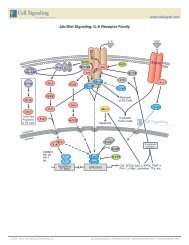
Pathway Description: <strong>Angiogenesis</strong> results in the formation of new blood vessels,<br />
and can be induced by tumor growth, tissue wound, and inflammation. Rapid tumor cell<br />
growth creates intracellular hypoxia. Hypoxia-inducible factor (HIF) is a transcription factor<br />
that responds to changing intracellular oxygen concentration. Under typical oxygen levels<br />
(normoxia), HIF is hydroxylated and acetylated, modifications that target the transcription<br />
factor for VHL mediated ubiquitin degradation. During hypoxia, HIF accumulates and is<br />
transported to the nucleus where it induces expression of numerous target gene products.<br />
Secreted growth factors (such as VEGF, FGF, and TGF) induce signaling pathways (including<br />
PLCγ, PI3K, Src, Smad signaling) that result in endothelial cell proliferation, increased<br />
vascular permeability and cell migration. In addition to hypoxia, PI3K and Ras pathways can<br />
increase HIF expression by promoting HIF translation.<br />
Pericytes are support cells that provide structural support for newly formed blood vessels,<br />
promote endothelial cell survival, guide sprouting vessels, and regulate vasoconstriction and<br />
dilation. This is done through a reciprocal signaling mechanism in which PDGF-BB secreted<br />
into the matrix by endothelial cells acts as a ligand for PDGF receptor-β located on the<br />
pericyte membrane. In return, pericytes produce and secrete VEGF that signals through the<br />
endothelial VEGF receptor.<br />
Extracellular matrix proteases and regulators induce tissue matrix remodeling in preparation<br />
for migration of endothelial cells from existing vessels to form new tubing. Tissue wounding,<br />
ischemia, or inflammation recruit macrophages and bone marrow-derived inflammatory cells<br />
(BDMC) to wound areas, and secrete a similar panel of proteins to induce angiogenesis.<br />
For Selected Reviews see www.cellsignal.com<br />
14<br />
Direct Stimulatory Modification<br />
Direct Inhibitory Modification<br />
Multistep Stimulatory Modification<br />
Multistep Inhibitory Modification<br />
Tentative Stimulatory Modification<br />
Tentative Inhibitory Modification<br />
Transcriptional Stimulatory Modification<br />
Transcriptional Inhibitory Modification
Please visit www.cellsignal.com for a complete product listing.<br />
Wnt/b-Catenin Signaling<br />
OFF-State<br />
RTK<br />
Src<br />
Fer<br />
Proteasomal<br />
Degradation<br />
Cytoplasm<br />
p120<br />
β-catenin<br />
α-catenin<br />
Nucleus<br />
OFF<br />
Cadherin<br />
β-TrCP<br />
ub β-catenin<br />
DKKs<br />
GBP<br />
PP2A<br />
Axin<br />
APC GSK-3<br />
WTX<br />
β-catenin<br />
CK1<br />
Groucho HDAC<br />
LEF/TCF<br />
Hesx1<br />
LRP<br />
SFRP<br />
Frizzled<br />
Tankyrase<br />
Dvl<br />
mTOR Pathway<br />
HDAC<br />
Groucho β-catenin<br />
Prop1<br />
ON-State<br />
Rac1<br />
LRP<br />
CK1 Axin<br />
GSK-3<br />
CYLD<br />
β-catenin<br />
WIF<br />
SFRP<br />
Wnt<br />
TGF-β/BMP<br />
Frizzled<br />
Dvl<br />
ub<br />
Dvl<br />
Naked<br />
PAR-1<br />
ub TAB2<br />
TAK1<br />
TAB1<br />
NLK<br />
RA<br />
RAR<br />
ICAT<br />
Chibby<br />
Pygo<br />
BCL9<br />
NLK<br />
β-catenin<br />
CBP Cdk8 Target Genes:<br />
Migration<br />
LEF/TCF<br />
Myc, Cyclin D1,<br />
TCF-1, PPAR-δ, Adhesion<br />
MMP-7, Axin-2,<br />
CD44, etc.<br />
β-catenin<br />
Pit1<br />
Prop1<br />
Pathway Description: The Wnt/β-Catenin pathway regulates cell fate decisions during<br />
development of vertebrates and invertebrates. The Wnt-ligand is a secreted glycoprotein<br />
that binds to Frizzled receptors, which triggers a cascade resulting in displacement of<br />
the multifunctional kinase GSK-3β from the APC/Axin/GSK-3β-complex. In the absence<br />
of Wnt-signal (Off-state), β-catenin, an integral cell-cell adhesion adaptor protein as<br />
well as transcriptional co-regulator, is targeted for degradation by the APC/Axin/GSK-<br />
3β-complex. Appropriate phosphorylation of β-catenin by coordinated action of CK1<br />
and GSK-3β leads to its ubiquitination and proteasomal degradation through the β-TrCP/<br />
SKP complex. In the presence of Wnt binding (On-state), Dishevelled (Dvl) is activated by<br />
phosphorylation and poly-ubiquitination, which in turn recruits GSK-3β away from the<br />
degradation complex. This allows for stabilization of β-catenin levels, Rac1-dependent<br />
nuclear translocation and recruitment to the LEF/TCF DNA-binding factors where it<br />
acts as an activator for transcription by displacement of Groucho-HDAC co-repressors.<br />
Additionally, in complex with the homeodomain factor Prop1, β-catenin has also<br />
been shown to act in context-dependent activation as well as repression complexes.<br />
Importantly, point-mutations in β-catenin lead to its deregulated stabilization. APC<br />
and Axin mutations also have been documented in some tumors, underscoring the<br />
deregulation of this pathway in human cancer. During development, the Wnt/β-catenin<br />
pathway integrates signals from many other pathways including retinoic acid, FGF,<br />
TGF-β, and BMP in many different cell-types and tissues. In addition, GSK-3β is also<br />
involved in glycogen metabolism and other key pathways, which has made its inhibition<br />
relevant to diabetes and neurodegenerative disorders.<br />
For Selected Reviews see www.cellsignal.com<br />
© 2003 – 2010 Cell Signaling Technology, Inc.<br />
Hippo Signaling<br />
Low Cell density<br />
YAP<br />
TF<br />
TAZ<br />
TF<br />
DCHS1/2<br />
MER KIBRA FRMD6<br />
FRMD6 KIBRA MER<br />
YAP<br />
FAT4<br />
Growth and<br />
Proliferation<br />
Growth and<br />
Proliferation<br />
Nucleus<br />
TAZ<br />
YAP<br />
SAV1<br />
Mst1/2<br />
MOB1<br />
LATS1/2<br />
Cytoplasm<br />
Cytoplasm<br />
TAZ<br />
14-3-3<br />
High Cell density<br />
Rassf<br />
CK1<br />
Ubiquitination<br />
Degradation<br />
SAV1<br />
Mst1/2<br />
MOB1<br />
LATS1/2<br />
Transcription Factors (TF) include: TEAD, TEF, RunX1, RunX2, p73 PPARγ and others<br />
YAP/TAZ<br />
YAP/TAZ<br />
DCHS1/2<br />
14-3-3<br />
FAT4<br />
DvI<br />
YAP/TAZ<br />
FAT4<br />
Wnt/β-Catenin Signaling<br />
TF<br />
Growth and<br />
Proliferation<br />
Cytoplasm<br />
Cytoplasm<br />
Nucleus<br />
© 2012 Cell Signaling Technology, Inc.<br />
Pathway Description: Hippo signaling is an evolutionarily conserved pathway that<br />
controls cell proliferation, apoptosis, and organ size in response to changing cell<br />
density levels. At relative low cell density, transcription co-activators YAP and TAZ bind<br />
transcription factors to induce expression of genes that favor cell growth and proliferation.<br />
Transcription factors activated following interaction with YAP and/or TAZ include TEAD,<br />
Runx2, p73, and TBX5. Interaction with p73 follows DNA damage and may promote<br />
apoptosis; most other activated transcription factors likely activate transcription of genes<br />
favoring cell growth and proliferation. As cell density increases, interaction between<br />
membrane-bound upstream hippo pathway regulators trigger activation of cytoplasmic<br />
kinases Mst1/2 and LATS1/2. Activated Mst kinase (the eponymous Hippo in Drosophila)<br />
associates with the adaptor WW45 and activates the downstream LATS kinase, which<br />
phosphorylates YAP and TAZ. Phosphorylation of these co-activators allows binding of<br />
the cytoplasmic anchor 14-3-3 protein. Prevented from entering the nucleus, YAP and<br />
TAZ can no longer help promote transcription of genes that favor increased cell growth<br />
and proliferation. Several parts of the pathway in mammalian cells remain unclear, but<br />
are suggested by better-characterized Drosophila counterparts. Cell surface protein<br />
interactions may involve Dachsous and Fat cadherins; the mechanism of Mst activation<br />
by upstream regulators Merlin and FRMD6 (Expanded in Drosophila) also remains<br />
unclear. Kinases PKA and PAK may inhibit Merlin while activated Fat receptor may inhibit<br />
cytoplasmic Dachsous.<br />
For Selected Reviews see www.cellsignal.com<br />
Separation of Subunits or Cleavage Products<br />
Joining of Subunits<br />
Translocation<br />
Kinase<br />
Phosphatase<br />
Transcription Factor<br />
Receptor<br />
Cyclin, pro-apoptotic<br />
Caspase Enzyme pro-survival<br />
GAP<br />
GEF<br />
GTPase<br />
G-protein, ribosomal subunit<br />
15<br />
Hippo Signaling • created November 2010
USA Headquarters<br />
Cell Signaling Technology, Inc.<br />
Tel: 978-867-2300<br />
E-mail: info@cellsignal.com<br />
www.cellsignal.com<br />
International Subsidiaries<br />
Cell Signaling Technology (China) Limited<br />
Tel: (86) 21-5835-6288<br />
E-mail: info@cst-c.com.cn<br />
www.cellsignal.com<br />
Cell Signaling Technology Europe<br />
Tel: +31 (0)71 568 1060<br />
E-mail: info@cellsignal.eu<br />
www.cellsignal.com<br />
Cell Signaling Technology Japan, K.K.<br />
Tel: 03 (5652) 0213<br />
E-mail: info@cstj.co.jp<br />
www.cstj.co.jp<br />
International Distributors<br />
www.cellsignal.com<br />
ARGENTINA: Migliore Laclaustra S.r.l.<br />
Tel: 5411-43729045<br />
E-mail: info@migliorelaclaustra.com.ar<br />
AUSTRALIA: Genesearch Pty. Ltd.<br />
Toll Free: 1800 074 278<br />
www.genesearch.com.au<br />
BELGIUM/LUXEMBOURG: Bioké<br />
Tel: 0800-71640 / www.bioke.com<br />
BRAZIL: Uniscience Do Brazil<br />
Tel: (011) 3622 2320<br />
www.uniscience.com<br />
CANADA: New England Biolabs Ltd.<br />
Toll Free: 1-800-387-1095 / www.neb.ca<br />
CHILE: Genetica Y Technologia Ltda.<br />
Tel: 56-2-633 52 69 / www.genytec.cl<br />
COLOMBIA/PANAMA: Bio Products, Inc.<br />
dba Subiotec Ltda.<br />
Tel: 561-434-2121 / www.bioproducts.net<br />
CZECH REPUBLIC: Biotech A.s.<br />
Toll Free: +420 800124683<br />
www.biotech.cz<br />
DENMARK: BioNordika Denmark A/S<br />
Tel: +45 3956 2000 / www.bionordika.dk<br />
ESTONIA/LATVIA/LITHUANIA: BioNordika Baltic Oü<br />
Tel: +372 6306 520 / www.bionordika.ee<br />
FINLAND: Fisher Scientific Oy<br />
Tel: +358 9 802 76 280 / www.fishersci.fi<br />
FRANCE: Ozyme<br />
Tel: (1) 34 60 24 24 / www.ozyme.fr<br />
GERMANY/AUSTRIA: New England Biolabs GmbH<br />
Tel: +49/ (0) 69 305 23140 / www.neb-online.de<br />
GREECE: Bioline Scientific Douros<br />
Bro – E. Demagos O.e.<br />
Tel: 210-5226547 / E-mail: demagos@hol.gr<br />
Hong Kong: Gene Company Limited<br />
Tel: (852) 2896-6283 / www.genehk.com<br />
HUNGARY: Kvalitex Kft.<br />
Tel: (36) 1340-4700 / www.kvalitex.hu<br />
ICELAND: Groco ehf<br />
Tel: +354-568-8533 / www.groco.is<br />
INDIA: <strong>Lab</strong>mate (Asia) Pvt Ltd.<br />
Tel: 44 222 000 66 / www.labmateasia.com<br />
INDONESIA: P T Research Biolabs<br />
Tel: 62-21-5859365<br />
E-mail: Indonesia@researchbiolabs.com<br />
REPUBLIC OF IRELAND: Isis Ltd.<br />
Tel: (1) 286 7777 / www.isisco.ie<br />
ISRAEL: Eldan Electronic Instruments Co., Ltd<br />
Tel: (3) 9371132 / www.eldan.biz<br />
ITALY: Euroclone<br />
Toll Free: 800-315911 / www.euroclonegroup.it<br />
KOREA: Koram Biotech Corp.<br />
Tel: (02) 556-0311 / www.korambiotech.com<br />
MALAYSIA: Research Biolabs Sdn Bhd<br />
Tel: 60358829588 / www.researchbiolabs.com<br />
MEXICO: Quimica Valaner S.a. De C.v.<br />
Tel: 5525-5725 / www.valaner.com<br />
THE NETHERLANDS: Bioké<br />
Tel: +31 (0)71 568 1000 / www.bioke.com<br />
New Zealand: Biolab Ltd<br />
Tel: (09) 980-6700 / www.biolabgroup.com<br />
NORWAY: BioNordika Norway AS<br />
Tel: 47 67 11 14 60 / www.bionordika.no<br />
POLAND: <strong>Lab</strong>-<strong>JOT</strong><br />
Tel: +48 22 2034155 / www.labjot.com<br />
PORTUGAL: Izasa Lisbon<br />
Tel: (21) 424 73 64 / www.izasa.es<br />
SINGAPORE: Research Biolabs Pte Ltd<br />
Tel: +65 6777 5366 / www.researchbiolabs.com<br />
SLOVAK REPUBLIC: Biotech S.r.o.<br />
Tel: (07) 54774488 / E-mail: biotech@biotech.cz<br />
SOUTH AFRICA: <strong>Lab</strong>oratory Specialist Services cc<br />
Tel: +27 (0)21 7887755 / www.lss.co.za<br />
SPAIN: Izasa, S.a.<br />
Tel: (34) 902 20 30 70 / www.izasa.es<br />
SWEDEN: BioNordika Sweden AB<br />
Tel: 46 8 30 60 10 / www.bionordika.se<br />
SWITZERLAND: Bioconcept<br />
Tel: (061) 486 80 80 / www.bioconcept.ch<br />
TAIWAN: Taigen Bioscience Corp.<br />
Tel: (02) 28802913 / www.taigen.com<br />
THAILAND: Theera Trading Co. Ltd.<br />
Tel: (02) 412-5672 / www.theetrad.com<br />
TURKEY: Sacem Hayat Teknolojileri<br />
Tel: +90 312 231 52 72 / www.sacem.com.tr<br />
UNITED KINGDOM: New England Biolabs (UK) Ltd.<br />
Toll Free: 0800 318486 / www.neb.uk.com<br />
URUGUAY: Tanirel SA<br />
Tel: 00598 24804895 / E-mail: ventas@tanirel.<br />
com.uy<br />
VENEZUELA: Bioproducts, Inc. DBA Corporacion<br />
Internacional De Tecnologia, S.a. (Corpointer)<br />
Tel: 561-434-2121 / www.bioproducts.net<br />
Printed in the USA on 55% recycled paper (30% post-consumer waste fiber) using soy inks and processed chlorine free.<br />
© 01/2012 Cell Signaling Technology, Inc.