IF_2012 - 7-2012.pdf - Lab-JOT
IF_2012 - 7-2012.pdf - Lab-JOT
IF_2012 - 7-2012.pdf - Lab-JOT

Create successful ePaper yourself
Turn your PDF publications into a flip-book with our unique Google optimized e-Paper software.
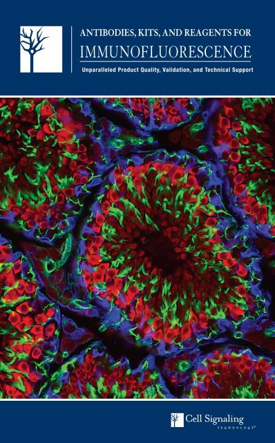
ANTIBODIES, KITS, AND REAGENTS FOR<br />
IMMUNOFLUORESCENCE<br />
Unparalleled Product Quality, Validation, and Technical Support
XP ®<br />
Monoclonal Antibodies<br />
XP® monoclonal antibodies are a line of high quality<br />
rabbit monoclonal antibodies exclusively available<br />
from Cell Signaling Technology (CST). Any product<br />
labeled with XP has been carefully selected based on<br />
superior performance in the most relevant applications.<br />
These antibodies are generated using XMT® technology,<br />
a proprietary monoclonal method developed at CST.<br />
The technology provides access to a broad range of<br />
antibody-producing B cells unattainable with traditional<br />
monoclonal technologies, allowing more comprehensive<br />
screening and the identification of XP monoclonal<br />
antibodies with exceptional specificity, sensitivity,<br />
and performance.<br />
eXceptional specificity<br />
As with all of our antibodies, the antibody is specific to your<br />
target of interest, saving you valuable time and resources.<br />
+ eXceptional sensitivity<br />
The antibody will provide a stronger signal for your target<br />
protein in cells and tissues, allowing you to monitor<br />
expression of low levels of endogenous proteins, saving<br />
you valuable materials.<br />
+ eXceptional stability and reproducibility<br />
XMT technology combined with our stringent quality<br />
control ensures maximum lot-to-lot consistency and the<br />
most reproducible results.<br />
= eXceptional Performance <br />
XMT technology coupled with our extensive antibody<br />
validation and stringent quality control delivers<br />
XP monoclonal antibodies with eXceptional Performance<br />
in the widest range of applications.<br />
kDa<br />
200<br />
140<br />
100<br />
80<br />
A<br />
HT-29<br />
SK-BR-3<br />
T-47D<br />
Pro Met<br />
Met<br />
B<br />
C<br />
60<br />
50<br />
40<br />
30<br />
20<br />
200<br />
140<br />
100<br />
80<br />
D<br />
E<br />
60<br />
50<br />
40<br />
30<br />
β-Actin<br />
Events<br />
20<br />
Met<br />
Met (D1C2) XP ® Rabbit mAb #8198: Western blot analysis (A) of extracts from HT-29 (Met+), SK-BR-3 (Met-), and T-47D (Met-)<br />
cells using #8198 (upper) or β-Actin Antibody #4967 (lower). Confocal <strong>IF</strong> analysis of HT-29 (B) and T-47D (C) cells using #8198<br />
(green). Actin filaments were labeled with DY-554 phalloidin (red). Blue pseudocolor = DRAQ5 ® #4084 (fluorescent DNA dye).<br />
Flow cytometric analysis (D) of T-47D (blue) and HT-29 (green) cells using #8198. IHC analysis (E) of paraffin-embedded human<br />
hepatocellular carcinoma using #8198.<br />
Visit our website for more experimental details, additional information,<br />
and a complete list of available XP monoclonal antibodies.
Cover Image<br />
Vimentin (D21H3) XP ® Rabbit<br />
mAb (Alexa Fluor ® 647 Conjugate)<br />
#9856 (blue pseudocolor). Miwi<br />
(D92B7) XP ® Rabbit mAb #6915<br />
(red pseudocolor). Actin filaments<br />
were labeled using DY-554<br />
phalloidin (green pseudocolor).<br />
Antibodies, Kits, and Reagents for<br />
Immunofluorescence<br />
Cell Signaling Technology (CST) provides the highest quality activation<br />
state and total protein antibodies available for use in immunofluorescence<br />
(<strong>IF</strong>). Our in-house <strong>IF</strong> group has validated each <strong>IF</strong>-recommended CST <br />
antibody using multiple approaches. Technical support is provided by the<br />
same scientists who validate these products.<br />
4<br />
Antibody Validation for Immunofluorescence<br />
6 Alexa Fluor® Conjugates for Immunofluorescence<br />
WINNER!<br />
Life Science<br />
Industry Awards ®<br />
:: Best Antibodies<br />
:: Best Breakthrough Products<br />
for Cancer Research<br />
8<br />
10<br />
12<br />
13<br />
14<br />
15<br />
16<br />
17<br />
Complementary Reagents and Controls<br />
Organelle Markers<br />
Tyrosine Kinases<br />
MAP Kinase Signaling<br />
PI3K/Akt Signaling<br />
Development and Differentiation<br />
Embryonic Stem Cell Markers<br />
Lineage-specific Markers<br />
20 Neuroscience<br />
Application Key:<br />
W Western / IP Immunoprecipitation / IHC Immunohistochemistry<br />
<strong>IF</strong> Immunofluorescence / F Flow Cytometry<br />
ChIP Chromatin Immunoprecipitation / HCA High Content Analysis<br />
(-IC Immunocytochemistry, -P Paraffin, -F Frozen) / E-P Peptide ELISA<br />
Reactivity Key:<br />
H human / M mouse / R rat / Hm hamster / Mk monkey / C chicken<br />
Mi mink / Dm D. melanogaster / X Xenopus / Z zebra fish / B bovine<br />
Dg dog / Pg pig / Sc S. cerevisiae / Ce C. elegans<br />
All all species expected / ( ) 100% sequence homology<br />
For research use only. Not for use in diagnostic procedures.<br />
22<br />
24<br />
27<br />
28<br />
30<br />
32<br />
34<br />
36<br />
37<br />
38<br />
39<br />
Immunology and Inflammation<br />
Chromatin and Epigenetic Regulation<br />
Apoptosis and Autophagy<br />
Translational Control<br />
Cellular Metabolism<br />
Cell Cycle, Checkpoint Control, and DNA Damage<br />
Cytoskeletal Regulation and Adhesion<br />
Epitope Tag Antibodies<br />
Protein Folding and Stability<br />
Motif and Other Antibodies<br />
Immunofluorescence Protocol
Antibody Validation for Immunofluorescence<br />
Scientists at Cell Signaling Technology (CST) have validated over 800 activation state-specific (e.g., phosphorylation-specific) and total protein<br />
antibodies for immunofluorescence (<strong>IF</strong>) applications, such as manual fluorescence microscopy or automated imaging and laser scanning high<br />
content platforms. All CST antibodies approved for use in immunofluorescent assays have undergone a rigorous validation process.<br />
In addition, our <strong>IF</strong> team has created multiple custom protocols for optimal fixation and staining of specific products; all protocols are<br />
available on our website and are available as links from product pages.<br />
Phosphatase Treatment and Knockout Cells:<br />
Cells are subjected to phosphatase treatment to verify phospho-specificity.<br />
Target specificity is also verified with the use of known knockout or null cell lines.<br />
Tissue:<br />
Antibody performance is assessed on appropriate tissues.<br />
Untreated<br />
LY294002- and<br />
Wortmannin-treated<br />
l Phosphatase-treated<br />
MEF wild-type<br />
MEF GSK3β -/-<br />
Pdx1 Antibody #2437: Confocal <strong>IF</strong> analysis of normal rat pancreas<br />
using #2437 (green, left) or Insulin (C27C9) Rabbit mAb #3014<br />
(green, right). Keratin filaments were labeled with Pan-Keratin (C11)<br />
Mouse mAb (Alexa Fluor ® 647 Conjugate) #4528 (blue pseudocolor).<br />
Red = Propidium Iodide (PI)/RNase Staining Solution #4087<br />
(fluorescent DNA dye).<br />
Subcellular Localization:<br />
Appropriate cell lines and tissues are used to verify<br />
subcellular localization.<br />
PC-3<br />
Mitochondria<br />
Endoplasmic Reticulum<br />
Phospho-GSK-3β (Ser9) (D85E12) XP ® Rabbit mAb #5558: Confocal <strong>IF</strong> analysis of wildtype<br />
mouse embryonic fibroblasts (MEFs) (top row), GSK-3β (-/-) MEFs (middle row), or PC-3<br />
cells (bottom row), untreated (left), LY294002 and Wortmannin-treated (#9901 and #9951<br />
respectively; center) or λ phosphatase-treated (right), using #5558 (green). Blue pseudocolor<br />
= DRAQ5 ® #4084 (fluorescent DNA dye). (MEF wild-type and GSK-3β (-/-) cells were kindly<br />
provided by Dr. Jim Woodgett, University of Toronto, Canada).<br />
COX IV (3E11) Rabbit mAb<br />
#4850 (green)<br />
ERp72 (D70D12) XP ® Rabbit<br />
mAb #5033 (green)<br />
High Content Screening<br />
Our High Content Screening (HCS) group tests <strong>IF</strong>-validated antibodies on in-house<br />
HCS platforms, which include the TTP <strong>Lab</strong>Tech Acumen ® eX 3 and Cellomics ®<br />
ArrayScan ® V TI . This allows us to assess antibody performance on HCS platforms<br />
to provide you with the best possible support for your HCS assays.<br />
Intercellular Junctions<br />
Mitotic Chromatin<br />
Phospho-p44/42 MAPK (Erk1/2)<br />
(Tyr202/Thr204) (green)<br />
-U0126/+TPA<br />
+U0126/+TPA<br />
Normalized Mean Fluorescence Intensity<br />
100<br />
80<br />
60<br />
40<br />
20<br />
Phospho-CREB<br />
Phospho-p44/42 MAPK<br />
Phospho-p90RSK<br />
0 0.1 0.5 1 5 10 50 100<br />
U0126 (µM)<br />
Phospho-p44/42 MAPK (Erk1/2) (Thr202/Tyr204) (D13.14.4E) XP ® Rabbit mAb #4370,<br />
Phospho-p90RSK (Thr573) Antibody #9346, and Phospho-CREB (Ser133) (87G3) Rabbit<br />
mAb #9198: A549 cells were concurrently exposed to U0126 (MEK1/2 Inhibitor) #9903 and<br />
the phorbol ester TPA #4174 to assess the effect on MAPK signaling. Phospho-p44/42 MAPK<br />
(Erk1/2), detected with #4370, and two downstream signaling proteins, phospho-p90RSK and<br />
phospho-CREB, detected with #9346 and #9198, were used as readouts. U0126 was titrated<br />
as an 8-point dose curve in triplicate to monitor the reproducibility and validity of the assay. The<br />
signal for each antibody was analyzed using an Acumen ® eX 3 and images were acquired with a<br />
Cellomics ® ArrayScan ® V TI . With increasing concentrations of U0126, a significant decrease in<br />
phospho-p44/42 MAPK (Erk1/2) signal (~10-fold), as well as phospho-p90RSK and phospho-<br />
CREB (>2-fold), was observed as compared to the TPA-stimulated control.<br />
ZO-3 (D57G7) XP ® Rabbit<br />
mAb #3704 (green)<br />
Phospho-Histone H3 (Ser10)<br />
(D2C8) XP ® Rabbit mAb #3377<br />
(green)<br />
Other Validation Steps Include:<br />
:: Requirement of threshold signal-to-noise ratio in<br />
antibody:isotype comparison and minimum foldinduction<br />
for phospho-specific antibodies ensures<br />
the greatest possible sensitivity.<br />
:: Activation state specificity, target expression, or<br />
translocation are examined using ligands or inhibitors<br />
to modulate pathway activity.<br />
:: Stringent testing ensures lot-to-lot consistency.<br />
:: Cells are subjected to siRNA treatment or overexpression<br />
of the target protein to verify target specificity.<br />
:: Cell lines or tissues with known target expression<br />
levels are used to verify specificity.<br />
4 Application and Reactivity Keys, pg 3. Monoclonal Antibody. Please see page 2 for details.
Optimization of Staining Protocols<br />
The standard CST immunofluorescence staining protocol<br />
includes formaldehyde fixation and detergent permeabilization.<br />
Since most antibodies work well with this protocol,<br />
two or more antibodies can be multiplexed on the same<br />
cells or tissue. However, some antibodies may work better<br />
with, or even require, different fixation and/or permeabilization<br />
methods. For example, our PDI and β-Actin<br />
antibodies benefit from methanol permeabilization (see<br />
images right). If an alternative protocol is recommended<br />
for a particular antibody, it will be clearly noted under the<br />
recommended dilutions section on the product datasheet.<br />
Please feel free to contact our immunofluorescence specialists<br />
at <strong>IF</strong>@cellsignal.com with any questions regarding<br />
treatment, fixation, or staining protocols.<br />
PDI Antibody #2446 and β-Actin (8H10D10) Mouse mAb #3700: Confocal <strong>IF</strong><br />
analysis of NIH/3T3 cells, permeabilized with methanol (left) or 0.3% Triton X-100<br />
(right), using #2446 (green) and #3700 (red). Blue pseudocolor = DRAQ5 ® #4084<br />
(fluorescent DNA dye).<br />
PathScan ® Multiplex <strong>IF</strong> Kits<br />
PathScan ® Multiplex <strong>IF</strong> Kits from CST provide a novel multiplex<br />
assay to simultaneously assess signaling through key pathway<br />
nodes using automated high content platforms (both imaging and<br />
laser scanning) or manual immunofluorescence microscopy. The<br />
kits contain a cocktail of three high quality primary antibodies,<br />
as well as a detection cocktail utilizing the Alexa Fluor ® series<br />
of fluorescent dyes. Antibody and dye pairings have been preoptimized<br />
and each kit contains enough reagents for 100 assays<br />
(based on a working volume of 100 μl/test).<br />
:: Kits allow the analysis of multiple pathway endpoints<br />
within a single sample, saving time and reagents.<br />
:: Kits are produced and optimized in-house with the<br />
highest quality antibodies, providing you with the<br />
greatest possible specificity and sensitivity.<br />
:: Technical support is provided by our in-house <strong>IF</strong> group,<br />
who developed the products and knows them best,<br />
ensuring a timely and accurate response.<br />
Applications<br />
Reactivity<br />
#7851 PathScan ® Apoptosis and Proliferation Multiplex <strong>IF</strong> Kit <strong>IF</strong>-IC H, Mk<br />
Kit includes primary antibody and detection cocktails to<br />
simultaneously detect levels of phospho-Histone H3 (Ser10),<br />
cleaved PARP (Asp214), and α-Tubulin<br />
#7967 PathScan ® EGF Receptor Activation Multiplex <strong>IF</strong> Kit <strong>IF</strong>-IC, <strong>IF</strong>-P H, Mk, (M)<br />
Kit includes primary antibody and detection cocktails to<br />
simultaneously detect levels of phospho-EGF Receptor<br />
(Tyr1068), total EGF Receptor, and phospho-p44/42 MAPK<br />
(Erk1/2) (Thr202/Tyr204)<br />
#8999 PathScan ® Signaling Nodes Multiplex <strong>IF</strong> Kit <strong>IF</strong>-IC H, M, R, Mk<br />
Kit includes primary antibody and detection cocktails to<br />
simultaneously detect levels of phospho-Akt (Ser473),<br />
phospho-S6 Ribosomal Protein (Ser235/236), and<br />
phospho-p44/42 MAPK (Erk1/2) (Thr202/Tyr204)<br />
PathScan ® Signaling Nodes Multiplex <strong>IF</strong> Kit<br />
#8999: Confocal <strong>IF</strong> analysis of MCF7 cells, serumstarved<br />
(upper) or insulin-treated (lower), using<br />
#8999. Red = phospho-Akt (Ser473), green =<br />
phospho-p44/42 MAPK (Erk1/2) (Thr202/Tyr204),<br />
and blue pseudocolor = phospho-S6 Ribosomal<br />
Protein (Ser235/236).<br />
PathScan ® Multiplex <strong>IF</strong> Kits: Some kit components are provided under an agreement between Life Technologies<br />
Corporation and Cell Signaling Technology, Inc., and the manufacture, use, sale or import of antibody conjugate in<br />
this product is subject to one or more US patents and corresponding non-US equivalents, owned or controlled by<br />
Life Technologies Corporation or its affiliates. The purchase of this product conveys to the buyer the non-transferable<br />
right to use the purchased amount of the product and components of the product only in research conducted by<br />
the buyer (whether the buyer is an academic or for-profit entity), for immunocytochemistry, high content screening<br />
(HCS) analysis, or flow cytometry applications. Buyer’s use of this product or its components (1) in manufacturing;<br />
(2) to provide a service, information, or data to an unaffiliated third party for payment; (3) for therapeutic, diagnostic<br />
or prophylactic purposes; (4) resale, whether or not such product or its components are resold for use in research; or<br />
for any other commercial purpose is prohibited. For information on purchasing a license to this product for purposes<br />
other than research, contact Life Technologies Corporation, Cellular Analysis Business Unit, Business Development,<br />
29851 Willow Creek Road, Eugene, OR 97402, Tel: (541) 465-8300. Fax: (541) 335-0354.<br />
Unparalleled Product Quality, Validation, and Technical Support www.cellsignal.com<br />
5
Alexa Fluor ® Conjugates<br />
for Immunofluorescence<br />
The superior brightness and photostability<br />
of Alexa Fluor ® dyes combined with the<br />
highest quality antibodies from<br />
Cell Signaling Technology results in the<br />
brightest signal with the lowest background.<br />
All Alexa Fluor conjugates recommended for<br />
immunofluorescence (<strong>IF</strong>) are validated by<br />
our in-house <strong>IF</strong> specialists.<br />
:: Technical support provided by our<br />
<strong>IF</strong> specialists translates into a thorough,<br />
fast, and accurate response.<br />
:: High quality custom conjugations<br />
to our off-the-shelf antibodies are<br />
available upon request.<br />
Detection Dye Ex (max)<br />
Em (max)<br />
Alexa Fluor ® 488 495 nm 519 nm<br />
Alexa Fluor ® 555 555 nm 565 nm<br />
Alexa Fluor ® 594 590 nm 617 nm<br />
Alexa Fluor ® 647 650 nm 668 nm<br />
Unconjugated Antibody Reactivity 488 555 594 647<br />
#4970 β-Actin (13E5) Rabbit mAb H, M, R, Mk, B,<br />
Pg, (C, Dg)<br />
#8844 #9470 #8584<br />
#4691 Akt (pan) (C67E7) Rabbit mAb H, M, R, Mk, Dm #5084 #5186<br />
#5153 Androgen Receptor (D6F11) XP ® Rabbit mAb H #7395 #7397<br />
#3195 E-Cadherin (24E10) Rabbit mAb H, M, (Dg, Pg) #3199 #4295<br />
#9661 Cleaved Caspase-3 (Asp175) Antibody H, M, R, Mk, (B,<br />
Dg, Pg)<br />
#9669<br />
#2677 β-Catenin (L54E2) Mouse mAb (<strong>IF</strong> Preferred) H, (M, R, Mk, Pg) #2849 #5612 #4627<br />
#3570 CD44 (156-3C11) Mouse mAb H #3516<br />
#5664 CNPase (D83E10) XP ® Rabbit mAb H, M, R #5714 #5715 #5716<br />
#4850 COX IV (3E11) Rabbit mAb H, R, Mk, Z, B, Pg #4853 #7561<br />
#9198 Phospho-CREB (Ser133) (87G3) Rabbit mAb H, M, R #9187<br />
#2368 DYKDDDDK Tag Antibody (Binds to same<br />
epitope as Sigma’s Anti-FLAG ® M2 Antibody)<br />
All #5407 #3768 #9696 #3916<br />
#4267 EGF Receptor (D38B1) XP ® Rabbit mAb H, M, Mk #5616 #5108 #5588<br />
#2929 EpCAM (VU1D9) Mouse mAb H #5198 #5488 #7319 #5447<br />
#2118 GAPDH (14C10) Rabbit mAb H, M, R, Mk #3906 #3964 #3907<br />
#3670 GFAP (GA5) Mouse mAb H, M, R #3655 #3656 #8152 #3657<br />
#2624 GST (26H1) Mouse mAb All #3368 #3720 #9440 #3445<br />
#2367 HA-Tag (6E2) Mouse mAb All #2350 #3444<br />
#2024 Hexokinase I (C35C4) Rabbit mAb H, M #3689 #3540<br />
#9718 Phospho-Histone H2A.X (Ser139)<br />
(20E3) Rabbit mAb<br />
#3377 Phospho-Histone H3 (Ser10) (D2C8) XP ®<br />
Rabbit mAb<br />
#9701 Phospho-Histone H3 (Ser10) Antibody H, M, R, Mk, C,<br />
Dm, Z, Sc, (X)<br />
#9649 Acetyl-Histone H3 (Lys9) (C5B11)<br />
Rabbit mAb<br />
H, M, R, Mk #9719 #8228 #9720<br />
H, M, R, Mk, Z #3465 #3475 #8481 #3458<br />
H, M, R, Mk,<br />
Z, (Sc)<br />
#9708 #9716<br />
#9683 #5489 #4484<br />
#4545 Pan-Keratin (C11) Mouse mAb H, R, Mk #4523 #3478 #4528<br />
#3308 c-Kit (Ab81) Mouse mAb H #3310<br />
Neurofilament-L (C28E10) Rabbit mAb (Alexa<br />
Fluor ® 488 Conjugate) #8024: Confocal <strong>IF</strong> analysis<br />
of normal rat cerebellum using #8024 (green)<br />
and GFAP (GA5) Mouse mAb #3670 (red). Blue<br />
pseudocolor = DRAQ5 ® #4084 (fluorescent DNA dye).<br />
Vimentin (D21H3) XP ® Rabbit mAb (Alexa Fluor ® 555 Conjugate) #9855: Confocal <strong>IF</strong> analysis of SNB19<br />
(left) or MCF7 (right) cells using #9855 (red). Blue pseudocolor = DRAQ5 ® #4084 (fluorescent DNA dye).<br />
Cleaved PARP (Asp214) (D64E10) XP ® Rabbit<br />
mAb (Alexa Fluor ® 488 Conjugate) #9148:<br />
Confocal <strong>IF</strong> analysis of HeLa cells, untreated (left) or<br />
treated with Staurosporine #9953 (1 μM, 3 hr; right),<br />
using #9148 (green). Actin filaments were labeled<br />
with DY-554 phalloidin (red).<br />
Phospho-NDRG1 (Thr346) (D98G11) XP ® Rabbit mAb (Alexa Fluor ® 647 Conjugate) #7497:<br />
Confocal <strong>IF</strong> analysis of C2C12 cells, insulin-treated (left) or treated with LY294002 #9901 (right), using<br />
#7497 (blue pseudocolor) and Lamin A/C (4C11) Mouse mAb #4777 (green).<br />
6 Application and Reactivity Keys, pg 3. Monoclonal Antibody. Please see page 2 for details.
Unconjugated Antibody Reactivity 488 555 594 647<br />
#2276 Myc-Tag (9B11) Mouse mAb All #2279 #3756 #9483 #2233<br />
#4903 Nanog (D73G4) XP ® Rabbit mAb H, (Mk) #5448<br />
#5482 Phospho-NDRG1 (Thr346) (D98G11) XP ®<br />
Rabbit mAb<br />
H, M, R, Mk #6992 #9851 #7497<br />
#2837 Neurofilament-L (C28E10) Rabbit mAb H, M, R #8024 #8039 #8590<br />
#2840 Oct-4A (C30A3) Rabbit mAb H, M #5177 #4439 #5263<br />
#2947 p21 Waf1/Cip1 (12D1) Rabbit mAb H, Mk #5487 #8493 #8587<br />
#4511 Phospho-p38 MAPK (Thr180/Tyr182)<br />
(D3F9) XP ® Rabbit mAb<br />
H, M, R, Mk, Sc,<br />
(Hm, C, Z, B, Pg)<br />
#9836<br />
#9286 Phospho-p53 (Ser15) (16G8) Mouse mAb H #9235 #9481<br />
#2527 p53 (7F5) Rabbit mAb H, Mk #5429 #5395<br />
#2524 p53 (1C12) Mouse mAb H, M, R, Mk #2015 #2533<br />
#5625 Cleaved PARP (Asp214) (D64E10) XP ®<br />
Rabbit mAb<br />
H, Mk #9148 #6894<br />
#3501 PDI (C81H6) Rabbit mAb H, M, R, Mk #5051 #8448 #8463<br />
#4858 Phospho-S6 Ribosomal Protein<br />
(Ser235/236) (D57.2.2E) XP ® Rabbit mAb<br />
#4856 Phospho-S6 Ribosomal Protein<br />
(Ser235/236) (2F9) Rabbit mAb<br />
#5364 Phospho-S6 Ribosomal Protein<br />
(Ser240/244) (D68F8) XP ® Rabbit mAb<br />
H, M, R, Mk,<br />
Sc, (C)<br />
H, M, R #4854<br />
H, M, R, Mk #5018<br />
#4803 #3985 #9865 #4851<br />
#2317 S6 Ribosomal Protein (54D2) Mouse mAb H, M, R, Mk, Dm #5317 #6989 #5548<br />
#4755 SSEA4 (MC813) Mouse mAb H #5835<br />
#3579 Sox2 (D6D9) XP ® Rabbit mAb H, (Mk, B, Dg) #5049 #5179 #5067<br />
#9167 Phospho-Stat1 (Tyr701) (58D6) Rabbit mAb H, M #8183<br />
#2808 Survivin (71G4B7) Rabbit mAb H, M, R #2810 #4580<br />
#2125 α-Tubulin (11H10) Rabbit mAb H, M, R, Mk, Dm,<br />
B, (Dg)<br />
#2128 β-Tubulin (9F3) Rabbit mAb H, M, R, Mk, Z,<br />
B, (C)<br />
#5063 #5059 #5046<br />
#3623 #2116 #7634 #3624<br />
#9411 Phospho-Tyrosine Mouse mAb (P-Tyr-100) All #9415<br />
#5741 Vimentin (D21H3) XP ® Rabbit mAb H, M, R, Mk #9854 #9855 #9856<br />
Alexa Fluor ® 594<br />
p21 Waf1/Cip1 (12D1) Rabbit mAb (Alexa Fluor ®<br />
555 Conjugate) #8493: Confocal <strong>IF</strong> analysis of<br />
MCF7 cells using #8493 (red) and Phospho-Histone<br />
H3 (Ser10) (D2C8) XP ® Rabbit mAb (Alexa Fluor ®<br />
488 Conjugate) #3465 (green). Blue pseudocolor =<br />
DRAQ5 ® #4084 (fluorescent DNA dye).<br />
CNPase (D83E10) XP ® Rabbit mAb (Alexa Fluor ®<br />
647 Conjugate) #5716: Confocal <strong>IF</strong> analysis of<br />
rat cerebellum using #5716 (blue pseudocolor) and<br />
β3-Tubulin (TU-20) Mouse mAb #4466 (green). Red =<br />
Propidium Iodide (PI)/RNase Staining Solution #4087<br />
(fluorescent DNA dye).<br />
Red-fluorescent Alexa Fluor ® 594 is useful for multiplex labeling in combination<br />
with green-fluorescent dyes. Alexa Fluor ® 594 produces a brighter signal than<br />
similar red dyes and has a higher degree of labeling (DOL).<br />
EpCAM (VU1D9) Mouse mAb (Alexa Fluor ® 594 Conjugate) #7319: Confocal <strong>IF</strong> analysis of<br />
HT-29 (left) and HeLa (right) cells using #7319 (red). Actin filaments were labeled with Alexa Fluor ®<br />
488 phalloidin (green). Blue pseudocolor = DRAQ5 ® #4084 (fluorescent DNA dye).<br />
β-Catenin (L54E2) Mouse mAb (Alexa Fluor ®<br />
555 Conjugate) #5612: Confocal <strong>IF</strong> analysis of HeLa<br />
(upper) and NCI-H28 (lower) cells using #5612 (red)<br />
and a Histone H3 antibody (green).<br />
Unparalleled Product Quality, Validation, and Technical Support www.cellsignal.com<br />
7
Complementary Reagents and Controls<br />
for Immunofluorescence<br />
Cell Signaling Technology (CST) offers a wide selection of secondary antibodies, DNA dyes, activators and inhibitors, as well as experimental<br />
controls. These same reagents are also used in-house for antibody validation in immunofluorescent analysis and therefore work optimally with<br />
our primary antibodies. Technical support is provided by the scientists who work regularly with the reagents and know them best.<br />
Conjugated Secondary Antibodies<br />
Alexa Fluor ®<br />
#4408 Anti-mouse IgG (H+L), F(ab’) 2 Fragment (Alexa Fluor ® 488 Conjugate)<br />
#4409 Anti-mouse IgG (H+L), F(ab’) 2 Fragment (Alexa Fluor ® 555 Conjugate)<br />
#4410 Anti-mouse IgG (H+L), F(ab’) 2 Fragment (Alexa Fluor ® 647 Conjugate)<br />
#4412 Anti-rabbit IgG (H+L), F(ab’) 2 Fragment (Alexa Fluor ® 488 Conjugate)<br />
#4413 Anti-rabbit IgG (H+L), F(ab’) 2 Fragment (Alexa Fluor ® 555 Conjugate)<br />
#4414 Anti-rabbit IgG (H+L), F(ab’) 2 Fragment (Alexa Fluor ® 647 Conjugate)<br />
#4416 Anti-rat IgG (H+L), (Alexa Fluor ® 488 Conjugate)<br />
#4417 Anti-rat IgG (H+L), (Alexa Fluor ® 555 Conjugate)<br />
#4418 Anti-rat IgG (H+L), (Alexa Fluor ® 647 Conjugate)<br />
DyLight ®<br />
#5470 Anti-mouse IgG (H+L) (DyLight ® 680 Conjugate)<br />
#5257 Anti-mouse IgG (H+L) (DyLight ® 800 Conjugate)<br />
#5366 Anti-rabbit IgG (H+L) (DyLight ® 680 Conjugate)<br />
#5151 Anti-rabbit IgG (H+L) (DyLight ® 800 Conjugate)<br />
Excitation/Emission Table<br />
Detection Dye<br />
(nm)<br />
Ex (max)<br />
(nm)<br />
Em (max)<br />
Alexa Fluor ® 488 495 519<br />
Alexa Fluor ® 555 555 565<br />
Alexa Fluor ® 647 650 668<br />
DyLight ® 680 692 712<br />
DyLight ® 800 777 794<br />
Anti-rabbit IgG (H+L), F(ab’) 2<br />
Fragment (Alexa Fluor ®<br />
555 Conjugate) #4413 and<br />
Anti-mouse IgG (H+L), F(ab’) 2<br />
Fragment (Alexa Fluor ® 488<br />
Conjugate) #4408: Confocal <strong>IF</strong><br />
analysis of mouse cerebellum<br />
using α-Synuclein Antibody (<strong>IF</strong><br />
Preferred) #2628 detected with<br />
#4413 (red) and Neurofilament-L<br />
(DA2) Mouse mAb #2835<br />
detected with #4408 (green).<br />
Blue pseudocolor = DRAQ5 ®<br />
#4084 (fluorescent DNA dye).<br />
Anti-rat IgG (H+L) (Alexa<br />
Fluor ® 555 Conjugate) #4417<br />
and Anti-rabbit IgG (H+L),<br />
F(ab’) 2 Fragment (Alexa<br />
Fluor ® 488 Conjugate)<br />
#4412: Confocal <strong>IF</strong> analysis of<br />
HeLa cells using RPA32 (4E4)<br />
Rat mAb #2208 detected with<br />
#4417 (red) and COX IV (3E11)<br />
Rabbit mAb #4850 detected<br />
with #4412 (green). Actin<br />
filaments were labeled with<br />
Alexa Fluor ® 647 phalloidin<br />
(blue pseudocolor).<br />
Fluorescent DNA Dyes<br />
Applications<br />
#4083 DAPI <strong>IF</strong>-F, <strong>IF</strong>-IC, <strong>IF</strong>-P All<br />
#4084 DRAQ5 ® <strong>IF</strong>-F, <strong>IF</strong>-IC, <strong>IF</strong>-P, F All<br />
#4082 Hoechst 33342 <strong>IF</strong>-F, <strong>IF</strong>-IC, <strong>IF</strong>-P All<br />
#4087 Propidium Iodide (PI)/RNase Staining<br />
Solution<br />
Product References:<br />
<strong>IF</strong>-F, <strong>IF</strong>-IC, F, HCA<br />
Reactivity<br />
(null)<br />
#4084 DRAQ5 ® : Galitovskiy, V. et al. (2011) Cytokine-induced alterations of α7 nicotinic<br />
receptor in colonic CD4 T cells mediate dichotomous response to nicotine in murine models of<br />
Th1/Th17- versus Th2-mediated colitis. J. Immunol. 187, 2677-2687.<br />
Propidium Iodide (PI)/<br />
RNase Staining Solution<br />
#4087: Confocal <strong>IF</strong> analysis<br />
of rat cerebellum using S6<br />
Ribosomal Protein (54D2)<br />
Mouse mAb (Alexa Fluor ®<br />
647 Conjugate) #5548 (blue<br />
pseudocolor) and β3-Tubulin<br />
(D71G9) XP ® Rabbit mAb<br />
#5568 (green). Red = #4087.<br />
DRAQ5 ® #4084: Confocal<br />
<strong>IF</strong> analysis of mouse<br />
testis using Miwi (G82)<br />
Antibody #2079 (green) and<br />
Pan-Keratin (C11) Mouse<br />
mAb #4545 (red). Blue<br />
pseudocolor = #4084.<br />
8 Application and Reactivity Keys, pg 3. Monoclonal Antibody. Please see page 2 for details.
Isotype Controls<br />
Applications<br />
#5415 Mouse (G3A1) mAb IgG1 Isotype Control IP, IHC-P, <strong>IF</strong>-F, <strong>IF</strong>-IC,<br />
F, ChIP<br />
New #9641 Mouse (G3A1) mAb IgG1 Isotype Control<br />
(Alexa Fluor ® 555 Conjugate)<br />
<strong>IF</strong>-IC<br />
#3900 Rabbit (DA1E) mAb IgG XP ® Isotype Control IHC-P, <strong>IF</strong>-IC, F, ChIP<br />
New #3969 Rabbit (DA1E) mAb IgG XP ® Isotype<br />
Control (Alexa Fluor ® 555 Conjugate)<br />
<strong>IF</strong>-IC<br />
Rabbit (DA1E) mAb IgG XP ® Isotype Control #3900: Confocal <strong>IF</strong> analysis of<br />
A549 cells using β-Tubulin (9F3) Rabbit mAb #2128 (green, left) compared to<br />
concentration matched #3900 (green, right). Blue pseudocolor = DRAQ5 ® #4084<br />
(fluorescent DNA dye).<br />
SignalSilence ® siRNA<br />
SignalSilence ® siRNA duplexes from CST allow the researcher to<br />
specifically inhibit protein expression in human or mouse systems.<br />
These products utilize RNA interference, a method by which gene<br />
expression can be selectively silenced through the delivery of double<br />
stranded RNA molecules into the cell. Often two equally potent<br />
siRNAs are available for each target (siRNA I and II). A fluoresceinlabeled<br />
non-targeted siRNA control allows the user to monitor<br />
transfection efficiency, and an unconjugated control siRNA can be<br />
used to control for specificity.<br />
#6201 SignalSilence ® Control siRNA (Fluorescein Conjugate)<br />
#6568 SignalSilence ® Control siRNA (Unconjugated)<br />
:: siRNA duplexes are designed, produced, and<br />
purified in-house – siRNA products are held to<br />
the same stringent quality control standards as<br />
CST antibody products.<br />
:: siRNA duplexes are used in-house for antibody<br />
validation – effective knockdown is assessed at<br />
the protein level.<br />
:: Technical support is provided by the same<br />
scientists who produce and validate the<br />
products – you have access to our knowledgeable<br />
technical support scientists to discuss<br />
transfection methods or any other questions.<br />
Activators and Inhibitors<br />
#9944 AICAR<br />
#9841 Bisindolylmaleimide I, Hydrochloride<br />
#9972 Brefeldin A<br />
#9902 Calyculin A (Serine/Threonine<br />
Phosphatase Inhibitor)<br />
#9973 Cyclosporin A<br />
#9886 Docetaxel<br />
#9974 FK-506<br />
#3828 Forskolin<br />
#9843 Geldanamycin<br />
#9844 H-89, Dihydrochloride<br />
#9995 Ionomycin, Calcium Salt<br />
#9901 LY294002 (PI3 Kinase Inhibitor)<br />
#9996 Oligomycin<br />
#9807 Paclitaxel<br />
#9900 PD98059 (MEK1 Inhibitor)<br />
#9904 Rapamycin (FRAP/mTOR Inhibitor)<br />
#9885 Roscovitine<br />
#5633 SB203580 (p38 Inhibitor)<br />
#9953 Staurosporine<br />
#4174 TPA (12-O-Tetradecanoylphorbol-<br />
13-Acetate)<br />
#9950 Trichostatin A (TSA)<br />
#9842 Tyrphostin AG 1478<br />
#9903 U0126 (MEK1/2 Inhibitor)<br />
#9951 Wortmannin<br />
Staurosporine #9953: Confocal <strong>IF</strong> analysis of HeLa cells,<br />
untreated (left) or treated with #9953 (right), using the Path-<br />
Scan ® Apoptosis and Proliferation Multiplex <strong>IF</strong> Kit #7851. Red<br />
= α-tubulin, green = phospho-Histone H3 (Ser10), and blue =<br />
cleaved PARP (Asp214).<br />
Also available as Alexa Fluor ® conjugate. See page 6 for details.<br />
Unparalleled Product Quality, Validation, and Technical Support www.cellsignal.com<br />
9
Organelle Markers<br />
(Organelle marker expression should always be confirmed by the user in the model system utilized.)<br />
VE-Cadherin (D87F2) XP ® Rabbit mAb #2500:<br />
Confocal <strong>IF</strong> analysis of HUVE cells using #2500 (green).<br />
Actin filaments were labeled with DY-554 phalloidin<br />
(red). Blue pseudocolor = DRAQ5 ® #4084 (fluorescent<br />
DNA dye).<br />
Ribosomal Protein S3 (D50G7) XP ® Rabbit<br />
mAb #9538: Confocal <strong>IF</strong> analysis of C6 cells using<br />
#9538 (green). Blue pseudocolor = DRAQ5 ® #4084<br />
(fluorescent DNA dye).<br />
Intercellular Junctions Applications Reactivity<br />
#3195 E-Cadherin (24E10) Rabbit mAb W, IHC-P, IHC-F, H, M, (Dg, Pg)<br />
<strong>IF</strong>-IC, F<br />
#2189 P-Cadherin (C13F9) Rabbit mAb W, <strong>IF</strong>-IC H, (Mk)<br />
#2500 VE-Cadherin (D87F2) XP ® Rabbit mAb W, IP, <strong>IF</strong>-IC, F H, Dm, B, Pg,<br />
(Mk)<br />
#3512 Connexin 43 Antibody W, IHC-P, IHC-F,<br />
<strong>IF</strong>-F, <strong>IF</strong>-IC<br />
H, M, R, Mk, Z,<br />
(Dg, Pg)<br />
#2847 ZO-2 Antibody W, <strong>IF</strong>-IC H, M, R, Mk,<br />
B, Dg<br />
#3704 ZO-3 (D57G7) XP ® Rabbit mAb W, <strong>IF</strong>-IC H<br />
Cytoskeleton<br />
#4970 β-Actin (13E5) Rabbit mAb W, IHC-P, IHC-F,<br />
<strong>IF</strong>-IC, F<br />
H, M, R, Mk, B,<br />
Pg, (C, Dg)<br />
New #8456 Pan-Actin (D18C11) Rabbit mAb W, IHC-P, <strong>IF</strong>-IC H, M, R, Mk<br />
#5332 Desmin (D93F5) XP ® Rabbit mAb W, <strong>IF</strong>-F, <strong>IF</strong>-IC H, M, R, (Mk)<br />
#4545 Pan-Keratin (C11) Mouse mAb W, IHC-P, <strong>IF</strong>-IC, H, R, Mk<br />
<strong>IF</strong>-P, F<br />
#4898 Keratin 7 (R458) Antibody W, <strong>IF</strong>-IC H<br />
#4543 Keratin 17 (D73C7) XP ® Rabbit mAb W, <strong>IF</strong>-IC, F H, M, R, Mk, (Dg)<br />
#2125 α-Tubulin (11H10) Rabbit mAb W, IHC-P, <strong>IF</strong>-IC, F H, M, R, Mk, Dm,<br />
B, (Dg)<br />
#2128 β-Tubulin (9F3) Rabbit mAb W, IHC-P, <strong>IF</strong>-IC, F H, M, R, Mk, Z,<br />
B, (C)<br />
#5741 Vimentin (D21H3) XP ® Rabbit mAb W, IHC-P, <strong>IF</strong>-IC, F H, M, R, Mk<br />
Cytoplasm<br />
New #8727 MEK1/2 (D1A5) Rabbit mAb W, <strong>IF</strong>-IC, F H, M, R, Mk, (Hm,<br />
Dm, X, Z, B, Dg,<br />
Pg, Ce)<br />
Ribosomes<br />
#9538 Ribosomal Protein S3 (D50G7) XP ® Rabbit mAb W, IP, <strong>IF</strong>-IC H, M, R, Mk<br />
#2415 Ribosomal Protein L7a (E109) Antibody W, <strong>IF</strong>-IC H, M, R, Mk<br />
#2217 S6 Ribosomal Protein (5G10) Rabbit mAb W, IHC-P, <strong>IF</strong>-F, <strong>IF</strong>-IC H, M, R, Mk<br />
Endoplasmic Reticulum<br />
#2679 Calnexin (C5C9) Rabbit mAb W, IHC-P, <strong>IF</strong>-IC H, Mk<br />
#5033 ERp72 (D70D12) XP ® Rabbit mAb W, <strong>IF</strong>-IC, F H, M, R, Mk<br />
#3501 PDI (C81H6) Rabbit mAb W, IHC-P, <strong>IF</strong>-IC H, M, R, Mk<br />
ERp72 (D70D12) XP ® Rabbit mAb #5033: Confocal<br />
<strong>IF</strong> analysis of PANC-1 cells using #5033 (green) and<br />
β-Actin (8H10D10) Mouse mAb #3700 (red). Blue<br />
pseudocolor = DRAQ5 ® #4084 (fluorescent DNA dye).<br />
Golgi<br />
#2869 Syntaxin 6 (C34B2) Rabbit mAb W, IP, <strong>IF</strong>-IC H, M, R, Mk<br />
#6960 RCAS1 Antibody W, IP, <strong>IF</strong>-IC, F H, M, R, Mk<br />
Mitochondria<br />
#5318 A<strong>IF</strong> (D39D2) XP ® Rabbit mAb W, IP, <strong>IF</strong>-IC H, M, R, Mk,<br />
(B, Dg)<br />
#4850 COX IV (3E11) Rabbit mAb W, IP, IHC-P, IHC-F, H, R, Mk, Z, B, Pg<br />
<strong>IF</strong>-IC, F<br />
#2024 Hexokinase I (C35C4) Rabbit mAb W, IP, IHC-P, <strong>IF</strong>-IC H, M<br />
Endosomes<br />
New #8522 Caveolin-2 (D4A6) XP ® Rabbit mAb W, IP, <strong>IF</strong>-IC H, Mk<br />
Rab11 (D4F5) XP ® Rabbit mAb #5589: Confocal<br />
<strong>IF</strong> analysis of A549 cells using #5589 (green). Actin<br />
filaments were labeled wtih DY-554 phalloidin (red). Blue<br />
pseudocolor = DRAQ5 ® #4084 (fluorescent DNA dye).<br />
#3267 Caveolin-1 (D46G3) XP ® Rabbit mAb W, IP, IHC-P,<br />
<strong>IF</strong>-IC, F<br />
H, M, R, Hm, Mk,<br />
B, Dg<br />
#4796 Clathrin Heavy Chain (D3C6) XP ® Rabbit mAb W, IP, <strong>IF</strong>-IC H, M, R, Mk<br />
#3288 EEA1 (C45B10) Rabbit mAb W, IP, <strong>IF</strong>-IC H, M, R<br />
#3547 Rab5 (C8B1) Rabbit mAb W, <strong>IF</strong>-IC H, M, R, Mk<br />
#9367 Rab7 (D95F2) XP ® Rabbit mAb W, IP, <strong>IF</strong>-IC H, M, R, Mk<br />
#5118 Rab9 (D52G8) XP ® Rabbit mAb W, IP, <strong>IF</strong>-IC H, M, R, Mk<br />
#5589 Rab11 (D4F5) XP ® Rabbit mAb W, IP, <strong>IF</strong>-IC H, M, R, Mk, (Hm)<br />
10 Application and Reactivity Keys, pg 3. Monoclonal Antibody. Please see page 2 for details.
Autophagosomes Applications Reactivity<br />
#2010 Atg12 Antibody (Human Specific) W, IP, <strong>IF</strong>-IC H<br />
#2011 Atg12 Antibody (Mouse Specific) W, IP, <strong>IF</strong>-IC M<br />
#4599 LC3A (D50G8) XP ® Rabbit mAb W, IP, IHC-P,<br />
<strong>IF</strong>-IC, F<br />
#3868 LC3B (D11) XP ® Rabbit mAb W, IP, IHC-P,<br />
<strong>IF</strong>-IC, F<br />
H, M, R, (Mk, Dg)<br />
H, M, R, (Mk,<br />
B, Pg)<br />
Nucleus<br />
#2196 ESET (C1C12) Rabbit mAb W, IP, <strong>IF</strong>-IC H, Mk<br />
#2718 Histone H2A.Z Antibody W, IP, <strong>IF</strong>-IC H, M, R, Mk, Z,<br />
(C, X, B)<br />
#4499 Histone H3 (D1H2) XP ® Rabbit mAb W, IHC-P, <strong>IF</strong>-IC H, M, R, Mk, (Hm,<br />
C, Dm, X, Z, B)<br />
#2184 LSD1 (C69G12) Rabbit mAb W, IP, IHC-P, IHC-F,<br />
<strong>IF</strong>-IC<br />
H, M, R, Mk<br />
LC3A (D50G8) XP ® Rabbit mAb #4599: Confocal <strong>IF</strong><br />
analysis of chloroquine-treated HeLa cells using #4599<br />
(green). Actin filaments were labeled with DY-554<br />
phalloidin (red). Blue pseudocolor = DRAQ5 ® #4084<br />
(fluorescent DNA dye).<br />
Nuclear Envelope<br />
#5019 ASH2L (D93F6) XP ® Rabbit mAb W, IP, <strong>IF</strong>-IC H, M, R, Mk, (Dm)<br />
#4777 Lamin A/C (4C11) Mouse mAb W, IP, IHC-P, <strong>IF</strong>-F, H, M, R, Mk<br />
<strong>IF</strong>-IC, F<br />
#2598 NUP98 (C39A3) Rabbit mAb W, IP, <strong>IF</strong>-IC H, M, R, Mk<br />
Nucleolus<br />
#3833 e<strong>IF</strong>6 (D16E9) XP ® Rabbit mAb W, IP, <strong>IF</strong>-IC H, M, R, Mk<br />
#2639 Fibrillarin (C13C3) Rabbit mAb W, <strong>IF</strong>-IC H, M, R, Mk<br />
Centromeres<br />
#2186 CENP-A Antibody W, <strong>IF</strong>-IC H<br />
#2048 CENP-A (C51A7) Rabbit mAb<br />
(Mouse Specific; <strong>IF</strong> Preferred)<br />
Mitosis<br />
#2914 Phospho-Aurora A (Thr288)/Aurora B (Thr232)/<br />
Aurora C (Thr198) (D13A11) XP ® Rabbit mAb<br />
W, <strong>IF</strong>-IC M<br />
W, <strong>IF</strong>-IC, F H, M, R<br />
#4718 Aurora A/AIK (1G4) Rabbit mAb W, IP, <strong>IF</strong>-IC H, Mk<br />
#2187 Phospho-CENP-A (Ser7) Antibody W, IP, <strong>IF</strong>-IC H, (Mk)<br />
#3377 Phospho-Histone H3 (Ser10) (D2C8) XP ®<br />
Rabbit mAb<br />
W, <strong>IF</strong>-IC, F H, M, R, Mk, Z<br />
Phospho-Aurora A (Thr288)/Aurora B (Thr232)/<br />
Aurora C (Thr198) (D13A11) XP ® Rabbit mAb<br />
#2914: Confocal <strong>IF</strong> analysis of HT-1080 cells using<br />
#2914 (green), β-Tubulin (9F3) Rabbit mAb (Alexa Fluor ®<br />
555 Conjugate) #2116 (red), and Phospho-Histone H3<br />
(Ser10) (6G3) Mouse mAb #9706 (blue pseudocolor).<br />
Cellular Localization <strong>IF</strong> Antibody Sampler Kit #4753<br />
This kit offers an economical alternative to purchasing individual<br />
organelle marker antibodies. These antibodies may also be used<br />
as western blot controls for fractionated cell lysates.<br />
Kit Components<br />
:: β-Tubulin (9F3) Rabbit mAb #2128<br />
:: COX IV (3E11) Rabbit mAb #4850<br />
:: NUP98 (C39A3) Rabbit mAb #2598<br />
:: CENP-A Antibody #2186<br />
:: Fibrillarin (C13C3) Rabbit mAb #2639<br />
:: LC3B (D11) XP ® Rabbit mAb #3868<br />
:: Rab5 (C8B1) Rabbit mAb #3547<br />
:: Calnexin (C5C9) Rabbit mAb #2679<br />
:: Histone H3 (D1H2) XP ® Rabbit mAb #4499<br />
Autophagosomes<br />
LC3B (D11) XP ® Rabbit mAb #3868: Confocal <strong>IF</strong> analysis of HeLa cells,<br />
untreated (left) or chloroquine-treated (right), using #3868 (green). Actin<br />
filaments were labeled with DY-554 phalloidin (red). Blue pseudocolor =<br />
DRAQ5 ® #4084 (fluorescent DNA dye).<br />
Please visit our website for more details<br />
about this kit.<br />
Also available as Alexa Fluor ® conjugate. See page 6 for details.<br />
Unparalleled Product Quality, Validation, and Technical Support www.cellsignal.com<br />
11
Tyrosine Kinases<br />
Applications<br />
Reactivity<br />
#2862 c-Abl Antibody W, IP, <strong>IF</strong>-IC H, M, R<br />
#3902 Bcr Antibody W, <strong>IF</strong>-IC, F H, M, R, Pg<br />
#5583 DDR1 (D1G6) XP ® Rabbit mAb W, IP, IHC-P, <strong>IF</strong>-IC H, M, R, Mk<br />
New #6997 EphA2 (D4A2) XP ® Rabbit mAb W, IP, <strong>IF</strong>-IC H, M, R, Mk<br />
#2237 Phospho-EGF Receptor (Tyr1045) Antibody W, <strong>IF</strong>-IC H, R<br />
#3777 Phospho-EGF Receptor (Tyr1068) (D7A5) XP ® Rabbit mAb W, IHC-P, <strong>IF</strong>-IC, F H, M, R, Mk<br />
#2085 EGF Receptor (E746-A750del Specific) (6B6) XP ®<br />
Rabbit mAb<br />
#3197 EGF Receptor (L858R Mutant Specific) (43B2)<br />
Rabbit mAb<br />
W, IP, IHC-P,<br />
<strong>IF</strong>-IC, <strong>IF</strong>-P, F<br />
W, IP, IHC-P,<br />
<strong>IF</strong>-IC, <strong>IF</strong>-P, F<br />
#4267 EGF Receptor (D38B1) XP ® Rabbit mAb W, IP, IHC-P, H, M, Mk<br />
<strong>IF</strong>-IC, F<br />
#3284 Phospho-FAK (Tyr925) Antibody W, IP, <strong>IF</strong>-IC H, (M, R, C)<br />
#4574 FGF Receptor 3 (C51F2) Rabbit mAb W, IP, IHC-P, <strong>IF</strong>-IC H<br />
New #8562 FGF Receptor 4 (D3B12) XP ® Rabbit mAb W, IP, <strong>IF</strong>-IC H<br />
H<br />
H<br />
Met (D1C2) XP ® Rabbit mAb #8198: Confocal <strong>IF</strong><br />
analysis of HT-29 (upper) and T-47D (lower) cells<br />
using #8198 (green). Actin filaments were labeled with<br />
DY-554 phalloidin (red). Blue pseudocolor = DRAQ5 ®<br />
#4084 (fluorescent DNA dye).<br />
#2165 HER2/ErbB2 (29D8) Rabbit mAb W, IP, IHC-P, H, (M, R)<br />
IHC-F, <strong>IF</strong>-IC, F<br />
#3074 c-Kit (D13A2) XP ® Rabbit mAb W, IP, <strong>IF</strong>-IC H, M<br />
#3308 c-Kit (Ab81) Mouse mAb W, IP, <strong>IF</strong>-IC, F H<br />
#2787 Lck (73A5) Rabbit mAb W, <strong>IF</strong>-IC, F H<br />
#4319 Mer (D21F11) XP ® Rabbit mAb W, IP, <strong>IF</strong>-IC H<br />
New #3077 Phospho-Met (Tyr1234/1235) (D26) XP ® Rabbit mAb W, IP, IHC-P, H, M, R<br />
IHC-F, <strong>IF</strong>-IC, F<br />
New #8198 Met (D1C2) XP ® Rabbit mAb<br />
W, IHC-P, IHC-F, H<br />
<strong>IF</strong>-IC, F<br />
#3174 PDGF Receptor α (D1E1E) XP ® Rabbit mAb W, IP, IHC-P, H, M<br />
<strong>IF</strong>-IC, F<br />
#5241 PDGF Receptor α (D13C6) XP ® Rabbit mAb W, IHC-P, <strong>IF</strong>-IC, F H<br />
#3169 PDGF Receptor β (28E1) Rabbit mAb W, IP, IHC-P, H, M, R<br />
IHC-F, <strong>IF</strong>-IC<br />
New #8131 Phospho-PZR (Tyr241) (D6F9) Rabbit mAb W, IP, <strong>IF</strong>-IC, F H, M, R, B<br />
New #8088 Phospho-PZR (Tyr263) (D6A5) Rabbit mAb W, IP, <strong>IF</strong>-IC, F H, M, R, B<br />
New #9893 PZR (D17B10) XP ® Rabbit mAb W, IP, <strong>IF</strong>-IC H<br />
#3223 Ret (C31B4) Rabbit mAb W, IP, <strong>IF</strong>-IC, F H<br />
#2109 Src (36D10) Rabbit mAb W, IP, IHC-P, IHC-<br />
F, <strong>IF</strong>-F, <strong>IF</strong>-IC, F<br />
H, M, R, Hm,<br />
Mk, B, Pg, (C)<br />
#2108 Src Antibody W, IP, <strong>IF</strong>-IC, F H, M, R,<br />
Mk, (C)<br />
#2478 Phospho-VEGF Receptor 2 (Tyr1175) (19A10)<br />
W, IHC-P, <strong>IF</strong>-IC H, M<br />
Rabbit mAb<br />
#2479 VEGF Receptor 2 (55B11) Rabbit mAb W, IP, IHC-P, <strong>IF</strong>-F, H, M<br />
<strong>IF</strong>-IC<br />
#2701 Phospho-Zap-70 (Tyr319)/Syk (Tyr352) Antibody W, IP, <strong>IF</strong>-IC, F H, M<br />
#3165 Zap-70 (D1C10E) XP ® Rabbit mAb W, IP, <strong>IF</strong>-F, F H, M<br />
EGF Receptor (D38B1) XP ® Rabbit mAb #4267:<br />
Confocal <strong>IF</strong> analysis of A549 cells, untreated (upper)<br />
or treated with Human Epidermal Growth Factor (hEGF)<br />
#8916 (lower), using #4267 (green). Blue pseudocolor<br />
= DRAQ5 ® #4084 (fluorescent DNA dye).<br />
Product References:<br />
#2085 EGF Receptor (E746-A750del Specific) (6B6) XP ® Rabbit mAb: Yu, J. et al. (2009) Mutation-specific<br />
antibodies for the detection of EGFR mutations in non-small-cell lung cancer. Clin. Cancer Res. 15, 3023-3028.<br />
#3197 EGF Receptor (L858R Mutant Specific) (43B2) Rabbit mAb: Yu, J. et al. (2009) Mutation-specific antibodies<br />
for the detection of EGFR mutations in non-small-cell lung cancer. Clin. Cancer Res. 15, 3023-3028.<br />
#2478 Phospho-VEGF Receptor 2 (Tyr1175) (19A10) Rabbit mAb: Kappas, N.C. et al. (2008) The VEGF receptor<br />
Flt-1 spatially modulates Flk-1 signaling and blood vessel branching. J. Cell Biol. 181, 847-858.<br />
#2479 VEGF Receptor 2 (55B11) Rabbit mAb: Antonescu, C.R. et al. (2009) KDR activating mutations in human<br />
angiosarcomas are sensitive to specific kinase inhibitors. Cancer Res. 15, 7175-7179.<br />
EphA2 (D4A2) XP ® Rabbit mAb<br />
#6997: Confocal <strong>IF</strong> analysis of A549<br />
cells using #6997 (green). Blue<br />
pseudocolor = DRAQ5 ® #4084<br />
(fluorescent DNA dye).<br />
FGF Receptor 4 (D3B12) XP ® Rabbit mAb<br />
#8562: Confocal <strong>IF</strong> analysis of Hep G2 cells using<br />
#8562 (green). Blue pseudocolor = DRAQ5 ® #4084<br />
(fluorescent DNA dye).<br />
12 Application and Reactivity Keys, pg 3. Monoclonal Antibody. Please see page 2 for details.
MAP Kinase Signaling<br />
Applications<br />
#9221 Phospho-ATF-2 (Thr71) Antibody W, IP, IHC-P,<br />
IHC-F, <strong>IF</strong>-IC, F<br />
Reactivity<br />
H, M, R, Mk<br />
#5122 FAM129B Antibody W, IP, <strong>IF</strong>-IC, F H, Mk<br />
#2251 FosB (5G4) Rabbit mAb W, IP, IHC-P, H, M, R<br />
<strong>IF</strong>-IC, F<br />
#2263 FosB Antibody W, IP, <strong>IF</strong>-IC H, M, R<br />
#5348 Phospho-c-Fos (Ser32) (D82C12)<br />
XP ® Rabbit mAb<br />
W, IP, <strong>IF</strong>-IC, F H, M, R, (Hm, Mk,<br />
B, Pg)<br />
#2250 c-Fos (9F6) Rabbit mAb W, <strong>IF</strong>-IC, F H, M, R, (Hm,<br />
B, Pg)<br />
New #5519 JIP4/SPAG9 (D72F4) XP ® Rabbit mAb W, IP, <strong>IF</strong>-IC, F<br />
H, M, R, Mk<br />
#9261 Phospho-c-Jun (Ser63) II Antibody W, IP, <strong>IF</strong>-IC, F H, M, R, Mk, Pg<br />
#3270 Phospho-c-Jun (Ser73) (D47G9)<br />
XP ® Rabbit mAb<br />
W, IP, IHC-P,<br />
<strong>IF</strong>-IC, F<br />
H, M, R, Mk<br />
#9164 Phospho-c-Jun (Ser73) Antibody W, <strong>IF</strong>-IC H, M, R, Mk<br />
#2994 Phospho-c-Jun (Ser243) Antibody W, <strong>IF</strong>-IC H, M, R, Mk<br />
#9165 c-Jun (60A8) Rabbit mAb W, IP, IHC-P,<br />
IHC-F, <strong>IF</strong>-IC<br />
H, M, R, Mk<br />
#3007 Phospho-MAPKAPK-2 (Thr334) W, IHC-P, <strong>IF</strong>-IC, F H, M, R, Mk<br />
(27B7) Rabbit mAb<br />
#5030 MEF2C (D80C1) XP ® Rabbit mAb W, IP, <strong>IF</strong>-IC H, M<br />
#9127 Phospho-MEK1 (Thr286) Antibody W, IP, <strong>IF</strong>-IC, F H, R, Mk, (M)<br />
New #8727 MEK1/2 (D1A5) Rabbit mAb W, <strong>IF</strong>-IC, F H, M, R, Mk, (Hm,<br />
Dm, X, Z, B, Dg,<br />
Pg, Ce)<br />
#4694 MEK1/2 (L38C12) Mouse mAb W, IHC-P, <strong>IF</strong>-IC, F H, M, R, Mk<br />
#3679 MSK2 (D41A4) XP ® Rabbit mAb W, IP, <strong>IF</strong>-IC H<br />
#3729 OSR1 Antibody W, <strong>IF</strong>-IC, F H, Mk, B<br />
#4511 Phospho-p38 MAPK (Thr180/<br />
Tyr182) (D3F9) XP ® Rabbit mAb<br />
#9215 Phospho-p38 MAPK (Thr180/<br />
Tyr182) (3D7) Rabbit mAb<br />
W, IP, IHC-P,<br />
<strong>IF</strong>-IC, F<br />
H, M, R, Mk, Sc,<br />
(Hm, C, Z, B, Pg)<br />
W, <strong>IF</strong>-IC, F H, M, R, Mk, Dm,<br />
Pg, Sc, (Hm, Z, B)<br />
Applications<br />
Reactivity<br />
#4631 Phospho-p38 MAPK (Thr180/<br />
Tyr182) (12F8) Rabbit mAb<br />
W, IHC-P, <strong>IF</strong>-IC H, M, R, Mk, Dm,<br />
(Hm, Mi, Z)<br />
#9211 Phospho-p38 MAPK (Thr180/<br />
Tyr182) Antibody<br />
W, IP, <strong>IF</strong>-IC, F H, M, R, Mk, Dm,<br />
Pg, Sc, (Hm, Z, B)<br />
#9216 Phospho-p38 MAPK (Thr180/<br />
Tyr182) (28B10) Mouse mAb<br />
W, IP, <strong>IF</strong>-IC, F H, M, R, Mk,<br />
Sc, (Z)<br />
New #8690 p38 MAPK (D13E1) XP ® Rabbit mAb W, IHC-P, <strong>IF</strong>-IC H, M, R, Mk,<br />
Hm, Pg<br />
#9212 p38 MAPK Antibody W, IHC-P, <strong>IF</strong>-IC, F H, M, R, Mk, (C)<br />
#9228 p38α MAPK (L53F8) Mouse mAb W, <strong>IF</strong>-IC, F H, M, R, Mk, Sc<br />
#4370 Phospho-p44/42 MAPK (Erk1/2)<br />
(Thr202/Tyr204) (D13.14.4E)<br />
XP ® Rabbit mAb<br />
W, IP, IHC-P,<br />
<strong>IF</strong>-IC, F<br />
H, M, R, Hm, Mk,<br />
Mi, Dm, Z, B, Dg,<br />
Pg, Sc, (Ce)<br />
#4377 Phospho-p44/42 MAPK (Erk1/2) W, <strong>IF</strong>-IC, F H, M, R, Mk, Mi,<br />
(Thr202/Tyr204) (197G2) Rabbit mAb<br />
Dm, Z, Pg<br />
#9101 Phospho-p44/42 MAPK (Erk1/2)<br />
(Thr202/Tyr204) Antibody<br />
#4695 p44/42 MAPK (Erk1/2) (137F5)<br />
Rabbit mAb<br />
W, IP, <strong>IF</strong>-IC, F H, M, R, Hm, Mk,<br />
Mi, Dm, Z, B, Pg,<br />
Ce, (C)<br />
W, IP, IHC-P,<br />
<strong>IF</strong>-IC, F<br />
#9102 p44/42 MAPK (Erk1/2) Antibody W, IP, IHC-P,<br />
<strong>IF</strong>-IC, F<br />
#4696 p44/42 MAPK (Erk1/2) (L34F12)<br />
Mouse mAb<br />
H, M, R, Hm, Mk,<br />
Mi, Dm, Z, B, Dg,<br />
Pg, Ce, (C)<br />
H, M, R, Hm, Mk,<br />
Mi, Z, B, Pg, Sc<br />
W, IHC-P, <strong>IF</strong>-IC, F H, M, R, Mk, Mi,<br />
Z, Pg<br />
#9346 Phospho-p90RSK (Thr573) Antibody W, <strong>IF</strong>-IC H, M, R, (Hm,<br />
C, X, Z)<br />
#9355 RSK1/RSK2/RSK3 (32D7)<br />
W, IP, <strong>IF</strong>-IC H, M, R, Mk<br />
Rabbit mAb<br />
New #5528 RSK2 (D21B2) XP ® Rabbit mAb W, <strong>IF</strong>-IC, F H, M, R, Mk, B,<br />
Pg, (Dg)<br />
#9255 Phospho-SAPK/JNK (Thr183/Tyr185) W, IP, <strong>IF</strong>-IC, F H, M, R, Hm, Sc<br />
(G9) Mouse mAb<br />
#2281 SPAK Antibody W, <strong>IF</strong>-F H, M, R, Mk<br />
#3225 TAB1 Antibody W, <strong>IF</strong>-IC, F H, M, R, Mk<br />
λ Phosphatase untreated<br />
RSK2 (D21B2) XP ® Rabbit mAb<br />
#5528: Confocal <strong>IF</strong> analysis of MCF7<br />
cells, untreated (left) or treated with<br />
sodium arsenite (500 µM, 1 hr;<br />
right) using #5528 (green). Blue<br />
pseudocolor = DRAQ5 ® #4084<br />
(fluorescent DNA dye). Localization of<br />
RSK2 to stress granules in arsenitetreated<br />
cells is similar to that observed<br />
by Eisinger-Mathason et. al. (2008)<br />
Mol. Cell 1, 722-736.<br />
Phospho-p44/42 MAPK (Erk1/2) S6 Ribosomal Protein DRAQ5 ®<br />
Merge<br />
Phospho-p44/42 MAPK (Erk1/2) (Thr202/Tyr204) (D13.14.4E) XP ® Rabbit mAb #4370: Confocal <strong>IF</strong> analysis of Drosophila<br />
egg chambers, untreated (upper) or l phosphatase-treated (lower), using #4370 (green) and S6 Ribosomal Protein (54D2)<br />
Mouse mAb #2317 (red). Blue pseudocolor = DRAQ5 ® #4084 (fluorescent DNA dye).<br />
Product References:<br />
#2250 c-Fos (9F6) Rabbit mAb:<br />
Le, H.Y. et al. (2008) Eena promotes myeloid<br />
proliferation through stimulating ERK1/2<br />
phosphorylation in zebrafish. J. Biol. Chem.<br />
283, 17652-17661.<br />
#4511 Phospho-p38 MAPK (Thr180/<br />
Tyr182) (D3F9) XP ® Rabbit mAb:<br />
Bruchas, M.R. et al. (2011) Selective<br />
p38α MAPK deletion in serotonergic neurons<br />
produces stress resilience in models of<br />
depression and addiction. Neuron 71,<br />
498-511.<br />
#9211 Phospho-p38 MAPK (Thr180/<br />
Tyr182) Antibody: Matsumoto, M. et al.<br />
(2004) Essential role of p38 mitogenactivated<br />
protein kinase in cathepsin K<br />
gene expression during osteoclastogenesis<br />
through association of NFATc1 and PU.1.<br />
J. Biol. Chem. 279, 45969-45979.<br />
#9212 p38 MAPK Antibody: Matsumoto,<br />
M. et al. (2004) Essential role of p38<br />
mitogen-activated protein kinase in<br />
cathepsin K gene expression during<br />
osteoclastogenesis through association<br />
of NFATc1 and PU.1. J. Biol. Chem. 279,<br />
45969-45979.<br />
#4370 Phospho-p44/42 MAPK (Erk1/2)<br />
(Thr202/Tyr204) (D13.14.4E) XP ® Rabbit<br />
mAb: Kippenberger, S. et al. (2010) Ligation<br />
of beta4 integrins activates PKB/Akt and<br />
ERK1/2 by distinct pathways-relevance of<br />
the keratin filament. Biochim. Biophys. Acta<br />
1803, 940-950.<br />
#9101 Phospho-p44/42 MAPK (Erk1/2)<br />
(Thr202/Tyr204) Antibody: Tsuda, M. et al.<br />
(2008) Activation of dorsal horn microglia<br />
contributes to diabetes-induced tactile<br />
allodynia via extracellular signal-regulated<br />
protein kinase signaling. Glia 56, 378-386.<br />
Also available as Alexa Fluor ® conjugate. See page 6 for details.<br />
Unparalleled Product Quality, Validation, and Technical Support www.cellsignal.com<br />
13
PI3K/Akt Signaling<br />
Growth factors, Hormones, Cytokines,<br />
Neurotrophins, Oxidative Stress, etc.<br />
Receptor<br />
Membrane<br />
Src<br />
P P P P P P P<br />
P P P<br />
P<br />
Tyr376 P<br />
PDK1 PIP 3 PI3K P<br />
Thr308 Tyr373 P<br />
P<br />
P PIP 3 P<br />
Akt/PKB<br />
Ser241<br />
P Thr450<br />
PTEN<br />
P<br />
SHIP<br />
P Ser473<br />
P<br />
Tyr326<br />
Thr308<br />
P<br />
Akt/PKB<br />
P Ser473<br />
mTOR<br />
SIN1<br />
Rictor<br />
GβL<br />
NO<br />
synthesis<br />
Neuroprotection<br />
Proliferation<br />
Cell Migration<br />
and Adhesion<br />
Insulin<br />
Action<br />
Protein Synthesis<br />
Survival<br />
Applications<br />
Reactivity<br />
#2965 Phospho-Akt (Thr308) (C31E5E) Rabbit mAb W, <strong>IF</strong>-IC, F H, M, R, Hm, Mk<br />
#4060 Phospho-Akt (Ser473) (D9E) XP ® Rabbit mAb W, IP, IHC-P, IHC-<br />
F, <strong>IF</strong>-IC, F<br />
H, M, R, Hm, Mk,<br />
Dm, Z, B, (C, X,<br />
Dg, Pg)<br />
#4058 Phospho-Akt (Ser473) (193H12) Rabbit mAb W, IP, <strong>IF</strong>-IC, F H, M, R<br />
#9271 Phospho-Akt (Ser473) Antibody W, IP, <strong>IF</strong>-IC, F H, M, R, Hm, Dm, B,<br />
Dg, Pg, (Mk, C, X)<br />
#4685 Akt (pan) (11E7) Rabbit mAb W, IP, IHC-P, H, M, R, Mk<br />
<strong>IF</strong>-IC, F<br />
#4691 Akt (pan) (C67E7) Rabbit mAb W, IP, IHC-P, H, M, R, Mk, Dm<br />
<strong>IF</strong>-IC, F<br />
#9272 Akt Antibody W, IP, <strong>IF</strong>-IC, F H, M, R, Hm, Mk, C,<br />
Dm, B, Pg, (Dg)<br />
#2920 Akt (pan) (40D4) Mouse mAb W, IP, IHC-P, H, M, R, Mk<br />
<strong>IF</strong>-IC, F<br />
#2966 Akt (5G3) Mouse mAb IP, <strong>IF</strong>-IC, F H, M, R, Hm<br />
#2880 FoxO1 (C29H4) Rabbit mAb W, IHC-P, <strong>IF</strong>-IC H, M, R, Mk<br />
#2497 FoxO3a (75D8) Rabbit mAb W, <strong>IF</strong>-IC H, M, R, Mk<br />
#4818 GSK-3α (D80D1) XP ® Rabbit mAb <strong>IF</strong>-IC, F H, M, (R)<br />
#5676 GSK-3α/β (D75D3) XP ® Rabbit mAb W, IP, <strong>IF</strong>-IC H, M, R, Hm, Mk<br />
#5558 Phospho-GSK-3β (Ser9) (D85E12) XP ®<br />
W, IP, <strong>IF</strong>-IC, F H, M, R, Hm<br />
Rabbit mAb<br />
#9323 Phospho-GSK-3β (Ser9) (5B3) Rabbit mAb W, IHC-P, <strong>IF</strong>-IC H, M, R, Mk<br />
New #9832 GSK-3β (3D10) Mouse mAb W, IP, <strong>IF</strong>-IC, F H, M, R, Hm, Mk<br />
#5482 Phospho-NDRG1 (Thr346) (D98G11) XP ®<br />
Rabbit mAb<br />
W, IHC-P, <strong>IF</strong>-IC,<br />
F<br />
H, M, R, Mk<br />
#4902 PI4 Kinase Antibody W, IP, <strong>IF</strong>-IC H, M, R<br />
#2727 SHIP1 (C40G9) Rabbit mAb W, IP, <strong>IF</strong>-IC, F H<br />
#2726 SHIP1 (P290) Antibody W, IP, <strong>IF</strong>-IC, F H<br />
#2839 SHIP2 (C76A7) Rabbit mAb W, IP, <strong>IF</strong>-IC, F H<br />
#4308 Tuberin/TSC2 (D93F12) XP ® Rabbit mAb W, IP, <strong>IF</strong>-IC H, M, R, Hm, Mk<br />
#4912 YAP Antibody W, IP, IHC-P, H, M, R, Mk<br />
<strong>IF</strong>-IC, F<br />
#4202 YB1 (D299) Antibody W, IHC-P, <strong>IF</strong>-IC H, M, R, Mk, (X, B)<br />
Product References:<br />
#4058 Phospho-Akt (Ser473) (193H12) Rabbit mAb: Kleiman, L.B. et al. (2011) Rapid phospho-turnover by receptor<br />
tyrosine kinases impacts downstream signaling and drug binding. Mol. Cell 43, 723-737.<br />
#9271 Phospho-Akt (Ser473) Antibody: Hoshino, Y. et al. (2004) Phosphatidylinositol 3-kinase and Akt participate in<br />
the FSH-induced meiotic maturation of mouse oocytes. Mol. Reprod. Dev. 69, 77-86.<br />
#2497 FoxO3a (75D8) Rabbit mAb: Sykes, S.M. et al. (2011) AKT/FOXO signaling enforces reversible differentiation<br />
blockade in myeloid leukemias. Cell 146, 697-708.<br />
Phospho-Akt (Ser473) (D9E) XP ® Rabbit mAb<br />
#4060: Confocal <strong>IF</strong> analysis of C2C12 cells, treated<br />
with LY294002 #9901 (upper) or insulin (lower), using<br />
#4060 (green). Actin filaments were labeled with<br />
Alexa Fluor ® 555 phalloidin (red). Blue pseudocolor =<br />
DRAQ5 ® #4084 (fluorescent DNA dye).<br />
GSK-3β (3D10) Mouse mAb #9832: Confocal <strong>IF</strong><br />
analysis of wild-type mouse embryonic fibroblasts<br />
(MEFs) (left) and GSK-3β (-/-) MEFs (right) using<br />
#9832 (green). Red = Propidium Iodide (PI)/RNase<br />
Staining Solution #4087 (fluorescent DNA dye). (MEF<br />
wild-type and GSK-3β (-/-) cells were kindly provided<br />
by Dr. Jim Woodgett, University of Toronto, Canada).<br />
Phospho-NDRG1 (Thr346) (D98G11) XP ® Rabbit<br />
mAb (Alexa Fluor ® 488 Conjugate) #6992:<br />
Confocal <strong>IF</strong> analysis of C2C12 cells, treated with<br />
insulin (left) or LY294002 #9901 (right) using #6992<br />
(green). Actin filaments were labeled with DY-554<br />
phalloidin (red). Blue pseudocolor = DRAQ5 ® #4084<br />
(fluorescent DNA dye).<br />
14 Application and Reactivity Keys, pg 3. Monoclonal Antibody. Please see page 2 for details.
Development and Differentiation<br />
Applications<br />
Reactivity<br />
#3903 AFP (3H8) Mouse mAb W, <strong>IF</strong>-IC H, M<br />
#3215 AP-2α (C83E10) Rabbit mAb W, <strong>IF</strong>-IC H, M, R, Mk<br />
#2509 AP-2β Antibody W, IP, <strong>IF</strong>-IC H, M, R<br />
#2320 AP-2γ Antibody W, <strong>IF</strong>-IC H<br />
#4176 Phospho-β-Catenin (Ser675) (D2F1) XP ®<br />
Rabbit mAb<br />
W, IP, <strong>IF</strong>-F, <strong>IF</strong>-IC H, (M, R, C, X, Z)<br />
#9581 β-Catenin Antibody (Amino-terminal Antigen) W, IP, <strong>IF</strong>-F H, M, R, Mk<br />
#2677 β-Catenin (L54E2) Mouse mAb (<strong>IF</strong> Preferred) IP, <strong>IF</strong>-IC, F H, (M, R, Mk, Pg)<br />
#4115 CDCP1 Antibody W, IP, <strong>IF</strong>-IC H<br />
#2005 FoxP1 Antibody W, IP, IHC-P,<br />
<strong>IF</strong>-IC, F<br />
H, M, (R)<br />
#2230 LEF1 (C12A5) Rabbit mAb W, IP, <strong>IF</strong>-IC, F H, M, R<br />
#4608 MAML1 Antibody W, IP, <strong>IF</strong>-IC H<br />
#4380 Notch1 (D6F11) XP ® Rabbit mAb W, <strong>IF</strong>-IC, F H, M, R<br />
#4530 Notch2 (D67C8) XP ® Rabbit mAb W, IP, <strong>IF</strong>-IC H, R<br />
#5732 Notch2 (D76A6) XP ® Rabbit mAb W, IP, <strong>IF</strong>-IC, F H, M, R<br />
New #9878 Phospho-Numb (Ser276) (D5C2) Rabbit mAb W, <strong>IF</strong>-IC H, (M, R, C, X, Z, B)<br />
#2756 Numb (C29G11) Rabbit mAb W, IP, <strong>IF</strong>-IC, F H, M, R, Mk<br />
#2761 Numb (C44B4) Rabbit mAb W, IP, <strong>IF</strong>-IC, F H<br />
#9585 Slug (C19G7) Rabbit mAb W, IP, IHC-P, <strong>IF</strong>-IC H, M<br />
#9516 Phospho-Smad1/5 (Ser463/465) (41D10)<br />
Rabbit mAb<br />
W, <strong>IF</strong>-IC, F H, M, R<br />
New #6944 Smad1 (D59D7) XP ® Rabbit mAb W, IP, <strong>IF</strong>-IC, F, ChIP H, M, (Mk, X, B)<br />
#9510 Phospho-Smad2 (Ser465/467)/ Smad3<br />
(Ser423/425) (D6G10) XP ® Rabbit mAb<br />
<strong>IF</strong>-IC, F<br />
#5339 Smad2 (D43B4) XP ® Rabbit mAb W, IP, <strong>IF</strong>-IC, F, ChIP H, M, R, Mk<br />
#3122 Smad2 (86F7) Rabbit mAb W, IP, <strong>IF</strong>-IC H, Mk<br />
New #8685 Smad2/3 (D7G7) XP ® Rabbit mAb W, IP, <strong>IF</strong>-IC, F, ChIP H, M, R, Mk<br />
H<br />
Phospho-Numb (Ser276) (D5C2) Rabbit mAb<br />
#9878: Confocal <strong>IF</strong> analysis of HeLa cells, untreated<br />
(upper) or treated with either TPA #4174 (middle) or<br />
TPA and λ phosphatase (lower), using #9878 (green).<br />
Actin filaments were labeled with DY-554 phalloidin<br />
(red). Blue pseudocolor = DRAQ5 ® #4084 (fluorescent<br />
DNA dye).<br />
#5678 Smad2/3 Antibody W, IP, <strong>IF</strong>-IC, F, ChIP H, M, R, Mk, (X)<br />
#9523 Smad3 (C67H9) Rabbit mAb W, IP, <strong>IF</strong>-IC, F, ChIP H, M, R, Mk, (X,<br />
Z, B)<br />
#9513 Smad3 Antibody W, IP, <strong>IF</strong>-IC H, M, R<br />
#4973 SnoN Antibody W, <strong>IF</strong>-IC H, (Mk)<br />
#2203 TCF1 (C63D9) Rabbit mAb W, IP, IHC-P,<br />
<strong>IF</strong>-IC, F<br />
#5868 T<strong>IF</strong>1β (4E1) Mouse mAb W, <strong>IF</strong>-IC H<br />
#4681 TLE1/2/3/4 Antibody W, <strong>IF</strong>-IC H, M, Mk, Dm,<br />
(R, X, Z)<br />
H, M<br />
β-Catenin (L54E2) Mouse mAb<br />
(<strong>IF</strong> Preferred) #2677: Confocal <strong>IF</strong><br />
analysis of HeLa cells using #2677<br />
(green). Actin filaments were labeled<br />
with DY-554 phalloidin (red). Blue<br />
pseudocolor = DRAQ5 ® #4084<br />
(fluorescent DNA dye).<br />
Smad1 (D59D7) XP ® Rabbit<br />
mAb #6944: Confocal <strong>IF</strong> analysis<br />
of HT-1080 cells, treated with<br />
human BMP2 #4697, using #6944<br />
(green). Actin filaments were<br />
labeled with DY-554 phalloidin<br />
(red).<br />
Smad2/3 (D7G7) XP ® Rabbit mAb #8685: Confocal<br />
<strong>IF</strong> analysis of HeLa cells, serum-starved (upper), treated<br />
with hTGF-β3 #8425 (100 ng/ml, 30 min; middle), or<br />
pre-treated with SB43152 (10 µg/mL, 1 hr) followed<br />
by hTGF-β3 #8425 (100 ng/ml, 30 min; lower), using<br />
#8685 (green). Actin filaments were labeled with<br />
DY-554 phalloidin (red).<br />
Also available as Alexa Fluor ® conjugate. See page 6 for details.<br />
Unparalleled Product Quality, Validation, and Technical Support www.cellsignal.com<br />
15
Embryonic Stem Cell Markers<br />
Induced Pluripotent<br />
Stem (iPS) Cells Applications Reactivity<br />
#9092 StemLight ® iPS Cell Reprogramming Antibody Kit <strong>IF</strong>-IC H<br />
New #8641 LIN28A (D1A1A) XP ® Rabbit mAb W, <strong>IF</strong>-IC, F H, M, (R, Mk)<br />
#3695 LIN28A (D84C11) XP ® Rabbit mAb W, <strong>IF</strong>-IC, F H, (R, Mk)<br />
New #8706 LIN28A (D9F5) Rabbit mAb W, IP, <strong>IF</strong>-IC H, M, (R, Mk)<br />
#3978 LIN28A (A177) Antibody W, IP, IHC-P, <strong>IF</strong>-IC, F H, M, (Mk)<br />
#5930 LIN28A (6D1F9) Mouse mAb W, <strong>IF</strong>-IC H<br />
#5605 c-Myc (D84C12) XP ® Rabbit mAb W, IP, <strong>IF</strong>-IC, F H, M, R, (Mk,<br />
Dg, Pg)<br />
#4903 Nanog (D73G4) XP ® Rabbit mAb W, IHC-P, <strong>IF</strong>-IC, F H, (Mk)<br />
#3580 Nanog Antibody W, <strong>IF</strong>-IC, F, ChIP H<br />
#4893 Nanog (1E6C4) Mouse mAb W, IHC-P, <strong>IF</strong>-IC, F H<br />
#2840 Oct-4A (C30A3) Rabbit mAb W, <strong>IF</strong>-IC, F H, M<br />
#2890 Oct-4A (C52G3) Rabbit mAb W, IHC-P, <strong>IF</strong>-IC, F, H<br />
ChIP<br />
#2750 Oct-4 Antibody W, IHC-P, <strong>IF</strong>-IC, F, H, (Mk)<br />
ChIP<br />
#3579 Sox2 (D6D9) XP ® Rabbit mAb W, IHC-P, <strong>IF</strong>-IC, F H, (Mk, B, Dg)<br />
#4900 Sox2 (L1D6A2) Mouse mAb W, <strong>IF</strong>-IC, F H, M, (R, B, Dg)<br />
LIN28A (D1A1A) XP ® Rabbit mAb #8641: Confocal <strong>IF</strong><br />
analysis of NTERA-2 cl.D1 (upper) and HeLa (lower) cells<br />
using #8641 (green). Actin filaments were labeled with<br />
DY-554 phalloidin (red). Blue pseudocolor = DRAQ5 ®<br />
#4084 (fluorescent DNA dye).<br />
Oct-4A (C30A3) Rabbit mAb (Alexa Fluor ® 555<br />
Conjugate) #4439: Confocal <strong>IF</strong> analysis of<br />
NTERA-2 cl.D1 (upper) and HeLa (lower) cells using<br />
#4439 (red). Actin filaments were labeled with<br />
Alexa Fluor ® 488 phalloidin (green).<br />
Blastocyst<br />
#9656 StemLight ® Pluripotency Antibody Kit <strong>IF</strong>-IC H<br />
#9094 StemLight ® Pluripotency Surface Marker Antibody Kit <strong>IF</strong>-IC H<br />
#9093 StemLight ® Pluripotency Transcription Factor <strong>IF</strong>-IC<br />
H<br />
Antibody Kit<br />
#3195 E-Cadherin (24E10) Rabbit mAb W, IHC-P, IHC-F, H, M, (Dg, Pg)<br />
<strong>IF</strong>-IC, F<br />
#2818 Cripto Antibody (Mouse Specific) W, IP, <strong>IF</strong>-IC M<br />
#4903 Nanog (D73G4) XP ® Rabbit mAb W, IHC-P, <strong>IF</strong>-IC, F H, (Mk)<br />
#3580 Nanog Antibody W, <strong>IF</strong>-IC, F, ChIP H<br />
#4893 Nanog (1E6C4) Mouse mAb W, IHC-P, <strong>IF</strong>-IC, F H<br />
#2840 Oct-4A (C30A3) Rabbit mAb W, <strong>IF</strong>-IC, F H, M<br />
#2890 Oct-4A (C52G3) Rabbit mAb W, IHC-P, <strong>IF</strong>-IC, F, ChIP H<br />
#2750 Oct-4 Antibody W, IHC-P, <strong>IF</strong>-IC, F, ChIP H, (Mk)<br />
New #8459 Sall4 (D16H12) Rabbit mAb W, <strong>IF</strong>-IC H<br />
#5339 Smad2 (D43B4) XP ® Rabbit mAb W, IP, <strong>IF</strong>-IC, F, ChIP H, M, R, Mk<br />
#3122 Smad2 (86F7) Rabbit mAb W, IP, <strong>IF</strong>-IC H, Mk<br />
#3579 Sox2 (D6D9) XP ® Rabbit mAb W, IHC-P, <strong>IF</strong>-IC, F H, (Mk, B, Dg)<br />
#4900 Sox2 (L1D6A2) Mouse mAb W, <strong>IF</strong>-IC, F H, M, (R, B, Dg)<br />
#4744 SSEA1 (MC480) Mouse mAb IHC-P, <strong>IF</strong>-IC, F M<br />
#4755 SSEA4 (MC813) Mouse mAb <strong>IF</strong>-IC, F H<br />
#9139 Stat3 (124H6) Mouse mAb W, IP, IHC-P, <strong>IF</strong>-IC, H, M, R, Mk<br />
F, ChIP<br />
#3737 SUZ12 (D39F6) XP ® Rabbit mAb W, IP, <strong>IF</strong>-IC, ChIP H, M, R, Mk<br />
#4746 TRA-1-60(S) (TRA-1-60(S)) Mouse mAb W, IHC-P, <strong>IF</strong>-IC, F H<br />
#4745 TRA-1-81 (TRA-1-81) Mouse mAb IHC-P, <strong>IF</strong>-IC, F H<br />
Product References:<br />
#4755 SSEA4 (MC813) Mouse mAb: Krupnick, J.G. et al. (1994) Globo-series carbohydrate antigens are expressed in<br />
different forms on human and murine teratocarcinoma-derived cells. Int. J. Cancer 59, 692-698.<br />
#4745 TRA-1-81 (TRA-1-81) Mouse mAb: Andrews, P.W. et al. (1996) Comparative analysis of cell surface antigens<br />
expressed by cell lines derived from human germ cell tumours. Int. J. Cancer 66, 806-816.<br />
SUZ12 (D39F6) XP ® Rabbit mAb #3737:<br />
Confocal <strong>IF</strong> analysis of mouse embryonic stem<br />
cells growing on mouse embryonic fibroblast (MEF)<br />
feeder cells using #3737 (green). Actin filaments<br />
were labeled with DY-554 phalloidin (red).<br />
Sox2 (D6D9) XP ® Rabbit mAb #3579: Confocal <strong>IF</strong><br />
analysis of NTERA-2 cl.D1 cells using #3579 (green).<br />
Actin filaments were labeled with DY-554 phalloidin (red).<br />
16 Application and Reactivity Keys, pg 3. Monoclonal Antibody. Please see page 2 for details.
Lineage-specific Markers<br />
Primordial Germ Cell Applications Reactivity<br />
#9115 Blimp-1/PRDI-BF1 (C14A4) Rabbit mAb W, IP, <strong>IF</strong>-IC H, M, (Mk)<br />
New #8227 DDX4 Antibody W, <strong>IF</strong>-F H, M, R, (Mk)<br />
New #5940 Mili (D14F5) XP ® Rabbit mAb W, IP, IHC-P, <strong>IF</strong>-F M<br />
#2071 Mili Antibody W, IP, IHC-P, <strong>IF</strong>-F M<br />
New #6915 Miwi (D92B7) XP ® Rabbit mAb W, IP, <strong>IF</strong>-F M, (H, R)<br />
#2079 Miwi (G82) Antibody W, IP, IHC-P, <strong>IF</strong>-F M<br />
#2840 Oct-4A (C30A3) Rabbit mAb W, <strong>IF</strong>-IC, F H, M<br />
#2890 Oct-4A (C52G3) Rabbit mAb W, IHC-P, <strong>IF</strong>-IC, F, H<br />
ChIP<br />
#2750 Oct-4 Antibody W, IHC-P, <strong>IF</strong>-IC, F, H, (Mk)<br />
ChIP<br />
#4744 SSEA1 (MC480) Mouse mAb IHC-P, <strong>IF</strong>-IC, F M<br />
#5868 T<strong>IF</strong>1β (4E1) Mouse mAb W, <strong>IF</strong>-IC H<br />
Ectoderm (Neuronal Lineage)<br />
Neural Stem Cell<br />
#2088 LEDGF (C57G11) Rabbit mAb W, IHC-P, <strong>IF</strong>-IC, F H, M, R, (Mk)<br />
#5663 Musashi-1 (D46A8) XP ® Rabbit mAb W, <strong>IF</strong>-F H, M, R<br />
#2154 Musashi Antibody W, <strong>IF</strong>-F H, M, R, (Z)<br />
#4760 Nestin (Rat-401) Mouse mAb IHC-P, <strong>IF</strong>-F R<br />
#8238 p75NTR (D4B3) XP ® Rabbit mAb W, IP, <strong>IF</strong>-IC H, M, R<br />
#4194 Sox1 Antibody W, <strong>IF</strong>-F M, R, (H)<br />
#3579 Sox2 (D6D9) XP ® Rabbit mAb W, IHC-P, <strong>IF</strong>-IC, F H, (Mk, B, Dg)<br />
#4900 Sox2 (L1D6A2) Mouse mAb W, <strong>IF</strong>-IC, F H, M, (R, B, Dg)<br />
#5741 Vimentin (D21H3) XP ® Rabbit mAb W, IHC-P, <strong>IF</strong>-IC, F H, M, R, Mk<br />
Mili (D14F5) XP ® Rabbit mAb #5940: Confocal<br />
<strong>IF</strong> analysis of mouse testis using #5940 (green) and<br />
Pan-Keratin (C11) Mouse mAb #4545 (red). Blue<br />
pseudocolor = DRAQ5 ® #4084 (fluorescent DNA dye).<br />
Neural Crest<br />
#4744 SSEA1 (MC480) Mouse mAb IHC-P, <strong>IF</strong>-IC, F M<br />
Glial Progenitor<br />
#4574 FGF Receptor 3 (C51F2) Rabbit mAb W, IP, IHC-P, <strong>IF</strong>-IC H<br />
#4760 Nestin (Rat-401) Mouse mAb IHC-P, <strong>IF</strong>-F R<br />
Astrocyte<br />
#4574 FGF Receptor 3 (C51F2) Rabbit mAb W, IP, IHC-P, <strong>IF</strong>-IC H<br />
#3670 GFAP (GA5) Mouse mAb W, IHC-P, <strong>IF</strong>-F H, M, R<br />
#2808 Survivin (71G4B7) Rabbit mAb W, IP, IHC-P, IHC-F,<br />
<strong>IF</strong>-IC, F<br />
H, M, R<br />
Miwi (D92B7) XP ® Rabbit mAb #6915: Confocal <strong>IF</strong><br />
analysis of mouse testis using #6915. Actin filaments<br />
were labeled with DY-554 phalloidin (red). Blue<br />
pseudocolor = DRAQ5 ® #4084 (fluorescent DNA dye).<br />
StemLight ® Kits<br />
The StemLight ® Kits contain panels of antibodies<br />
for the detection of various proteins used to mark<br />
pluripotency, or combinations of proteins that have<br />
been used to reprogram somatic cells to induced<br />
pluripotent stem (iPS) cells. These kits can be<br />
used to track pluripotency status through surface<br />
markers or transcription factors. They can also be<br />
used to determine the efficiency of expression of<br />
the reprogramming factors following transfection,<br />
viral transduction, or other means of protein delivery.<br />
The kit components are pre-optimized for parallel<br />
use in immunofluorescent analysis at a standard<br />
dilution, but components are also validated for use in<br />
other applications–please refer to individual product<br />
datasheet information for application-specific<br />
recommendations. Enough reagents are provided for<br />
100 immunofluorescent assays, based on a working<br />
volume of 100 μl.<br />
Applications<br />
#9092 StemLight ® iPS Cell Reprogramming Antibody Kit <strong>IF</strong>-IC H<br />
#9656 StemLight ® Pluripotency Antibody Kit <strong>IF</strong>-IC H<br />
#9094 StemLight ® Pluripotency Surface Marker Antibody Kit <strong>IF</strong>-IC H<br />
#9093 StemLight ® Pluripotency Transcription Factor<br />
Antibody Kit<br />
Reactivity<br />
StemLight ® Pluripotency Surface Marker Antibody Kit #9094: Confocal <strong>IF</strong> analysis<br />
of NTERA-2 cl.D1 cells using TRA-1-60(S) (TRA-1-60(S)) Mouse mAb (left), TRA-1-81<br />
(TRA-1-81) Mouse mAb (center), and SSEA4 (MC813) Mouse mAb (right). Actin<br />
filaments were labeled with DY-554 phalloidin (red). Blue pseudocolor = DRAQ5 ® #4084<br />
(fluorescent DNA dye).<br />
<strong>IF</strong>-IC<br />
H<br />
Also available as Alexa Fluor ® conjugate. See page 6 for details.<br />
Unparalleled Product Quality, Validation, and Technical Support www.cellsignal.com<br />
17
Lineage-specific Markers Continued<br />
20x<br />
Ectoderm (Neuronal Lineage) Continued Applications Reactivity<br />
Oligodendrocyte<br />
#3570 CD44 (156-3C11) Mouse mAb W, IP, IHC-P, <strong>IF</strong>-IC, F H<br />
#5640 CD44 (8E2) Mouse mAb W, IP, <strong>IF</strong>-IC, F H, M, R<br />
#5664 CNPase (D83E10) XP ® Rabbit mAb W, IP, <strong>IF</strong>-F H, M, R<br />
#3493 RIP (D94C12) XP ® Rabbit mAb W, IP, <strong>IF</strong>-IC, F H, M, R, Hm, Mk<br />
Neuronal Progenitor<br />
#9894 CEND1 Antibody W, <strong>IF</strong>-F H, M, R<br />
#4541 Phospho-MAP2 (Ser136) Antibody W, <strong>IF</strong>-F H, R, Mk, (M)<br />
#4542 MAP2 Antibody W, <strong>IF</strong>-F, <strong>IF</strong>-IC H, M, R, Mk<br />
#4760 Nestin (Rat-401) Mouse mAb IHC-P, <strong>IF</strong>-F R<br />
4x<br />
CNPase (D83E10) XP ® Rabbit mAb #5664: Confocal<br />
<strong>IF</strong> analysis of rat cerebellum using #5664 (green) and<br />
α/β-Synuclein (Syn205) Mouse mAb #2644 (red). Blue<br />
pseudocolor = DRAQ5 ® #4084 (fluorescent DNA dye).<br />
Neural Stem Cell<br />
Neuron<br />
#8070 AMPA Receptor (GluR 4) (D41A11) XP ® Rabbit mAb W, IP, <strong>IF</strong>-F H, M, R<br />
#9894 CEND1 Antibody W, <strong>IF</strong>-F H, M, R<br />
#9393 CRMP-2 Antibody W, <strong>IF</strong>-IC H, M, R<br />
#3453 Phospho-Doublecortin (Ser334) Antibody W, IP, <strong>IF</strong>-F H, M, R<br />
#4604 Doublecortin Antibody W, IP, <strong>IF</strong>-F, F H, M, R, Mk, Dm<br />
#8945 GAP43 (D9C8) Rabbit mAb W, IP, <strong>IF</strong>-F H, M, R<br />
#4541 Phospho-MAP2 (Ser136) Antibody W, <strong>IF</strong>-F H, R, Mk, (M)<br />
#4542 MAP2 Antibody W, <strong>IF</strong>-F, <strong>IF</strong>-IC H, M, R, Mk<br />
#4760 Nestin (Rat-401) Mouse mAb IHC-P, <strong>IF</strong>-F R<br />
#2836 Neurofilament-H (RMdO 20) Mouse mAb W, IP, IHC-P, <strong>IF</strong>-F H, M, R<br />
#2837 Neurofilament-L (C28E10) Rabbit mAb W, IHC-P, <strong>IF</strong>-F H, M, R<br />
#2835 Neurofilament-L (DA2) Mouse mAb W, IHC-P, <strong>IF</strong>-F H, M, R<br />
#2838 Neurofilament-M (RMO 14.9) Mouse mAb W, IP, IHC-P, <strong>IF</strong>-IC H, M, R<br />
#3366 Neuropilin-2 (D39A5) XP ® Rabbit mAb W, IP, IHC-P, <strong>IF</strong>-F M, R<br />
#3450 PSD95 (D27E11) XP ® Rabbit mAb W, <strong>IF</strong>-F H, M, R<br />
#2507 PSD95 Antibody W, IP, <strong>IF</strong>-F H, M, R<br />
#4191 Phospho-Stathmin (Ser38) (D19H10) Rabbit mAb W, IP, IHC-P, <strong>IF</strong>-IC H, Mk<br />
#4265 STOP (175) Mouse mAb W, <strong>IF</strong>-F H, M, R<br />
#5297 Synapsin-1 (D12G5) XP ® Rabbit mAb W, IP, IHC-P, <strong>IF</strong>-F H, M, R<br />
#2869 Syntaxin 6 (C34B2) Rabbit mAb W, IP, <strong>IF</strong>-IC H, M, R, Mk<br />
#4179 α-Synuclein (D37A6) XP ® Rabbit mAb W, IP, IHC-P, <strong>IF</strong>-F M, R<br />
#2628 α-Synuclein Antibody (<strong>IF</strong> Preferred) W, <strong>IF</strong>-F H, M, R<br />
#2647 α-Synuclein (Syn204) Mouse mAb W, IHC-P, <strong>IF</strong>-P H<br />
#2644 α/β-Synuclein (Syn205) Mouse mAb W, IP, IHC-P, <strong>IF</strong>-F H, M, R<br />
#5461 Synaptophysin (D35E4) XP ® Rabbit mAb W, IP, <strong>IF</strong>-F H, M, R<br />
#4019 Tau (Tau46) Mouse mAb W, IHC-P, <strong>IF</strong>-F, <strong>IF</strong>-P H, M, R, (B)<br />
#5568 β3-Tubulin (D71G9) XP ® Rabbit mAb W, IP, <strong>IF</strong>-F H, M, R<br />
#4466 β3-Tubulin (TU-20) Mouse mAb W, IHC-P, <strong>IF</strong>-F H, M, R<br />
#2791 Phospho-Tyrosine Hydroxylase (Ser40) Antibody W, <strong>IF</strong>-IC R, (H, M)<br />
#2792 Tyrosine Hydroxylase Antibody W, <strong>IF</strong>-IC H, M, R<br />
#5741 Vimentin (D21H3) XP ® Rabbit mAb W, IHC-P, <strong>IF</strong>-IC, F H, M, R, Mk<br />
Neural Crest<br />
Glial Progenitor<br />
Neuronal Progenitor<br />
Astrocyte<br />
Oligodendrocyte<br />
Neuron<br />
Vimentin (D21H3) XP ® Rabbit mAb (Alexa Fluor ®<br />
488 Conjugate) #9854: Confocal <strong>IF</strong> analysis of SNB19<br />
cells using #9854 (green). Actin filaments were labeled<br />
with DY-554 phalloidin (red). Blue pseudocolor = DRAQ5 ®<br />
#4084 (fluorescent DNA dye).<br />
18 Application and Reactivity Keys, pg 3. Monoclonal Antibody. Please see page 2 for details.
Mesoderm (Mesenchymal Lineage) Applications<br />
Reactivity<br />
Mesenchymal Stem Cell<br />
#3074 c-Kit (D13A2) XP ® Rabbit mAb W, IP, <strong>IF</strong>-IC H, M<br />
#3308 c-Kit (Ab81) Mouse mAb W, IP, <strong>IF</strong>-IC, F H<br />
#5420 SPARC Antibody W, IP, <strong>IF</strong>-IC H, M, Mk<br />
4x<br />
Adipogenesis<br />
#2295 C/EBPα Antibody W, <strong>IF</strong>-IC H, M, R<br />
#2443 PPARγ (81B8) Rabbit mAb W, IP, <strong>IF</strong>-IC H, M, (R)<br />
#2435 PPARγ (C26H12) Rabbit mAb W, IHC-P, <strong>IF</strong>-IC H, M, (R)<br />
Myogenesis<br />
#5332 Desmin (D93F5) XP ® Rabbit mAb W, <strong>IF</strong>-F, <strong>IF</strong>-IC H, M, R, (Mk)<br />
#4024 Desmin Antibody W, <strong>IF</strong>-F M, R, (H, Mk)<br />
Osteo- and Chondrogenesis<br />
#4442 OB-Cadherin (P707) Antibody W, IP, <strong>IF</strong>-IC H, M, R, (Mk)<br />
20x<br />
Mesoderm (Hematopoietic Lineage)<br />
Hemangioblast<br />
#4336 AML1 (D33G6) XP ® Rabbit mAb W, IHC-P, <strong>IF</strong>-IC, F H, Mk<br />
#4334 AML1 Antibody W, <strong>IF</strong>-IC, F H, Mk<br />
#4589 GATA-1 (D24E4) XP ® Rabbit mAb W, IP, <strong>IF</strong>-IC, F H<br />
#3535 GATA-1 (D52H6) XP ® Rabbit mAb W, IP, IHC-P, <strong>IF</strong>-IC, F H, M, R<br />
#2479 VEGF Receptor 2 (55B11) Rabbit mAb W, IP, IHC-P, <strong>IF</strong>-F, <strong>IF</strong>-IC H, M<br />
Hematopoietic Stem Cell<br />
#4336 AML1 (D33G6) XP ® Rabbit mAb W, IHC-P, <strong>IF</strong>-IC, F H, Mk<br />
#4334 AML1 Antibody W, <strong>IF</strong>-IC, F H, Mk<br />
#4115 CDCP1 Antibody W, IP, <strong>IF</strong>-IC H<br />
#4589 GATA-1 (D24E4) XP ® Rabbit mAb W, IP, <strong>IF</strong>-IC, F H<br />
#3535 GATA-1 (D52H6) XP ® Rabbit mAb W, IP, IHC-P, <strong>IF</strong>-IC, F H, M, R<br />
#5852 GATA-3 (D13C9) XP ® Rabbit mAb W, <strong>IF</strong>-IC, F H, (Mk)<br />
#3074 c-Kit (D13A2) XP ® Rabbit mAb W, IP, <strong>IF</strong>-IC H, M<br />
#3308 c-Kit (Ab81) Mouse mAb W, IP, <strong>IF</strong>-IC, F H<br />
#2258 PU.1 (9G7) Rabbit mAb W, IP, IHC-P, <strong>IF</strong>-IC, H, M, (Mk, Pg)<br />
F, ChIP<br />
#2266 PU.1 Antibody W, IP, IHC-P, <strong>IF</strong>-IC,<br />
F, ChIP<br />
H, M, (Mk, Pg)<br />
Neurofilament-L (C28E10) Rabbit mAb (Alexa<br />
Fluor ® 555 Conjugate) #8039: Confocal <strong>IF</strong> analysis<br />
of rat cerebellum using #8039 (red) and GFAP (GA5)<br />
Mouse mAb #3670 (green). Blue pseudocolor =<br />
DRAQ5 ® #4084 (fluorescent DNA dye).<br />
Angiogenesis<br />
#2500 VE-Cadherin (D87F2) XP ® Rabbit mAb W, IP, <strong>IF</strong>-IC, F H, Dm, B, Pg, (Mk)<br />
#2158 VE-Cadherin Antibody W, <strong>IF</strong>-IC H, Dm, B<br />
#3528 CD31 (PECAM-1) (89C2) Mouse mAb W, IP, IHC-P, <strong>IF</strong>-IC, F H<br />
#2479 VEGF Receptor 2 (55B11) Rabbit mAb W, IP, IHC-P, <strong>IF</strong>-F, <strong>IF</strong>-IC H, M<br />
Endoderm<br />
Endodermal Progenitor<br />
#5851 GATA-6 (D61E4) XP ® Rabbit mAb W, <strong>IF</strong>-IC H, (M, R, Dg, Pg)<br />
#5868 T<strong>IF</strong>1β (4E1) Mouse mAb W, <strong>IF</strong>-IC H<br />
Hepatogenesis<br />
#3903 AFP (3H8) Mouse mAb W, <strong>IF</strong>-IC H, M<br />
#3180 Fatty Acid Synthase (C20G5) Rabbit mAb W, IP, IHC-P, IHC-F, H, M, R, (B)<br />
<strong>IF</strong>-IC<br />
#3143 FoxA2/HNF3β Antibody W, IP, <strong>IF</strong>-IC H, (M, R)<br />
#3113 HNF4α (C11F12) Rabbit mAb W, IHC-P, <strong>IF</strong>-IC H<br />
Pancreatic Cell<br />
#4593 C-Peptide Antibody IHC-P, IHC-F, <strong>IF</strong>-F, <strong>IF</strong>-IC H, M, R<br />
#2760 Glucagon Antibody IHC-P, IHC-F, <strong>IF</strong>-F H, M, R<br />
#3014 Insulin (C27C9) Rabbit mAb IHC-P, <strong>IF</strong>-F, <strong>IF</strong>-IC, F H, M, R<br />
#4590 Insulin Antibody IHC-P, <strong>IF</strong>-F, <strong>IF</strong>-IC, F H, M, R<br />
New #8138 Insulin (L6B10) Mouse mAb W, IP, <strong>IF</strong>-F R, (H, M)<br />
VE-Cadherin (D87F2) XP ® Rabbit mAb #2500:<br />
Confocal <strong>IF</strong> analysis of HUVE (upper) and HeLa (lower)<br />
cells using #2500 (green). Actin filaments were labeled<br />
with DY-554 phalloidin (red). Blue pseudocolor =<br />
DRAQ5 ® #4084 (fluorescent DNA dye).<br />
Also available as Alexa Fluor ® conjugate. See page 6 for details.<br />
Unparalleled Product Quality, Validation, and Technical Support www.cellsignal.com<br />
19
Neuroscience<br />
Applications<br />
Reactivity<br />
New #8070 AMPA Receptor (GluR 4) (D41A11) XP ® Rabbit mAb W, IP, <strong>IF</strong>-F H, M, R<br />
AMPA Receptor (GluR 4) (D41A11) XP ® Rabbit<br />
mAb #8070: Confocal <strong>IF</strong> analysis of rat retina using<br />
#8070 (green). Blue pseudocolor = DRAQ5 ® #4084<br />
(fluorescent DNA dye).<br />
20x<br />
4x<br />
CEND1 Antibody #9894: Confocal <strong>IF</strong> analysis of P14<br />
rat brain using #9894 (green) and Nestin (Rat-401)<br />
Mouse mAb #4760 (red). Blue pseudocolor = DRAQ5 ®<br />
#4084 (fluorescent DNA dye).<br />
Product References:<br />
#2452 APP Antibody: Muresan, Z. and Muresan, V. (2004)<br />
A phosphorylated, carboxy-terminal fragment of beta-amyloid<br />
precursor protein localizes to the splicing factor compartment.<br />
Hum. Mol. Genet. 13, 475-488.<br />
#2454 β-Amyloid Antibody: Saiz-Sanchez, D. et al. (2010)<br />
Somatostatin, tau, and beta-amyloid within the anterior olfactory<br />
nucleus in Alzheimer disease. Exp. Neurol. 223, 347-350.<br />
#9198 Phospho-CREB (Ser133) (87G3) Rabbit mAb:<br />
Gaddini, L. et al. (2009) Early effects of high glucose in retinal<br />
tissue cultures Renin-Angiotensin system-dependent and<br />
-independent signaling. Neurobiol. Dis. 35, 278-285.<br />
#3670 GFAP (GA5) Mouse mAb: Davies, J.E. et al. (2008)<br />
Transplanted astrocytes derived from BMP- or CNTF-treated<br />
glial-restricted precursors have opposite effects on recovery and<br />
allodynia after spinal cord injury. J. Biol. 7, 24.<br />
#2154 Musashi Antibody: Shiras, A. et al. (2007)<br />
Spontaneous transformation of human adult nontumorigenic<br />
stem cells to cancer stem cells is driven by genomic instability in<br />
a human model of glioblastoma. Stem Cells 25, 1478-1489.<br />
#3823 Phospho-APP (Thr668) Antibody W, IP, <strong>IF</strong>-IC H, (M, R)<br />
#2452 APP Antibody W, <strong>IF</strong>-IC H, M, R, Mk<br />
#2450 APP/β-Amyloid (NAB228) Mouse mAb W, IHC-P, <strong>IF</strong>-P H, Mk, B, (Dg,<br />
Pg)<br />
#3858 APPL1 (D83H4) XP ® Rabbit mAb W, IP, <strong>IF</strong>-IC H, M, R, Mk<br />
#2454 β-Amyloid Antibody W, IHC-P, <strong>IF</strong>-P H<br />
New #6897 Bassoon (D63B6) Rabbit mAb W, <strong>IF</strong>-F M, R, (H)<br />
New #9894 CEND1 Antibody W, <strong>IF</strong>-F H, M, R<br />
#5664 CNPase (D83E10) XP ® Rabbit mAb W, IP, <strong>IF</strong>-F H, M, R<br />
#9198 Phospho-CREB (Ser133) (87G3) Rabbit mAb W, IHC-P, IHC-F,<br />
<strong>IF</strong>-F, <strong>IF</strong>-IC, F,<br />
ChIP<br />
#4820 CREB (D76D11) Rabbit mAb W, IP, <strong>IF</strong>-F, <strong>IF</strong>-IC,<br />
F, ChIP<br />
#9197 CREB (48H2) Rabbit mAb W, IP, IHC-P,<br />
IHC-F, <strong>IF</strong>-F, <strong>IF</strong>-IC,<br />
F, ChIP<br />
H, M, R<br />
H, M, R, Hm,<br />
Mk, Dm<br />
H, M, R, Mk,<br />
Dm<br />
#9393 CRMP-2 Antibody W, <strong>IF</strong>-IC H, M, R<br />
#2306 DARPP-32 (19A3) Rabbit mAb W, IP, IHC-P, <strong>IF</strong>-F M, R, (H)<br />
#3453 Phospho-Doublecortin (Ser334) Antibody W, IP, <strong>IF</strong>-F H, M, R<br />
#4604 Doublecortin Antibody W, IP, <strong>IF</strong>-F, F H, M, R, Mk,<br />
Dm<br />
#5684 EAAT1 (D44E2) XP ® Rabbit mAb W, IP, <strong>IF</strong>-F H, M, R<br />
#3838 EAAT2 Antibody W, <strong>IF</strong>-F M, R, (H)<br />
#4153 EGR1 (15F7) Rabbit mAb W, IP, IHC-P, H, M, R, (B)<br />
<strong>IF</strong>-IC, ChIP<br />
#4154 EGR1 (44D5) Rabbit mAb W, IP, <strong>IF</strong>-IC, F, H, M, R, (B)<br />
ChIP<br />
#5843 GAD2 (D5G2) XP ® Rabbit mAb W, IP, <strong>IF</strong>-F H, M, R<br />
#3988 GAD2 Antibody W, <strong>IF</strong>-F M, R, (H)<br />
New #8945 GAP43 (D9C8) Rabbit mAb W, IP, <strong>IF</strong>-F H, M, R<br />
#3670 GFAP (GA5) Mouse mAb W, IHC-P, <strong>IF</strong>-F H, M, R<br />
#3369 GSTP1 (3F2) Mouse mAb W, IHC-P, <strong>IF</strong>-IC H, Mk<br />
#5656 Huntingtin (D7F7) XP ® Rabbit mAb W, IP, <strong>IF</strong>-F H, M, R<br />
#4541 Phospho-MAP2 (Ser136) Antibody W, <strong>IF</strong>-F H, R, Mk, (M)<br />
#4542 MAP2 Antibody W, <strong>IF</strong>-F, <strong>IF</strong>-IC H, M, R, Mk<br />
#5663 Musashi-1 (D46A8) XP ® Rabbit mAb W, <strong>IF</strong>-F H, M, R<br />
#2154 Musashi Antibody W, <strong>IF</strong>-F H, M, R, (Z)<br />
#3402 Myosin Va Antibody W, IP, <strong>IF</strong>-F H, M, R, (Mk, C)<br />
New #8237 NCS1 (D12D2) XP ® Rabbit mAb W, IP, <strong>IF</strong>-IC H, M, R, Hm,<br />
Mk<br />
#4760 Nestin (Rat-401) Mouse mAb IHC-P, <strong>IF</strong>-F R<br />
#2836 Neurofilament-H (RMdO 20) Mouse mAb W, IP, IHC-P, <strong>IF</strong>-F H, M, R<br />
#2837 Neurofilament-L (C28E10) Rabbit mAb W, IHC-P, <strong>IF</strong>-F H, M, R<br />
#2835 Neurofilament-L (DA2) Mouse mAb W, IHC-P, <strong>IF</strong>-F H, M, R<br />
#2838 Neurofilament-M (RMO 14.9) Mouse mAb W, IP, IHC-P, <strong>IF</strong>-IC H, M, R<br />
#3366 Neuropilin-2 (D39A5) XP ® Rabbit mAb W, IP, IHC-P, <strong>IF</strong>-F M, R<br />
#3394 NHERF1 (A310) Antibody W, <strong>IF</strong>-IC H<br />
New #8351 NKCC1 (D13A9) Rabbit mAb W, IP, <strong>IF</strong>-IC H, (B, Pg)<br />
#3381 Phospho-NMDAR1 (Ser890) Antibody W, <strong>IF</strong>-F H, M, R<br />
#4231 nNOS (C7D7) Rabbit mAb W, IP, IHC-P, <strong>IF</strong>-F H, M, R<br />
#3451 Phospho-μ-Opioid Receptor (Ser375) Antibody W, IP, <strong>IF</strong>-F M, (H)<br />
Phospho-CREB (Ser133)<br />
(87G3) Rabbit mAb (Alexa<br />
Fluor ® 488 Conjugate)<br />
#9187: Confocal <strong>IF</strong> analysis of<br />
P14 rat cerebellum, untreated<br />
(left) or phosphatase-treated<br />
(right), using #9187 (green) and<br />
GFAP (GA5) Mouse mAb #3670<br />
(red). Blue pseudocolor =<br />
DRAQ5 ® (fluorescent DNA dye).<br />
20 Application and Reactivity Keys, pg 3. Monoclonal Antibody. Please see page 2 for details.
α-Synuclein<br />
α-synuclein is a 140 amino acid<br />
protein expressed abundantly<br />
in the brain and recognized<br />
as a neuron marker. Research<br />
literature has shown α-synuclein<br />
to be the main component of<br />
pathogenic Lewy bodies and Lewy<br />
neurites. 1 Researchers have also<br />
demonstrated that mutations of the<br />
α-synuclein gene have been linked<br />
to Parkinson disease. 1<br />
Cerebellum<br />
Hippocampus<br />
Striatum<br />
1<br />
Goldberg, M.S. and Lansbury Jr., P.T. (2000)<br />
Nat. Cell Biol. 2, 115-119.<br />
α-Synuclein (D37A6) XP ® Rabbit mAb #4179: Confocal <strong>IF</strong> analysis of rat cerebellum, hippocampus, and<br />
striatum using #4179 (green). Blue pseudocolor = DRAQ5 ® #4084 (fluorescent DNA dye).<br />
Applications<br />
Reactivity<br />
#2680 p35/25 (C64B10) Rabbit mAb W, IP, IHC-P, <strong>IF</strong>-F H, M, R<br />
New #8238 p75NTR (D4B3) XP ® Rabbit mAb W, IP, <strong>IF</strong>-IC H, M, R<br />
#3450 PSD95 (D27E11) XP ® Rabbit mAb W, <strong>IF</strong>-F H, M, R<br />
#2507 PSD95 Antibody W, IP, <strong>IF</strong>-F H, M, R<br />
#5657 RAIG1 Antibody W, <strong>IF</strong>-IC H<br />
New #5659 Non-phospho-STEP (Ser221) (D74H3) XP ® Rabbit mAb W, IP, <strong>IF</strong>-F H, M, R<br />
#4396 STEP (23E5) Mouse mAb W, IP, <strong>IF</strong>-F M, R<br />
#4265 STOP (175) Mouse mAb W, <strong>IF</strong>-F H, M, R<br />
#5297 Synapsin-1 (D12G5) XP ® Rabbit mAb W, IP, IHC-P, <strong>IF</strong>-F H, M, R<br />
#5461 Synaptophysin (D35E4) XP ® Rabbit mAb W, IP, <strong>IF</strong>-F H, M, R<br />
#4179 α-Synuclein (D37A6) XP ® Rabbit mAb W, IP, IHC-P, <strong>IF</strong>-F M, R<br />
#2628 α-Synuclein Antibody (<strong>IF</strong> Preferred) W, <strong>IF</strong>-F H, M, R<br />
#2647 α-Synuclein (Syn204) Mouse mAb W, IHC-P, <strong>IF</strong>-P H<br />
#2644 α/β-Synuclein (Syn205) Mouse mAb W, IP, IHC-P, <strong>IF</strong>-F H, M, R<br />
#3347 SYT1 Antibody W, <strong>IF</strong>-IC, F H, M, R, Mk<br />
#4019 Tau (Tau46) Mouse mAb W, IHC-P, <strong>IF</strong>-F, H, M, R, (B)<br />
<strong>IF</strong>-P<br />
#3448 TDP43 (G400) Antibody W, IP, <strong>IF</strong>-IC H, M, R<br />
#3449 TDP43 (A260) Antibody W, <strong>IF</strong>-IC H, M, R<br />
#4609 Trk (pan) (C17F1) Rabbit mAb W, IP, <strong>IF</strong>-IC, F H, M, R<br />
#3376 TrkC (C44H5) Rabbit mAb W, IP, <strong>IF</strong>-IC H, M, R<br />
#5568 β3-Tubulin (D71G9) XP ® Rabbit mAb W, IP, <strong>IF</strong>-F H, M, R<br />
#4466 β3-Tubulin (TU-20) Mouse mAb W, IHC-P, <strong>IF</strong>-F H, M, R<br />
#2791 Phospho-Tyrosine Hydroxylase (Ser40) Antibody W, <strong>IF</strong>-IC R, (H, M)<br />
#2792 Tyrosine Hydroxylase Antibody W, <strong>IF</strong>-IC H, M, R<br />
XP ®<br />
Monoclonal Antibodies,<br />
one antibody, multiple applications<br />
p75NTR (D4B3) XP ® Rabbit mAb #8238: Confocal<br />
<strong>IF</strong> analysis of SK-N-MC cells using #8238 (green).<br />
Actin filaments were labeled with DY-554 phalloidin<br />
(red). Blue pseudocolor = DRAQ5 ® #4084 (fluorescent<br />
DNA dye).<br />
Product References:<br />
#2835 Neurofilament-L (DA2) Mouse mAb: Evans, J. et al.<br />
(2001) Characterization of mitotic neurons derived from adult<br />
rat hypothalamus and brain stem. J. Neurophysiol. 87, 1076-<br />
1085. / Haddad, L.A. et al. (2002) The TSC1 tumor suppressor<br />
hamartin interacts with neurofilament-L and possibly functions<br />
as a novel integrator of the neuronal cytoskeleton. J. Biol. Chem.<br />
277, 44180-44186. / Shaw, G. et al. (2002) Preferential<br />
transformation of human neuronal cells by human adenoviruses<br />
and the origin of HEK 293 cells. FASEB J. 16, 869-871.<br />
#4265 STOP (175) Mouse mAb: Baratier, J. et al. (2006)<br />
Cloning, expression, and properties of the microtubulestabilizing<br />
protein STOP. J. Biol. Chem. 281, 19561-19569. /<br />
Bosc, C. et al. (1996) Phosphorylation of microtubule-associated<br />
protein STOP by calmodulin kinase II. Proc. Natl. Acad. Sci. USA<br />
93, 2125-2130.<br />
#4019 Tau (Tau46) Mouse mAb: Bramblett, G. T. et al. (1993)<br />
New phosphorylation sites identified in hyperphosphorylated<br />
tau (paired helical filament-tau) from Alzheimer’s disease<br />
brain using nanoelectrospray mass spectrometry. Neuron 10,<br />
1089-1099. / Saiz-Sanchez, D. et al. Somatostatin, tau, and<br />
beta-amyloid within the anterior olfactory nucleus in Alzheimer<br />
disease. (2010) Exp. Neurol. 223, 347-350.<br />
Also available as Alexa Fluor ® conjugate. See page 6 for details.<br />
Unparalleled Product Quality, Validation, and Technical Support www.cellsignal.com<br />
21
Immunology and Inflammation<br />
Phospho-HS1 (Tyr397) (D12C1) XP ® Rabbit mAb<br />
#8714: Confocal <strong>IF</strong> analysis of Ramos cells, untreated<br />
(left) or treated with IgM (12 μg/mL, 10 min; right)<br />
using #8714 (green). Blue pseudocolor= DRAQ5 ®<br />
#4084 (fluorescent DNA dye).<br />
NFAT1 (D43B1) XP ® Rabbit mAb #5861: Confocal<br />
<strong>IF</strong> analysis of MCF7 cells, untreated (left) or treated<br />
with Ionomycin #9995 (1 μM, 1 hr; right), using #5861<br />
(green). Actin filaments were labeled with DY-554<br />
phalloidin (red). Blue pseudocolor = DRAQ5 ® #4084<br />
(fluorescent DNA dye).<br />
Phospho-TBK1/NAK (Ser172) (D52C2) XP ®<br />
Rabbit mAb #5483: Confocal <strong>IF</strong> analysis of THP-1<br />
cells differentiated with TPA #4174 (80nM, overnight)<br />
followed by treatment with LPS (1µg/ml, 1hr; left) or<br />
LPS with λ phosphatase treatment (right) using #5483<br />
(green). Actin filaments were labeled with DY-554<br />
Phalloidin (red). Blue pseudocolor = DRAQ5 ® #4084<br />
(fluorescent DNA dye).<br />
Product References:<br />
#4964 IRF-4 Antibody: Staudt, V. et al. (2010) Interferonregulatory<br />
factor 4 is essential for the developmental program<br />
of T helper 9 cells. Immunity 33, 192-202.<br />
#3033 Phospho-NF-κB p65 (Ser536) (93H1) Rabbit mAb:<br />
Yadav, U.C. et al. (2009) Prevention of endotoxin-induced uveitis<br />
in rats by benfotiamine, a lipophilic analogue of vitamin B1.<br />
Invest. Ophthalmol. Vis. Sci. 50, 2276-2282. / Xie, S. et al.<br />
(2010) IL-17 activates the canonical NF-kappaB signaling<br />
pathway in autoimmune B cells of BXD2 mice to upregulate<br />
the expression of regulators of G-protein signaling 16.<br />
J. Immunol. 184, 2289-2296.<br />
#4764 NF-κB p65 (C22B4) Rabbit mAb: Stairs, D.B. et al.<br />
(2011) Deletion of p120-catenin results in a tumor microenvironment<br />
with inflammation and cancer that establishes<br />
it as a tumor suppressor gene. Cancer Cell 19, 470-483.<br />
Applications Reactivity<br />
#4327 Phospho-AML1 (Ser249) Antibody W, IP, <strong>IF</strong>-IC, F H<br />
#4336 AML1 (D33G6) XP ® Rabbit mAb W, IHC-P, <strong>IF</strong>-IC, F H, Mk<br />
#4334 AML1 Antibody W, <strong>IF</strong>-IC, F H, Mk<br />
#9115 Blimp-1/PRDI-BF1 (C14A4) Rabbit mAb W, IP, <strong>IF</strong>-IC H, M, (Mk)<br />
#3574 CD19 Antibody W, IP, <strong>IF</strong>-IC, F H, M<br />
#3528 CD31 (PECAM-1) (89C2) Mouse mAb W, IP, IHC-P, <strong>IF</strong>-IC, F H<br />
#5640 CD44 (8E2) Mouse mAb W, IP, <strong>IF</strong>-IC, F H, M, R<br />
#3570 CD44 (156-3C11) Mouse mAb W, IP, IHC-P, <strong>IF</strong>-IC, F H<br />
#5173 Phospho-CD79A (Tyr182) Antibody W, IP, <strong>IF</strong>-IC, F H, (M, R)<br />
#3351 CD79A Antibody W, IHC-P, <strong>IF</strong>-IC H<br />
#2593 Evi-1 (C50E12) Rabbit mAb W, IP, <strong>IF</strong>-IC, F H<br />
#4589 GATA-1 (D24E4) XP ® Rabbit mAb W, IP, <strong>IF</strong>-IC, F H<br />
#3535 GATA-1 (D52H6) XP ® Rabbit mAb W, IP, IHC-P, <strong>IF</strong>-IC, F H, M, R<br />
New #8714 Phospho-HS1 (Tyr397) (D12C1) XP ® Rabbit mAb W, <strong>IF</strong>-IC, F H, (M, R)<br />
#3890 HS1 (D83A8) XP ® Rabbit mAb (Human Specific) W, IP, IHC-P, <strong>IF</strong>-IC, F H<br />
#4814 IκBα (L35A5) Mouse mAb (Amino-terminal W, IP, IHC-P, <strong>IF</strong>-IC, F H, M, R, Mk, B, Pg<br />
Antigen)<br />
#5443 Ikaros Antibody W, IP, IHC-P, <strong>IF</strong>-IC H, M, R<br />
#3416 IKKε (D61F9) XP ® Rabbit mAb W, IP, <strong>IF</strong>-IC, F M, R<br />
#4504 IRAK1 (D51G7) XP ® Rabbit mAb W, IP, <strong>IF</strong>-IC H, M, Mk<br />
#4359 IRAK1 Antibody (Human Specific) W, <strong>IF</strong>-IC H, Mk<br />
#4367 IRAK2 Antibody W, <strong>IF</strong>-IC H, M, R, Mk<br />
#4369 IRAK-M Antibody W, <strong>IF</strong>-IC H, Mk<br />
#4964 IRF-4 Antibody W, IP, <strong>IF</strong>-IC, F, ChIP H<br />
#3257 IRF-5 Antibody W, IP, <strong>IF</strong>-IC H<br />
#3230 Jak2 (D2E12) XP ® Rabbit mAb W, IP, IHC-P, <strong>IF</strong>-IC, F H, M, R, (Hm, Mk, C,<br />
X, B, Dg, Pg)<br />
#3753 JunB (C37F9) Rabbit mAb W, IP, IHC-P, <strong>IF</strong>-IC H, M, R, Mk<br />
#3746 JunB (G53) Antibody W, IP, <strong>IF</strong>-IC H<br />
#3755 JunB (P169) Antibody W, IP, <strong>IF</strong>-IC H, M, (R)<br />
#2088 LEDGF (C57G11) Rabbit mAb W, IHC-P, <strong>IF</strong>-IC, F H, M, R, (Mk)<br />
#3993 MAVS Antibody W, <strong>IF</strong>-IC H<br />
#4983 MAVS Antibody (Rodent Specific) W, IP, <strong>IF</strong>-IC M, R<br />
New #5861 NFAT1 (D43B1) XP ® Rabbit mAb W, IP, <strong>IF</strong>-IC, F H, M<br />
#4389 NFAT1 Antibody W, IP, <strong>IF</strong>-IC H, M, (R)<br />
#3033 Phospho-NF-κB p65 (Ser536) (93H1) Rabbit mAb W, IP, <strong>IF</strong>-IC, F H, M, R, Hm, Mk,<br />
Pg, (Dg)<br />
New #8242 NF-κB p65 (D14E12) XP ® Rabbit mAb<br />
W, IP, IHC-P, <strong>IF</strong>-IC, H, M, R, Hm, Mk, Dg<br />
F, ChIP<br />
#4764 NF-κB p65 (C22B4) Rabbit mAb W, IHC-P, <strong>IF</strong>-IC, F H, M, R, Mk, B, (Dg)<br />
New #6956 NF-κB p65 (L8F6) Mouse mAb<br />
W, IP, IHC-P, <strong>IF</strong>-IC,<br />
F, ChIP<br />
H, M, R, Hm, Mk, Mi,<br />
B, Dg, Pg<br />
#3187 NQO1 (A180) Mouse mAb W, IHC-P, <strong>IF</strong>-IC H<br />
New #8970 PAX5 (D19F8) XP ® Rabbit mAb W, IP, <strong>IF</strong>-IC, F H, M, (X)<br />
#4342 Pbx1 Antibody W, IP, <strong>IF</strong>-IC H, M, (R)<br />
#3550 PIAS1 (D33A7) XP ® Rabbit mAb W, <strong>IF</strong>-IC, F H, M, R, Mk<br />
#4730 Pim-2 (D1D2) XP ® Rabbit mAb W, IP, IHC-P, <strong>IF</strong>-IC H<br />
#2258 PU.1 (9G7) Rabbit mAb W, IP, IHC-P, <strong>IF</strong>-IC, H, M, (Mk, Pg)<br />
F, ChIP<br />
#2266 PU.1 Antibody W, IP, IHC-P, <strong>IF</strong>-IC, H, M, (Mk, Pg)<br />
F, ChIP<br />
#6960 RCAS1 Antibody W, IP, <strong>IF</strong>-IC, F H, M, R, Mk<br />
#4727 c-Rel Antibody W, IP, IHC-P, <strong>IF</strong>-IC, F H, Mk<br />
#5025 Phospho-RelB (Ser552) (D41B9) XP ® Rabbit mAb W, IP, <strong>IF</strong>-IC, F H, M, (R, Mk, B, Dg)<br />
#4999 Phospho-RelB (Ser552) Antibody W, IP, <strong>IF</strong>-IC, F H, M, (R, Mk, B, Dg)<br />
#3493 RIP (D94C12) XP ® Rabbit mAb W, IP, <strong>IF</strong>-IC, F H, M, R, Hm, Mk<br />
New #9167 Phospho-Stat1 (Tyr701) (58D6) Rabbit mAb W, IP, IHC-P, IHC-F, H, M<br />
<strong>IF</strong>-IC, F, ChIP<br />
#9177 Phospho-Stat1 (Ser727) Antibody W, <strong>IF</strong>-IC, F, ChIP H, M, R, (B)<br />
#9145 Phospho-Stat3 (Tyr705) (D3A7) XP ® Rabbit mAb W, IP, IHC-P, IHC-F,<br />
<strong>IF</strong>-IC, F, ChIP<br />
H, M, R, Mk, (Hm,<br />
B, Pg)<br />
#9131 Phospho-Stat3 (Tyr705) Antibody W, IP, <strong>IF</strong>-IC, ChIP H, M, R, Mk, (C,<br />
B, Dg)<br />
#4113 Phospho-Stat3 (Tyr705) (M9C6) Mouse mAb W, IP, IHC-P, <strong>IF</strong>-IC, F H, M, R, Mk<br />
#9134 Phospho-Stat3 (Ser727) Antibody W, IP, <strong>IF</strong>-IC, ChIP H, M, R, (B)<br />
#9139 Stat3 (124H6) Mouse mAb W, IP, IHC-P, <strong>IF</strong>-IC, H, M, R, Mk<br />
F, ChIP<br />
#4322 Phospho-Stat5 (Tyr694) (D47E7) XP ® Rabbit mAb W, IP, <strong>IF</strong>-IC, F H, M, (R, Mk, B)<br />
#9314 Phospho-Stat5 (Tyr694) (C71E5) Rabbit mAb W, IHC-P, <strong>IF</strong>-IC, F H, M, (R, Mk, B)<br />
22 Application and Reactivity Keys, pg 3. Monoclonal Antibody. Please see page 2 for details.
Applications<br />
Reactivity<br />
#9361 Phospho-Stat6 (Tyr641) Antibody W, IP, <strong>IF</strong>-IC, F H, (B)<br />
#5483 Phospho-TBK1/NAK (Ser172) (D52C2) XP ®<br />
Rabbit mAb<br />
W, IP, <strong>IF</strong>-IC, F H, (M, R, Mk, X,<br />
B, Dg)<br />
#5251 Phospho-TCTP (Ser46) Antibody W, IHC-P, <strong>IF</strong>-IC, F H, M, R, Mk<br />
#4715 TRAF1 (45D3) Rabbit mAb W, IP, IHC-P, <strong>IF</strong>-IC, F H, (Mk)<br />
#4710 TRAF1 (1F3) Rat mAb W, IP, IHC-P, <strong>IF</strong>-IC H, M, R<br />
#2701 Phospho-Zap-70 (Tyr319)/Syk (Tyr352) Antibody W, IP, <strong>IF</strong>-IC, F H, M<br />
#3165 Zap-70 (D1C10E) XP ® Rabbit mAb W, IP, <strong>IF</strong>-F, F H, M<br />
Product References:<br />
#9145 Phospho-Stat3 (Tyr705) (D3A7) XP ® Rabbit mAb:<br />
Stairs, D.B. et al. (2011) Deletion of p120-catenin results in<br />
a tumor microenvironment with inflammation and cancer that<br />
establishes it as a tumor suppressor gene. Cancer Cell 19,<br />
470-483.<br />
#9131 Phospho-Stat3 (Tyr705) Antibody: Bartoli, M. et al.<br />
(2000) Vascular endothelial growth factor activates STAT proteins<br />
in aortic endothelial cells. J. Biol. Chem. 275, 33189-33192.<br />
/ Guo, Z. et al. (2004) CDC91L1 (PIG-U) is a newly discovered<br />
oncogene in human bladder cancer. Nat. Med. 10, 374-381.<br />
Cytokines and Growth Factors<br />
The world’s highest quality antibody provider has now extended its<br />
expertise to Cytokine and Growth Factor production.<br />
Cell Signaling Technology (CST) offers a growing<br />
selection of cytokines and growth factors. These<br />
reagents are produced and bioassayed in-house<br />
and are held to the same unparalleled quality standards<br />
as the CST antibodies you know and trust.<br />
:: Produced and bioassayed in-house with the<br />
highest purity and bioactivity.<br />
:: Comparison of multiple lots, stringent production<br />
specifications, and rigorous quality control<br />
ensure maximum lot-to-lot consistency.<br />
:: Products are produced in the appropriate system<br />
to optimize natural folding and conformation.<br />
:: Validation includes the use of CST antibodies to<br />
assess downstream signaling events.<br />
:: Multi-milligram quantities always available.<br />
:: Carrier or carrier-free formulation available.<br />
Human Tumor Necrosis Factor-α (hTNF-α)<br />
#8902 validation includes:<br />
A<br />
C<br />
kDa<br />
116<br />
97<br />
66<br />
55<br />
37<br />
31<br />
22<br />
14<br />
6<br />
3<br />
OD 595 nm<br />
Purity<br />
0.4<br />
0.2<br />
+ –<br />
Bioactivity<br />
hTNF-α<br />
B<br />
kDa<br />
100<br />
80<br />
60<br />
50<br />
100<br />
80<br />
60<br />
50<br />
Western Blot<br />
–<br />
0.01<br />
0.1<br />
1<br />
10<br />
Phospho-NF-κB<br />
p65 (Ser536)<br />
(93H1) Rabbit mAb<br />
#3033<br />
NF-κB p65<br />
Antibody<br />
#3034<br />
hTNF-α (ng/ml)<br />
Human Tumor Necrosis Factor-α (hTNF-α) #8902:<br />
(A) 6 µg reduced (+) and non-reduced (-) hTNF-α; hTNF-α<br />
does not contain tags or additional amino acids. (B)<br />
Western blot analysis of HeLa cells treated with hTNF-α<br />
for 20 minutes. (C) The viability of L-929 cells treated with<br />
increasing amounts of hTNF-α in the presence of 2 ng/ml<br />
actinomycin D was determined. (D) HT-1080 cells were<br />
treated with increasing concentrations of hTNF-α to assess<br />
the effect on nuclear translocation of NF-κB, detected<br />
using NF-κB p65 (D14E12) XP ® Rabbit mAb #8242. All<br />
data points were performed in triplicate to ensure validity<br />
and reproducibility of the assay. The signal was analyzed<br />
using an Acumen ® eX 3 . Inset: Confocal <strong>IF</strong> analysis of<br />
HT-1080 cells, untreated (left) or treated with hTNF-α (20<br />
ng/ml, 20 min; right), using NF-κB p65 (D14E12) XP ®<br />
Rabbit mAb #8242 (green). Actin filaments were labeled<br />
with DY-554 phalloidin (red). Blue pseudocolor = DRAQ5 ®<br />
#4084 (fluorescent DNA dye).<br />
D<br />
Normalized<br />
Nuclear Fluorescence Intensity<br />
0<br />
170<br />
160<br />
150<br />
140<br />
130<br />
120<br />
110<br />
0 .01 .1 1 10 100<br />
hTNF-α (ng/ml)<br />
Downstream Signaling<br />
untreated<br />
hTNF-α-treated<br />
100<br />
0.001 0.01 0.1<br />
hTNF-α (ng/ml)<br />
1 10<br />
Also available as Alexa Fluor ® conjugate. See page 6 for details.<br />
Unparalleled Product Quality, Validation, and Technical Support www.cellsignal.com<br />
23
Chromatin and Epigenetic Regulation<br />
CtBP1 (D2D6) Rabbit mAb #8684: Confocal <strong>IF</strong><br />
analysis of 293T cells using #8684 (green). Actin<br />
filaments were labeled with DY-554 phalloidin (red).<br />
DBC1 (3G4) Mouse mAb #5857: Confocal <strong>IF</strong> analysis<br />
of HeLa cells using #5857 (green). Actin filaments<br />
were labeled with DY-554 phalloidin (red).<br />
Applications<br />
#2990 ASF1A (C6E10) Rabbit mAb W, IP, IHC-P,<br />
<strong>IF</strong>-IC<br />
Reactivity<br />
#2902 ASF1B (C70E2) Rabbit mAb W, IP, <strong>IF</strong>-IC H, Mk<br />
H, M, Mk, (C, B)<br />
#5019 ASH2L (D93F6) XP ® Rabbit mAb W, IP, <strong>IF</strong>-IC H, M, R, Mk, (Dm)<br />
#6964 Bmi1 (D20B7) XP ® Rabbit mAb W, IP, <strong>IF</strong>-IC, ChIP H, Mk<br />
#5856 Bmi1 (D42B3) Rabbit mAb W, IP, <strong>IF</strong>-IC, ChIP H, M, R, Mk<br />
#3508 Brg1 (A52) Antibody W, <strong>IF</strong>-IC H, M, Mk, (R)<br />
New #7389 CBP (D6C5) Rabbit mAb W, IP, <strong>IF</strong>-IC, ChIP H, M, R, Mk<br />
#2187 Phospho-CENP-A (Ser7) Antibody W, IP, <strong>IF</strong>-IC H, (Mk)<br />
#2048 CENP-A (C51A7) Rabbit mAb<br />
(Mouse Specific; <strong>IF</strong> Preferred)<br />
W, <strong>IF</strong>-IC M<br />
#2186 CENP-A Antibody W, <strong>IF</strong>-IC H<br />
#5480 CHAF1A (D77D5) XP ® Rabbit mAb W, IP, <strong>IF</strong>-IC H, Mk<br />
New #8684 CtBP1 (D2D6) Rabbit mAb<br />
W, IP, IHC-P,<br />
<strong>IF</strong>-IC<br />
H, M, Mk<br />
#3417 CTCF (D1A7) XP ® Rabbit mAb W, IP, <strong>IF</strong>-IC, ChIP H, R, Mk, (B)<br />
#3418 CTCF (D31H2) XP ® Rabbit mAb W, IP, IHC-P,<br />
<strong>IF</strong>-IC, ChIP<br />
H, M, R, Mk, (B)<br />
#2899 CTCF Antibody W, IP, <strong>IF</strong>-IC, ChIP H, M, R, Mk<br />
#4880 Phospho-DBC1 (Thr454) Antibody W, IP, <strong>IF</strong>-IC H<br />
New #5693 DBC1 Antibody W, IP, <strong>IF</strong>-IC H, M, R, Mk<br />
New #5857 DBC1 (3G4) Mouse mAb W, IP, <strong>IF</strong>-IC H, M, R, Mk<br />
#5032 DNMT1 (D63A6) XP ® Rabbit mAb W, <strong>IF</strong>-IC H, M, R, Mk, (Hm,<br />
B, Dg)<br />
#2196 ESET (C1C12) Rabbit mAb W, IP, <strong>IF</strong>-IC H, Mk<br />
HDAC6<br />
HDAC6 is a class II histone deacetylase enzyme localized to the cytoplasm<br />
and associated with the microtubule network. It is involved in the regulation of<br />
many cellular processes, including cell migration, immune synapse formation,<br />
viral infection, and degradation of misfolded proteins. HDAC6 contains two<br />
tandem catalytic domains that facilitate the deacetylation of multiple protein<br />
substrates, including histones and non-histone proteins such as tubulin,<br />
cortactin, and HSP90.<br />
Despite the ability to deacetylate histone proteins in vitro, there is no evidence<br />
for HDAC6-mediated deacetylation of histones in vivo. In addition to its role as<br />
a protein deacetylase, HDAC6 functions as a component of the aggresome,<br />
a proteinaceous inclusion body that forms in response to an accumulation of<br />
misfolded or partially denatured proteins. Formation of the aggresome is a<br />
protective response that sequesters cytotoxic protein aggregates for eventual<br />
autophagic clearance from the cell. HDAC6 contains a zinc finger ubiquitinbinding<br />
domain, allowing binding of both mono- and poly-ubiquitinated<br />
proteins and dynein motors, facilitating the transport of misfolded proteins to<br />
the aggresome.<br />
HDAC6 is also required for subsequent recruitment of the autophagic machinery<br />
and clearance of aggresomes from the cell. Thus, researchers concluded<br />
that HDAC6 plays a key role in the protection against the deleterious effects<br />
of pathological protein aggregation that occurs in various diseases, such as<br />
neurodegenerative Huntington’s disease. 1<br />
HDAC6 (D2E5) Rabbit mAb #7558: Confocal <strong>IF</strong><br />
analysis of A549 cells, untreated (upper) or treated with<br />
MG132 (5 μM, 24 hr; lower), using #7558 (green). Actin<br />
filaments were labeled with DY-554 phalloidin (red). Blue<br />
pseudocolor = DRAQ5 ® #4084 (fluorescent DNA dye).<br />
1<br />
Iwata, A. et al. (2005) J. Biol. Chem. 280, 40282-40292.<br />
24 Application and Reactivity Keys, pg 3. Monoclonal Antibody. Please see page 2 for details.
Applications<br />
#5246 Ezh2 (D2C9) XP ® Rabbit mAb W, IP, IHC-P,<br />
<strong>IF</strong>-IC, ChIP<br />
Reactivity<br />
H, M, R, Mk<br />
#3147 Ezh2 (AC22) Mouse mAb W, <strong>IF</strong>-IC H, M, R, Mk<br />
#3306 G9a/EHMT2 (C6H3) Rabbit mAb W, <strong>IF</strong>-IC H, M, R, Mk, (B, Pg)<br />
#3305 GCN5L2 (C26A10) Rabbit mAb W, IP, <strong>IF</strong>-IC H, M, R, Mk, (B)<br />
#5356 HDAC1 (10E2) Mouse mAb W, IP, <strong>IF</strong>-IC H, M, R, Mk<br />
#2540 HDAC2 Antibody W, <strong>IF</strong>-IC H, M, R, Mk<br />
#5113 HDAC2 (3F3) Mouse mAb W, IP, <strong>IF</strong>-IC H, M, R, Mk<br />
#3815 Phospho-HDAC3 (Ser424) Antibody W, IP, IHC-P,<br />
<strong>IF</strong>-IC<br />
H, M, R, (Mk, C, X)<br />
#3949 HDAC3 (7G6C5) Mouse mAb W, IP, <strong>IF</strong>-IC H, M, R, Mk<br />
New #7558 HDAC6 (D2E5) Rabbit mAb<br />
#9718 Phospho-Histone H2A.X (Ser139) (20E3)<br />
Rabbit mAb<br />
W, IP, IHC-P,<br />
<strong>IF</strong>-IC<br />
H, Mk<br />
W, IHC-P, <strong>IF</strong>-IC, F H, M, R, Mk<br />
#2577 Phospho-Histone H2A.X (Ser139) Antibody W, IHC-P, <strong>IF</strong>-IC, F H, M, R<br />
#5438 Phospho-Histone H2A.X (Ser139/Tyr142) Antibody W, IP, <strong>IF</strong>-IC, F H, M, R, Mk<br />
New #7631 Histone H2A.X (D17A3) XP ® Rabbit mAb W, IHC-P, <strong>IF</strong>-IC H, M, R, Mk<br />
#2718 Histone H2A.Z Antibody W, IP, <strong>IF</strong>-IC H, M, R, Mk, Z, (C,<br />
X, B)<br />
#4827 MacroH2A1.2 Antibody W, <strong>IF</strong>-IC H, M, R, Mk, (C, B)<br />
#5410 Acetyl-Histone H2B (Lys12) Antibody W, IP, <strong>IF</strong>-IC H, M, R, Mk<br />
#5435 Acetyl-Histone H2B (Lys15) Antibody W, <strong>IF</strong>-IC H, M, R, Mk, (B, Pg)<br />
#9649 Acetyl-Histone H3 (Lys9) (C5B11) Rabbit mAb W, IHC-P, <strong>IF</strong>-IC,<br />
F, ChIP<br />
H, M, R, Mk, Z, (Sc)<br />
#4318 Acetyl-Histone H3 (Lys14) Antibody W, <strong>IF</strong>-IC H, M, R, Mk, (Dm, Ce)<br />
New #8173 Acetyl-Histone H3 (Lys27) (D5E4) XP ® Rabbit mAb W, <strong>IF</strong>-IC, ChIP H, M, R, Mk, (Hm,<br />
X, Z)<br />
#4243 Acetyl-Histone H3 (Lys56) Antibody W, IP, <strong>IF</strong>-IC H, M, R, Mk<br />
#5326 Mono-Methyl-Histone H3 (Lys4) (D1A9) XP ®<br />
Rabbit mAb<br />
W, <strong>IF</strong>-IC, ChIP H, M, R, Mk<br />
#9723 Mono-Methyl-Histone H3 (Lys4) Antibody W, IP, <strong>IF</strong>-IC H, M, R, Mk, (X, Z)<br />
#9725 Di-Methyl-Histone H3 (Lys4) (C64G9)<br />
Rabbit mAb<br />
W, IP, IHC-P,<br />
<strong>IF</strong>-IC, ChIP<br />
#9726 Di-Methyl-Histone H3 (Lys4) Antibody W, IP, IHC-P,<br />
<strong>IF</strong>-IC, ChIP<br />
#9751 Tri-Methyl-Histone H3 (Lys4) (C42D8)<br />
Rabbit mAb<br />
W, IHC-P, <strong>IF</strong>-IC,<br />
ChIP<br />
#9727 Tri-Methyl-Histone H3 (Lys4) Antibody W, IP, IHC-P,<br />
<strong>IF</strong>-IC, ChIP<br />
#4658 Di-Methyl-Histone H3 (Lys9) (D85B4) XP ®<br />
Rabbit mAb<br />
#9753 Di-Methyl-Histone H3 (Lys9) Antibody W, IP, IHC-P,<br />
<strong>IF</strong>-IC, ChIP<br />
#5327 Di/Tri-Methyl-Histone H3 (Lys9) (6F12)<br />
Mouse mAb<br />
#4473 Pan-Methyl-Histone H3 (Lys9) (D54) XP ®<br />
Rabbit mAb<br />
H, M, R, Mk<br />
H, M, R, Mk, (X, Z)<br />
H, M, R, Mk, Dm,<br />
Sc, (X, Z)<br />
H, M, R, Mk, (X, Z)<br />
W, IP, <strong>IF</strong>-IC, ChIP H, M, R, Mk, (Dm, X,<br />
Z, B, Pg, Sc, Ce)<br />
H, M, R, Mk, Dm, Sc<br />
W, IP, <strong>IF</strong>-IC, ChIP H, M, R, Mk<br />
W, IP, <strong>IF</strong>-IC, ChIP H, M, R, Mk, (C, Dm,<br />
X, Z, B, Pg, Sc, Ce)<br />
#4069 Pan-Methyl-Histone H3 (Lys9) Antibody W, IP, <strong>IF</strong>-IC, ChIP H, M, R, Mk, Z<br />
#9728 Di-Methyl-Histone H3 (Lys27) (D18C8) XP ®<br />
Rabbit mAb<br />
#9733 Tri-Methyl-Histone H3 (Lys27) (C36B11)<br />
Rabbit mAb<br />
#2901 Di-Methyl-Histone H3 (Lys36) (C75H12)<br />
Rabbit mAb<br />
W, <strong>IF</strong>-IC, ChIP H, M, R, Mk<br />
W, IP, IHC-P,<br />
<strong>IF</strong>-IC, ChIP<br />
H, M, R, Mk, (X, Z)<br />
W, IHC-P, <strong>IF</strong>-IC H, M, R, Mk<br />
#9763 Tri-Methyl-Histone H3 (Lys36) Antibody W, IHC-P, <strong>IF</strong>-IC H, M, R, Mk<br />
#3377 Phospho-Histone H3 (Ser10) (D2C8) XP ®<br />
Rabbit mAb<br />
#3642 Phospho-Histone H3 (Ser10) (D2C8) XP ® Rabbit<br />
mAb (Biotinylated)<br />
#9701 Phospho-Histone H3 (Ser10) Antibody W, IP, IHC-P,<br />
IHC-F, <strong>IF</strong>-IC, F<br />
W, <strong>IF</strong>-IC, F H, M, R, Mk, Z<br />
W, <strong>IF</strong>-F, <strong>IF</strong>-IC, F H, M, R, Mk<br />
#9706 Phospho-Histone H3 (Ser10) (6G3) Mouse mAb W, <strong>IF</strong>-F, <strong>IF</strong>-IC, F H, M, R<br />
H, M, R, Mk, C, Dm,<br />
Z, Sc, (X)<br />
Histone H2A.X (D17A3) XP ® Rabbit mAb #7631:<br />
Confocal <strong>IF</strong> analysis of MCF7 cells using #7631<br />
(green). Actin filaments were labeled with DY-554<br />
phalloidin (red).<br />
Phospho-Histone H3 (Ser10) (D2C8) XP ® Rabbit<br />
mAb (Alexa Fluor ® 594 Conjugate) #8481:<br />
Confocal <strong>IF</strong> analysis of HeLa cells using #8481 (red).<br />
Actin filaments were labeled with Alexa Fluor ® 488<br />
phalloidin (green). Blue pseudocolor = DRAQ5 ® #4084<br />
(fluorescent DNA dye).<br />
Mono-Methyl-Histone H3 (Lys4) (D1A9) XP ®<br />
Rabbit mAb #5326: Confocal <strong>IF</strong> analysis of HeLa cells<br />
using #5326 (green) and MEK1/2 (L38C12) Mouse<br />
mAb #4694 (blue pseudocolor). Actin filaments were<br />
labeled with DY-554 phalloidin (red).<br />
Product References:<br />
#2902 ASF1B (C70E2) Rabbit mAb: Jasencakova, Z. et al.<br />
(2010) Replication stress interferes with histone recycling and<br />
predeposition marking of new histones. Mol. Cell 37, 736-743.<br />
#9719 Phospho-Histone H2A.X (Ser139) (20E3) Rabbit<br />
mAb (Alexa Fluor ® 488 Conjugate): Pei, X.Y. et al. (2011)<br />
Cytokinetically quiescent (G0/G1) human multiple myeloma cells<br />
are susceptible to simultaneous inhibition of Chk1 and MEK1/2.<br />
Blood 118, 5189-5200.<br />
#2577 Phospho-Histone H2A.X (Ser139) Antibody: Tran,<br />
J.C. et al. (2011) Mapping intact protein isoforms in discovery<br />
mode using top-down proteomics. Nature 480, 254-258.<br />
Also available as Alexa Fluor ® conjugate. See page 6 for details.<br />
Unparalleled Product Quality, Validation, and Technical Support www.cellsignal.com<br />
25
Chromatin and Epigenetic Regulation Continued<br />
MeCP2 (D4F3) XP ® Rabbit mAb #3456: Confocal <strong>IF</strong><br />
analysis of SH-SY5Y cells using #3456 (green). Actin<br />
filaments were labeled with DY-554 phalloidin (red).<br />
Menin (D45B1) XP ® Rabbit mAb #6891: Confocal<br />
<strong>IF</strong> analysis of COS-7 cells using #6891 (green). Actin<br />
filaments were labeled with DY-554 phalloidin (red). Blue<br />
pseudocolor = DRAQ5 ® #4084 (fluorescent DNA dye).<br />
Product References:<br />
#9701 Phospho-Histone H3 (Ser10) Antibody: Li, J. et al.<br />
(2001) Transcriptional induction of MKP-1 in response to stress is<br />
associated with histone H3 phosphorylation-acetylation. Mol. Cell.<br />
Biol. 21, 8213-8224. / Li, T. et al. (2004) Failure to proliferate<br />
and mitotic arrest of CDK11(p110/p58)-null mutant mice at the<br />
blastocyst stage of embryonic cell development. Mol. Cell. Biol.<br />
24, 3188-3197. / Cheung, C.H. et al. (2011) BPR1K653, a novel<br />
Aurora kinase inhibitor, exhibits potent anti-proliferative activity in<br />
MDR1 (P-gp170)-mediated multidrug-resistant cancer cells.<br />
PLoS One 6, e23485.<br />
#9706 Phospho-Histone H3 (Ser10) (6G3) Mouse mAb: Kaitna,<br />
S. et al. (2002) The Aurora B Kinase AIR-2 regulates kinetochores<br />
during mitosis and is required for separation of homologous<br />
chromosomes during meiosis. Curr. Biol. 12,<br />
798-812. / Crosio, C. et al. (2002) Mitotic phosphorylation of<br />
histone H3: spatio-temporal regulation by mammalian Aurora<br />
kinases. Mol. Cell. Biol. 22, 874-885.<br />
#9733 Tri-Methyl-Histone H3 (Lys27) (C36B11) Rabbit mAb:<br />
Kim, K.Y. et al. (2011) Neuronal maturation defect in induced<br />
pluripotent stem cells from patients with Rett syndrome. Proc. Natl.<br />
Acad. Sci. U.S.A. 108, 14169-14174.<br />
#3625 NUT (C52B1) Rabbit mAb: Yan, J. et al. (2011)<br />
Perturbation of BRD4 protein function by BRD4-NUT protein<br />
abrogates cellular differentiation in NUT midline carcinoma.<br />
J. Biol. Chem. 286, 27663-27675.<br />
Applications<br />
Reactivity<br />
#9764 Phospho-Histone H3 (Thr11) Antibody W, IP, <strong>IF</strong>-IC, F H, M, R, (X)<br />
#9713 Phospho-Histone H3 (Ser28) Antibody W, IP, <strong>IF</strong>-F,<br />
<strong>IF</strong>-IC, F<br />
H, M, Hm, Dm, (R, C,<br />
X, Z, B)<br />
#4499 Histone H3 (D1H2) XP ® Rabbit mAb W, IHC-P, <strong>IF</strong>-IC H, M, R, Mk, (Hm, C,<br />
Dm, X, Z, B)<br />
#9672 Acetyl-Histone H4 (Lys5) Antibody W, IP, IHC-P,<br />
<strong>IF</strong>-IC, ChIP<br />
H, M, R, Mk, (C, Dm,<br />
X, Z, B, Pg, Ce)<br />
#3935 HMGB1 Antibody W, <strong>IF</strong>-IC H, M, R, Mk, (Hm,<br />
B, Pg)<br />
New #5692 HMGN1 Antibody W, <strong>IF</strong>-IC H, R, Mk, (B)<br />
New #9437 HMGN2 (D9B9) XP ® Rabbit mAb W, IP, <strong>IF</strong>-IC H, M, R, Mk, (B,<br />
Dg, Pg)<br />
#2623 HP1α (C7F11) Rabbit mAb W, IP, IHC-P,<br />
<strong>IF</strong>-IC<br />
#2616 HP1α Antibody W, IP, IHC-P,<br />
<strong>IF</strong>-IC, F<br />
H, M, R, Mk<br />
H, M, R, Mk, (B)<br />
#2600 Phospho-HP1γ (Ser83) Antibody W, IP, <strong>IF</strong>-IC H, M, R, Mk, (Dm, B)<br />
#2619 HP1γ Antibody W, IP, <strong>IF</strong>-IC, F H, M, R, Mk<br />
#3314 JMJD1B (C69G2) Rabbit mAb W, IP, <strong>IF</strong>-IC H, Mk<br />
#2621 JMJD1B/JHDM2B Antibody W, IP, <strong>IF</strong>-IC H, M, R, Mk<br />
#5377 JMJD1B (6A1-1F5) Mouse mAb W, IP, <strong>IF</strong>-IC H, M, R, Mk<br />
#5328 JMJD2A (C37E5) Rabbit mAb W, IP, IHC-P,<br />
<strong>IF</strong>-IC<br />
H, M, R, Mk<br />
#3393 JMJD2A (C70G6) Rabbit mAb W, IP, <strong>IF</strong>-IC H, M, R, (Mk)<br />
#2184 LSD1 (C69G12) Rabbit mAb W, IP, IHC-P,<br />
IHC-F, <strong>IF</strong>-IC<br />
#2139 LSD1 Antibody W, IP, IHC-P,<br />
<strong>IF</strong>-IC, F<br />
#3456 MeCP2 (D4F3) XP ® Rabbit mAb W, IP, IHC-P,<br />
<strong>IF</strong>-IC<br />
H, M, R, Mk<br />
H, M, R, Mk<br />
H, M, R, Mk<br />
New #6891 Menin (D45B1) XP ® Rabbit mAb W, <strong>IF</strong>-IC H, M, R, Mk, (B, Pg)<br />
#2823 MEP50 Antibody W, IP, <strong>IF</strong>-IC H<br />
#3625 NUT (C52B1) Rabbit mAb W, IP, IHC-P, <strong>IF</strong>-F H, R, (Mk)<br />
#3934 PHF20 (D96F6) XP ® Rabbit mAb W, IP, <strong>IF</strong>-IC H, M, R, Mk, (B)<br />
#2449 PRMT1 (A33) Antibody W, IP, <strong>IF</strong>-IC H, M, R, Mk, (B)<br />
#3379 PRMT4/CARM1 (C31G9) Rabbit mAb W, IP, <strong>IF</strong>-IC H, M, R, Mk<br />
#4321 RAD21 (D213) Antibody W, IP, <strong>IF</strong>-IC H, M, R, Mk, (C,<br />
B, Pg)<br />
#6882 RBAP46 (V415) Antibody W, IP, <strong>IF</strong>-IC H, M, R, Mk, (Pg)<br />
#5694 RING1B (D22F2) XP ® Rabbit mAb W, IP, <strong>IF</strong>-IC, ChIP H, M, R, Mk<br />
#3650 SATB1 (L745) Antibody W, IP, <strong>IF</strong>-IC H, (Mk)<br />
#2813 SET7/SET9 Antibody W, <strong>IF</strong>-IC H, M, R, Mk<br />
#2996 SET8 (C18B7) Rabbit mAb W, <strong>IF</strong>-IC H, M, R, Mk, (B, Pg)<br />
#2493 SirT1 (D739) Antibody W, IP, <strong>IF</strong>-IC H, Mk<br />
#2028 SirT1 Antibody (Mouse Specific) W, IP, <strong>IF</strong>-IC M<br />
New #8469 SirT1 (1F3) Mouse mAb W, IP, <strong>IF</strong>-IC H, M, R, Mk<br />
#2590 SirT6 Antibody W, IP, <strong>IF</strong>-IC H<br />
#4029 Phospho-SMC1 (Ser360) Antibody W, <strong>IF</strong>-IC H, M, R, (Mk, C, X,<br />
B, Sc)<br />
#5696 SMC3 (D47B5) Rabbit mAb W, IP, <strong>IF</strong>-IC H, M, R, Mk, (C,<br />
X, Z, B)<br />
#5882 STAG2 (D25A4) XP ® Rabbit mAb W, IP, <strong>IF</strong>-IC H, M, R, Mk, (C,<br />
B, Pg)<br />
#3737 SUZ12 (D39F6) XP ® Rabbit mAb W, IP, <strong>IF</strong>-IC, ChIP H, M, R, Mk<br />
#5868 T<strong>IF</strong>1β (4E1) Mouse mAb W, <strong>IF</strong>-IC H<br />
Unparalleled Product Quality, Validation, and Technical Support<br />
26 Application and Reactivity Keys, pg 3. Monoclonal Antibody. Please see page 2 for details.
Apoptosis<br />
Cleaved Caspase-3 (Asp175) (D3E9) Rabbit mAb<br />
#9579: Confocal <strong>IF</strong> analysis of HeLa cells, untreated (left)<br />
or treated with Staurosporine #9953 (1 μM, 4 hr; right),<br />
using #9579 (green). Actin filaments were labeled with<br />
DY-554 phalloidin (red). Blue pseudocolor = DRAQ5 ®<br />
#4084 (fluorescent DNA dye).<br />
Cleaved PARP (Asp214) (D64E10) XP ® Rabbit mAb<br />
(Alexa Fluor ® 555 Conjugate) #6894: Confocal <strong>IF</strong><br />
analysis of HeLa cells, untreated (left) or treated with<br />
Staurosporine #9953 (right) using #6894 (red) and<br />
α-Tubulin (DM1A) Mouse mAb #3873 (green). Blue<br />
pseudocolor = DRAQ5 ® #4084 (fluorescent DNA dye).<br />
Product References:<br />
#9669 Cleaved Caspase-3 (Asp175) Antibody (Alexa<br />
Fluor ® 488 Conjugate): Pei, X.Y. et al. (2011) Cytokinetically<br />
quiescent (G0/G1) human multiple myeloma cells are<br />
susceptible to simultaneous inhibition of Chk1 and MEK1/2.<br />
Blood 118, 5189-5200.<br />
#9507 Cleaved Caspase-9 (Asp353) Antibody (Rat<br />
Specific): Sánchez-Gómez, M.V. et al. (2003) Caspasedependent<br />
and caspase-independent oligodendrocyte death<br />
mediated by AMPA and kainate receptors. J. Neurosci. 23,<br />
9519-9528.<br />
#9544 Cleaved PARP (Asp214) Antibody (Mouse<br />
Specific): Garnier, P. et al. (2003) Ischemic preconditioning<br />
by caspase cleavage of poly(ADP-ribose) polymerase-1.<br />
J. Neurosci. 23, 7967-7973.<br />
Applications<br />
Reactivity<br />
#4934 Acinus Antibody W, <strong>IF</strong>-IC H, M, R, Mk<br />
#5318 A<strong>IF</strong> (D39D2) XP ® Rabbit mAb W, IP, <strong>IF</strong>-IC H, M, R, Mk, (B, Dg)<br />
#4642 A<strong>IF</strong> Antibody W, IP, IHC-P, <strong>IF</strong>-IC H, M, R<br />
#2300 Aven Antibody W, <strong>IF</strong>-IC, F H, M, R, Mk<br />
#2827 Phospho-Bcl-2 (Ser70) (5H2) Rabbit mAb W, <strong>IF</strong>-IC, F H<br />
#2764 Bcl-xL (54H6) Rabbit mAb W, IP, IHC-P, IHC-F, H, M, R, Mk<br />
<strong>IF</strong>-IC, F<br />
#2933 Bim (C34C5) Rabbit mAb W, IP, IHC-P, <strong>IF</strong>-IC, F H, M, R, (Mk, B, Dg)<br />
#2819 Bim Antibody W, IP, <strong>IF</strong>-IC, F H, M, R, (Mk)<br />
New #9579 Cleaved Caspase-3 (Asp175) (D3E9) Rabbit mAb IHC-P, <strong>IF</strong>-IC, F H, (M, R, Mk, B, Pg)<br />
#9664 Cleaved Caspase-3 (Asp175) (5A1E) Rabbit mAb W, IP, IHC-P, IHC-F,<br />
<strong>IF</strong>-IC, F<br />
H, M, R, Mk, (Dg)<br />
#9661 Cleaved Caspase-3 (Asp175) Antibody W, IHC-P, IHC-F,<br />
<strong>IF</strong>-IC, F<br />
New #8592 Cleaved Caspase-8 (Asp387) (D5B2) XP ® Rabbit<br />
mAb (Mouse Specific)<br />
W, IP, <strong>IF</strong>-IC, F M<br />
H, M, R, Mk, (B,<br />
Dg, Pg)<br />
#9496 Cleaved Caspase-8 (Asp391) (18C8) Rabbit mAb W, IHC-P, <strong>IF</strong>-IC, F H<br />
#9509 Cleaved Caspase-9 (Asp353) Antibody (Mouse W, <strong>IF</strong>-IC M<br />
Specific)<br />
#9507 Cleaved Caspase-9 (Asp353) Antibody (Rat Specific) W, <strong>IF</strong>-IC R<br />
#4533 Daxx (25C12) Rabbit mAb W, <strong>IF</strong>-IC H, M, R, (Mk, B, Dg)<br />
New #8023 Fas (4C3) Mouse mAb W, IP, <strong>IF</strong>-IC, F H<br />
#2035 Cleaved Lamin A (Small Subunit) Antibody W, IHC-P, <strong>IF</strong>-IC H, M, R<br />
#2036 Cleaved Lamin A (Small Subunit) (30H5) Mouse mAb W, <strong>IF</strong>-IC H, M, R<br />
#2026 Phospho-Lamin A/C (Ser22) Antibody W, <strong>IF</strong>-IC H, M, R<br />
#4777 Lamin A/C (4C11) Mouse mAb W, IP, IHC-P, <strong>IF</strong>-F, H, M, R, Mk<br />
<strong>IF</strong>-IC, F<br />
#5369 LAP2α (3A3) Mouse mAb W, <strong>IF</strong>-IC H, Mk<br />
#5471 Livin (D61D1) XP ® Rabbit mAb W, IP, <strong>IF</strong>-IC H<br />
#4739 Max (S20) Antibody W, <strong>IF</strong>-IC H, M, R, (Mk, B)<br />
#5605 c-Myc (D84C12) XP ® Rabbit mAb W, IP, <strong>IF</strong>-IC, F H, M, R, (Mk, Dg, Pg)<br />
#3960 Nur77 (D63C5) XP ® Rabbit mAb W, IP, <strong>IF</strong>-IC, F H, (Mk)<br />
#2328 PAR-4 Antibody W, IP, <strong>IF</strong>-IC, F H, M, R, Mk<br />
#5625 Cleaved PARP (Asp214) (D64E10) XP ®<br />
W, IP, IHC-P, <strong>IF</strong>-IC, F H, Mk<br />
Rabbit mAb<br />
#9541 Cleaved PARP (Asp214) Antibody (Human Specific) W, IHC-P, <strong>IF</strong>-IC, F H<br />
#9544 Cleaved PARP (Asp214) Antibody (Mouse Specific) W, <strong>IF</strong>-IC M<br />
#9532 PARP (46D11) Rabbit mAb W, IP, <strong>IF</strong>-IC H, M, R, Mk<br />
#9535 PDCD4 (D29C6) XP ® Rabbit mAb W, IP, IHC-P, <strong>IF</strong>-IC H, M, R<br />
#3693 Perforin Antibody (Mouse Specific) W, <strong>IF</strong>-IC, F M<br />
#2954 Smac/Diablo Mouse mAb W, IP, IHC-P, <strong>IF</strong>-IC H, Mk<br />
#2808 Survivin (71G4B7) Rabbit mAb W, IP, IHC-P, IHC-F, H, M, R<br />
<strong>IF</strong>-IC, F<br />
#3219 TRAIL (C92B9) Rabbit mAb W, IP, IHC-P, <strong>IF</strong>-IC, F H<br />
Autophagy<br />
Applications<br />
Reactivity<br />
#2010 Atg12 Antibody (Human Specific) W, IP, <strong>IF</strong>-IC H<br />
#2011 Atg12 Antibody (Mouse Specific) W, IP, <strong>IF</strong>-IC M<br />
New #8089 Atg16L1 (D6D5) Rabbit mAb W, IP, <strong>IF</strong>-IC H, M, R, (Mk)<br />
#4599 LC3A (D50G8) XP ® Rabbit mAb W, IP, IHC-P, <strong>IF</strong>-IC, F H, M, R, (Mk, Dg)<br />
#4108 LC3A/B Antibody W, <strong>IF</strong>-IC, F H, M, R, (Mk, C, X, Z, Dg)<br />
#3868 LC3B (D11) XP ® Rabbit mAb W, IP, IHC-P, <strong>IF</strong>-IC, F H, M, R, (Mk, B, Pg)<br />
#2775 LC3B Antibody W, <strong>IF</strong>-IC, F H, M, R, (Mk, B, Pg)<br />
New #7695 SQSTM1/p62 (D10E10) Rabbit mAb (<strong>IF</strong> Preferred) IP, <strong>IF</strong>-IC H<br />
LC3B (D11) XP ® Rabbit mAb #3868: Confocal <strong>IF</strong><br />
analysis of HeLa cells, untreated (left) or chloroquinetreated<br />
(right), using #3868 (green). Actin filaments<br />
were labeled with DY-554 phalloidin (red). Blue<br />
pseudocolor = DRAQ5 ® #4084 (fluorescent DNA dye).<br />
Product References:<br />
#2775 LC3B Antibody: Deuretzbacher, A. et al. (2009) Beta1 integrin-dependent engulfment of Yersinia enterocolitica<br />
by macrophages is coupled to the activation of autophagy and suppressed by type III protein secretion. J. Immunol. 183,<br />
5847-5860. / Wen, H. et al. (2011) Fatty acid-induced NLRP3-ASC inflammasome activation interferes with insulin<br />
signaling. Nat. Immunol. 12, 408-415.<br />
Also available as Alexa Fluor ® conjugate. See page 6 for details.<br />
Unparalleled Product Quality, Validation, and Technical Support www.cellsignal.com<br />
27
Translational Control<br />
Applications<br />
Reactivity<br />
#2855 Phospho-4E-BP1 (Thr37/46) (236B4) Rabbit mAb W, IHC-P, <strong>IF</strong>-IC, F H, M, R, Mk,<br />
Dm<br />
#9644 4E-BP1 (53H11) Rabbit mAb W, IP, IHC-P, H, M, R, Mk<br />
<strong>IF</strong>-IC, F<br />
#2119 BRF1/2 Antibody W, <strong>IF</strong>-IC, F H, M, R, Mk,<br />
(C, B)<br />
#2679 Calnexin (C5C9) Rabbit mAb W, IHC-P, <strong>IF</strong>-IC H, Mk<br />
#2433 Calnexin Antibody W, IHC-P, <strong>IF</strong>-IC H<br />
#3256 CLK3 Antibody W, <strong>IF</strong>-IC H, M, R, Mk<br />
New #9877 DDX5 (D15E10) XP ® Rabbit mAb W, IP, <strong>IF</strong>-IC H, M, R, Mk, (B)<br />
DDX5 (D15E10) XP ® Rabbit mAb #9877: Confocal<br />
<strong>IF</strong> analysis of HeLa cells using #9877 (green). Actin<br />
filaments were labeled with DY-554 phalloidin (red). Blue<br />
pseudocolor = DRAQ5 ® #4084 (fluorescent DNA dye).<br />
hnRNP A0 (D8A3) XP ® Rabbit mAb #5545: Confocal<br />
<strong>IF</strong> analysis of HeLa cells using #5545 (green). Actin<br />
filaments were labeled with DY-554 phalloidin (red).<br />
Product References:<br />
#2332 eEF2 Antibody: Kimball , S. R. et al. (2003)<br />
Mammalian stress granules represent sites of accumulation<br />
of stalled translation initiation complexes. Am. J. Physiol.<br />
Cell Physiol. 284, 273-284.<br />
#9407 DDX6/RCK Antibody W, <strong>IF</strong>-IC, F H, M, R, Mk<br />
#2548 EDC4/Ge-1 Antibody W, IP, <strong>IF</strong>-IC H, M, Mk<br />
#2551 eEF1A Antibody W, <strong>IF</strong>-IC H, M, R, Mk<br />
#2332 eEF2 Antibody W, <strong>IF</strong>-IC H, M, R, Mk,<br />
Dm, (Hm, C)<br />
#3692 eEF2k Antibody W, IP, <strong>IF</strong>-IC H, R, Mk<br />
#3411 e<strong>IF</strong>3A (D51F4) XP ® Rabbit mAb W, IP, IHC-P, <strong>IF</strong>-IC H, M, R, Mk<br />
#2538 e<strong>IF</strong>3A Antibody W, IP, <strong>IF</strong>-IC H, M, R, Mk<br />
#3413 e<strong>IF</strong>3H (D9C1) XP ® Rabbit mAb W, IP, IHC-P, <strong>IF</strong>-IC H, M, R, Mk<br />
#2490 e<strong>IF</strong>4A1 Antibody W, <strong>IF</strong>-IC H, M, R, Mk<br />
#5399 Phospho-e<strong>IF</strong>4B (Ser406) Antibody W, IP, <strong>IF</strong>-IC H, M, (R)<br />
#2441 Phospho-e<strong>IF</strong>4G (Ser1108) Antibody W, IP, <strong>IF</strong>-IC H, M, R, Hm,<br />
Mk, B<br />
#2469 e<strong>IF</strong>4G (C45A4) Rabbit mAb W, IHC-P, <strong>IF</strong>-IC, F H, M, R, Mk<br />
#2498 e<strong>IF</strong>4G Antibody W, IHC-P, <strong>IF</strong>-IC, F H, M, R, Mk<br />
#2858 e<strong>IF</strong>4GI Antibody W, IHC-P, <strong>IF</strong>-IC H, M, R<br />
#5169 e<strong>IF</strong>4G2/p97 (D88B6) XP ® Rabbit mAb W, IP, <strong>IF</strong>-IC, F H, M, R, Mk<br />
#3469 e<strong>IF</strong>4H (D85F2) XP ® Rabbit mAb W, IP, <strong>IF</strong>-IC H, M, R, Mk<br />
#2444 e<strong>IF</strong>4H Antibody W, <strong>IF</strong>-IC H, M, R, Mk<br />
#3833 e<strong>IF</strong>6 (D16E9) XP ® Rabbit mAb W, IP, <strong>IF</strong>-IC H, M, R, Mk<br />
#3263 e<strong>IF</strong>6 Antibody W, IP, IHC-P, <strong>IF</strong>-IC H, M, R<br />
#5033 ERp72 (D70D12) XP ® Rabbit mAb W, <strong>IF</strong>-IC, F H, M, R, Mk<br />
New #7104 FMRP (D14F4) Rabbit mAb W, <strong>IF</strong>-IC H, M, R, Mk<br />
#4317 FMRP Antibody W, IP, <strong>IF</strong>-IC H, M, R, Mk<br />
New #7098 FXR2 (D85D6) Rabbit mAb W, <strong>IF</strong>-IC H, M, R, Mk<br />
#3593 Grp75 (D13H4) XP ® Rabbit mAb W, IP, IHC-P, <strong>IF</strong>-IC H, M, Mk<br />
#2816 Grp75 Antibody W, <strong>IF</strong>-IC H, M, R, Mk<br />
Fragile X<br />
Fragile X syndrome, which research literature has shown to be a frequent cause of<br />
inherited mental retardation, often results from expansion of the CGG trinucleotide<br />
repeat in the gene that encodes the Fragile X mental retardation protein (FMRP). 1 FMRP<br />
(also known as FMR1) and its two autosomal homologs (FXR1 and FXR2) all bind RNA<br />
and play a role in the pathogenesis of Fragile X syndrome. In addition, these related<br />
proteins can associate with one another and form homodimers. FMRP can act as a<br />
translation regulator and is a component of RNAi effector complexes (RISC), suggesting<br />
a role in gene silencing. In Drosophila, dFMRP associates with Argonaute 2 (Ago2) and<br />
Dicer and coimmunoprecipitates with miRNA and siRNA. These results suggest that<br />
Fragile X syndrome is related to abnormal translation caused by a defect in RNAi-related<br />
pathways. In addition, FMRP, FXR1, and FXR2 are components of stress granules (SG)<br />
and have been implicated in the translational regulation of mRNAs.<br />
1<br />
Verkerk, A.J. et al. (1991) Cell 65, 905-914.<br />
FMRP Antibody #4317: Confocal <strong>IF</strong> analysis of C2C12 cells, untreated (upper) or<br />
MG-132-treated (10 μg/mL, 3 hr; lower), using #4317 (green). Actin filaments<br />
were labeled with DY-554 phalloidin (red). Blue pseudocolor = DRAQ5 ® #4084<br />
(fluorescent DNA dye).<br />
28 Application and Reactivity Keys, pg 3. Monoclonal Antibody. Please see page 2 for details.
Applications<br />
Reactivity<br />
New #5545 hnRNP A0 (D8A3) XP ® Rabbit mAb W, IP, <strong>IF</strong>-IC, F H, M, R, Mk<br />
#4046 hnRNP A0 Antibody W, IP, <strong>IF</strong>-IC H, M, R, Mk<br />
New #8443 hnRNP A1 (D21H11) Rabbit mAb W, IP, <strong>IF</strong>-IC, F H, M, R, Mk<br />
#4296 hnRNP A1 (K350) Antibody W, IP, <strong>IF</strong>-IC H, M, R, Mk<br />
#5380 hnRNP A1 (R196) Antibody W, IP, <strong>IF</strong>-IC H, M, R, Mk<br />
#4675 hnRNP K (R332) Antibody W, IP, <strong>IF</strong>-IC, F H, M, R, Mk<br />
#3434 Hydroxy-H<strong>IF</strong>-1α (Pro564) (D43B5) XP ® Rabbit mAb W, IP, <strong>IF</strong>-IC H, (M, R, Mk, C,<br />
X, Z, Pg)<br />
#3414 H<strong>IF</strong>-1β/ARNT (C15A11) Rabbit mAb W, IP, <strong>IF</strong>-IC H, Mk<br />
#3718 H<strong>IF</strong>-1β/ARNT Antibody W, <strong>IF</strong>-IC H, M, R, Mk<br />
#5681 IWS1 Antibody W, IP, <strong>IF</strong>-IC H, M, R<br />
New #8047 KEAP1 (D6B12) Rabbit mAb W, <strong>IF</strong>-IC H, M, R, (Mk,<br />
B, Pg)<br />
New #8975 LAMTOR1/C11orf59 (D11H6) XP ® Rabbit mAb<br />
W, IP, IHC-P, <strong>IF</strong>-IC H, M, R, Mk<br />
#2066 MRPL11 (D68F2) XP ® Rabbit mAb W, IP, IHC-P, <strong>IF</strong>-IC H, M, R, Mk<br />
#5536 Phospho-mTOR (Ser2448) (D9C2) XP ® Rabbit mAb W, IP, <strong>IF</strong>-IC H, M, Mk, (R)<br />
#2983 mTOR (7C10) Rabbit mAb W, IHC-P, <strong>IF</strong>-IC, F H, M, R, Mk<br />
#3505 Asymmetric-Methyl-PABP1 (Arg455/Arg460)<br />
W, IP, IHC-P, <strong>IF</strong>-IC H, M, R, Mk, (C)<br />
(C60A10) Rabbit mAb<br />
#3501 PDI (C81H6) Rabbit mAb W, IHC-P, <strong>IF</strong>-IC H, M, R, Mk<br />
#2446 PDI Antibody W, IHC-P, <strong>IF</strong>-IC H, M, R, Mk<br />
#3072 PKR Antibody W, IP, <strong>IF</strong>-IC H<br />
#9480 RagC (D8H5) Rabbit mAb W, IP, <strong>IF</strong>-IC, F H, M, R, Mk<br />
#3360 RagC Antibody W, IP, <strong>IF</strong>-IC H, M, R, Mk<br />
#6960 RCAS1 Antibody W, IP, <strong>IF</strong>-IC, F H, M, R, Mk<br />
#2415 Ribosomal Protein L7a (E109) Antibody W, <strong>IF</strong>-IC H, M, R, Mk<br />
#9538 Ribosomal Protein S3 (D50G7) XP ® Rabbit mAb W, IP, <strong>IF</strong>-IC H, M, R, Mk<br />
#2579 Ribosomal Protein S3 Antibody W, <strong>IF</strong>-IC H, M, R, Mk<br />
#4858 Phospho-S6 Ribosomal Protein (Ser235/236)<br />
(D57.2.2E) XP ® Rabbit mAb<br />
#4856 Phospho-S6 Ribosomal Protein (Ser235/236) (2F9)<br />
Rabbit mAb<br />
#4857 Phospho-S6 Ribosomal Protein (Ser235/236) (91B2)<br />
Rabbit mAb<br />
#2211 Phospho-S6 Ribosomal Protein (Ser235/236)<br />
Antibody<br />
#5364 Phospho-S6 Ribosomal Protein (Ser240/244) (D68F8)<br />
XP ® Rabbit mAb<br />
W, IHC-P, IHC-F,<br />
<strong>IF</strong>-IC, F<br />
H, M, R, Mk,<br />
Sc, (C)<br />
W, <strong>IF</strong>-IC, F H, M, R<br />
W, IHC-P, IHC-F,<br />
<strong>IF</strong>-IC<br />
W, IP, IHC-P,<br />
IHC-F, <strong>IF</strong>-IC, F<br />
W, IP, IHC-P,<br />
<strong>IF</strong>-IC, F<br />
#2217 S6 Ribosomal Protein (5G10) Rabbit mAb W, IHC-P, <strong>IF</strong>-F,<br />
<strong>IF</strong>-IC<br />
#2317 S6 Ribosomal Protein (54D2) Mouse mAb W, IP, IHC-P,<br />
<strong>IF</strong>-IC, F<br />
H, M, R<br />
H, M, R, Mk,<br />
Sc, (C, X)<br />
H, M, R, Mk<br />
H, M, R, Mk<br />
H, M, R, Mk,<br />
Dm<br />
#5439 SKAR α/β (D65E8) XP ® Rabbit mAb W, <strong>IF</strong>-IC H, M, R, Mk<br />
New #8509 TIAR (D32D3) XP ® Rabbit mAb W, IP, <strong>IF</strong>-IC, F H, M, R, Mk<br />
Phospho-S6 Ribosomal Protein (Ser235/236)<br />
(D57.2.2E) XP ® Rabbit mAb (Alexa Fluor ® 594<br />
Conjugate) #9865: Confocal <strong>IF</strong> analysis of HeLa cells,<br />
treated with LY294002 #9901, Rapamycin #9904,<br />
and U0126 #9903 (upper), or with insulin (lower) using<br />
#9865 (red). Actin filaments were labeled with Alexa<br />
Fluor ® 488 phalloidin (green). Blue pseudocolor =<br />
DRAQ5 ® #4084 (fluorescent DNA dye).<br />
New #8611 TIAR (D26E4) Rabbit mAb W, IP, <strong>IF</strong>-IC H, M, R, Mk<br />
#5137 TIAR Antibody W, IP, <strong>IF</strong>-IC H, M, R, Mk<br />
TIAR (D32D3) XP ® Rabbit mAb #8509: Confocal <strong>IF</strong><br />
analysis of HeLa cells, untreated (upper) or UV-treated<br />
(lower), using #8509 (green). Actin filaments were<br />
labeled with DY-554 phalloidin (red). Blue pseudocolor<br />
= DRAQ5 ® #4084 (fluorescent DNA dye).<br />
KEAP1 (D6B12) Rabbit mAb #8047:<br />
Confocal <strong>IF</strong> analysis of OVCAR8 cells using<br />
#8047 (green). Blue pseudocolor = DRAQ5 ®<br />
#4084 (fluorescent DNA dye).<br />
LAMTOR1/C11orf59 (D11H6) XP ® Rabbit<br />
mAb #8975: Confocal <strong>IF</strong> analysis of HeLa cells<br />
using #8975 (green). Actin filaments were<br />
labeled with DY-554 phalloidin. Blue pseudocolor<br />
= DRAQ5 ® #4084 (fluorescent DNA dye).<br />
Unparalleled Product<br />
Quality, Validation, and<br />
Technical Support<br />
Also available as Alexa Fluor ® conjugate. See page 6 for details.<br />
Unparalleled Product Quality, Validation, and Technical Support www.cellsignal.com<br />
29
Cellular Metabolism<br />
ASCT2 (D7C12) Rabbit mAb #8057: Confocal <strong>IF</strong><br />
analysis of HT-29 cells using #8057 (green). Blue<br />
pseudocolor = DRAQ5 ® #4084 (fluorescent DNA dye).<br />
Applications<br />
Reactivity<br />
#3676 Acetyl-CoA Carboxylase (C83B10) Rabbit mAb W, IHC-P, <strong>IF</strong>-IC, F H, M, R, Hm<br />
#3662 Acetyl-CoA Carboxylase Antibody W, IP, IHC-P,<br />
<strong>IF</strong>-IC, F<br />
H, M, R, Mk, B,<br />
(C, Dm)<br />
#4190 Acetyl-CoA Carboxylase 1 Antibody W, IP, <strong>IF</strong>-IC H, M, R<br />
New #6922 ACO2 Antibody W, <strong>IF</strong>-IC H, M, R, Mk<br />
#3207 AMACR (2A10) Mouse mAb W, IP, <strong>IF</strong>-IC H, M<br />
#4150 AMPKβ1/2 (57C12) Rabbit mAb W, IHC-P, <strong>IF</strong>-IC, F H, M, R, Hm, Mk<br />
New #8057 ASCT2 (D7C12) Rabbit mAb W, IP, <strong>IF</strong>-IC, F H, M, Mk<br />
#5345 ASCT2 (V501) Antibody W, IP, <strong>IF</strong>-IC, F H, M, R<br />
#2439 ATGL (30A4) Rabbit mAb W, IP, IHC-P, <strong>IF</strong>-IC M<br />
#2138 ATGL Antibody W, IP, IHC-P, <strong>IF</strong>-IC M, (R)<br />
#2295 C/EBPα Antibody W, <strong>IF</strong>-IC H, M, R<br />
#2895 CHOP (L63F7) Mouse mAb W, IP, <strong>IF</strong>-IC H, M, R<br />
#4850 COX IV (3E11) Rabbit mAb W, IP, IHC-P,<br />
IHC-F, <strong>IF</strong>-IC, F<br />
#4844 COX IV Antibody W, IP, IHC-P,<br />
<strong>IF</strong>-IC, F<br />
H, R, Mk, Z,<br />
B, Pg<br />
#3544 FABP4 (D25B3) XP ® Rabbit mAb W, IP, <strong>IF</strong>-IC M, (H)<br />
#3180 Fatty Acid Synthase (C20G5) Rabbit mAb W, IP, IHC-P, IHC-F,<br />
<strong>IF</strong>-IC<br />
H, M, R, Mk, B<br />
H, M, R, (B)<br />
New #8186 FoxA2/HNF3β (D56D6) XP ® Rabbit mAb W, IP, <strong>IF</strong>-IC H, M, R<br />
#3143 FoxA2/HNF3β Antibody W, IP, <strong>IF</strong>-IC H, (M, R)<br />
#5174 GAPDH (D16H11) XP ® Rabbit mAb W, IHC-P, <strong>IF</strong>-IC H, M, R, Mk<br />
#2118 GAPDH (14C10) Rabbit mAb W, IHC-P, <strong>IF</strong>-IC, F H, M, R, Mk<br />
#5014 GAPDH (14C10) Rabbit mAb (Biotinylated) W, <strong>IF</strong>-IC, F H, M, R, Mk<br />
New #8233 Glucagon (D16G10) XP ® Rabbit mAb IHC-P, IHC-F, <strong>IF</strong>-F H, M, R<br />
FoxA2/HNF3β (D56D6) XP ® Rabbit mAb #8186:<br />
Confocal <strong>IF</strong> analysis of Hep G2 cells using #8186<br />
(green). Actin filaments were labeled with DY-554<br />
phalloidin (red).<br />
Glucagon (D16G10) XP ® Rabbit mAb #8233:<br />
Confocal <strong>IF</strong> analysis of mouse pancreas using #8233<br />
(green). Actin filaments were labeled with DY-554<br />
phalloidin (red). Blue pseudocolor = DRAQ5 ® #4084<br />
(fluorescent DNA dye).<br />
Product References:<br />
#2295 C/EBPα Antibody: Loo, L.H. et al. (2009) Heterogeneity<br />
in the physiological states and pharmacological responses of<br />
differentiating 3T3-L1 preadipocytes. J. Cell Biol. 187, 375-384.<br />
#4844 COX IV Antibody: Endo, H. et al. (2006) Mitochondrial<br />
translocation of p53 underlies the selective death of<br />
hippocampal CA1 neurons after global cerebral ischaemia.<br />
Biochem. Soc. Trans. 34, 1283-1286.<br />
#4137 Phospho-HSL (Ser565) Antibody: Loo, L.H. et<br />
al. (2009) Heterogeneity in the physiological states and<br />
pharmacological responses of differentiating 3T3-L1<br />
preadipocytes. J. Cell Biol. 187, 375-384.<br />
#4107 HSL Antibody: Loo, L.H. et al. (2009) Heterogeneity<br />
in the physiological states and pharmacological responses of<br />
differentiating 3T3-L1 preadipocytes. J. Cell Biol. 187, 375-384.<br />
#2760 Glucagon Antibody IHC-P, IHC-F, <strong>IF</strong>-F H, M, R<br />
#2024 Hexokinase I (C35C4) Rabbit mAb W, IP, IHC-P, <strong>IF</strong>-IC H, M<br />
#2867 Hexokinase II (C64G5) Rabbit mAb W, IHC-P, <strong>IF</strong>-IC H, M, R, Mk<br />
#3113 HNF4α (C11F12) Rabbit mAb W, IHC-P, <strong>IF</strong>-IC H<br />
#4139 Phospho-HSL (Ser563) Antibody W, <strong>IF</strong>-IC M, (H, R)<br />
#4137 Phospho-HSL (Ser565) Antibody W, <strong>IF</strong>-IC M, (H, R)<br />
#4126 Phospho-HSL (Ser660) Antibody W, <strong>IF</strong>-IC M, R<br />
#4107 HSL Antibody W, IP, <strong>IF</strong>-IC M, (H)<br />
#3014 Insulin (C27C9) Rabbit mAb IHC-P, <strong>IF</strong>-F, <strong>IF</strong>-IC, F H, M, R<br />
#4590 Insulin Antibody IHC-P, <strong>IF</strong>-F, <strong>IF</strong>-IC, F H, M, R<br />
New #6918 IRAP (D7C5) XP ® Rabbit mAb W, IP, <strong>IF</strong>-IC H, M, R, Mk<br />
#3808 IRAP Antibody W, IP, <strong>IF</strong>-IC H, M, (R)<br />
New #9876 IRAP (3E1) Mouse mAb W, <strong>IF</strong>-IC M, (R)<br />
#3582 LDHA (C4B5) Rabbit mAb W, IHC-P, <strong>IF</strong>-IC H, Mk<br />
#5195 Lipin 1 Antibody W, IP, <strong>IF</strong>-IC H, M<br />
#4446 MRP2 (R260) Antibody W, IP, <strong>IF</strong>-IC H<br />
IRAP (D7C5) XP ® Rabbit<br />
mAb #6918: Confocal <strong>IF</strong><br />
analysis of differentiated<br />
3T3-L1 cells, treated with<br />
LY294002 #9901 (50 μM,<br />
2 hr; left) or insulin (100 nM,<br />
30 min; right) using #6918<br />
(red). Blue pseudocolor =<br />
DRAQ5 ® #4084 (fluorescent<br />
DNA dye).<br />
30 Application and Reactivity Keys, pg 3. Monoclonal Antibody. Please see page 2 for details.
Applications<br />
Reactivity<br />
New #6924 PCK2 Antibody W, IP, <strong>IF</strong>-IC H, M, R, Mk<br />
#5679 Pdx1 (D59H3) XP ® Rabbit mAb W, IP, <strong>IF</strong>-IC R, (H)<br />
#2437 Pdx1 Antibody W, IP, <strong>IF</strong>-F, <strong>IF</strong>-IC M, R, (H)<br />
#4593 C-Peptide Antibody IHC-P, IHC-F, <strong>IF</strong>-F,<br />
<strong>IF</strong>-IC<br />
#9349 Perilipin (D1D8) XP ® Rabbit mAb W, IP, IHC-P, <strong>IF</strong>-F,<br />
<strong>IF</strong>-IC<br />
H, M, R<br />
#3470 Perilipin (D418) Antibody W, IHC-P, <strong>IF</strong>-IC M, (H)<br />
#3467 Perilipin (K117) Antibody W, <strong>IF</strong>-IC M, (H, Pg)<br />
#3190 PKM1/2 (C103A3) Rabbit mAb W, <strong>IF</strong>-IC H, M, R, Mk<br />
#4053 PKM2 (D78A4) XP ® Rabbit mAb W, IP, IHC-P, <strong>IF</strong>-IC H, M, R, Mk<br />
#2784 Pyruvate Dehydrogenase Antibody W, <strong>IF</strong>-IC H, M, R, Mk<br />
#2794 SCD1 (C12H5) Rabbit mAb W, IP, IHC-P, <strong>IF</strong>-IC M<br />
#2438 SCD1 (M38) Antibody W, <strong>IF</strong>-IC M, (H)<br />
#2283 SCD1 (R347) Antibody W, <strong>IF</strong>-IC M, (H)<br />
#5839 SDHA Antibody W, <strong>IF</strong>-IC H, M, R, Mk<br />
New #8449 StAR (D10H12) XP ® Rabbit mAb W, IP, <strong>IF</strong>-IC H, M, R<br />
H, M<br />
PCK2<br />
mitochondria<br />
New #8071 Succinyl-CoA Synthetase (D8A11) Rabbit mAb W, IP, <strong>IF</strong>-IC H, M, R, Mk<br />
merge<br />
StAR (D10H12) XP ® Rabbit<br />
mAb #8449: Confocal <strong>IF</strong><br />
analysis of MLTC-1 (left)<br />
and C2C12 (right) cells<br />
using #8449 (green). Blue<br />
pseudocolor = DRAQ5 ®<br />
#4084 (fluorescent DNA dye).<br />
PCK2 Antibody #6924: Confocal <strong>IF</strong> analysis of C2C12<br />
cells using #6924 (green) showing colocalization with<br />
mitochondria that were labeled with MitoTracker ® Red<br />
CMXRos (red). Blue pseudocolor = DRAQ5 ® #4084<br />
(fluorescent DNA dye).<br />
Perilipin<br />
Triacylglycerol is stored in lipid droplets<br />
as a primary energy reserve. Perilipin is<br />
localized at the periphery of lipid droplets<br />
and serves as a protective coating against<br />
lipases. Research evidence suggests that<br />
PKA regulates lipolysis by phosphorylating<br />
perilipin. 1-4 Phosphorylation of perilipin<br />
results in the conformational change that<br />
exposes lipid droplets to endogenous<br />
lipases, such as hormone-sensitive<br />
lipases. Hence, perilipin plays a pivotal<br />
role in lipid storage.<br />
1<br />
Greenberg, A.S. et al. (1991) J. Biol. Chem. 266, 11341-11346.<br />
2<br />
Brasaemle, D.L. (2007) J. Lipid Res. 48, 2547-2559.<br />
Perilipin (D1D8) XP ® Rabbit mAb #9349: Confocal <strong>IF</strong> analysis of frozen mouse brown adipose<br />
tissue (left) and 3T3-L1 adipocytes (right) using #9349 (green). Blue pseudocolor = DRAQ5 ® #4084<br />
(fluorescent DNA dye).<br />
3<br />
Egan, J.J. et al. (1990) J. Biol. Chem. 265, 18769-18775.<br />
4<br />
Brasaemle, D.L. et al. (2009) Mol. Cell Biochem. 326, 15-21.<br />
Also available as Alexa Fluor ® conjugate. See page 6 for details.<br />
Unparalleled Product Quality, Validation, and Technical Support www.cellsignal.com<br />
31
Cell Cycle, Checkpoint Control, and<br />
DNA Damage<br />
CDT1 (D10F11) Rabbit mAb #8064: Confocal <strong>IF</strong><br />
analysis of HT-29 (upper) and HUVE (lower) cells using<br />
#8064 (green). Actin filaments were labeled with<br />
DY-554 phalloidin (red). Blue pseudocolor = DRAQ5 ®<br />
#4084 (fluorescent DNA dye).<br />
MCM7 (D10A11) XP ® Rabbit mAb #3735: Confocal<br />
<strong>IF</strong> analysis of COS-7 cells using #3735 (green). Actin<br />
filaments were labeled with DY-554 phalloidin (red).<br />
NuMA (D49H4) Rabbit mAb #8967: Confocal <strong>IF</strong><br />
analysis of MCF7 cells using #8967 (green). Actin<br />
filaments were labeled with DY-554 phalloidin (red). Blue<br />
pseudocolor = DRAQ5 ® #4084 (fluorescent DNA dye).<br />
Applications<br />
Reactivity<br />
#2675 Phospho-53BP1 (Ser1778) Antibody W, <strong>IF</strong>-IC, F H, Mk<br />
#4937 53BP1 Antibody W, IHC-P, <strong>IF</strong>-IC H, Mk<br />
#4128 Ape1 Antibody W, <strong>IF</strong>-F, <strong>IF</strong>-IC H, M, R, (Mk)<br />
#2737 ATRIP Antibody W, IP, <strong>IF</strong>-IC H<br />
#3079 Phospho-Aurora A (Thr288) (C39D8) Rabbit mAb W, <strong>IF</strong>-IC H<br />
#2914 Phospho-Aurora A (Thr288)/Aurora B (Thr232)/Aurora<br />
C (Thr198) (D13A11) XP ® Rabbit mAb<br />
W, <strong>IF</strong>-IC, F H, M, R<br />
#4718 Aurora A/AIK (1G4) Rabbit mAb W, IP, <strong>IF</strong>-IC H, Mk<br />
#5292 BrdU (Bu20a) Mouse mAb IHC-P, <strong>IF</strong>-IC, F All<br />
#4539 Phospho-cdc2 (Tyr15) (10A11) Rabbit mAb W, IP, <strong>IF</strong>-IC, F H, M, R, Mk<br />
#9527 Phospho-cdc25C (Thr48) Antibody W, <strong>IF</strong>-IC H<br />
#2316 CDK9 (C12F7) Rabbit mAb W, IP, IHC-P,<br />
IHC-F, <strong>IF</strong>-IC, F<br />
New #8064 CDT1 (D10F11) Rabbit mAb W, IP, <strong>IF</strong>-IC H, Mk<br />
H, M, R, Hm,<br />
Mk, B, Dg<br />
#2348 Phospho-Chk1 (Ser345) (133D3) Rabbit mAb W, <strong>IF</strong>-IC, F H, M, R, Mk<br />
#2661 Phospho-Chk2 (Thr68) Antibody W, IP, <strong>IF</strong>-IC, F H, Mk<br />
#3440 Chk2 (1C12) Mouse mAb W, IHC-P, <strong>IF</strong>-IC H, Mk<br />
#4138 Cyclin B1 Antibody W, <strong>IF</strong>-IC H, M, R, Hm,<br />
Mk<br />
#3300 Phospho-Cyclin D1 (Thr286) (D29B3) XP ® Rabbit mAb W, IP, <strong>IF</strong>-IC, F H, (Mk)<br />
#5166 EAPP (1E4) Mouse mAb W, <strong>IF</strong>-IC H, M, R, Mk<br />
#5436 FoxM1 (D12D5) XP ® Rabbit mAb W, <strong>IF</strong>-IC H<br />
#4944 HR6A/HR6B Antibody W, <strong>IF</strong>-IC, F H, M, R, Mk, (C,<br />
Dm, X, Z)<br />
#2786 INCENP (A841) Antibody W, <strong>IF</strong>-IC, F H<br />
#2807 INCENP (P240) Antibody W, <strong>IF</strong>-IC, F H<br />
#2180 Ku80 (C48E7) Rabbit mAb W, IP, IHC-P,<br />
IHC-F, <strong>IF</strong>-IC<br />
#2753 Ku80 Antibody W, IP, IHC-P,<br />
<strong>IF</strong>-IC, F<br />
H, Mk<br />
H, Mk, (M, R)<br />
#9159 Phospho-LATS1 (Thr1079) Antibody W, <strong>IF</strong>-IC H, M, Mk, (R,<br />
C, Z, B)<br />
#3619 MCM2 (D7G11) XP ® Rabbit mAb W, IP, IHC-P,<br />
<strong>IF</strong>-IC, ChIP<br />
H, M, R, Mk<br />
#4007 MCM2 Antibody W, IP, IHC-P, <strong>IF</strong>-IC H, M, R, Hm,<br />
Mk<br />
#4012 MCM3 Antibody W, IP, IHC-P, <strong>IF</strong>-IC H, M, R, Hm,<br />
Mk<br />
#3735 MCM7 (D10A11) XP ® Rabbit mAb W, IP, IHC-P, <strong>IF</strong>-IC H, M, R, Hm,<br />
Mk, Dg<br />
#3515 MLH1 (4C9C7) Mouse mAb W, IP, <strong>IF</strong>-IC, F H, Mk<br />
#2850 MSH2 (3A2) Mouse mAb W, IP, <strong>IF</strong>-IC H<br />
#5424 MSH6 (D60G2) XP ® Rabbit mAb W, <strong>IF</strong>-IC H, Mk<br />
#3996 MSH6 (L990) Antibody W, <strong>IF</strong>-IC H, Mk<br />
#3517 Phospho-NPM (Thr95) Antibody W, IP, <strong>IF</strong>-IC, F H, (M, R, Mk)<br />
#3541 Phospho-NPM (Thr199) Antibody W, IP, IHC-P, <strong>IF</strong>-IC H, M, R<br />
New #8967 NuMA (D49H4) Rabbit mAb W, <strong>IF</strong>-IC H, Mk<br />
#3888 NuMA Antibody W, <strong>IF</strong>-IC H, Mk<br />
Product References:<br />
#2661 Phospho-Chk2 (Thr68) Antibody: Lukas, C. et al. (2003) Distinct spatiotemporal dynamics of mammalian<br />
checkpoint regulators induced by DNA damage. Nat. Cell Biol. 5, 255-260. / Castedo, M. et al. (2004) The cell cycle<br />
checkpoint kinase Chk2 is a negative regulator of mitotic catastrophe. Oncogene 23, 4353-4361.<br />
#2947 p21 Waf1/Cip1 (12D1) Rabbit mAb: Chew, Y.C. et al. (2011) Protein kinase C (PKC) delta suppresses keratinocyte<br />
proliferation by increasing p21(Cip1) level by a KLF4 transcription factor-dependent mechanism. J. Biol. Chem. 286,<br />
28772-28782.<br />
#9284 Phospho-p53 (Ser15) Antibody: Kruman, I.I. et al. (2000) Homocysteine elicits a DNA damage response in<br />
neurons that promotes apoptosis and hypersensitivity to excitotoxicity. J. Neurosci. 20, 6920-6926. / O’Driscoll, M. et al.<br />
(2003) A splicing mutation affecting expression of ataxia-telangiectasia and Rad3-related protein (ATR) results in Seckel<br />
syndrome. Nat. Genet. 33, 497-501. / Castedo, M. et al. (2004) The cell cycle checkpoint kinase Chk2 is a negative<br />
regulator of mitotic catastrophe. Oncogene 23, 4353-4361. / Castedo, M. et al. (2001) Human immunodeficiency virus 1<br />
envelope glycoprotein complex-induced apoptosis involves mammalian target of rapamycin/FKBP12-rapamycin-associated<br />
protein-mediated p53 phosphorylation. J. Exp. Med. 194, 1097-1110.<br />
32 Application and Reactivity Keys, pg 3. Monoclonal Antibody. Please see page 2 for details.
Applications<br />
Reactivity<br />
#4736 ORC2 (3G6) Rat mAb W, IP, <strong>IF</strong>-IC H, Mk<br />
#2947 p21 Waf1/Cip1 (12D1) Rabbit mAb W, IP, IHC-P,<br />
<strong>IF</strong>-IC, F<br />
H, Mk<br />
#3686 p27 Kip1 (D69C12) XP ® Rabbit mAb W, IP, <strong>IF</strong>-IC H, R, Mk<br />
#3698 p27 Kip1 (SX53G8.5) Mouse mAb W, <strong>IF</strong>-IC H, M, R, Mk<br />
#9284 Phospho-p53 (Ser15) Antibody W, IP, <strong>IF</strong>-IC, ChIP H, M, R, Mk<br />
#9286 Phospho-p53 (Ser15) (16G8) Mouse mAb W, <strong>IF</strong>-IC, F H<br />
#9289 Phospho-p53 (Ser37) Antibody W, <strong>IF</strong>-IC, F H, Mk<br />
#2521 Phospho-p53 (Ser46) Antibody W, IP, <strong>IF</strong>-IC, F H, Mk<br />
#2676 Phospho-p53 (Thr81) Antibody W, IHC-P, <strong>IF</strong>-IC H, Mk<br />
#2527 p53 (7F5) Rabbit mAb W, IHC-P, <strong>IF</strong>-IC,<br />
F, ChIP<br />
H, Mk<br />
#2524 p53 (1C12) Mouse mAb W, IP, <strong>IF</strong>-IC, ChIP H, M, R, Mk<br />
#2558 Phospho-p57 Kip2 (Thr310) Antibody W, <strong>IF</strong>-IC H<br />
#2557 p57 Kip2 Antibody W, IP, <strong>IF</strong>-IC H<br />
#4892 p63-α Antibody W, <strong>IF</strong>-IC H, (C)<br />
#4941 Phospho-PBK/TOPK (Thr9) Antibody W, <strong>IF</strong>-IC, F H, M<br />
#2586 PCNA (PC10) Mouse mAb W, IP, IHC-P, <strong>IF</strong>-IC H, M, R, Mk,<br />
B, Pg<br />
New #5524 PITSLRE/CDK11 (D88B3) Rabbit mAb W, IP, <strong>IF</strong>-IC H, M, Mk, (R)<br />
#3258 PLK4 Antibody W, <strong>IF</strong>-IC H, M, R, (Mk)<br />
#3425 Rad52 Antibody W, <strong>IF</strong>-IC H, M, R, Mk,<br />
(Hm)<br />
#9309 Rb (4H1) Mouse mAb W, IP, IHC-P,<br />
<strong>IF</strong>-IC, F, ChIP<br />
#4839 RecQL1 (Q1N3) Mouse mAb W, <strong>IF</strong>-IC H<br />
H, Mk, B, Pg<br />
#2208 RPA32 (4E4) Rat mAb W, IP, <strong>IF</strong>-IC H, M, R, Hm,<br />
Mk<br />
#2267 RPA70 Antibody W, IP, <strong>IF</strong>-IC, F H, R, Mk<br />
#2198 RPA70 (4D9) Rat mAb W, IP, <strong>IF</strong>-IC H, Mk<br />
#9592 SMG-1 (V72) Antibody W, IP, <strong>IF</strong>-IC H, Mk<br />
New #8842 Phospho-TACC3 (Ser558) (D8H10) XP ® Rabbit mAb W, <strong>IF</strong>-IC H<br />
p21 Waf1/Cip1 (12D1) Rabbit mAb (Alexa Fluor ®<br />
647 Conjugate) #8587: Confocal <strong>IF</strong> analysis of MCF7<br />
cells using #8587 (blue pseudocolor) and Phospho-<br />
Histone H3 (Ser10) (D2C8) XP ® Rabbit mAb (Alexa<br />
Fluor ® 488 Conjugate) #3465 (green). Actin filaments<br />
were labeled with DY-554 phalloidin (red).<br />
New #8069 TACC3 (D9E4) XP ® Rabbit mAb W, IP, <strong>IF</strong>-IC H<br />
#4124 T<strong>IF</strong>1β (C42G12) Rabbit mAb W, IHC-P, <strong>IF</strong>-IC, F H, M, R, Mk<br />
#4123 T<strong>IF</strong>1β Antibody W, IP, <strong>IF</strong>-IC H, M, R, Mk<br />
#4125 TLK1 Antibody W, <strong>IF</strong>-IC H, M, R<br />
#3255 TTK Antibody W, IP, <strong>IF</strong>-IC H, (Mk)<br />
#2648 VCP Antibody W, <strong>IF</strong>-IC, F H, M, R, Mk, (X,<br />
Z, B, Pg, Sc)<br />
#2735 XRCC1 Antibody W, IP, <strong>IF</strong>-IC H<br />
Phospho-p53 (Ser15) (16G8) Mouse mAb (Alexa<br />
Fluor ® 555 Conjugate) #9481: Confocal <strong>IF</strong> analysis<br />
of HT-29 cells, untreated (upper) or UV-treated (lower),<br />
using #9481 (red). Actin filaments were labeled with<br />
Alexa Fluor ® 488 phalloidin (green).<br />
TACC3 (D9E4) XP ® Rabbit mAb #8069: Confocal<br />
<strong>IF</strong> analysis of HT-29 cells using #8069 (green). Blue<br />
pseudocolor = DRAQ5 ® #4084 (fluorescent DNA dye).<br />
Phospho-TACC3 (Ser558) (D8H10) XP ® Rabbit<br />
mAb #8842: Confocal <strong>IF</strong> analysis of HT-29 cells using<br />
#8842 (green). Blue pseudocolor = DRAQ5 ® #4084<br />
(fluorescent DNA dye).<br />
Also available as Alexa Fluor ® conjugate. See page 6 for details.<br />
Unparalleled Product Quality, Validation, and Technical Support www.cellsignal.com<br />
33
Cytoskeletal Regulation and Adhesion<br />
CD2AP (A599) Antibody #5478: Confocal <strong>IF</strong> analysis<br />
of A-431 cells using #5478 (green). Actin filaments<br />
were labeled with DY-554 phalloidin (red). Blue<br />
pseudocolor = DRAQ5 ® #4084 (fluorescent DNA dye).<br />
Desmin (D93F5) XP ® Rabbit mAb #5332: Confocal<br />
<strong>IF</strong> analysis of C2C12 cells (upper) and mouse heart<br />
tissue (lower) using #5332 (green). Blue pseudocolor =<br />
DRAQ5 ® #4084 (fluorescent DNA dye).<br />
DRP1 (D6C7) Rabbit mAb #8570: Confocal <strong>IF</strong> analysis<br />
of ACHN cells using #8570 (green). Actin filaments<br />
were labeled with DY-554 phalloidin (red). Blue<br />
pseudocolor = DRAQ5 ® #4084 (fluorescent DNA dye).<br />
Pan-Keratin (C11) Mouse mAb (Alexa Fluor ®<br />
488 Conjugate) #4523: Confocal <strong>IF</strong> analysis of<br />
paraffin-embedded human breast carcinoma using<br />
#4523 (green). Blue pseudocolor = DRAQ5 ® #4084<br />
(fluorescent DNA dye).<br />
Applications<br />
Reactivity<br />
New #8456 Pan-Actin (D18C11) Rabbit mAb W, IHC-P, <strong>IF</strong>-IC H, M, R, Mk<br />
#4970 β-Actin (13E5) Rabbit mAb W, IHC-P, IHC-F,<br />
<strong>IF</strong>-IC, F<br />
H, M, R, Mk, B,<br />
Pg, (C, Dg)<br />
#3700 β-Actin (8H10D10) Mouse mAb W, IHC-P, <strong>IF</strong>-IC, F H, M, R, Hm, Mk<br />
#3134 α-Actinin Antibody W, <strong>IF</strong>-IC H, M, R, Hm,<br />
Mk<br />
#5601 βIG-H3 (D31B8) XP ® Rabbit mAb W, IP, <strong>IF</strong>-IC H<br />
#2719 βIG-H3 Antibody W, IP, <strong>IF</strong>-IC H, (Mk)<br />
#3195 E-Cadherin (24E10) Rabbit mAb W, IHC-P, IHC-F, H, M, (Dg, Pg)<br />
<strong>IF</strong>-IC, F<br />
#4442 OB-Cadherin (P707) Antibody W, IP, <strong>IF</strong>-IC H, M, R, (Mk)<br />
#2189 P-Cadherin (C13F9) Rabbit mAb W, <strong>IF</strong>-IC H, (Mk)<br />
#2130 P-Cadherin Antibody W, IP, <strong>IF</strong>-IC, F H<br />
#2500 VE-Cadherin (D87F2) XP ® Rabbit mAb W, IP, <strong>IF</strong>-IC, F H, Dm, B, Pg, (Mk)<br />
#2158 VE-Cadherin Antibody W, <strong>IF</strong>-IC H, Dm, B<br />
#2980 Caldesmon-1 Antibody W, <strong>IF</strong>-IC H, M, R, Mk<br />
#3236 α-E-Catenin Antibody W, <strong>IF</strong>-IC H, M, R<br />
#2163 α-N-Catenin (C12G4) Rabbit mAb W, IP, <strong>IF</strong>-IC H, M, R<br />
#2131 α-N-Catenin Antibody W, IP, <strong>IF</strong>-IC, F H, M, R<br />
#2309 γ-Catenin Antibody W, IP, IHC-P, <strong>IF</strong>-IC H, M, R, Hm, Mk<br />
#2911 Phospho-Catenin δ-1 (Tyr228) Antibody W, <strong>IF</strong>-IC H<br />
#3267 Caveolin-1 (D46G3) XP ® Rabbit mAb W, IP, IHC-P,<br />
<strong>IF</strong>-IC, F<br />
#3238 Caveolin-1 Antibody W, IP, IHC-P,<br />
IHC-F, <strong>IF</strong>-IC, F<br />
H, M, R, Hm,<br />
Mk, B, Dg<br />
H, M, R, Hm, Z,<br />
B, Pg<br />
New #5478 CD2AP (A599) Antibody W, IP, <strong>IF</strong>-IC H, M, (Mk)<br />
#2383 CEA/CD66e (CB30) Mouse mAb W, IHC-P, <strong>IF</strong>-IC, F H<br />
#4796 Clathrin Heavy Chain (D3C6) XP ® Rabbit mAb W, IP, <strong>IF</strong>-IC H, M, R, Mk<br />
#2410 Clathrin Heavy Chain (P1663) Antibody W, <strong>IF</strong>-IC H, M, R, Mk, B<br />
#3313 Phospho-Cofilin (Ser3) (77G2) Rabbit mAb W, <strong>IF</strong>-IC H, M, R, Mk, B<br />
#5175 Cofilin (D3F9) XP ® Rabbit mAb W, <strong>IF</strong>-IC H, M, R, Mk, Dg<br />
#3512 Connexin 43 Antibody W, IHC-P, IHC-F,<br />
<strong>IF</strong>-F, <strong>IF</strong>-IC<br />
H, M, R, Mk, Z,<br />
(Dg, Pg)<br />
#4573 Cool2/αPix (C23D2) Rabbit mAb W, <strong>IF</strong>-IC H, M, (R, Dg)<br />
#3503 Cortactin (H222) Antibody W, IP, <strong>IF</strong>-IC H, Mk, B<br />
#5332 Desmin (D93F5) XP ® Rabbit mAb W, <strong>IF</strong>-F, <strong>IF</strong>-IC H, M, R, (Mk)<br />
#4024 Desmin Antibody W, <strong>IF</strong>-F M, R, (H, Mk)<br />
New #8570 DRP1 (D6C7) Rabbit mAb W, IP, <strong>IF</strong>-IC H, M, R, Mk<br />
#3288 EEA1 (C45B10) Rabbit mAb W, IP, <strong>IF</strong>-IC H, M, R<br />
#2411 EEA1 Antibody W, IP, <strong>IF</strong>-IC H, M, R, Mk<br />
#2428 EML4 Antibody W, IP, <strong>IF</strong>-IC H<br />
#2929 EpCAM (VU1D9) Mouse mAb W, IHC-P, <strong>IF</strong>-IC, F H<br />
#3149 Phospho-Ezrin (Thr567)/Radixin (Thr564)/Moesin<br />
(Thr558) (41A3) Rabbit mAb<br />
W, IHC-P, <strong>IF</strong>-IC H, M, R, Mk,<br />
Dm, B, (X, Dg)<br />
#3145 Ezrin Antibody W, IP, IHC-P, H, M, R, Mk, B<br />
<strong>IF</strong>-IC, F<br />
#2639 Fibrillarin (C13C3) Rabbit mAb W, <strong>IF</strong>-IC H, M, R, Mk<br />
#3436 Flotillin-2 (C42A3) Rabbit mAb W, IP, <strong>IF</strong>-IC H, M, R, Mk<br />
#4708 Integrin β5 Antibody W, IP, <strong>IF</strong>-IC H<br />
#4545 Pan-Keratin (C11) Mouse mAb W, IHC-P, <strong>IF</strong>-IC, H, R, Mk<br />
<strong>IF</strong>-P, F<br />
#4898 Keratin 7 (R458) Antibody W, <strong>IF</strong>-IC H<br />
Product References:<br />
#3195 E-Cadherin (24E10) Rabbit mAb: Stairs, D.B. et al. (2011) Deletion of p120-catenin results in a tumor microenvironment<br />
with inflammation and cancer that establishes it as a tumor suppressor gene. Cancer Cell 19, 470-483.<br />
#3267 Caveolin-1 (D46G3) XP ® Rabbit mAb: Béliveau, F. et al. (2011) Essential role of endocytosis of the type II<br />
transmembrane serine protease TMPRSS6 in regulating its functionality. J. Biol. Chem. 286, 29035-29043.<br />
#3512 Connexin 43 Antibody: Zhang, Q. et al. (2006) Descending vasa recta endothelium is an electrical syncytium.<br />
Am. J. Physiol. Regul. Integr. Comp. Physiol. 291, R1688-1699.<br />
#4796 Clathrin Heavy Chain (D3C6) XP ® Rabbit mAb: Béliveau, F. et al. (2011) Essential role of endocytosis of the<br />
type II transmembrane serine protease TMPRSS6 in regulating its functionality. J. Biol. Chem. 286, 29035-29043.<br />
#2411 EEA1 Antibody: Fort, A.G. et al. (2011) In vitro motility of liver connexin vesicles along microtubules utilizes<br />
kinesin motors. J. Biol. Chem. 286, 22875-22885.<br />
#3145 Ezrin Antibody: Canals, D. et al. (2010) Differential effects of ceramide and sphingosine 1-phosphate on ERM<br />
phosphorylation: probing sphingolipid signaling at the outer plasma membrane. J. Biol. Chem. 285, 32476-32485.<br />
#3149 Phospho-Ezrin (Thr567)/Radixin (Thr564)/Moesin (Thr558) (41A3) Rabbit mAb: Lavialle, M. et al. (2011)<br />
Structural plasticity of perisynaptic astrocyte processes involves ezrin and metabotropic glutamate receptors. Proc. Natl.<br />
Acad. Sci. U.S.A. 108, 12915-12919.<br />
34 Application and Reactivity Keys, pg 3. Monoclonal Antibody. Please see page 2 for details.
Myosin IIc<br />
Applications<br />
Reactivity<br />
#4543 Keratin 17 (D73C7) XP ® Rabbit mAb W, <strong>IF</strong>-IC, F H, M, R, Mk,<br />
(Dg)<br />
#4548 Keratin 18 (DC10) Mouse mAb W, IHC-P, <strong>IF</strong>-IC, F H<br />
#3389 LPP (8B3A11) Mouse mAb W, IP, IHC-P, <strong>IF</strong>-IC H, M, Hm, Mk<br />
#3150 Moesin (Q480) Antibody W, <strong>IF</strong>-IC, F H, M, R, B<br />
#4538 MUC1 (VU4H5) Mouse mAb W, IP, IHC-P, H<br />
<strong>IF</strong>-IC, F<br />
#3403 Myosin IIa Antibody W, <strong>IF</strong>-IC H, M, R<br />
#3404 Myosin IIb Antibody W, <strong>IF</strong>-IC H, M, Mk, (R)<br />
New #8189 Myosin IIc (D4A7) Rabbit mAb W, <strong>IF</strong>-IC H, M, R, Mk<br />
#3402 Myosin Va Antibody W, IP, <strong>IF</strong>-F H, M, R, (Mk, C)<br />
#3674 Phospho-Myosin Light Chain 2 (Thr18/Ser19)<br />
Antibody<br />
W, <strong>IF</strong>-IC H, M, (R, C, X,<br />
Z, B, Pg)<br />
#3671 Phospho-Myosin Light Chain 2 (Ser19) Antibody W, <strong>IF</strong>-IC H, M, R, Dm, (C,<br />
X, Z, B, Pg)<br />
#3675 Phospho-Myosin Light Chain 2 (Ser19) Mouse mAb W, <strong>IF</strong>-IC H, M, R, B, Pg<br />
#3924 NSF (D31C7) XP ® Rabbit mAb W, IP, <strong>IF</strong>-F H, M, R, Mk<br />
#2145 NSF Antibody W, IP, <strong>IF</strong>-F H, M, R, Hm,<br />
Mk<br />
#3053 NTF2 (5A3) Mouse mAb W, <strong>IF</strong>-IC H, M, R, Mk<br />
#2598 NUP98 (C39A3) Rabbit mAb W, IP, <strong>IF</strong>-IC H, M, R, Mk<br />
New #8190 α-Parvin (D7F9) XP ® Rabbit mAb W, IP, <strong>IF</strong>-IC H, M, R, Mk, Dg<br />
β-Actin<br />
merge<br />
#4026 α-Parvin Antibody W, <strong>IF</strong>-IC H, M, R, Hm,<br />
Mk, Dg<br />
#2541 Phospho-Paxillin (Tyr118) Antibody W, <strong>IF</strong>-IC H, M, R, Mk<br />
#5213 PCM-1 (G2000) Antibody W, IP, <strong>IF</strong>-IC, F H, M, (Mk)<br />
#5259 PCM-1 (Q15) Antibody W, IP, <strong>IF</strong>-IC, F H, (Mk)<br />
#2863 Plectin-1 Antibody W, <strong>IF</strong>-IC H, Mk<br />
#3237 Profilin-1 Antibody W, <strong>IF</strong>-IC, F H, M, R, B<br />
#3547 Rab5 (C8B1) Rabbit mAb W, <strong>IF</strong>-IC H, M, R, Mk<br />
#2143 Rab5 Antibody W, <strong>IF</strong>-IC H, M, R, Hm,<br />
Mk<br />
#9367 Rab7 (D95F2) XP ® Rabbit mAb W, IP, <strong>IF</strong>-IC H, M, R, Mk<br />
New #6975 Rab8 (D22D8) XP ® Rabbit mAb W, IP, <strong>IF</strong>-IC H, M, R, Mk<br />
Myosin IIc (D4A7) Rabbit mAb #8189: Confocal<br />
<strong>IF</strong> analysis of COS-7 cells using #8189 (green) and<br />
β-Actin (8H10D10) Mouse mAb #3700 (red). Blue<br />
pseudocolor = DRAQ5 ® #4084 (fluorescent DNA dye).<br />
#5118 Rab9 (D52G8) XP ® Rabbit mAb W, IP, <strong>IF</strong>-IC H, M, R, Mk<br />
New #8127 Rab10 (D36C4) XP ® Rabbit mAb W, IP, <strong>IF</strong>-IC H, M, R, Mk<br />
#5589 Rab11 (D4F5) XP ® Rabbit mAb W, IP, <strong>IF</strong>-IC H, M, R, Mk,<br />
(Hm)<br />
#4314 Rab25 Antibody W, IP, <strong>IF</strong>-IC H<br />
#5134 RCC1 (D15H6) Rabbit mAb W, <strong>IF</strong>-IC H, M, R, Mk<br />
#3589 RCC1 Antibody W, <strong>IF</strong>-IC H, M, R, Mk<br />
#5104 RCC2 (D14F3) Rabbit mAb W, <strong>IF</strong>-IC H, M, R, Mk<br />
#3667 RCC2 Antibody W, <strong>IF</strong>-IC H, M, R, Mk<br />
#2564 RhoGDI Antibody W, <strong>IF</strong>-IC, F H, M, R, B<br />
#3669 p115 RhoGEF (D25D2) XP ® Rabbit mAb W, IP, <strong>IF</strong>-IC H, M, R, Mk<br />
#4191 Phospho-Stathmin (Ser38) (D19H10) Rabbit mAb W, IP, IHC-P, <strong>IF</strong>-IC H, Mk<br />
#2869 Syntaxin 6 (C34B2) Rabbit mAb W, IP, <strong>IF</strong>-IC H, M, R, Mk<br />
#5335 Acetyl-α-Tubulin (Lys40) (D20G3) XP ® Rabbit mAb W, IP, <strong>IF</strong>-IC H, M, R, Mk, Z<br />
Rab8 (D22D8) XP ® Rabbit mAb #6975: Confocal<br />
<strong>IF</strong> analysis of MCF7 cells using #6975 (green). Actin<br />
filaments were labeled with DY-554 phalloidin (red).<br />
Blue pseudocolor = DRAQ5 ® #4084 (fluorescent<br />
DNA dye).<br />
Rab10 (D36C4) XP ® Rabbit mAb #8127:<br />
Confocal <strong>IF</strong> analysis of MCF7 cells using #8127<br />
(green). Actin filaments were labeled with DY-554<br />
phalloidin (red). Blue pseudocolor = DRAQ5 ®<br />
#4084 (fluorescent DNA dye).<br />
α-Parvin (D7F9) XP ® Rabbit mAb #8190: Confocal<br />
<strong>IF</strong> analysis of HeLa cells using #8190 (green). Actin<br />
filaments were labeled with DY-554 phalloidin (red). Blue<br />
pseudocolor= DRAQ5 ® #4084 (fluorescent DNA dye).<br />
Product References:<br />
#3674 Phospho-Myosin Light Chain 2 (Thr18/Ser19)<br />
Antibody: Birukova, A. A. et al. (2004) Microtubule disassembly<br />
induces cytoskeletal remodeling and lung vascular barrier<br />
dysfunction: role of Rho-dependent mechanisms. J. Cellular<br />
Phys. 201, 55-50. / Bhadriraju, K. et al. (2007) Quantifying<br />
myosin light chain phosphorylation in single adherent cells<br />
with automated fluorescence microscopy. BMC Cell Biol. 8,<br />
43. / Ponsaerts, R. et al. (2008) The myosin II ATPase inhibitor<br />
blebbistatin prevents thrombin-induced inhibition of intercellular<br />
calcium wave propagation in corneal endothelial cells. Invest.<br />
Ophthalmol. Vis. Sci. 49, 4816-4827.<br />
#3671 Phospho-Myosin Light Chain 2 (Ser19) Antibody:<br />
John, G. R. et al. (2004) Interleukin-1beta induces a reactive<br />
astroglial phenotype via deactivation of the Rho GTPase-Rock<br />
axis. J. Neurosci. 24, 2837-2845. / Kamijo, K. et al. (2006)<br />
Dissecting the role of Rho-mediated signaling in contractile ring<br />
formation. Mol. Biol. Cell 17, 43-55.<br />
#3675 Phospho-Myosin Light Chain 2 (Ser19) Mouse<br />
mAb: Sakurada, K. et al. (1998) Dynamics of myosin light chain<br />
phosphorylation at Ser19 and Thr18/Ser19 in smooth muscle<br />
cells in culture. Am. J. Physiol. 274, 1563-1572. / Totsukawa,<br />
F. et al. (2000) Distinct roles of ROCK (Rho-kinase) and MLCK<br />
in spatial regulation of MLC phosphorylation for assembly of<br />
stress fibers and focal adhesions in 3T3 fibroblasts. J. Cell Biol.<br />
150, 797-806.<br />
Also available as Alexa Fluor ® conjugate. See page 6 for details.<br />
Unparalleled Product Quality, Validation, and Technical Support www.cellsignal.com<br />
35
Cytoskeletal Regulation and Adhesion Continued<br />
β-Tubulin (9F3) Rabbit mAb (Alexa Fluor ® 594<br />
Conjugate) #7634: Confocal <strong>IF</strong> analysis of C2C12<br />
cells using #7634 (red). Blue pseudocolor = DRAQ5 ®<br />
#4084 (fluorescent DNA dye).<br />
Applications<br />
Reactivity<br />
#2125 α-Tubulin (11H10) Rabbit mAb W, IHC-P, <strong>IF</strong>-IC, F H, M, R, Mk,<br />
Dm, B, (Dg)<br />
#2144 α-Tubulin Antibody W, IHC-P, <strong>IF</strong>-IC, F H, M, R, Mk,<br />
Dm, B, (X)<br />
#3873 α-Tubulin (DM1A) Mouse mAb W, IP, IHC-P, <strong>IF</strong>-IC H, M, R, Mk<br />
#2148 α/β-Tubulin Antibody W, IHC-P, <strong>IF</strong>-IC, F H, M, R, Mk,<br />
Z, B<br />
#2128 β-Tubulin (9F3) Rabbit mAb W, IHC-P, <strong>IF</strong>-IC, F H, M, R, Mk, Z,<br />
B, (C)<br />
#2146 β-Tubulin Antibody W, IHC-P, <strong>IF</strong>-IC, F H, M, R, Mk, Z,<br />
B, (X)<br />
#3132 VASP (9A2) Rabbit mAb W, IP, <strong>IF</strong>-IC H, M, R, Hm,<br />
Mk, B<br />
#2369 Villin-1 (R814) Antibody W, <strong>IF</strong>-IC H, M, (R)<br />
#3877 Phospho-Vimentin (Ser56) Antibody W, <strong>IF</strong>-IC H, M, R, Mk<br />
#5741 Vimentin (D21H3) XP ® Rabbit mAb W, IHC-P, <strong>IF</strong>-IC, F H, M, R, Mk<br />
#3932 Vimentin (R28) Antibody W, <strong>IF</strong>-IC H, M, R, Mk<br />
#3659 WAVE-2 (D2C8) XP ® Rabbit mAb W, IP, <strong>IF</strong>-IC H, M, R, Mk<br />
#2847 ZO-2 Antibody W, <strong>IF</strong>-IC H, M, R, Mk,<br />
B, Dg<br />
#3704 ZO-3 (D57G7) XP ® Rabbit mAb W, <strong>IF</strong>-IC H<br />
Product References:<br />
#2128 β-Tubulin (9F3) Rabbit mAb: Guo, A. et al. (2008) Signaling networks assembled by oncogenic EGFR and<br />
c-Met. Proc. Natl. Acad. Sci. U.S.A 105, 692-697.<br />
ZO-3 (D57G7) XP ® Rabbit mAb #3704: Confocal<br />
<strong>IF</strong> analysis of MCF7 cells using #3704 (green). Blue<br />
pseudocolor = DRAQ5 ® #4084 (fluorescent DNA dye).<br />
Acetyl-α-Tubulin (Lys40)<br />
(D20G3) XP ® Rabbit mAb<br />
#5335: Confocal <strong>IF</strong> analysis of<br />
HeLa cells, untreated (left) or<br />
treated with Trichostatin A (TSA)<br />
#9950 (right), using #5335<br />
(green). Actin filaments were<br />
labeled with DY-554 phalloidin<br />
(red). Blue pseudocolor =<br />
DRAQ5 ® #4084 (fluorescent<br />
DNA dye).<br />
Epitope Tag Antibodies<br />
#2368 DYKDDDDK Tag Antibody (Binds<br />
to same epitope as Sigma’s<br />
Anti-FLAG ® M2 Antibody)<br />
Applications<br />
W, IP, IHC-P, <strong>IF</strong>-IC All<br />
#2372 β-Gal (14B7) Mouse mAb W, IP, <strong>IF</strong>-IC, F All<br />
#2956 GFP (D5.1) XP ® Rabbit mAb W, IHC-P, <strong>IF</strong>-IC, F All<br />
#2448 Glu-Glu Tag Antibody W, IP, <strong>IF</strong>-IC All<br />
#2625 GST (91G1) Rabbit mAb W, IP, <strong>IF</strong>-IC All<br />
#2624 GST (26H1) Mouse mAb W, IP, <strong>IF</strong>-IC All<br />
#3724 HA-Tag (C29F4) Rabbit mAb W, IP, IHC-P, <strong>IF</strong>-IC, F All<br />
#2367 HA-Tag (6E2) Mouse mAb W, IHC-P, <strong>IF</strong>-IC, F All<br />
#2365 His-Tag Antibody W, IP, <strong>IF</strong>-IC All<br />
#2396 MBP (8G1) Mouse mAb W, IP, <strong>IF</strong>-IC All<br />
#2278 Myc-Tag (71D10) Rabbit mAb W, IP, <strong>IF</strong>-IC, F All<br />
#2272 Myc-Tag Antibody W, <strong>IF</strong>-IC, F All<br />
#2276 Myc-Tag (9B11) Mouse mAb W, IP, IHC-P, <strong>IF</strong>-IC, F All<br />
Reactivity<br />
Product References:<br />
#2368 DYKDDDDK Tag Antibody (Binds to same epitope as Sigma’s Anti-FLAG ® M2<br />
Antibody): Forrester, M.T. et al. (2011) Bacterial flavohemoglobin: a molecular tool to probe<br />
mammalian nitric oxide biology. Biotechniques 50, 41-45.<br />
#3724 HA-Tag (C29F4) Rabbit mAb: Corcoran, K. et al. (2011) Ubiquitin-mediated<br />
regulation of CD86 protein expression by the ubiquitin ligase membrane-associated RING-<br />
CH-1 (MARCH1). J. Biol. Chem. 286, 37168-37180.<br />
#2367 HA-Tag (6E2) Mouse mAb: Klein, R.M. et al. (2008) B-RAF regulation of Rnd3<br />
participates in actin cytoskeletal and focal adhesion organization. Mol. Biol. Cell 19, 498-<br />
508. / Corcoran, K. et al. (2011) Ubiquitin-mediated regulation of CD86 protein expression<br />
by the ubiquitin ligase membrane-associated RING-CH-1 (MARCH1). J. Biol. Chem. 286,<br />
37168-37180.<br />
#2272 Myc-Tag Antibody: Zhang, H. et al. (2002) Enzymes of the SUMO modification<br />
pathway localize to filaments of the nuclear pore complex. Mol. Cell. Biol. 22, 6498-6508. /<br />
Shimizu, K. et al. (2002) The fifth essential DNA polymerase phi in Saccharomyces cerevisiae<br />
is localized to the nucleolus and plays an important role in synthesis of rRNA. Proc. Natl. Acad.<br />
Sci. USA 99, 9133-9138.<br />
36 Application and Reactivity Keys, pg 3. Monoclonal Antibody. Please see page 2 for details.
Protein Folding and Stability<br />
Applications<br />
Reactivity<br />
#2556 Calpain 1 Large Subunit (Mu-type) Antibody W, <strong>IF</strong>-F H, M, R<br />
#2539 Calpain 2 Large Subunit (M-type) Antibody W, IP, <strong>IF</strong>-F, F H, M, R<br />
#4793 CDC37 (D11A3) XP ® Rabbit mAb W, IP, <strong>IF</strong>-IC H, M, R, Mk<br />
#6895 COPS5 Antibody W, IP, <strong>IF</strong>-IC H, M, R, Mk, (X,<br />
Z, B, Dg, Pg)<br />
#4833 HAUSP (D17C6) XP ® Rabbit mAb W, IP, <strong>IF</strong>-IC H, M, R, Mk<br />
New #5670 HOP (D10E2) XP ® Rabbit mAb W, <strong>IF</strong>-IC, F H, Mk, B<br />
#4356 HSF1 Antibody W, IP, IHC-P, H, M, R, Mk<br />
<strong>IF</strong>-IC, F, ChIP<br />
#2401 Phospho-HSP27 (Ser82) Antibody W, IHC-P, IHC-F, H, M, R, Mk<br />
<strong>IF</strong>-IC<br />
#2406 Phospho-HSP27 (Ser82) Antibody II W, IHC-P, IHC-F, H, M, R, Mk<br />
<strong>IF</strong>-IC, F<br />
#2442 HSP27 Antibody (Rodent Preferred) W, <strong>IF</strong>-IC, F M, R<br />
#2402 HSP27 (G31) Mouse mAb W, IHC-P, <strong>IF</strong>-IC H, Mk<br />
#4868 HSP40 Antibody W, IP, <strong>IF</strong>-IC, F H, M, R, Mk<br />
#4870 HSP60 (D307) Antibody W, <strong>IF</strong>-IC, F H, M, R, Mk,<br />
Dm<br />
#4869 HSP60 (D85) Antibody W, <strong>IF</strong>-IC, F H, M, R, Mk,<br />
Dm<br />
#4876 HSP70 (D69) Antibody W, IHC-P, IHC-F, H, M, R, Mk<br />
<strong>IF</strong>-IC, F<br />
#4873 HSP70 (6B3) Rat mAb W, IP, IHC-P, H, Mk<br />
<strong>IF</strong>-IC, F<br />
#4877 HSP90 (C45G5) Rabbit mAb W, IHC-P, <strong>IF</strong>-IC, F H, M, R, Mk, (B)<br />
#2740 NEDD4 Antibody W, <strong>IF</strong>-IC, F H, M, R, Mk<br />
#4013 NEDD4L Antibody W, IP, <strong>IF</strong>-IC H, M, R, Mk<br />
#2409 PA28β Antibody W, IP, <strong>IF</strong>-IC H, M, R, Mk<br />
#2412 PA28γ Antibody W, IP, IHC-P, <strong>IF</strong>-IC H, M, R, Mk<br />
#2455 PSMA2 Antibody W, <strong>IF</strong>-IC H, M, R, Mk<br />
#2457 PSMA5 (K231) Antibody W, <strong>IF</strong>-IC H, M, R, Mk<br />
#5591 SENP3 (D20A10) XP ® Rabbit mAb W, IP, <strong>IF</strong>-IC H, M, R, Mk<br />
#2156 Skp1 Antibody W, <strong>IF</strong>-IC, F H, M, R<br />
#4313 Skp2 (L70) Antibody W, <strong>IF</strong>-IC H, M, R, Mk<br />
#4358 Skp2 Antibody W, <strong>IF</strong>-IC H, Mk<br />
#4930 SUMO-1 Antibody W, IP, IHC-P, <strong>IF</strong>-IC H, M, R, Mk<br />
#4971 SUMO-2/3 (18H8) Rabbit mAb W, <strong>IF</strong>-IC H, R, (M)<br />
#4786 Ubc9 (D26F2) XP ® Rabbit mAb W, <strong>IF</strong>-IC H, M, R, Mk<br />
#4890 UBE1a Antibody W, IP, IHC-P, H, M, R<br />
<strong>IF</strong>-IC, F<br />
#4891 UBE1a/b Antibody W, <strong>IF</strong>-IC H, M, R<br />
#3936 Ubiquitin (P4D1) Mouse mAb W, IHC-P, <strong>IF</strong>-IC All<br />
New #8501 USP10 (D7A5) Rabbit mAb W, IP, <strong>IF</strong>-IC H, M, R, Mk<br />
#5553 USP10 Antibody W, IP, <strong>IF</strong>-IC H, M, R, Mk<br />
HOP (D10E2) XP ® Rabbit mAb #5670: Confocal<br />
<strong>IF</strong> analysis of HeLa cells using #5670 (green). Actin<br />
filaments were labeled with DY-554 phalloidin (red). Blue<br />
pseudocolor = DRAQ5 ® #4084 (fluorescent DNA dye).<br />
Phospho-HSP27 (Ser82) Antibody #2401: Confocal<br />
<strong>IF</strong> analysis of HeLa cells, anisomycin-treated (upper) or<br />
untreated (lower), using #2401 (green). Actin filaments<br />
were labeled with Alexa Fluor ® 555 phalloidin (red). Blue<br />
pseudocolor = DRAQ5 ® #4084 (fluorescent DNA dye).<br />
Product References:<br />
#2401 Phospho-HSP27 (Ser82) Antibody: Hofmann, M. et al. (2004) Mechanical pressure-induced phosphorylation of<br />
p38 mitogen-activated protein kinase in epithelial cells via Src and protein kinase C. Biochemical and Biophysical Research<br />
Communications 316, 673-679.<br />
Ubc9 (D26F2) XP ® Rabbit mAb #4786: Confocal<br />
<strong>IF</strong> analysis of A172 cells using #4786 (green). Actin<br />
filaments were labeled with DY-554 phalloidin (red).<br />
USP10 (D7A5) Rabbit mAb #8501: Confocal <strong>IF</strong><br />
analysis of HCT 116 cells using #8501 (green).<br />
Blue pseudocolor = DRAQ5 ® #4084 (fluorescent<br />
DNA dye).<br />
CDC37 (D11A3) XP ® Rabbit mAb #4793: Confocal<br />
<strong>IF</strong> analysis of HeLa cells, serum-starved (upper) or<br />
treated with hEGF #8916 (100 ng/mL, 5 min; lower),<br />
using #4793 (green). Blue pseudocolor = DRAQ5 ®<br />
#4084 (fluorescent DNA dye).<br />
Also available as Alexa Fluor ® conjugate. See page 6 for details.<br />
Unparalleled Product Quality, Validation, and Technical Support www.cellsignal.com<br />
37
Motif and Other Antibodies<br />
Motif Antibodies Applications Reactivity<br />
#9441 Acetylated-Lysine Antibody W, IP, IHC-P, <strong>IF</strong>-IC,<br />
ChIP, E-P<br />
#9411 Phospho-Tyrosine Mouse mAb (P-Tyr-100) W, IP, IHC-P, <strong>IF</strong>-F,<br />
<strong>IF</strong>-IC, <strong>IF</strong>-P, F, E-P<br />
Calcium, cAMP, and Lipid Signaling<br />
New #5203 AKAP1 (D9C5) XP ® Rabbit mAb W, IP, <strong>IF</strong>-IC H, M, R<br />
All<br />
All<br />
AKAP1 (D9C5) XP ® Rabbit mAb #5203: Confocal<br />
<strong>IF</strong> analysis of MCF7 cells using #5203 (green). Actin<br />
filaments were labeled with DY-554 phalloidin (red). Blue<br />
pseudocolor = DRAQ5 ® #4084 (fluorescent DNA dye).<br />
S100A10 (4E7E10) Mouse mAb #5529: Confocal <strong>IF</strong><br />
analysis of HEK001 cells using #5529 (green). Actin<br />
filaments were labeled with DY-554 phalloidin (red). Blue<br />
pseudocolor = DRAQ5 ® #4084 (fluorescent DNA dye).<br />
#2614 Pan-Calcineurin A Antibody W, IP, <strong>IF</strong>-IC, F H, M, R, Mk, Dm,<br />
(C, X, B, Pg)<br />
#5607 MARCKS (D88D11) XP ® Rabbit mAb W, IHC-P, <strong>IF</strong>-IC, F H, Mk<br />
#4782 PKA C-α Antibody W, IP, <strong>IF</strong>-IC, F H, M, R<br />
#3927 PKA RI-α/β Antibody W, <strong>IF</strong>-IC H, M, R<br />
#2056 PKCα Antibody W, IP, <strong>IF</strong>-IC, F H, M, R, Mk, (Dg)<br />
New #8188 PKD2 (D1A7) Rabbit mAb W, <strong>IF</strong>-IC H, Mk, Pg<br />
New #8713 Phospho-PLCγ1 (Ser1248) (D25A9) Rabbit mAb W, IP, IHC-P, <strong>IF</strong>-IC, F H, M, Mk, (R)<br />
New #5529 S100A10 (4E7E10) Mouse mAb W, IHC-P, <strong>IF</strong>-IC, F H, Mk<br />
New #5668 STIM1 (D88E10) Rabbit mAb W, IP, <strong>IF</strong>-IC H, M, R, Mk, B<br />
#4917 STIM2 Antibody W, <strong>IF</strong>-IC H, M, R, Mk<br />
#3557 TGM2 (D11A6) XP ® Rabbit mAb W, IHC-P, <strong>IF</strong>-IC H, M, R, Mk<br />
Phosphatases<br />
#9527 Phospho-cdc25C (Thr48) Antibody W, <strong>IF</strong>-IC H<br />
#2041 PP2A A Subunit (81G5) Rabbit mAb W, IHC-P, <strong>IF</strong>-IC H, M, R, Mk<br />
#2039 PP2A A Subunit Antibody W, <strong>IF</strong>-IC, F H, M, R, Mk, Dm<br />
#4953 PP2A B Subunit Antibody W, IP, IHC-P, <strong>IF</strong>-IC, F H, M, R, Mk, (C)<br />
#2038 PP2A C Subunit Antibody W, IP, IHC-P, <strong>IF</strong>-IC, F H, M, R, Mk, Dm,<br />
(C, Pg)<br />
#3549 PP2C-α (D18C10) XP ® Rabbit mAb W, IP, IHC-P, <strong>IF</strong>-IC, F H, Mk<br />
#2727 SHIP1 (C40G9) Rabbit mAb W, IP, <strong>IF</strong>-IC, F H<br />
#2726 SHIP1 (P290) Antibody W, IP, <strong>IF</strong>-IC, F H<br />
#2839 SHIP2 (C76A7) Rabbit mAb W, IP, <strong>IF</strong>-IC, F H<br />
Adaptor Proteins<br />
#2747 c-Cbl Antibody W, IP, <strong>IF</strong>-IC H, M, R, Mk, (B)<br />
Nuclear Receptors<br />
#5153 Androgen Receptor (D6F11) XP ® Rabbit mAb W, IHC-P, <strong>IF</strong>-IC, F H<br />
#4161 Phospho-Glucocorticoid Receptor (Ser211) Antibody W, IP, <strong>IF</strong>-IC H, (M)<br />
#3960 Nur77 (D63C5) XP ® Rabbit mAb W, IP, <strong>IF</strong>-IC, F H, (Mk)<br />
#2435 PPARγ (C26H12) Rabbit mAb W, IHC-P, <strong>IF</strong>-IC H, M, (R)<br />
#2443 PPARγ (81B8) Rabbit mAb W, IP, <strong>IF</strong>-IC H, M, (R)<br />
#3157 Progesterone Receptor B (C1A2) Rabbit mAb W, IHC-P, <strong>IF</strong>-IC, F H<br />
New #8965 RARγ1 (D3A4) XP ® Rabbit mAb W, IP, IHC-P, <strong>IF</strong>-IC H, M, (R, Hm,<br />
B, Dg)<br />
#2979 Phospho-SRC-3 (Thr24) Antibody W, <strong>IF</strong>-IC H<br />
#2115 SRC-3 (11B1) Mouse mAb W, <strong>IF</strong>-IC, F H<br />
Androgen Receptor (D6F11) XP ® Rabbit mAb<br />
#5153: Confocal <strong>IF</strong> analysis of LNCaP (upper) and<br />
DU 145 (lower) cells using #5153 (green). Actin<br />
filaments were labeled with DY-554 phalloidin (red).<br />
Product References:<br />
#5668 STIM1 (D88E10) Rabbit mAb: Mungai, P.T. et al. (2011) Hypoxia triggers AMPK activation through reactive<br />
oxygen species-mediated activation of calcium release-activated calcium channels. Mol. Cell Biol. 31, 3531-3545.<br />
Cell Signaling Technology ® , CST, eXceptional Performance, XMT ® , XP ® , PathScan ® , SignalSilence ® , and StemLight ® are trademarks of Cell Signaling Technology, Inc. / Motif antibodies are covered by<br />
U.S. Patent Nos.: 6,441,140; 6,982,318; 7,259,022; 7,344,714; U.S.S.N. 11,484,485; and all foreign equivalents. Use of Motif antibodies from Cell Signaling Technology (CST) within certain methods (e.g.,<br />
U.S. Patent No.’s 7,198,896 & 7,300,753) may require a license from CST. For information regarding academic licensing terms please have your technology transfer office contact CST Legal Department at<br />
CST_ip@cellsignal.com. For information regarding commercial licensing terms please contact CST Pharma Services at ptmscan@cellsignal.com. Jak antibodies are sold under license from Chemicon International,<br />
Inc. relating to U.S. Patent No. 5,658,791. Selected Rabbit Monoclonals are produced under license (granting certain rights, including those under U.S. Patent No. 5,675,063 and in some instances<br />
7,429,487) from Epitomics, Inc. / Anti-FLAG ® is a trademark of Sigma-Aldrich. / Acumen ® is a trademark of TTP <strong>Lab</strong>tech, Ltd. / Cellomics ® and ArrayScan ® are trademarks of Cellomics, Inc. / DRAQ5 ® is a<br />
trademark of Biostatus Ltd. / Alexa Fluor ® , MitoTracker ® , and Prolong ® are trademarks of Molecular Probes, Inc. / DyLight ® is a trademark of Thermo Fisher Scientific, Inc. and its subsidiaries. / Milli-Q ® is<br />
a trademark of Millipore. / The Alexa Fluor ® dye conjugated secondary antibodies are sold under license from Life Technologies, Inc., for research use only in immunocytochemistry, immunohistochemistry,<br />
high content screening (HCS) analysis, or flow cytometry applications. / Life Science Industry Awards ® is a jointly-owned registered trademark of BioInformatics, LLC and The Scientist LLC.<br />
All content of this Brochure and Technical Reference is protected by U.S. and foreign intellectual property laws. You may not copy, modify, upload, download, post, transmit, republish or distribute any of the<br />
content without prior written permission from CST except for your own personal and non-commercial purposes. Except as provided in the preceding sentence, nothing contained in this Brochure and Technical<br />
Reference shall be construed as granting a license or other rights under any patent, trademark, copyright or other intellectual property of Cell Signaling Technology, Inc. or any third party. Unauthorized use<br />
of any CST trademark, service mark or logo may be a violation of federal and state trademark laws.<br />
38 Application and Reactivity Keys, pg 3. Monoclonal Antibody. Please see page 2 for details.
Immunofluorescence Protocol<br />
For the recommended immunofluorescence protocols, please visit the individual product page on our website.<br />
*IMPORTANT: Please refer to the APPLICATIONS section on the front page of the datasheet to determine if this product is<br />
validated and approved for use on cultured cell lines (<strong>IF</strong>-IC), paraffin-embedded samples (<strong>IF</strong>-P), or frozen tissue sections<br />
(<strong>IF</strong>-F).<br />
IMPORTANT: Please refer to the APPLICATIONS section on the front page of<br />
the datasheet to determine if this product is validated and approved for use on cultured<br />
cell lines (<strong>IF</strong>-IC), paraffin-embedded samples (<strong>IF</strong>-P), or frozen tissue sections (<strong>IF</strong>-F).<br />
Please see product datasheet for appropriate antibody dilution<br />
and unmasking solution.<br />
A. Solutions and Reagents<br />
NOTE: Prepare solutions with Milli-Q ® or equivalently purified water.<br />
• 10X Phosphate Buffered Saline (PBS): To prepare 1L add 80 g sodium chloride<br />
(NaCl), 2 g potassium chloride (KCl), 14.4 g sodium phosphate, dibasic (Na 2<br />
HPO 4<br />
)<br />
and 2.4 g potassium phosphate, monobasic (KH 2<br />
PO 4<br />
) to 1L dH 2<br />
O.. Adjust pH to 8.0.<br />
• Formaldehyde, 16%, methanol free, Polysciences, Inc. (cat# 18814), use fresh,<br />
store opened vials at 4°C in dark, dilute in PBS for use.<br />
• Blocking Buffer: (1X PBS / 5% normal goat serum (#5425) / 0.3% Triton X-100):<br />
To prepare 25 ml, add 2.5 ml 10X PBS, 1.25 ml normal serum from the same<br />
species as the secondary antibody (e.g., normal goat serum, normal donkey serum)<br />
and 21.25 ml dH 2<br />
O and mix well. While stirring, add 75 µl Triton X-100.<br />
• Antibody Dilution Buffer (1X PBS / 1% BSA / 0.3% Triton X-100):<br />
To prepare 40 ml, add 4 ml 10X PBS and 120 µl Triton X-100 to 0.4 g BSA. Bring to<br />
final volume of 40 ml with dH 2<br />
O and mix well.<br />
• Fluorochrome-conjugated secondary antibody (recommended secondary<br />
antibodies) NOTE: When using any primary or fluorochrome-conjugated secondary<br />
antibody for the first time, titrate the antibody to determine which dilution allows for<br />
the strongest specific signal with the least background for your sample.<br />
• Prolong ® Gold Antifade Reagent (Invitrogen, Eugene, OR, cat# P36930)<br />
Reagents specific to <strong>IF</strong>-P application:<br />
• Xylene<br />
• Ethanol, anhydrous denatured, histological grade, 100% and 95%.<br />
• Antigen Unmasking:<br />
• For Citrate: 10 mM Sodium Citrate Buffer: To prepare 1L add 2.94 g sodium<br />
citrate trisodium salt dihydrate (C 6<br />
H 5<br />
Na 3<br />
O 7<br />
•2H 2<br />
O) to 1L dH 2<br />
O. Adjust pH to 6.0.<br />
• For EDTA: 1 mM EDTA: To prepare 1L add 0.372 g EDTA<br />
(C 10<br />
H 14<br />
N 2<br />
O 8<br />
Na 2<br />
•2H 2<br />
O) to 1L dH 2<br />
O. Adjust pH to 8.0.<br />
B. Specimen Preparation<br />
I. Cultured Cell Lines (<strong>IF</strong>-IC)<br />
NOTE: Cells should be grown, treated, fixed and stained directly in multi-well plates,<br />
chamber slides or on coverslips.<br />
1. Aspirate liquid, then cover cells to a depth of 2–3 mm with 4% formaldehyde<br />
in PBS.<br />
NOTE: Formaldehyde is toxic, use only in fume hood.<br />
2. Allow cells to fix for 15 minutes at room temperature.<br />
3. Aspirate fixative, rinse three times in PBS for 5 minutes each.<br />
4. Proceed with Immunostaining (Section C).<br />
II. Paraffin Sections (<strong>IF</strong>-P)<br />
NOTE: Do not allow slides to dry at any time during this process.<br />
1. Deparaffinization/Rehydration:<br />
a. Incubate sections in three washes of xylene for 5 minutes each.<br />
b. Incubate sections in two washes of 100% ethanol for 10 minutes each.<br />
c. Incubate sections in two washes of 95% ethanol for 10 minutes each.<br />
d. Rinse sections twice in dH 2<br />
O for 5 minutes each.<br />
2. Antigen Unmasking: NOTE: Consult product datasheet for specific<br />
recommendation for the unmasking solution.<br />
a. For Citrate: Bring slides to a boil in 10 mM sodium citrate buffer pH 6.0, then<br />
maintain at a sub-boiling temperature for 10 minutes. Cool slides on bench top<br />
for 30 minutes.<br />
b. For EDTA: Bring slides to a boil in 1 mM EDTA pH 8.0 followed by<br />
15 minutes at a sub-boiling temperature. No cooling is necessary.<br />
3. Proceed with Immunostaining (Section C).<br />
III. Frozen/Cryostat Sections (<strong>IF</strong>-F)<br />
1. For fixed frozen tissue proceed with Immunostaining (Section C).<br />
2. For fresh, unfixed frozen tissue, please fix immediately, as follows:<br />
a. Cover sections with 2–4% formaldehyde in PBS. NOTE: Formaldehyde<br />
is toxic, use only in fume hood.<br />
b. Allow sections to fix for 15 minutes at room temperature.<br />
c. Rinse slides three times in PBS for 5 minutes each.<br />
d. Proceed with Immunostaining (Section C).<br />
C. Immunostaining<br />
NOTE: All subsequent incubations should be carried out at room temperature unless<br />
otherwise noted in a humid light-tight box or covered dish/plate to prevent drying and<br />
fluorochrome fading.<br />
1. Block specimen in Blocking Buffer for 60 minutes.<br />
2. While blocking, prepare primary antibody by diluting as indicated on datasheet in<br />
Antibody Dilution Buffer.<br />
3. Aspirate blocking solution, apply diluted primary antibody.<br />
4. Incubate overnight at 4°C.<br />
5. Rinse three times in PBS for 5 minutes each.<br />
NOTE: If using primary antibodies directly conjugated with Alexa Fluor ®<br />
fluorochromes, then skip to step C8.<br />
6. Incubate specimen in fluorochrome-conjugated secondary antibody* diluted in<br />
Antibody Dilution Buffer for 1–2 hours at room temperature in dark.<br />
7. Rinse in PBS as in step 5.<br />
8. Coverslip slides with Prolong ® Gold Antifade Reagent.<br />
9. For best results, examine specimens immediately using appropriate excitation<br />
wavelength. For long-term storage, store slides flat at 4°C protected from light.<br />
* Recommended secondary antibodies are listed on page 18.<br />
Recommended Secondary Antibodies<br />
#4408 Anti-mouse IgG (H+L), F(ab') 2<br />
Fragment (Alexa Fluor ®<br />
488 Conjugate)<br />
#4412 Anti-rabbit IgG (H+L), F(ab') 2<br />
Fragment (Alexa Fluor ®<br />
488 Conjugate)<br />
#4416 Anti-rat IgG (H+L) (Alexa Fluor ® 488 Conjugate)<br />
#4409 Anti-mouse IgG (H+L), F(ab') 2<br />
Fragment (Alexa Fluor ®<br />
555 Conjugate)<br />
#4413 Anti-rabbit IgG (H+L), F(ab') 2<br />
Fragment (Alexa Fluor ®<br />
555 Conjugate)<br />
#4417 Anti-rat IgG (H+L) (Alexa Fluor ® 555 Conjugate)<br />
#4410 Anti-mouse IgG (H+L), F(ab') 2<br />
Fragment (Alexa Fluor ®<br />
647 Conjugate)<br />
#4414 Anti-rabbit IgG (H+L), F(ab') 2<br />
Fragment (Alexa Fluor ®<br />
647 Conjugate)<br />
#4418 Anti-rat IgG (H+L) (Alexa Fluor ® 647 Conjugate)<br />
#5470 Anti-mouse IgG (H+L) (DyLight ® 680 Conjugate)<br />
#5366 Anti-rabbit IgG (H+L) (DyLight ® 680 Conjugate)<br />
#5257 Anti-mouse IgG (H+L) (DyLight ® 800 Conjugate)<br />
#5151 Anti-rabbit IgG (H+L) (DyLight ® 800 Conjugate)<br />
Also available as Alexa Fluor ® conjugate. See page 6 for details.<br />
Unparalleled Product Quality, Validation, and Technical Support www.cellsignal.com<br />
39
3 Trask Lane, Danvers, MA 01923 USA<br />
PRESORTED<br />
STANDARD<br />
U.S. POSTAGE<br />
PAID<br />
E. HAMPSTEAD, NH<br />
PERMIT NO. 65<br />
Scan the<br />
code to view<br />
our most<br />
up to date<br />
product list.<br />
USA Headquarters<br />
Cell Signaling Technology, Inc.<br />
3 Trask Lane, Danvers, MA 01923<br />
Tel: 978-867-2300 / E-mail: info@cellsignal.com<br />
www.cellsignal.com<br />
International Subsidiaries<br />
Cell Signaling Technology (China) Limited<br />
Tel: (86) 21-5835-6288 / E-mail: info@cst-c.com.cn<br />
www.cellsignal.com<br />
Cell Signaling Technology Europe<br />
Tel: +31 (0)71 568 1060<br />
E-mail: info@cellsignal.eu<br />
www.cellsignal.com<br />
Cell Signaling Technology Japan, K.K.<br />
E-mail: info@cstj.co.jp<br />
www.cstj.co.jp<br />
International Distributors<br />
ARGENTINA: Migliore Laclaustra S.r.l.<br />
Tel: 5411-43729045<br />
E-mail: info@migliorelaclaustra.com.ar<br />
AUSTRALIA: Genesearch Pty. Ltd.<br />
Toll Free: 1800 074 278 / www.genesearch.com.au<br />
BELGIUM/LUXEMBOURG: BIOKÉ<br />
Tel: 0800-71640 / www.bioke.com<br />
BRAZIL: Uniscience Do Brazil<br />
Tel: (011) 3622 2320 / www.uniscience.com<br />
CANADA: New England Biolabs Ltd.<br />
Toll Free: 1-800-387-1095 / www.neb.ca<br />
CHILE: Genetica Y Technologia Ltda.<br />
Tel: 56-2-633 52 69 / www.genytec.cl<br />
COLOMBIA/PANAMA: Bio Products, Inc.<br />
dba Subiotec Ltda.<br />
Tel: 561-434-2121 / www.bioproducts.net<br />
CZECH REPUBLIC: Biotech A.s.<br />
Toll Free: +420 800124683 / www.biotech.cz<br />
DENMARK: BioNordika Denmark A/S<br />
Tel: +45 3956 2000 / www.bionordika.dk<br />
ESTONIA/LATVIA/LITHUANIA: BioNordika Baltic Oü<br />
Tel: +372 6306 520 / www.bionordika.ee<br />
FINLAND: Fisher Scientific Oy<br />
Tel: +358 9 802 76 280 / www.fishersci.fi<br />
FRANCE: Ozyme<br />
Tel: (1) 34 60 24 24 / www.ozyme.fr<br />
GERMANY/AUSTRIA: New England Biolabs GmbH<br />
Tel: +49/ (0) 69 305 23140 / www.neb-online.de<br />
GREECE: Bioline Scientific Douros Bro –<br />
E. Demagos O.e.<br />
Tel: 210-5226547 / E-mail: demagos@hol.gr<br />
Hong Kong: Gene Company Limited<br />
Tel: (852) 2896-6283 / www.genehk.com<br />
HUNGARY: Kvalitex Kft.<br />
Tel: (36) 1340-4700 / www.kvalitex.hu<br />
ICELAND: Groco ehf<br />
Tel: +354-568-8533 / www.groco.is<br />
INDIA: <strong>Lab</strong>mate (Asia) Pvt Ltd.<br />
Tel: 44 222 000 66 / www.labmateasia.com<br />
INDONESIA: P T Research Biolabs<br />
Tel: 62-21-5859365<br />
E-mail: Indonesia@researchbiolabs.com<br />
REPUBLIC OF IRELAND: Isis Ltd.<br />
Tel: (1) 286 7777 / www.isisco.ie<br />
ISRAEL: Eldan Electronic Instruments Co., Ltd<br />
Tel: (3) 9371132 / www.eldan.biz<br />
ITALY: Euroclone<br />
Toll Free: 800-315911 / www.euroclonegroup.it<br />
KOREA: Koram Biotech Corp.<br />
Tel: (02) 556-0311 / www.korambiotech.com<br />
MALAYSIA: Research Biolabs Sdn Bhd<br />
Tel: 60358829588 / www.researchbiolabs.com<br />
MEXICO: Quimica Valaner S.a. De C.v.<br />
Tel: 5525-5725 / www.valaner.com<br />
THE NETHERLANDS: BIOKÉ<br />
Tel: +31 (0)71 568 1000 / www.bioke.com<br />
NEW ZEALAND: Biolab Ltd<br />
Tel: (09) 980-6700 / www.biolabgroup.com<br />
NORWAY: BioNordika Norway AS<br />
Tel: 47 67 11 14 60 / www.bionordika.no<br />
POLAND: <strong>Lab</strong>-<strong>JOT</strong><br />
Tel: +48 22 2034155 / www.labjot.com<br />
PORTUGAL: Izasa Lisbon<br />
Tel: (21) 424 73 64 / www.izasa.es<br />
SINGAPORE: Research Biolabs Pte Ltd<br />
Tel: +65 6777 5366 / www.researchbiolabs.com<br />
SLOVAK REPUBLIC: Biotech S.r.o.<br />
Tel: (07) 54774488 / E-mail: biotech@biotech.cz<br />
SOUTH AFRICA: <strong>Lab</strong>oratory Specialist Services cc<br />
Tel: +27 (0)21 7887755 / www.lss.co.za<br />
SPAIN: Izasa, S.a.<br />
Tel: (34) 902 20 30 70 / www.izasa.es<br />
SWEDEN: BioNordika Sweden AB<br />
Tel: 46 8 30 60 10 / www.bionordika.se<br />
SWITZERLAND: Bioconcept<br />
Tel: (061) 486 80 80 / www.bioconcept.ch<br />
TAIWAN: Taigen Bioscience Corp.<br />
Tel: (02) 28802913 / www.taigen.com<br />
THAILAND: Theera Trading Co. Ltd.<br />
Tel: (02) 412-5672 / www.theetrad.com<br />
TURKEY: Sacem Hayat Teknolojileri<br />
Tel: +90 312 231 52 72 / www.sacem.com.tr<br />
UNITED KINGDOM: New England Biolabs (UK) Ltd.<br />
Toll Free: 0800 318486 / www.neb.uk.com<br />
URUGUAY: Tanirel SA<br />
Tel: 00598 24804895 / E-mail: ventas@tanirel.com.uy<br />
VENEZUELA: Bioproducts, Inc. DBA Corporacion<br />
Internacional De Tecnologia, S.a. (Corpointer)<br />
Tel: 561-434-2121 / www.bioproducts.net<br />
Printed in the USA on 55% recycled paper (30% post-consumer waste fiber) using soy inks and processed chlorine free. © 2005–02/<strong>2012</strong> Cell Signaling Technology, Inc.