IF_2012 - 7-2012.pdf - Lab-JOT
IF_2012 - 7-2012.pdf - Lab-JOT
IF_2012 - 7-2012.pdf - Lab-JOT

You also want an ePaper? Increase the reach of your titles
YUMPU automatically turns print PDFs into web optimized ePapers that Google loves.
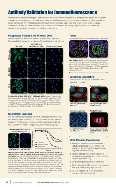
Antibody Validation for Immunofluorescence<br />
Scientists at Cell Signaling Technology (CST) have validated over 800 activation state-specific (e.g., phosphorylation-specific) and total protein<br />
antibodies for immunofluorescence (<strong>IF</strong>) applications, such as manual fluorescence microscopy or automated imaging and laser scanning high<br />
content platforms. All CST antibodies approved for use in immunofluorescent assays have undergone a rigorous validation process.<br />
In addition, our <strong>IF</strong> team has created multiple custom protocols for optimal fixation and staining of specific products; all protocols are<br />
available on our website and are available as links from product pages.<br />
Phosphatase Treatment and Knockout Cells:<br />
Cells are subjected to phosphatase treatment to verify phospho-specificity.<br />
Target specificity is also verified with the use of known knockout or null cell lines.<br />
Tissue:<br />
Antibody performance is assessed on appropriate tissues.<br />
Untreated<br />
LY294002- and<br />
Wortmannin-treated<br />
l Phosphatase-treated<br />
MEF wild-type<br />
MEF GSK3β -/-<br />
Pdx1 Antibody #2437: Confocal <strong>IF</strong> analysis of normal rat pancreas<br />
using #2437 (green, left) or Insulin (C27C9) Rabbit mAb #3014<br />
(green, right). Keratin filaments were labeled with Pan-Keratin (C11)<br />
Mouse mAb (Alexa Fluor ® 647 Conjugate) #4528 (blue pseudocolor).<br />
Red = Propidium Iodide (PI)/RNase Staining Solution #4087<br />
(fluorescent DNA dye).<br />
Subcellular Localization:<br />
Appropriate cell lines and tissues are used to verify<br />
subcellular localization.<br />
PC-3<br />
Mitochondria<br />
Endoplasmic Reticulum<br />
Phospho-GSK-3β (Ser9) (D85E12) XP ® Rabbit mAb #5558: Confocal <strong>IF</strong> analysis of wildtype<br />
mouse embryonic fibroblasts (MEFs) (top row), GSK-3β (-/-) MEFs (middle row), or PC-3<br />
cells (bottom row), untreated (left), LY294002 and Wortmannin-treated (#9901 and #9951<br />
respectively; center) or λ phosphatase-treated (right), using #5558 (green). Blue pseudocolor<br />
= DRAQ5 ® #4084 (fluorescent DNA dye). (MEF wild-type and GSK-3β (-/-) cells were kindly<br />
provided by Dr. Jim Woodgett, University of Toronto, Canada).<br />
COX IV (3E11) Rabbit mAb<br />
#4850 (green)<br />
ERp72 (D70D12) XP ® Rabbit<br />
mAb #5033 (green)<br />
High Content Screening<br />
Our High Content Screening (HCS) group tests <strong>IF</strong>-validated antibodies on in-house<br />
HCS platforms, which include the TTP <strong>Lab</strong>Tech Acumen ® eX 3 and Cellomics ®<br />
ArrayScan ® V TI . This allows us to assess antibody performance on HCS platforms<br />
to provide you with the best possible support for your HCS assays.<br />
Intercellular Junctions<br />
Mitotic Chromatin<br />
Phospho-p44/42 MAPK (Erk1/2)<br />
(Tyr202/Thr204) (green)<br />
-U0126/+TPA<br />
+U0126/+TPA<br />
Normalized Mean Fluorescence Intensity<br />
100<br />
80<br />
60<br />
40<br />
20<br />
Phospho-CREB<br />
Phospho-p44/42 MAPK<br />
Phospho-p90RSK<br />
0 0.1 0.5 1 5 10 50 100<br />
U0126 (µM)<br />
Phospho-p44/42 MAPK (Erk1/2) (Thr202/Tyr204) (D13.14.4E) XP ® Rabbit mAb #4370,<br />
Phospho-p90RSK (Thr573) Antibody #9346, and Phospho-CREB (Ser133) (87G3) Rabbit<br />
mAb #9198: A549 cells were concurrently exposed to U0126 (MEK1/2 Inhibitor) #9903 and<br />
the phorbol ester TPA #4174 to assess the effect on MAPK signaling. Phospho-p44/42 MAPK<br />
(Erk1/2), detected with #4370, and two downstream signaling proteins, phospho-p90RSK and<br />
phospho-CREB, detected with #9346 and #9198, were used as readouts. U0126 was titrated<br />
as an 8-point dose curve in triplicate to monitor the reproducibility and validity of the assay. The<br />
signal for each antibody was analyzed using an Acumen ® eX 3 and images were acquired with a<br />
Cellomics ® ArrayScan ® V TI . With increasing concentrations of U0126, a significant decrease in<br />
phospho-p44/42 MAPK (Erk1/2) signal (~10-fold), as well as phospho-p90RSK and phospho-<br />
CREB (>2-fold), was observed as compared to the TPA-stimulated control.<br />
ZO-3 (D57G7) XP ® Rabbit<br />
mAb #3704 (green)<br />
Phospho-Histone H3 (Ser10)<br />
(D2C8) XP ® Rabbit mAb #3377<br />
(green)<br />
Other Validation Steps Include:<br />
:: Requirement of threshold signal-to-noise ratio in<br />
antibody:isotype comparison and minimum foldinduction<br />
for phospho-specific antibodies ensures<br />
the greatest possible sensitivity.<br />
:: Activation state specificity, target expression, or<br />
translocation are examined using ligands or inhibitors<br />
to modulate pathway activity.<br />
:: Stringent testing ensures lot-to-lot consistency.<br />
:: Cells are subjected to siRNA treatment or overexpression<br />
of the target protein to verify target specificity.<br />
:: Cell lines or tissues with known target expression<br />
levels are used to verify specificity.<br />
4 Application and Reactivity Keys, pg 3. Monoclonal Antibody. Please see page 2 for details.