Bulletin of the Sea Fisheries Institute 2 (156) 2002 - CEEMaR
Bulletin of the Sea Fisheries Institute 2 (156) 2002 - CEEMaR
Bulletin of the Sea Fisheries Institute 2 (156) 2002 - CEEMaR

Create successful ePaper yourself
Turn your PDF publications into a flip-book with our unique Google optimized e-Paper software.
48 K. FORMICKI, A. KORZELECKA-ORKISZ, M. BONIS£AWSKA, A. TAÑSKI, W. WAWRZYNIAK and A. WINNICKI<br />
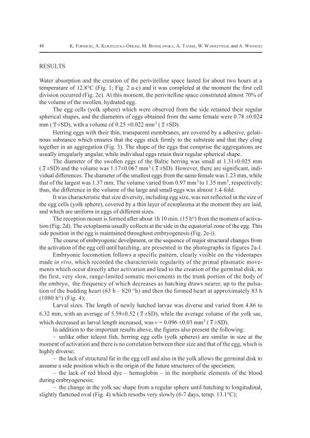
RESULTS<br />
Water absorption and <strong>the</strong> creation <strong>of</strong> <strong>the</strong> perivitelline space lasted for about two hours at a<br />
temperature <strong>of</strong> 12.8°C (Fig. 1; Fig. 2 a-c) and it was completed at <strong>the</strong> moment <strong>the</strong> first cell<br />
division occurred (Fig. 2e). At this moment, <strong>the</strong> perivitelline space constituted almost 70% <strong>of</strong><br />
<strong>the</strong> volume <strong>of</strong> <strong>the</strong> swollen, hydrated egg.<br />
The egg cells (yolk sphere) which were observed from <strong>the</strong> side retained <strong>the</strong>ir regular<br />
spherical shapes, and <strong>the</strong> diameters <strong>of</strong> eggs obtained from <strong>the</strong> same female were 0.78 ±0.024<br />
mm ( x ±SD), with a volume <strong>of</strong> 0.25 ±0.022 mm 3 ( x ±SD).<br />
Herring eggs with <strong>the</strong>ir thin, transparent membranes, are covered by a adhesive, gelatinous<br />
substance which ensures that <strong>the</strong> eggs stick firmly to <strong>the</strong> substrate and that <strong>the</strong>y cling<br />
toge<strong>the</strong>r in an aggregation (Fig. 3). The shape <strong>of</strong> <strong>the</strong> eggs that comprise <strong>the</strong> aggregations are<br />
usually irregularly angular, while individual eggs retain <strong>the</strong>ir regular spherical shape.<br />
The diameter <strong>of</strong> <strong>the</strong> swollen eggs <strong>of</strong> <strong>the</strong> Baltic herring was small at 1.31±0.025 mm<br />
( x ±SD) and <strong>the</strong> volume was 1.17±0.067 mm 3 ( x ±SD). However, <strong>the</strong>re are significant, individual<br />
differences. The diameter <strong>of</strong> <strong>the</strong> smallest eggs from <strong>the</strong> same female was 1.23 mm, while<br />
that <strong>of</strong> <strong>the</strong> largest was 1.37 mm. The volume varied from 0.97 mm 3 to 1.35 mm 3 , respectively;<br />
thus, <strong>the</strong> difference in <strong>the</strong> volume <strong>of</strong> <strong>the</strong> large and small eggs was almost 1.4-fold.<br />
It was characteristic that size diversity, including egg size, was not reflected in <strong>the</strong> size <strong>of</strong><br />
<strong>the</strong> egg cells (yolk sphere), covered by a thin layer <strong>of</strong> ectoplasma at <strong>the</strong> moment <strong>the</strong>y are laid,<br />
and which are uniform in eggs <strong>of</strong> different sizes.<br />
The reception mount is formed after about 1h 10 min. (15 h o ) from <strong>the</strong> moment <strong>of</strong> activation<br />
(Fig. 2d). The ectoplasma usually collects at <strong>the</strong> side in <strong>the</strong> equatorial zone <strong>of</strong> <strong>the</strong> egg. This<br />
side position in <strong>the</strong> egg is maintained throughout embryogenesis (Fig. 2e-i).<br />
The course <strong>of</strong> embryogenic develpment, or <strong>the</strong> sequence <strong>of</strong> major structural changes from<br />
<strong>the</strong> activation <strong>of</strong> <strong>the</strong> egg cell until hatching, are presented in <strong>the</strong> photographs in figures 2a-l.<br />
Embryonic locomotion follows a specific pattern, clearly visible on <strong>the</strong> videotapes<br />
made in vivo, which recorded <strong>the</strong> characteristic regularity <strong>of</strong> <strong>the</strong> primal plasmatic movements<br />
which occur directly after activation and lead to <strong>the</strong> creation <strong>of</strong> <strong>the</strong> germinal disk, to<br />
<strong>the</strong> first, very slow, range-limited somatic movements in <strong>the</strong> trunk portion <strong>of</strong> <strong>the</strong> body <strong>of</strong><br />
<strong>the</strong> embryo, <strong>the</strong> frequency <strong>of</strong> which decreases as hatching draws nearer, up to <strong>the</strong> pulsation<br />
<strong>of</strong> <strong>the</strong> budding heart (63 h – 820 °h) and <strong>the</strong>n <strong>the</strong> formed heart at approximately 83 h<br />
(1080 h°) (Fig. 4);<br />
Larval sizes. The length <strong>of</strong> newly hatched larvae was diverse and varied from 4.86 to<br />
6.32 mm, with an average <strong>of</strong> 5.59±0.52 ( x ±SD), while <strong>the</strong> average volume <strong>of</strong> <strong>the</strong> yolk sac,<br />
which decreased as larval length increased, was v = 0.096 ±0.03 mm 3 ( x ±SD).<br />
In addition to <strong>the</strong> important results above, <strong>the</strong> figures also present <strong>the</strong> following:<br />
– unlike o<strong>the</strong>r teleost fish, herring egg cells (yolk spheres) are similar in size at <strong>the</strong><br />
moment <strong>of</strong> activation and <strong>the</strong>re is no correlation between <strong>the</strong>ir size and that <strong>of</strong> <strong>the</strong> egg, which is<br />
highly diverse;<br />
– <strong>the</strong> lack <strong>of</strong> structural fat in <strong>the</strong> egg cell and also in <strong>the</strong> yolk allows <strong>the</strong> germinal disk to<br />
assume a side position which is <strong>the</strong> origin <strong>of</strong> <strong>the</strong> future structures <strong>of</strong> <strong>the</strong> specimen;<br />
– <strong>the</strong> lack <strong>of</strong> red blood dye – hemoglobin – in <strong>the</strong> morphotic elements <strong>of</strong> <strong>the</strong> blood<br />
during embryogenesis;<br />
– <strong>the</strong> change in <strong>the</strong> yolk sac shape from a regular sphere until hatching to longitudinal,<br />
slightly flattened oval (Fig. 4) which resorbs very slowly (6-7 days, temp. 13.1°C);