doppler evaluation of valvular stenosis #3 - Echo in Context
doppler evaluation of valvular stenosis #3 - Echo in Context
doppler evaluation of valvular stenosis #3 - Echo in Context

You also want an ePaper? Increase the reach of your titles
YUMPU automatically turns print PDFs into web optimized ePapers that Google loves.
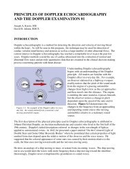
Figure 3. 25 Left panel shows an aortic stenotic jet <strong>in</strong><br />
relation to possible view<strong>in</strong>g directions us<strong>in</strong>g CW<br />
Doppler. Right panel shows spectral velocity trac<strong>in</strong>gs<br />
from each respective w<strong>in</strong>dow. The best record<strong>in</strong>g is<br />
from the right sternal w<strong>in</strong>dow. (Calibration marks =<br />
2m/s)<br />
Figure 3. 26 CW spectral velocity record<strong>in</strong>g from the<br />
suprasternal w<strong>in</strong>dow <strong>in</strong>to the ascend<strong>in</strong>g aorta from a<br />
patient with severe <strong>stenosis</strong>. Note the vary<strong>in</strong>g peak<br />
velocities with vary<strong>in</strong>g R-R <strong>in</strong>terval <strong>of</strong> the ECG.<br />
(Calibration marks = 1 m/s).<br />
In patients with aortic valve disease and<br />
<strong>stenosis</strong>, a careful exam<strong>in</strong>ation must be<br />
performed from all possible views as the<br />
abnormal jet may be directed anywhere <strong>in</strong> the<br />
aorta. In these cases, we are most <strong>in</strong>terested <strong>in</strong><br />
record<strong>in</strong>g the highest peak systolic velocity<br />
present. As previously po<strong>in</strong>ted out, the most<br />
faithful representation <strong>of</strong> flow will be obta<strong>in</strong>ed<br />
when the beam is parallel. The use <strong>of</strong> multiple<br />
positions for the record<strong>in</strong>g <strong>of</strong> peak systolic<br />
aortic velocity is very important <strong>in</strong> aortic<br />
<strong>stenosis</strong> s<strong>in</strong>ce this jet may be directed <strong>in</strong> a wide<br />
variety <strong>of</strong> orientations. This is especially true<br />
<strong>in</strong> older patients with acquired aortic <strong>stenosis</strong>.<br />
Figure 3.25 demonstrates one such direction <strong>of</strong><br />
flow and its relationship to various transducer<br />
positions for CW Doppler record<strong>in</strong>g. In this<br />
case, peak flow was best recorded by CW<br />
Doppler from the right parasternal approach,<br />
rather than from the suprasternal notch. The<br />
velocity pr<strong>of</strong>ile from the apex seems adequate<br />
but is slightly lower than the right sternal<br />
record<strong>in</strong>g. The record<strong>in</strong>g from the suprasternal<br />
notch is grossly <strong>in</strong>adequate and lacks a fully<br />
formed pr<strong>of</strong>ile. When exam<strong>in</strong><strong>in</strong>g for aortic<br />
<strong>stenosis</strong>, all available acoustic w<strong>in</strong>dows should<br />
be utilized.<br />
There will be times when the chang<strong>in</strong>g<br />
appearance <strong>of</strong> the spectral trace is not the result<br />
<strong>of</strong> an improper beam direction or<br />
misadjustment <strong>of</strong> system controls. Figure 3.26<br />
shows a CW record<strong>in</strong>g from the suprasternal<br />
notch with the beam directed toward the<br />
ascend<strong>in</strong>g aorta. The differ<strong>in</strong>g appearances <strong>of</strong><br />
the velocity pr<strong>of</strong>iles are a result <strong>of</strong> an irregular<br />
heart rate which leads to beat-to-beat changes<br />
<strong>in</strong> stroke volume and, consequently, aortic<br />
gradient. Stenotic jets, like regurgitant jets,<br />
readily change their configuration with cardiac<br />
rhythms.<br />
Aortic Valve Gradient at Catheterization<br />
Estimation <strong>of</strong> trans<strong>valvular</strong> aortic gradients <strong>in</strong> patients with aortic <strong>stenosis</strong> us<strong>in</strong>g Doppler has been<br />
<strong>in</strong> common use for some time. There is an abundance <strong>of</strong> papers <strong>in</strong> the literature discuss<strong>in</strong>g the<br />
relative merits and limitations <strong>of</strong> this approach. All are based upon correlations with pressure<br />
measurements obta<strong>in</strong>ed at catheterization.<br />
In Figure 3.27 three possible methods are shown for calculat<strong>in</strong>g pressure gradients across the aortic<br />
valve at catheterization. All depend upon the record<strong>in</strong>g <strong>of</strong> pressure from the left ventricle and<br />
aorta, or some peripheral artery. If a peripheral artery is used, it takes time for the systolic pulse to