doppler evaluation of valvular stenosis #3 - Echo in Context
doppler evaluation of valvular stenosis #3 - Echo in Context
doppler evaluation of valvular stenosis #3 - Echo in Context

You also want an ePaper? Increase the reach of your titles
YUMPU automatically turns print PDFs into web optimized ePapers that Google loves.
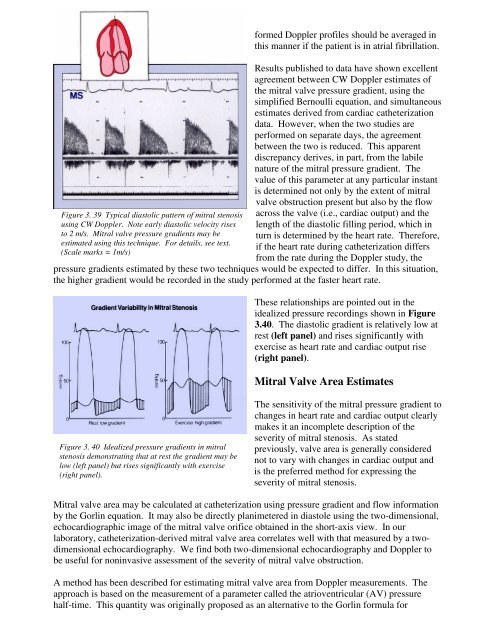
Figure 3. 39 Typical diastolic pattern <strong>of</strong> mitral <strong>stenosis</strong><br />
us<strong>in</strong>g CW Doppler. Note early diastolic velocity rises<br />
to 2 m/s. Mitral valve pressure gradients may be<br />
estimated us<strong>in</strong>g this technique. For details, see text.<br />
(Scale marks = 1m/s)<br />
formed Doppler pr<strong>of</strong>iles should be averaged <strong>in</strong><br />
this manner if the patient is <strong>in</strong> atrial fibrillation.<br />
Results published to data have shown excellent<br />
agreement between CW Doppler estimates <strong>of</strong><br />
the mitral valve pressure gradient, us<strong>in</strong>g the<br />
simplified Bernoulli equation, and simultaneous<br />
estimates derived from cardiac catheterization<br />
data. However, when the two studies are<br />
performed on separate days, the agreement<br />
between the two is reduced. This apparent<br />
discrepancy derives, <strong>in</strong> part, from the labile<br />
nature <strong>of</strong> the mitral pressure gradient. The<br />
value <strong>of</strong> this parameter at any particular <strong>in</strong>stant<br />
is determ<strong>in</strong>ed not only by the extent <strong>of</strong> mitral<br />
valve obstruction present but also by the flow<br />
across the valve (i.e., cardiac output) and the<br />
length <strong>of</strong> the diastolic fill<strong>in</strong>g period, which <strong>in</strong><br />
turn is determ<strong>in</strong>ed by the heart rate. Therefore,<br />
if the heart rate dur<strong>in</strong>g catheterization differs<br />
from the rate dur<strong>in</strong>g the Doppler study, the<br />
pressure gradients estimated by these two techniques would be expected to differ. In this situation,<br />
the higher gradient would be recorded <strong>in</strong> the study performed at the faster heart rate.<br />
These relationships are po<strong>in</strong>ted out <strong>in</strong> the<br />
idealized pressure record<strong>in</strong>gs shown <strong>in</strong> Figure<br />
3.40. The diastolic gradient is relatively low at<br />
rest (left panel) and rises significantly with<br />
exercise as heart rate and cardiac output rise<br />
(right panel).<br />
Mitral Valve Area Estimates<br />
Figure 3. 40 Idealized pressure gradients <strong>in</strong> mitral<br />
<strong>stenosis</strong> demonstrat<strong>in</strong>g that at rest the gradient may be<br />
low (left panel) but rises significantly with exercise<br />
(right panel).<br />
The sensitivity <strong>of</strong> the mitral pressure gradient to<br />
changes <strong>in</strong> heart rate and cardiac output clearly<br />
makes it an <strong>in</strong>complete description <strong>of</strong> the<br />
severity <strong>of</strong> mitral <strong>stenosis</strong>. As stated<br />
previously, valve area is generally considered<br />
not to vary with changes <strong>in</strong> cardiac output and<br />
is the preferred method for express<strong>in</strong>g the<br />
severity <strong>of</strong> mitral <strong>stenosis</strong>.<br />
Mitral valve area may be calculated at catheterization us<strong>in</strong>g pressure gradient and flow <strong>in</strong>formation<br />
by the Gorl<strong>in</strong> equation. It may also be directly planimetered <strong>in</strong> diastole us<strong>in</strong>g the two-dimensional,<br />
echocardiographic image <strong>of</strong> the mitral valve orifice obta<strong>in</strong>ed <strong>in</strong> the short-axis view. In our<br />
laboratory, catheterization-derived mitral valve area correlates well with that measured by a twodimensional<br />
echocardiography. We f<strong>in</strong>d both two-dimensional echocardiography and Doppler to<br />
be useful for non<strong>in</strong>vasive assessment <strong>of</strong> the severity <strong>of</strong> mitral valve obstruction.<br />
A method has been described for estimat<strong>in</strong>g mitral valve area from Doppler measurements. The<br />
approach is based on the measurement <strong>of</strong> a parameter called the atrioventricular (AV) pressure<br />
half-time. This quantity was orig<strong>in</strong>ally proposed as an alternative to the Gorl<strong>in</strong> formula for