Lab 15: Population Genetics - eScience Labs
Lab 15: Population Genetics - eScience Labs
Lab 15: Population Genetics - eScience Labs

Create successful ePaper yourself
Turn your PDF publications into a flip-book with our unique Google optimized e-Paper software.
<strong>Lab</strong> 11: Mitosis<br />
G 2 : the second growth phase in which cellular organelles (mitochondria, ribosomes, and centrioles)<br />
are replicated.<br />
M: mitotic division (prophase, metaphase, anaphase and telophase)<br />
A cell normally completes the cycle in 18‐24 hours, with mitosis occupying 1‐2<br />
hours of that time (Figure 2). Each stage is regulated by specialized proteins<br />
that coordinate the division and cell growth. Certain types of cancer are associated<br />
with the failure of these proteins.<br />
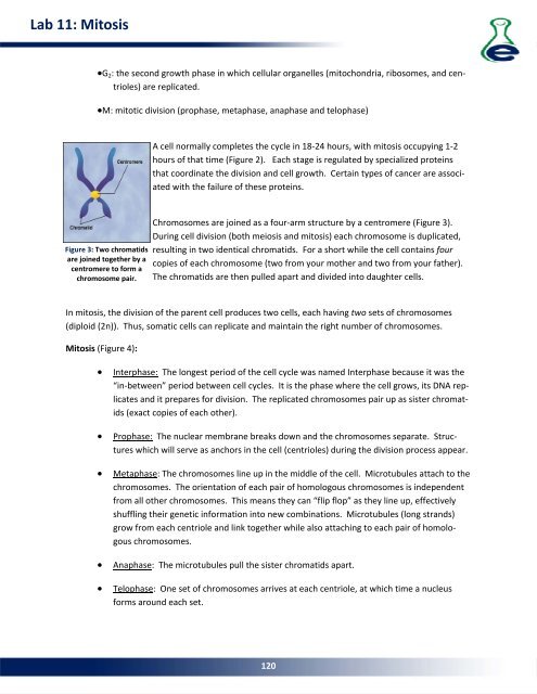
Figure 3: Two chromatids<br />
are joined together by a<br />
centromere to form a<br />
chromosome pair.<br />
Chromosomes are joined as a four‐arm structure by a centromere (Figure 3).<br />
During cell division (both meiosis and mitosis) each chromosome is duplicated,<br />
resulting in two identical chromatids. For a short while the cell contains four<br />
copies of each chromosome (two from your mother and two from your father).<br />
The chromatids are then pulled apart and divided into daughter cells.<br />
In mitosis, the division of the parent cell produces two cells, each having two sets of chromosomes<br />
(diploid (2n)). Thus, somatic cells can replicate and maintain the right number of chromosomes.<br />
Mitosis (Figure 4):<br />
<br />
<br />
<br />
<br />
<br />
Interphase: The longest period of the cell cycle was named Interphase because it was the<br />
“in‐between” period between cell cycles. It is the phase where the cell grows, its DNA replicates<br />
and it prepares for division. The replicated chromosomes pair up as sister chromatids<br />
(exact copies of each other).<br />
Prophase: The nuclear membrane breaks down and the chromosomes separate. Structures<br />
which will serve as anchors in the cell (centrioles) during the division process appear.<br />
Metaphase: The chromosomes line up in the middle of the cell. Microtubules attach to the<br />
chromosomes. The orientation of each pair of homologous chromosomes is independent<br />
from all other chromosomes. This means they can “flip flop” as they line up, effectively<br />
shuffling their genetic information into new combinations. Microtubules (long strands)<br />
grow from each centriole and link together while also attaching to each pair of homologous<br />
chromosomes.<br />
Anaphase: The microtubules pull the sister chromatids apart.<br />
Telophase: One set of chromosomes arrives at each centriole, at which time a nucleus<br />
forms around each set.<br />
120