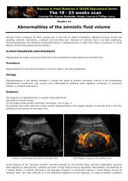
CHAPTER 4: SCREENING FOR CERVICAL CANCER
CHAPTER 4: SCREENING FOR CERVICAL CANCER
CHAPTER 4: SCREENING FOR CERVICAL CANCER

Create successful ePaper yourself
Turn your PDF publications into a flip-book with our unique Google optimized e-Paper software.
156 PS 13: Loop Electrosurgical Excision Procedure (LEEP)PS 13Practice Sheet 13: Loop Electrosurgical Excision Procedure (LEEP)PER<strong>FOR</strong>MING LEEPBefore the procedure1. Explain the procedure and why it is important to return for further management asrequested. Ensure that the woman has understood and obtain informed consent.2. Prepare the patient for a gynaecological examination.3. Attach a return electrode to the inner thigh.4. Insert a non-conducting speculum with an electrically insulating coating, or aspeculum covered with a latex condom.5. Look at the cervix, and note any abnormalities, such as discharge from the os,inflammation, bleeding or lesions. Record the findings.6. If there is no evidence of infection, proceed. If you note signs of infection, suspendthe procedure and treat the patient and her partner completely before making asecond attempt.During LEEP 187. Before each step, tell the woman what you will do and what she may feel.8. Wipe the cervix with a saline-soaked cotton swab.9. Apply 5% acetic acid and examine with the colposcope to determine the locationand extent of the lesion.10. Inject 3–5 ml of local anaesthetic (1% or 2% lidocaine with 1:100 0000epinephrine (to control bleeding)), using a long 27-gauge needle, just beneaththe cervical epithelium at the 12 o’clock, 3 o’clock, 6 o’clock and 9 o’clockpositions (in patients with cardiac problems, use lidocaine without epinephrine).11. Select the appropriate electrode to remove the entire abnormal area in a singlepass: for small low-grade lesions in nulliparous women, use an electrode 1.5 cmwide by 0.5 cm deep; for larger lesions and multiparous women, use one 2.0 cmwide by 0.8 cm deep.12. Turn the vacuum suction on and activate the generator.13. Excise the lesion: push the electrode perpendicularly into the tissue to a depthof 4–5 mm and draw it laterally across the cervix to the other side, producinga dome-shaped circle of tissue with the canal in the centre. Do not insert theelectrode deeper than 5 mm at the 3 o’clock and 9 o’clock positions, becausethis can damage the uterine arteries.18 In some cases, the patient may have a vasovagal reaction, with fainting and plummetingblood pressure. If this happens, stop the treatment immediately and raise the patient’s legs asmuch as possible.