BEILHARZ ET AL.unable to find <strong>an</strong>y combination of syn<strong>an</strong>amorphs thatwould equate with the cercosporoid fungus infectingphyllodes of A. crassicarpa <strong>an</strong>d described here. Thecorrect or most logical me<strong>an</strong>s of treating the nomenclatureof pleo<strong>an</strong>amorphic fungi remains somewhatsubjective, <strong>an</strong>d has not been defined in the Code ofBot<strong>an</strong>ical Nomenclature. The well-known genericname <strong>Passalora</strong> Fr. sensu Crous & Braun (2003) iscorrect for the hyphomycetous syn<strong>an</strong>amorph, <strong>an</strong>d it isparticularly useful in this case because most coloniesof the fungus show liberal <strong>an</strong>d conspicuous sporulationof this morph, whereas the cryptic Type 2 syn<strong>an</strong>amorphis less likely to be observed, <strong>an</strong>d isunlikely to be found in isolation. Gams (1982) suggestedthat the <strong>an</strong>amorph form with the greatest differentiationshould have priority (unless it is rare), aview which further supports the application of thename <strong>Passalora</strong> to the fungus on A. crassicarpa.The conidia of the Type 1 <strong>an</strong>d Type 2 syn<strong>an</strong>amorphsof the cercosporoid fungus from A. crassicarpa,although easily distinguished, show somesimilarity in morphology. The Type 2 conidia aresomewhat cercosporoid in type <strong>an</strong>d reminiscent ofsome species of Colletogloeum Petr., but the conidiomatado not fit with the acervular conidiomata of thatgenus. Critical differences between the two types ofconidia, including pigmentation, c<strong>an</strong> be linked to theirrelative positions in relation to the host tissue. Forexample, the thickened hila of Type 1 conidia areprobably associated with their readiness to secede(Beilharz 1994). In contrast, the broader, unthickenedhila of Type 2 conidia are more appropriate to passiverelease following breakdown of overlying fungal <strong>an</strong>dhost tissues.Acacia crassicarpa is a species that has becomeincreasingly import<strong>an</strong>t in pl<strong>an</strong>tations in various partsof South-East Asia, where it is grown specifically forthe production of pulp. The relatively recent outbreakof a serious <strong>leaf</strong> <strong>blight</strong> disease caused by a cercosporoidfungus has dem<strong>an</strong>ded <strong>an</strong> appropriate taxonomictreatment of this org<strong>an</strong>ism. This study represents acollaborative effort by a number of research groupswho have <strong>an</strong> interest in this fungus <strong>an</strong>d the diseasewith which it is associated. Here, we describe a potentiallydevastating, newly recognised disease of A.crassicarpa, <strong>an</strong>d describe the causal org<strong>an</strong>ism, a novelpleo<strong>an</strong>amorphic species of <strong>Passalora</strong>.MATERIALS AND METHODSIsolatesAt VPRI, cultures were derived from the Australi<strong>an</strong>specimen VPRI 21125 by the following me<strong>an</strong>s. Naturally-producedType 1 conidia were lifted from lesionsen masse with a fine needle <strong>an</strong>d jab-inoculated on to 2% potato-dextrose agar (PDA; Difco) plates emendedwith achromycin (0.05 mg/mL) (PDA+A). Similarly472harvested Type 1 conidia were also suspended in adrop of water containing a trace of Tween 80, streakedout on to 2 % tap water agar <strong>an</strong>d tr<strong>an</strong>sferred individuallyto PDA+A the following day, after germinating.Type 2 conidia formed in vitro in PDA cultures derivedfrom individual Type 1 conidia were used toprovide single-conidial isolates as described above. Inaddition, whole, pale, smooth protuber<strong>an</strong>t stromatalacking external conidiophores <strong>an</strong>d putatively containingType 2 conidia, were lifted from the phyllodesurface with a fine, sterile needle <strong>an</strong>d placed directlyonto PDA+A. All PDA+A cultures were tr<strong>an</strong>sferred toPDA after 3–7 d <strong>an</strong>d grown on for up to 2 mo in thedark at 22 °C. The choice of the various forms ofinoculum was determined by the ease with which theycould be harvested from infected phyllodes or cultures.For example, Type 1 conidia were abund<strong>an</strong>t onthe natural subtrate, but occurred in comparativelysmall numbers in culture, where they were liable to becontaminated with Type 2 conidia. Type 2 conidiawere abund<strong>an</strong>t in wet masses in culture; in nature,however, they tended to remain aggregated <strong>an</strong>d oftencould not be completely freed from excised conidiomata,despite the application of pressure on coverslipsor attempts to tease the elements apart in a drop ofwater on a microscope slide. Type 3 conidia were notseen in 2-mo-old cultures on PDA, <strong>an</strong>d Type 2 conidiafrom these cultures germinated normally on fresh agarplates. The Australi<strong>an</strong> specimens <strong>an</strong>d a dried cultureof VPRI 21125 have been deposited in herb. VPRI,Knoxfield, Victoria, Australia.At <strong>CBS</strong>, single Type 1 conidial isolates werederived from Indonesi<strong>an</strong> specimens <strong>an</strong>d cultivated on2 % malt extract agar (MEA; Difco) as described byCrous (1998). Colonies sporulated on MEA after 1–2mo incubation on the laboratory bench in daylight atroom temperature, forming conidiomata containingType 1 <strong>an</strong>d Type 2 conidia. After 3 mo incubation onMEA, conidiomata with Type 2 conidia were observedto also give rise to Type 3 conidia, a formobserved only in culture. Specimens <strong>an</strong>d cultures havebeen deposited in the herbarium <strong>an</strong>d culture collectionof <strong>CBS</strong> in Utrecht, the Netherl<strong>an</strong>ds.A phylogeny of the cercosporoid fungi occurringon Acacia, including <strong>Passalora</strong> <strong>perplexa</strong>, is presentedelsewhere in this volume (Crous et al. 2004).MorphologySlide preparations were made in lactic acid <strong>an</strong>d 50examples of each structure were measured under a×100 oil immersion lens using Olympus BH–2 (VPRI)or Zeiss Axioskop (<strong>CBS</strong>) light microscopes. The 95 %confidence intervals were also determined for conidialdimensions, with the extremes in conidium length <strong>an</strong>dwidth given in parentheses. Colony colour was determinedon 2 % MEA after 3 mo at 25 °C in the darkusing the colour designations of Rayner (1970).
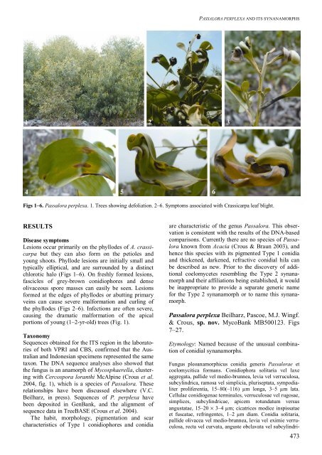
PASSALORA PERPLEXA AND ITS SYNANAMORPHSFigs 1–6. <strong>Passalora</strong> <strong>perplexa</strong>. 1. Trees showing defoliation. 2–6. Symptoms associated with Crassicarpa <strong>leaf</strong> <strong>blight</strong>.RESULTSDisease symptomsLesions occur primarily on the phyllodes of A. crassicarpabut they c<strong>an</strong> also form on the petioles <strong>an</strong>dyoung shoots. Phyllode lesions are initially small <strong>an</strong>dtypically elliptical, <strong>an</strong>d are surrounded by a distinctchlorotic halo (Figs 1–6). On freshly formed lesions,fascicles of grey-brown conidiophores <strong>an</strong>d denseolivaceous spore masses c<strong>an</strong> easily be seen. Lesionsformed at the edges of phyllodes or abutting primaryveins c<strong>an</strong> cause severe malformation <strong>an</strong>d curling ofthe phyllodes (Figs 2–6). Infections are often severe,causing the dramatic malformation of the apicalportions of young (1–2-yr-old) trees (Fig. 1).TaxonomySequences obtained for the ITS region in the laboratoriesof both VPRI <strong>an</strong>d <strong>CBS</strong>, confirmed that the Australi<strong>an</strong><strong>an</strong>d Indonesi<strong>an</strong> specimens represented the sametaxon. The DNA sequence <strong>an</strong>alyses also showed thatthe fungus is <strong>an</strong> <strong>an</strong>amorph of Mycosphaerella, clusteringwith Cercospora lor<strong>an</strong>thi McAlpine (Crous et al.2004, fig. 1), which is a species of <strong>Passalora</strong>. Theserelationships have been discussed elsewhere (V.C.Beilharz, in press). Sequences of P. <strong>perplexa</strong> havebeen deposited in GenB<strong>an</strong>k, <strong>an</strong>d the alignment ofsequence data in TreeBASE (Crous et al. 2004).The habit, morphology, pigmentation <strong>an</strong>d scarcharacteristics of Type 1 conidiophores <strong>an</strong>d conidiaare characteristic of the genus <strong>Passalora</strong>. This observationis consistent with the results of the DNA-basedcomparisons. Currently there are no species of <strong>Passalora</strong>known from Acacia (Crous & Braun 2003), <strong>an</strong>dhence this species with its pigmented Type 1 conidia<strong>an</strong>d thickened, darkened, refractive conidial hila c<strong>an</strong>be described as new. Prior to the discovery of additionalcoelomycetes resembling the Type 2 syn<strong>an</strong>amorph<strong>an</strong>d their affiliations being established, it wouldbe inappropriate to provide a separate generic namefor the Type 2 syn<strong>an</strong>amorph or to name this syn<strong>an</strong>amorph.<strong>Passalora</strong> <strong>perplexa</strong> Beilharz, Pascoe, M.J. Wingf.& Crous, sp. nov. MycoB<strong>an</strong>k MB500123. Figs7–27.Etymology: Named because of the unusual combinationof conidial syn<strong>an</strong>amorphs.Fungus pleo<strong>an</strong>amorphicus conidia generis <strong>Passalora</strong>e etcoelomycitica form<strong>an</strong>s. Conidiophora solitaria vel laxeaggregata, pallide vel medio-brunnea, levia vel verruculosa,subcylindrica, ramosa vel simplicia, pluriseptata, sympodialiterproliferentia, 15–80(–116) µm longa, 3–5 µm lata.Cellulae conidiogenae terminales, verruculosae vel rugosae,simplices, subcylindricae, apicem rotundatum versus<strong>an</strong>gustatae, 15–20 × 3–4 µm; cicatrices modice inspissataeet fuscatae, refringentes, 1–2 µm diam. Conidia solitaria,pallide olivacea vel medio-brunnea, levia vel eximie verruculosa,recta vel curvata, <strong>an</strong>guste obclavata vel subcylindri-473