

Passalora perplexa, an important pleoanamorphic leaf blight ... - CBS
Passalora perplexa, an important pleoanamorphic leaf blight ... - CBS
Passalora perplexa, an important pleoanamorphic leaf blight ... - CBS

Create successful ePaper yourself
Turn your PDF publications into a flip-book with our unique Google optimized e-Paper software.
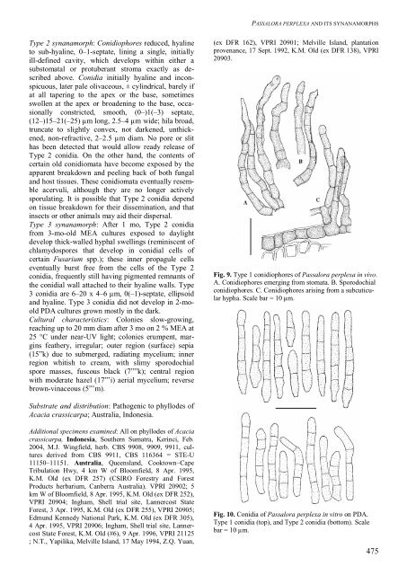
PASSALORA PERPLEXA AND ITS SYNANAMORPHSType 2 syn<strong>an</strong>amorph: Conidiophores reduced, hyalineto sub-hyaline, 0–1-septate, lining a single, initiallyill-defined cavity, which develops within either asubstomatal or protuber<strong>an</strong>t stroma exactly as describedabove. Conidia initially hyaline <strong>an</strong>d inconspicuous,later pale olivaceous, ± cylindrical, barely ifat all tapering to the apex or the base, sometimesswollen at the apex or broadening to the base, occasionallyconstricted, smooth, (0–)1(–3) septate,(12–)15–21(–25) µm long, 2.5–4 µm wide; hila broad,truncate to slightly convex, not darkened, unthickened,non-refractive, 2–2.5 µm diam. No pore or slithas been detected that would allow ready release ofType 2 conidia. On the other h<strong>an</strong>d, the contents ofcertain old conidiomata have become exposed by theapparent breakdown <strong>an</strong>d peeling back of both fungal<strong>an</strong>d host tissues. These conidiomata eventually resembleacervuli, although they are no longer activelysporulating. It is possible that Type 2 conidia dependon tissue breakdown for their dissemination, <strong>an</strong>d thatinsects or other <strong>an</strong>imals may aid their dispersal.Type 3 syn<strong>an</strong>amorph: After 1 mo, Type 2 conidiafrom 3-mo-old MEA cultures exposed to daylightdevelop thick-walled hyphal swellings (reminiscent ofchlamydospores that develop in conidial cells ofcertain Fusarium spp.); these inner propagule cellseventually burst free from the cells of the Type 2conidia, frequently still having pigmented remn<strong>an</strong>ts ofthe conidial wall attached to their hyaline walls. Type3 conidia are 6–20 x 4–6 µm, 0(–1)-septate, ellipsoid<strong>an</strong>d hyaline. Type 3 conidia did not develop in 2-mooldPDA cultures grown mostly in the dark.Cultural characteristics: Colonies slow-growing,reaching up to 20 mm diam after 3 mo on 2 % MEA at25 °C under near-UV light; colonies erumpent, marginsfeathery, irregular; outer region (surface) sepia(15”k) due to submerged, radiating mycelium; innerregion whitish to cream, with slimy sporodochialspore masses, fuscous black (7””k); central regionwith moderate hazel (17”’i) aerial mycelium; reversebrown-vinaceous (5”’m).(ex DFR 162), VPRI 20901; Melville Isl<strong>an</strong>d, pl<strong>an</strong>tationproven<strong>an</strong>ce, 17 Sept. 1992, K.M. Old (ex DFR 138), VPRI20903.Fig. 9. Type 1 conidiophores of <strong>Passalora</strong> <strong>perplexa</strong> in vivo.A. Conidiophores emerging from stomata. B. Sporodochialconidiophores. C. Conidiophores arising from a subcuticularhypha. Scale bar = 10 µm.Substrate <strong>an</strong>d distribution: Pathogenic to phyllodes ofAcacia crassicarpa; Australia, Indonesia.Additional specimens examined: All on phyllodes of Acaciacrassicarpa, Indonesia, Southern Sumatra, Kerinci, Feb.2004, M.J. Wingfield, herb. <strong>CBS</strong> 9908, 9909, 9911, culturesderived from <strong>CBS</strong> 9911, <strong>CBS</strong> 116364 = STE-U11150–11151. Australia, Queensl<strong>an</strong>d, Cooktown–CapeTribulation Hwy, 4 km W of Bloomfield, 8 Apr. 1995,K.M. Old (ex DFR 257) (CSIRO Forestry <strong>an</strong>d ForestProducts herbarium, C<strong>an</strong>berra Australia), VPRI 20902; 5km W of Bloomfield, 8 Apr. 1995, K.M. Old (ex DFR 252),VPRI 20904; Ingham, Shell trial site, L<strong>an</strong>nercost StateForest, 3 Apr. 1995, K.M. Old (ex DFR 255), VPRI 20905;Edmund Kennedy National Park, K.M. Old (ex DFR 305),4 Apr. 1995, VPRI 20906; Ingham, Shell trial site, L<strong>an</strong>nercostState Forest, K.M. Old (#6), 9 Apr. 1996, VPRI 21125; N.T., Yapilika, Melville Isl<strong>an</strong>d, 17 May 1994, Z.Q. Yu<strong>an</strong>,Fig. 10. Conidia of <strong>Passalora</strong> <strong>perplexa</strong> in vitro on PDA.Type 1 conidia (top), <strong>an</strong>d Type 2 conidia (bottom). Scalebar = 10 µm.475


