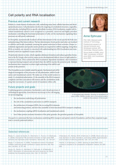
Developmental Biology UnitCell polarity and RNA localisationPrevious and current researchPolarity is a main feature of eukaryotic cells, underlying many basic cellular functions and developmentalprocesses. Cell polarisation involves the targeting of cytoskeletal structures, organelles,and molecules, including RNAs to specific subcellular locations. RNA localisation coupled with localisedtranslational control is now recognised as a powerful, conserved and highly prevalentmechanism controlling the functional polarisation of cells, yet the mechanisms regulating theseprocesses are still poorly understood.In Drosophila, asymmetrically localised cell fate determinants in the oocyte specify the body axesand patterning of the future embryo. The key determinants, bicoid, gurken and oskar, are localisedas mRNAs and locally translated, ensuring the spatial restriction of their activities. Proper cytoskeletalorganisation and specific motor proteins are required for mRNA targeting. Using theseRNAs as models, our research is concerned with understanding how RNA localisation and translationalcontrol are regulated in space and time.Of particular interest is oskar, which regulates abdomen formation and induces germline formationin the fly. Ectopic oskar activity causes severe developmental defects, hence its tight spatial restrictionis critical. This is achieved by RNA localisation-dependent translation: oskar translationis repressed during transport and activated when the mRNA reaches the posterior pole. Multiplemechanisms then cooperate to achieve tight anchoring of the mRNA andprotein at the posterior.Anne EphrussiPhD 1985, MassachusettsInstitute of Technology.Postdoctoral research atHarvard University andWhitehead Institute, MIT,Cambridge, Massachusetts.Group leader at <strong>EMBL</strong> since1992. Coordinator of EICATsince 2005; Unit Coordinatorsince 2007.The Drosophila oocyte is ideally suited for genetic, biochemical and cell biologicalinvestigation of the processes of cell polarisation, mRNA localisationand translational control. We make use of this model system tostudy (1) cytoskeletal polarisation, (2) the assembly of the RNA transportcomplexes and their association with motors and the cytoskeleton mediatingtheir movement, (3) spatial control of translation within cells.Future projects and goalsCombining genetics, proteomics, biochemistry, and a broad spectrum ofcell biological approaches, from electron microscopy to live cell imaging,we are investigating:• the mechanisms underlying cell polarisation;A Drosophila egg-chamber, showing co-localisation ofoskar mRNA, Staufen protein and a microtubule polaritymarker at the posterior of the oocyte.• the role of the cytoskeleton and motors in mRNA transport;• the architecture of transport RNPs: the cis-acting RNA elementsand interacting proteins, and how they assemble to form functional RNA transport complexes;• the mechanisms coupling mRNA localisation and translational control;• how Oskar protein nucleates formation of the polar granules, the germline granules of Drosophila.Our goal is to understand the basic mechanisms underlying RNA transport and spatial control of translation, and how they cooperate to generatea correctly patterned embryo.Selected referencesBesse, F., Lopez de Quinto, S., Marchand, V., Trucco, A. & Ephrussi,A. (2009). Drosophila PTB promotes formation of high-order RNPparticles and represses oskar translation. Genes Dev., 23, 195-207Besse, F. & Ephrussi, A. (2008). Translational control of localizedmRNAs: restricting protein synthesis in space and time. Nat. Rev.Mol. Cell Biol., 9, 971-80Vanzo, N., Oprins, A., Xanthakis, D., Ephrussi, A. & Rabouille, C.(2007). Stimulation of endocytosis and actin dynamics by Oskarpolarizes the Drosophila oocyte. Dev. Cell, 12, 53-555.Chekulaeva, M., Hentze, M.W. & Ephrussi, A. (2006). Bruno acts as adual repressor of oskar translation, promoting mRNA oligomerizationand formation of silencing particles. Cell, 12, 521–533.Hachet, O. & Ephrussi, A. (200). Splicing of oskar RNA in thenucleus is coupled to its cytoplasmic localization. Nature, 28, 959-96323
<strong>EMBL</strong> Research at a Glance 2009Detlev ArendtPhD 1999, Albert-Ludwigs-Universität, Freiburg.Postdoctoral research at<strong>EMBL</strong>.Team leader at <strong>EMBL</strong> since2002. Group leader andSenior Scientist since 2007.Academic Mentor, postdoctoraltraining since 2007.Evolution of the central nervous system in BilateriaPrevious and current researchWe are intrigued by one of the remaining great mysteries in animal evolution: how did our centralnervous system (CNS) come into existence? What did it look like at first and how did it function?We are especially interested in the CNS of an extinct animal known as Urbilateria, the lastcommon ancestor of humans, flies and most other ‘higher’ animals that live today, which livedsome 600 million years ago in the ocean.We have therefore chosen to work on a ‘living fossil’, the marine annelid Platynereis dumerilii, thatwe keep in laboratory culture. This species exhibits many ancient features in its lifestyle, anatomyand development. In bioinformatics comparisons we found that Platynereis also shows an ancestralgene inventory and gene structure.We combine morphological and molecular approaches in a novel evo-devo approach, the molecularcomparison of cell types. Animal nervous systems are made up of different sorts of sensoryneurons, motor- and interneurons. Each type displays a characteristic ‘molecular fingerprint’, aunique combination of specifying transcription factors and downstream effector genes such asreceptors, transmitters or neuropeptides. The comparison of molecular fingerprints allows thetracing of cell types through animal evolution. For example, in the Platynereis brain we have characteriseda special type of photoreceptor cell, a ‘ciliary photoreceptor’ that by molecular fingerprintcomparison relates to the rods and cones, the visual photoreceptors of the vertebrate retina. This has led to the fascinating hypothesis that thevertebrate eye evolved from within the Urbilaterian brain.Besides ciliary photoreceptors, the Platynereis brain harbours several neuron types that have a dual function: they are both sensory and neurosecretory.The ongoing molecular characterisation of these cell types again revealed striking parallels to vertebrate cell types, mostly situatedin the hypothalamus. Finally, we have also characterised themolecular architecture of the Platynereis trunk central nervous systemand discovered striking parallels to the molecular architecture of the vertebrateneural tube. Basically, it appears that the vertebrate neural tubehas evolved by the infolding of a pre-existing central nervous system thatwas in place already in the bilaterian ancestors.Finally, we have also established neurobiological assay systems for larvalswimming and for adult learning, combined with computer modelling ofthese and of other complex behavioural traits, in order to investigate thefunctions of conserved cell type and to gain insight into the neurobiologyof marine planktonic life.Future projects and goalsIt is now clear that our molecular fingerprint comparisons between annelid,vertebrate and insect have the potential to unravel the origin of thePlatynereis dumerilii (Polychaeta, Annelida, Lophotrochozoa).bilaterian central nervous system. We are excited by the prospect of furtherdeciphering the evolution of photoreceptor cells and of the diverse eye types that exist in animals. Also, we want to know the evolutionaryorigin of the most advanced brain part that ever evolved, the telencephalon. We have discovered neurons in Platynereis related totelencephalic neuron types by molecular fingerprint, and started to investigate them further.The clear picture is emerging that the Platynereis brain harbours many cell types so far known only for the vertebrates, but in a much moresimple, very different overall arrangement. This makes it an attractive goal to elucidate the functioning of these cell types in the ancient marineenvironment in order to gain insight into the evolutionary origins of the vertebrate brain.Selected referencesJekely, G., Colombelli, J., Hausen, H., Guy, K., Stelzer, E., Nédélec,F. & Arendt, D. (2008). Mechanism of phototaxis in marinezooplankton. Nature, 56, 395-399Arendt, D. (2008). The evolution of cell types in animals: emergingprinciples from molecular studies. Nat. Rev. Genet. 9, 868-822Denes, A.S., Jekely, G., Steinmetz, P.R., Raible, F., Snyman, H.,Prud’homme, B., Ferrier, D.E., Balavoine, G. & Arendt, D. (2007).Molecular architecture of annelid nerve cord supports commonorigin of nervous system centralization in bilateria. Cell, 129, 277-288Tessmar-Raible, K., Raible, F., Christodoulou, F., Guy, K., Rembold,M., Hausen, H. & Arendt, D. (2007). Conserved sensoryneurosecretorycell types in annelid and fish forebrain: insights intohypothalamus evolution. Cell, 129, 1389-100