ayout 1 - EMBL Grenoble
ayout 1 - EMBL Grenoble
ayout 1 - EMBL Grenoble

You also want an ePaper? Increase the reach of your titles
YUMPU automatically turns print PDFs into web optimized ePapers that Google loves.
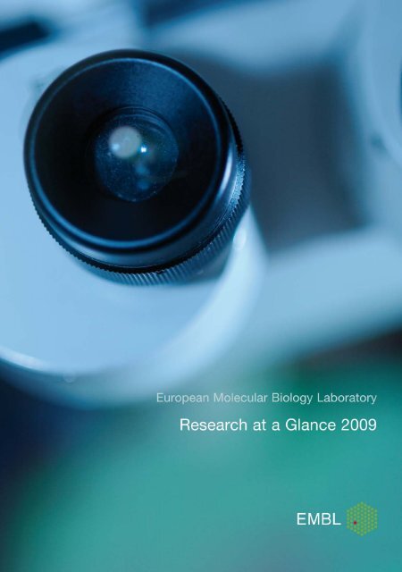
Contents4 Foreword by <strong>EMBL</strong>’s Director General5 <strong>EMBL</strong> Heidelberg, Germany7 Cell Biology and Biophysics Unit22 Developmental Biology Unit31 Gene Expression Unit41 Structural and Computational Biology Unit52 Directors’ Research55 Core Facilities63 <strong>EMBL</strong>-EBI, Hinxton, UKEuropean Bioinformatics Institute87 <strong>EMBL</strong> <strong>Grenoble</strong>, FranceStructural Biology97 <strong>EMBL</strong> Hamburg, GermanyStructural Biology108 <strong>EMBL</strong> Monterotondo, ItalyMouse Biology118 Index of group leaders
<strong>EMBL</strong> Research at a Glance 2009Foreword by <strong>EMBL</strong>’s Director General<strong>EMBL</strong> – Europe’s flagship laboratory for basicresearch in molecular biologyThe vision of the nations which founded the European Molecular Biology Laboratory was to create acentre of excellence where Europe’s best brains would come together to conduct basic research inmolecular biology. During the past three decades, <strong>EMBL</strong> has grown and developed substantially,and its member states now number twenty-one, including the first associate member state, Australia.Over the years, <strong>EMBL</strong> has become the flagship of European molecular biology and is ranked as oneof the top research institutes worldwide.<strong>EMBL</strong>’s missions are to perform cutting-edge research in molecular biology, to offer services to Europeanscientists, to provide advanced training to researchers at all levels, to develop new technologiesand instrumentation and to actively engage in technology transfer for the benefit of scientistsand society.In research, the five <strong>EMBL</strong> sites (a central laboratory in Heidelberg, with outstations in <strong>Grenoble</strong>,Hamburg, Hinxton and Monterotondo) put strong emphasis on interdisciplinarity and collaboration,and when the researchers leave to assume key positions in the member states, they export theirunique experience of working in a very energetic and international environment. Freedom, flexibilityand a regular turnover of staff allows <strong>EMBL</strong> to pursue the most exciting themes in molecular biologyas they arise. Our long-standing tradition of organising excellent courses, conferences andworkshops and an extensive outreach programme ensure that know-how spreads further and informsthe public about the impact modern biology has on our lives.In Research at a Glance you will find a concise overview of the work of our research groups and corefacilities. Science at <strong>EMBL</strong> covers themes ranging from studies of single molecules to an understandingof how they work together in complex systems to organise cells and organisms. Our researchis loosely structured under thematic units, giving scientists the intellectual freedom to pursuethe topics that most interest them.But what really distinguishes <strong>EMBL</strong> is the large number of inter-unit collaborations, bringing peoplewith common interests but distinct expertise together to tackle ambitious projects. Cross-unit networkingand training further support scientists working on interdisciplinary projects. Increasingly,our young scientists come with physics, chemistry, mathematics and computer science backgrounds,bringing in expertise that helps us to move into the growing field of systems biology.<strong>EMBL</strong> combines a critical mass of expertise and resources with organisational flexibility, enabling usto keep pace with today’s biology. The impact of the laboratory’s scientific work, the quality of itsservices and its continued attractiveness to world-leading young scientists are testimony to <strong>EMBL</strong>’ssuccess, and show that we are well-equipped for the future.Iain Mattaj<strong>EMBL</strong> Director General
<strong>EMBL</strong> Heidelberg, GermanyA city of about 140,000 inhabitants, Heidelberg is home to Germany’s oldest university, as well as leadingbiological and medical science institutes such as the Centre for Molecular Biology, the German CancerResearch Center (DKFZ) and the Max Planck Institute for Medical Research, making it an ideal site for<strong>EMBL</strong>’s main laboratory.Nestling in the wooded hills above the city, the complex is home to five of <strong>EMBL</strong>’s scientific units: GeneExpression, Cell Biology and Biophysics, Developmental Biology, Structural and Computational Biology andDirectors’ Research, as well as the Core Facilities and the central administration, from which service functionsare provided for the use of staff at all five <strong>EMBL</strong> sites. Heidelberg is also home to <strong>EMBL</strong>EM, the laboratory’stechnology transfer company.Today more than 900 personnel are located at <strong>EMBL</strong> Heidelberg, and the close proximity of the otherexcellent institutes has led to numerous long-term collaborations. <strong>EMBL</strong> shares a campus with its sisterorganisation, the European Molecular Biology Organization. The two share strong historical ties and worktogether in many ways; for example, they combine to stage many highly-recognised international courses andconferences. Integrated in the <strong>EMBL</strong> campus the newly-built Advanced Training Centre (ATC) will hoststate-of-the-art training facilities for practical courses and computer labs together with a 450-seat auditorium,setting the scene for a new area of scientific conferences at <strong>EMBL</strong> promoting advanced scientific training andeducation in Europe.
Cell Biology and Biophysics UnitThe cell is the basic unit of life. Interestingly, living cells occupy the precise midpoint between the molecularand macroscopic scales. Thus, in order to understand how organisms are built and how they function,we need to understand the molecular mechanisms and physical principles that give rise to cellular organisationand function.All cells (including prokaryotes) are divided into functional domains, each with different molecular compositions.In addition, eukaryotes have compartments such as the nucleus, the cytoskeleton and the endomembranesystem. These compartments are permanently renewed by mechanisms that are still poorlyunderstood.Research in the Cell Biology and Biophysics Unit focuses on the mechanisms and principles that underliethe organisation and function of these different compartments and the distribution of specific molecules toeach cellular sub-system. Cell biologists and physicists at <strong>EMBL</strong> are therefore trying to define the role oftargeting events, as well as that of more complex self-organisation processes in organising cellular space.These principles are best understood at transitions when the organisation of the cell undergoes dramaticchanges to carry out new functions. This is the case when cells divide, or when they change their fate duringthe development of the organism to form specific tissues and organs. Both opportunities are exploitedin the unit.As a cell prepares to divide, all the microtubules suddenly depolymerise to reassemble into the mitoticspindle. At the same time, the nucleus is disassembled, mitotic chromosomes are formed, the Golgi complexfragments and membrane traffic ceases. After segregation of the genome is achieved, cellular organisationis re-established. Thus every cell cycle provides the opportunity to study the principles of thebiogenesis of cellular compartments. Similarly, during development, when progenitor cells differentiateinto new cell types, not only do the daughter cells receive a complement of chromosomes and organellesfrom the parent cell, but the genetic program is changed. A reorganisation of cellular architecture takesplace, guided by rules that we begin to unravel. The elucidation of such rules and principles is a major challengeto contemporary biology.The areas that we are presently concentrating on aremembrane trafficking, cytoskeletal networks andchromosomes and the nucleus and their role inmitosis and meiosis as well as in development.New directions are therefore beingexplored at the interface between celland developmental biology to understandhow the cell organisation andcollective cell behaviour leads toorgan formation. Physicists andchemists working together withbiologists are trying to elucidatethe fundamental rules that governdynamic cell organisationand function while developingnew instruments and tools.Novel developments in microscopyand computer simulationsare a particular strength ofthe unit.Jan Ellenberg and Eric KarsentiJoint Coordinators, Cell Biology andBiophysics Unit
<strong>EMBL</strong> Research at a Glance 2009Jan EllenbergPhD 1998, Freie UniversitätBerlin.Postdoctoral research at theCell Biology and MetabolismBranch, NICHD, NIH,Bethesda.Group leader at <strong>EMBL</strong> since1999.Head of Gene ExpressionUnit since 2006. Joint UnitCoordinator of Cell Biologyand Biophysics Unit since2009.Functional dynamics of nuclear structure duringthe cell cyclePrevious and current researchThe genome of eukaryotic cells is compartmentalised inside the nucleus, delimited by the nuclearenvelope (NE) whose double membranes are continuous with the endoplasmatic reticulum (ER)and stabilised by the nuclear lamina filament meshwork. The NE is perforated by nuclear porecomplexes (NPCs), which allow selective traffic between nucleus and cytoplasm. In M-phase, mostmetazoan cells reversibly dismantle the highly ordered structure of the NE. Nuclear membranesthat surround chromatin in interphase are ‘replaced’ by cytoplasmic spindle microtubules, whichsegregate the condensed chromosomes in an ‘open’ division. After chromosome segregation thenucleus rapidly reassembles.The overall aim of our research is to elucidate the mechanisms underlying cell cycle remodellingof the nucleus in live cells. Breakdown and reassembly of the nucleus and the formation and correctmovement of compact mitotic chromosomes are essential but poorly understood processes.To study them, we are assaying fluorescently-tagged structural proteins and their regulators usingadvanced fluorescence microscopy methods coupled with computerised image processing andsimulations to extract biophysical parameters and build mechanistic models.In the past, we could definethe ER as the reservoir andmeans of partitioning fornuclear membrane proteinsin mitosis and found that nuclear breakdown is triggered by disassemblyof the NPC and then further facilitated by microtubule mediatedtearing of the nuclear lamina. During the meiotic division of starfishoocytes, we could show that long-range chromosome motion after nuclearbreakdown is driven by actin filaments. In mouse oocytes this occursonly after formation of an acentrosomal spindle, which we couldshow assembles by self-organisation of cytoplasmic microtubule asters.In mitotic cells, we have analysed chromosome dynamics during theirsegregation and could show that their overall spatial arrangement istransmitted through mitosis and that their maximal compaction isreached only at the end of anaphase, just before nuclear reformation.Future projects and goalsThe objective of our future work is to gain further mechanistic insightinto nuclear remodelling in live cells. In particular, we are focussing onthe mechanism of nuclear growth in interphase, nuclear disassemblyand reformation as well as chromosome condensation and positioningin somatic cells and microtubule-independent chromosome motion inoocytes. To rapidly obtain quantitative data from intact cells, we aim toautomate and standardise advanced fluorescence microscopy assays asAcentriolar microtubule organising centres (green)form a 3D network around the chromosomes (red)during spindle assembly in a mouse oocytemuch as possible. This enables us to apply them in higher throughput to all relevant proteins and achieve a systems level understanding ofthe transformations in nuclear structure during cell division. As a first step, we have developed high-throughput live cell imaging in combinationwith RNAi screening to identify novel genes that function in the above cell division processes.Selected referencesDultz, E., Zanin, E., Wurzenberger, C., Braun, M., Rabut, G., Sironi,L. & Ellenberg, J. (2008). Systematic kinetic analysis of mitotic disandreassembly of the nuclear pore in living cells. J. Cell Biol., 180,857-65Schuh, M. & Ellenberg, J. (2008). A new model for asymmetricspindle positioning in mouse oocytes. Curr. Biol., 18, 1986-92Schuh, M. & Ellenberg, J. (2007). Self-organization of MTOCsreplaces centrosome function during acentrosomal spindle assemblyin live mouse oocytes. Cell, 13, 8-98Neumann, B., Held, M., Liebel, U., Erfle, H., Rogers, P., Pepperkok,R. & Ellenberg, J. (2006). High-throughput RNAi screening by timelapseimaging of live human cells. Nat. Methods, 3, 385-908
<strong>EMBL</strong> Research at a Glance 2009Darren GilmourPhD 1996, CambridgeUniversity.Postdoctoral research at theMax Planck Institute forDevelopmental Biology,Tübingen.Group leader at <strong>EMBL</strong> since200.The role of collective cell migration during organmorphogenesisPrevious and current researchMorphogenesis is the generation of complex biological form through coordinated changes in thesize, shape and positioning of groups of cells. The guided migration of cohesive groups of cells isa hallmark of embryonic morphogenesis. While such collective migrations determine the shapeof most organ systems and are a common feature of wound repair, regeneration and cancer, theyare still poorly understood.The zebrafish lateral line primordium is a migrating cluster of some two hundred cells whosefunction is to generate and disperse mechanosensory organs throughout the embryonic skin. Cellsin this moving tissue must multitask – theymigrate, grow, divide and differentiate simultaneously.The lateral therefore providesa powerful model system for addressinghow complex form arises through the interplayof basic cellular behaviours. In recentyears we have developed a number of in vivoimaging and perturbation tools that allow this entire morphogenetic process to be addressedat sub-cellular resolution in the context of the intact, living embryo.Genetic screens have lead to the isolation of a number of signalling molecules requiredfor primordium migration. The primordium is guided by the chemokine Sdf1 and its receptorCxcr4, a signalling pathway that is known to regulate the invasive behaviour ofmany human tumours. Furthermore, cells within the primordium are assembled intorosette-like organ progenitors via a dynamic mesenchymal-epithelial transitionthat is driven through spots of FGF-ligand that repeatedly appear within the tissueas it migrates.Future projects and goalsOur aim is to understand how changes in cell migration and morphology spreadacross moving tissues during organogenesis. We are developing quantitative imagingmethods that allow us to precisely measure the activity of Cxcr4/Sdf1, FGFand other key chemical signalling systems with the aim of elucidating how localchanges in activity drive differences in cell behaviour. As these signalling systemsexert their effect via the cytoskeleton and cell cortex, we are also using a complementary,‘bottom-up’ approach that addresses how local changes cytoskeletal dynamicsregulate cell-cell interactions within tissues. Using biophysical tools suchas laser ablation in combination with advanced 3D imaging, we hope to addressthe role of mechanical forces in coordinating cell behaviour. These quantitativedata are being used to support the formulation of mathematically models thatwill accurately simulate this complex in vivo morphogenesis process.Figure 1: The zebrafish migratinglateral line organ allows collectivemigration to be easily studied in vivo.Figure 2: Transplanted wild-type cells (red) rescuethe migration of cxcr4 mutant primordia (green).Selected referencesLecaudey, V., Cakan-Akdogan, G., Norton, W.H. & Gilmour, D.(2008). Dynamic Fgf signaling couples morphogenesis and migrationin the zebrafish lateral line primordium. Development, 135, 2695-2705Pouthas, F., Girard, P., Lecaudey, V., Ly, T.B., Gilmour, D., Boulin,C., Pepperkok, R. & Reynaud, E.G. (2008). In migrating cells, theGolgi complex and the position of the centrosome depend ongeometrical constraints of the substratum. J. Cell Sci., 121, 206-21Valentin, G., Haas, P. & Gilmour, D. (2007). The chemokine SDF1acoordinates tissue migration through the spatially restrictedactivation of Cxcr7 and Cxcrb. Curr. Biol, 17, 1026-1031Haas, P. & Gilmour, D. (2006). Chemokine signaling mediates selforganizingtissue migration in the zebrafish lateral line. Dev. Cell, 10,673-68012
Cell Biology and Biophysics UnitChromosome structure and dynamicsPrevious and current researchChromosomes undergo enormous changes over the course of the cell cycle. DNA replication generatestwo identical copies of every chromosome, the so-called sister chromatids, which remaintightly connected with each other. As cells get ready to divide, sister chromatid pairs individualiseinto compact rod-shaped structures and their kinetochores attach to the mitotic spindle. Once allsister kinetochores have attached in a bipolar fashion, the connection between sister chromatidsis released to trigger their segregation towards opposite poles, followed by cytokinesis and chromosomedecompaction. This process ensures that every daughter cell inherits a complete set ofchromosomes. Errors during chromosome segregation lead to aneuploidy, a hallmark of most cancercells and the leading cause for spontaneous miscarriages.Even though the formation and segregation of mitotic chromosomes was first observed more than125 years ago, the underlying mechanisms are still poorly understood. The overall aim of our researchis to gain insight into the action of molecular machines that organise chromosomes priorto and during cell divisions. Recent research has identified two multi-subunit protein complexescalled cohesin and condensin as central players in shaping and segregating chromosomes. While cohesin is holding sister chromatids together,condensin is a key component in maintaining chromatids in a stable compact form.Both complexes are built of heterodimers of structural maintenance of chromosomes (SMC) and kleisin subunits that associate with additionalproteins. The discovery that cohesin’s kleisin subunit Scc1 connects the ABC ATPase domains of its Smc1 and Smc3 subunits to form a giganticring structure suggests that it might hold sister chromatids together by entrapping both sisters inside its ring (figure 1). Condensin mightact similarly by entrapping different regions of the same chromatid within a ring structure to form loops of chromatin (figure 2).We are investigating the molecular mechanisms of cohesin and condensin function using a combination of biochemistry, molecular biology,cell biology and, in collaboration, chemical and structural biology. In an independent project, we are exploring novel approaches to identifyadditional players that direct the formation of mitotic and meiotic chromosomes. For most of our studies we take advantage of the versatilityof the budding and fission yeast model systems. The high degree of conservation between not only the proteins involved but also the generalprinciples behind structuring chromosomes makes it very likely that discoveries made will be of universal significance for all eukaryotes.Future projects and goalsOur major goal is to elucidate the fundamental molecular mechanics behind the organisation of mitotic chromosomes on different levels. Wewill initially focus on the following three questions:• How does the condensin complex bind to chromosomes, how does it function on chromosomes, and how is its activity controlled?• How does the interplay of condensin with DNA and other chromosomal proteins ultimately shape a mitotic or meiotic chromosome?• What other key components are required for making a mitotic or meiotic chromosome?Christian HäringPhD 2003, IMP Vienna.Postdoctoral work at theUniversity of Oxford.Group leader at <strong>EMBL</strong> since2007.Figure 1: Model of the cohesin ring holding sister chromatids together.Figure 2: Model of the condensin ring structuring chromosomes.Selected referencesHaering, C.H., Farcas, A.M., Arumugam, P., Metson, J. & Nasmyth,K. (2008). The cohesin ring concatenates sister DNA molecules.Nature, 5, 297-301Nasmyth, K. & Haering, C.H. (2005). The structure and function ofSMC and kleisin complexes. Annu. Rev. Biochem., 7, 595-68Haering, C.H., Schoffnegger, D., Nishino, T., Helmhart, W., Nasmyth,K. & Lowe, J. (200). Structure and stability of cohesin’s Smc1-kleisin interaction. Mol. Cell, 15, 951-96Gruber, S., Haering, C.H. & Nasmyth, K. (2003). Chromosomalcohesin forms a ring. Cell, 112, 765-77713
<strong>EMBL</strong> Research at a Glance 2009Lars HufnagelPhD 2001, MPI for Dynamicsand Self-Organisation,Göttingen.Postdoctoral research at theKavli Institute for TheoreticalPhysics, Santa Barbara,California.Group leader at <strong>EMBL</strong>Heidelberg since 2007.Dynamics of cell growth and tissue architecturePrevious and current researchWe have recently investigated the interplay between the growth of Drosophila wing imaginal discsand the formation of the Dpp morphogen gradient. Our results suggest a new scenario of size determination,where disc size is determined relative to the fixed morphogen distribution. Our modelshows that a feedback of mechanical stress on cell growth can compensate for non-uniform distributionsof growth-stimulating morphogens and insures uniform growth throughout the disc.Furthermore, we have formulatedand analysed amodel describing the interactionof morphogens with glypicans and have compared its predictionto measurements of the effect of glypican Dally-like (Dlp) overexpressionon Wingless (Wg) morphogen signalling in Drosophila wing imaginaldiscs. The model explains the opposing effect that Dlpoverexpression has on Wg signalling in the distal and proximal regionsof the disc. Our model suggests that Dlp acts by allowing Wg to diffuseon cell surface while protecting it from loss and degradation, and thatDlp, rather than acting as Wg co-receptor, competes with receptors formorphogen binding.Currently, we are investigating the role of mechanical constraints on cellgrowth, apoptosis, orientation of division, intra-tissue rearrangementsand cell differentiation.Future projects and goalsOur research interests are focussed on the control and regulation of cellproliferation, apoptosis and cellular rearrangement processes in developingtissues, with a specific emphasis on epithelial tissues and the roleof mechanical interactions as a regulator.Two fundamental processes must occur concurrently in tissues during animal development. Firstly,tissues must grow rapidly to generate the final adult size of the organism, and cells have to stopgrowing and dividing once the final size is reached. Secondly, the tissue needs to be specified andpatterned with each cell adopting the appropriate fate and gene expression profile for its position.Both processes are intrinsically connected and need to be coordinated. Central to the formationof a tissue is the establishment, maintenance and remodelling of complex cell-cell interactionsthat supply mechanical integrity and stability. Tissue growth is a highly dynamic and heterogeneousprocess. It involves many spatial and temporal scales, and for a deeper understanding onehas to integrate information on a single cell level with cell-cell interactions and population effects.Bridging the scales from a single cell to thewhole tissue by combining cell culture andorgan growth experiments with modelling.We seek to characterise and quantify the spatiotemporal effects of mechanical stress, deformations and fluid flow-induced sheer stress on cellgrowth, gene expression and cellular polarity in two-dimensional epithelial tissues. To address this issue, we pursue an interdisciplinary approachcombining classical biological techniques with detailed modelling methods from various fields, ranging from statistical physics to appliedmathematics and computer science. Our research also relies on novel microscopy methods in conjunction with the development ofsophisticated image analysis tools. Furthermore, the group continues its current research on Drosophila wing development and has a specificinterest in the spread of pathogens in epithelial tissues.Selected referencesBrockmann, D. & Hufnagel, L. (2007). Front propagation in reactionsuperdiffusiondynamics: taming Lévy flights with fluctuations. Phys.Rev. Lett., 98, 178301Hufnagel, L., Teleman, A.A., Rouault, H., Cohen, S.M. & Shraiman,B.I. (2007). On the mechanism of wing size determination in flydevelopment. Proc. Natl. Acad. Sci. USA, 10, 3835-380Brockmann, D., Hufnagel, L. & Geisel, T. (2006). The scaling laws ofhuman travel. Nature, 39, 62-65.1Hufnagel, L., Kreuger, J., Cohen, S.M. & Shraiman, B.I. (2006). Onthe role of glypicans in the process of morphogen gradientformation. Dev. Biol., 300, 512-522.Brockmann, D., Hufnagel, L. & Geisel, T. (2005). Dynamics ofModern Epidemics. In ‘SARS: A Case Study in Emerging Infections’,McLean, A., May, R., Pattison, J. & Weiss, R. (eds), OxfordUniversity PressHufnagel, L., Brockmann, D. & Geisel, T. (200). Forecast andcontrol of epidemics in a globalized world. PNAS, 101, 1512–15129
Cell Biology and Biophysics UnitDynamics of membrane traffickingPrevious and current researchMany biological processes at the cellular level are based on complex networks of macromolecularinteractions. These networks have modular organisation, where the modules form dynamic molecularmachines that drive processes such as signalling, cell motility, cytokinesis and vesicle trafficking.Our laboratory’s long-term goal is to contribute to the understanding of the generalprinciples governing the assembly and function of these supramolecular machines.More specifically, we are interested in the formation of cargo-loaded transport vesicles, such asendocytic vesicles. The formation of the endocytic vesicle is driven by a highly dynamic molecularmachinery composed of more than 50 different protein species and several thousand individualprotein molecules. Our main experimental organism is budding yeast, Saccharomyces cerevisiae.We combine powerful yeast genetics with quantitative live-cell imaging methods, with which wehave shown that the endocytic proteins assemble at the endocytic sites in a highly regulated sequenceand form modular machinery that drives vesicle formation. Using mutant yeast strains wehave revealed specific roles for numerous proteins in this process.Marko KaksonenPhD 2002, University ofHelsinki.Postdoctoral research at theUniversity of California,Berkeley.Group leader at <strong>EMBL</strong> since2006.Future projects and goalsIn the future, we will continue to study the membrane trafficking events in budding yeast using live-cell imaging combined with yeast genetics.We will focus on the mechanisms of the assembly of the clathrin-based endocytic machinery and the mechanisms of selective recruitment ofcargo molecules into the endocytic vesicle. We will also extend our work to trafficking events at the Golgi complex. These membrane traffickingevents are highly conserved elemental processes that are involved in multiple biological phenomena ranging from cell polarisation toneural plasticity. As most of the yeast trafficking proteins are widely conserved in eukaryotes, we believe that the mechanisms we unravel inyeast cells will be applicable to eukaryotes in general.A yeast cell expressing fluorescentlylabelledendocytic proteins. The firsttwo images show Sla1 (green) andAbp1 (red) proteins. The last imageshows both channels merged. Thespots at the cell surface revealaccumulation of the proteins atendocytic sites. The proteincomposition of endocytic machinerychanges dynamically during vesicleformation.Selected referencesKaksonen, M., Toret, C.P. & Drubin, D.G. (2006). Harnessing actindynamics for clathrin-mediated endocytosis. Nat. Rev. Mol. CellBiol., 7, 0-1Liu, J., Kaksonen, M., Drubin, D.G. & Oster, G. (2006). Endocyticvesicle scission by lipid phase boundary forces. Proc. Natl. Acad.Sci. USA, 103, 10277-82Kaksonen, M., Toret, C.P. & Drubin, D.G. (2005). A modular designfor the clathrin- and actin-mediated endocytosis machinery. Cell, 12,305-20Kaksonen, M., Sun, Y. & Drubin, D.G. (2003). A pathway forassociation of receptors, adaptors, and actin during endocyticinternalization. Cell, 2, 115, 75-8715
<strong>EMBL</strong> Research at a Glance 2009Michael KnopPhD 1995, University ofStuttgart.Postdoctoral research at theMPI for Biochemistry, Munichand the Beatson Institute forCancer Research, Glasgow.Group leader at the MPI forBiochemistry, Munich.Group leader at <strong>EMBL</strong> since2001.Systems biology of meiosis and mating inbudding yeastPrevious and current researchOur group is interested in the various cellular processes that underlie the sexual cycle of buddingyeast (mating and meiosis). In the past we have addressed the meiosis specific pathways that regulatespore morphogenesis with respect to spindle pole body function, membrane formation andmorphogenesis and cytokinesis (figure 1).We mainly focussed on the processes thatregulate spore morphogenesis in comparisonto cell division by bud formation.Among other things, we concentrated onthe regulation of spindle pole function incontrolling vesicle fusion and in the initiationof spore morphogenesis and on membraneshaping of the spore.Mating is another important aspect of thelife cycle of yeast. How do yeast cells find amating partner? We study the MAP kinasesignal transduction pathway that underliessignal transduction during mating. We established Fluorescence (Cross-) CorrelationSpectroscopy (FCCS) and FLIM (fluorescence lifetime imaging, figure 2) to work withyeast cells. These new quantitative imaging methods enable us to measure protein complexformation and to visualise the activity of the MAP kinases. This yields importantnew insights into the dynamics and the spatial organisation of the signalling process.Future projects and goalsWe continue to use quantitative microscopy approaches and subsequently expand our investigation tothree interconnected MAP kinase signalling pathways by using semi-high throughput screening microscopyto quantify protein concentration, protein-protein interaction and protein localisation of allthe major components involved. We consider both quiescent and signalling conditions. The goal is toenhance our understanding of the spatial and dynamic organisation of the signalling processes. Thiswill help us to derive and further develop quantitative models of the processes that regulate signallingthrough theses pathways.Our work on meiosis has gradually shifted to questions that relate to the role and function of genomerecombination in meiosis. As a model, we use computer simulations of population of yeast-likegenomes that undergo yeast-like life cycles. Here we address the role of meiosis and recombination andthe impact of genome architecture on handling deleterious mutational load. To complement these approaches,we use yeast as a model for experimental evolutionary studies where we address the consequencesof random mutations on fitness, and on the role of meiosis and recombination to purgedeleterious load.Figure 1: Electron micrograph of a forming spore. Thepicture shows a spindle pole body (SPB) that is inprogress of forming a spore membrane.2.2 Lifetime (ns) 2.6Furthermore, we study a novel yeast species with similar live-cycle properties as S. cerevisiae, butwhich has one notable and most interesting difference: this species appears not to recombine its genome during meiosis. We use genome sequencingand experimental approaches to address how this species performs meiosis I and to understand the impact of absent recombinationon the evolution of the genome.Tau avg (ns)Figure 2: High relative Fus3 MAPkinase activity in the matingprojection (shmoo) of pheromonestimulated yeast cells. Fus3 activitywas detected using FLIM (incollaboration with Mark Hink andPhilippe Bastiaens).Selected referencesMaier, P., Rathfelder, N., Maeder, C.I., Colombelli, J., Stelzer, E.H.K.& Knop, M. (2008). The SpoMBe pathway drives membrane bendingnecessary for cytokinesis and spore formation in yeast meiosis.EMBO J., 27, 2363-237Maeder, C.I., Hink, M.A., Kinkhabwala, A., Mayr, R., Bastiaens, P.I. &Knop, M. (2007). Spatial regulation of Fus3 MAP kinase activitythrough a reaction-diffusion mechanism in yeast pheromonesignalling. Nat. Cell Biol., 9, 1319-1326Maier, P., Rathfelder, N., Finkbeiner, M.G., Taxis, C., Mazza, M.,Panse, S.L., Haguenauer-Tsapis, R. & Knop, M. (2007). Cytokinesisin yeast meiosis depends on the regulated removal of Ssp1p fromthe prospore membrane. EMBO J., 26, 183-52Knop, M. (2006). Evolution of the hemiascomycete yeasts: on lifestyles and the importance of inbreeding. Bioessays, 28, 696-70816
Cell Biology and Biophysics UnitCellular architecturePrevious and current researchModern microscopy has shown us the dynamic nature of biological organisation. During cell division,for example, chromosome segregation is accomplished by a structure called a mitotic spindle,made of chromosomes, microtubules (polar filaments) and numerous associated proteins. Allthese elements are connected into a structure which is solid and yet highly dynamic at the sametime: the main components – microtubules – are in rapid turnover. They grow, shrink and disappearin a matter of minutes, while the mitotic spindles can subsist for hours. In fact, none of themicrotubule associated proteins – such as molecular motors – remain for long, yet their permanentstochastic interactions at the molecular level result in a stable overall structure: a spindle conservesits shape and size, and applies precisely the balanced forces necessary to position andsegregate the chromosomes.The spindle is thus a fascinating structure, which illustrates a central question in biology: how canthe uncoordinated and inevitably imperfect actions of proteins and molecules result in a structureable to fulfil its biological function with the utmost accuracy?Obviously, some kind of averaging is going on, but deciphering how multiple elements (proteins)contribute to a system’s properties is not straightforward. It is a challenging problem for many reasons:1) there are many different types of protein implicated; 2) elements are not present in so many copies, such as to allow a simple statisticalaveraging; and 3) most of their interactions are dynamic and sometimes poorly characterised.Within the field of the cytoskeleton, we address these aspects in practical terms, by developing in vitro experiments and modelling tools. Thein vitro approach allows us to reduce the number of components in the system: we can either remove a specific protein, or start from scratchby mixing purified components. Modelling allows us to recapitulate the process of protein organisation in a framework in which all the interactionsare known exactly and can be specified at will. In practice, we develop innovative numerical methods to simulate the collective behaviourof multiple polar fibres and of theirassociated proteins. They are implemented ina simulation called cytosim, which is beingapplied to diverse problems of cytoskeletalorganisation. Simulations are often used tovalidate or refute pre-existing ideas, but theycan also be used in a more creative way: onecan generate systematically various propertiesfor the molecules, and automatically testtheir ability to form stable structures. Theanalysis of successful scenarios leads to theformulation of hypotheses, which can later betested experimentally.Future projects and goalsFrançois NédélecPhD 1998, Université Paris 11.Postdoctoral research at<strong>EMBL</strong>.BioMS group leader since2002.Joint appointment with theStructural and ComputationalBiology Unit.Simulation of the microtubule cytoskeleton in the fission yeast S. pombe.We will study systems in which experimentsand theory can be synergistically combined.We currently focus on chromosome-microtubule interactions using Xenopus egg extracts, and experimental system in which many parts ofmitosis can be recapitulated. We are generally interested in modelling cellular processes in which the cytoskeleton serves a major role, suchas the different stages of mitosis, the generation of cell shape in S. pombe, and the generation of asymmetry during cell division.Selected referencesAthale, C.A., Dinarina, A., Mora-Coral, M., Pugieux, C., Nédélec, F. &Karsenti, E. (2008). Regulation of Microtubule Dynamics by ReactionCascades Around Chromosomes. Science, 322, 123-127Jékely, G., Colombelli, J., Hausen, H., Guy, K., Stelzer, E., Nédélec,F. & Arendt, D. (2008). Mechanism of phototaxis in marinezooplankton. Nature, 56, 395-399Janson, M.E., Loughlin, R., Loiodice, I., Fu, C., Brunner, D., Nédélec,Fr. & Tran, P.T. (2007). Crosslinkers and motors organize dynamicmicrotubules to form stable bipolar arrays in fission yeast. Cell, 128,357-368Kozlowski, C., Srayko, M. & Nédélec, Fr. (2007). Cortical microtubulecontacts position the spindle in C. elegans embryos. Cell, 129, 99-51017
<strong>EMBL</strong> Research at a Glance 2009RainerPepperkokPhD 1992, University ofKaiserslautern.Postdoctoral work atUniversity of Geneva.Lab head at the ImperialCancer Research Fund,London.Team leader at <strong>EMBL</strong> since1998.Membrane traffic in the early secretory pathwayPrevious and current researchTransport between the endoplasmic reticulum (ER) and the Golgi complex in mammalian cells involvesat least four basic steps (see figure): 1) biogenesis of membrane bounded transport carriersat specialised domains (ER-exit sites) of the ER; 2) microtubule mediated transport of thecarriers to the Golgi complex; 3) docking and fusion of the carriers with the Golgi complex; and4) recycling of the transport machinery back to the ER. To warrant both the specificity of deliveryof cargo and the maintenance of the unique protein and lipid compositions of the organellesinvolved, these four steps must be tightly regulated and coordinated at the molecular level.The specific questions we are presently addressing in this context are: 1) what are the mechanismsunderlying the regulation of ER-exit sites biogenesis and function; 2) how are ER exit and microtubulemediated ER to Golgi transport coupled at the molecular level; 3) what are the mechanismsof Golgi biogenesis; and 4) which are themolecules regulating recycling of Golgiresident proteins to the ER.To investigate this, we develop computerautomated light microscopy approachesto directly visualise and quantify in livingcells the kinetics of secretory and organellemarkers simultaneously withvesicular coat molecules (COPI and COPII) and their regulators. We also use fluorescencerecovery after photobleaching (FRAP) and fluorescence resonance energytransfer measurements (FRET), together with mathematical modelling of the data inorder to understand the mechanistic of the temporal and spatial regulation of themolecular interactions involved. Our combined data suggest that secretory cargo,lipids and the microtubule motor associated dynactin complex play a critical role inthe stabilisation of the COPII vesicular coat complex to provide the time that is necessaryfor cargo selection and concentration at ER exit sites. In order to investigatethe mechanisms of Golgi biogenesis we have developed an approach, in which we removeby laser nanosurgery the entire Golgi complex from living cells and subsequentlyanalyse the “Golgi-less” karyoplast by time-lapse and electron microscopy.With this approach we could show that Golgi biogenesis in mammalian cells occursde novo from ER derived membranes.In order to identify putative molecules involved in this de novo Golgi biogenesis, wehave developed and applied functional assays to assess the effect of knock-ins bycDNA over-expression and knockdowns by RNAi, on processes such as constitutiveprotein transport, Golgi integrity and function of vesicular coat complexes. Toachieve the throughput that such genome-wide analyses require we have developeda fully automated high content screening microscopy platform including sample preparation, image acquisition and automated analysis of complexcellular phenotypes. We have applied this technology to genome-wide siRNA screens to identify and characterise comprehensively thegenes and their underlying functional networks involved in secretory membrane traffic and Golgi integrity.Future projects and goalsThe four steps involved in ER to Golgi transport inmammalian cells. (I): Biogenesis of COPII coatedvesicles occurs at specialised ER exit sites of the ER.(II): COPII vesicles homotypically fuse to form largervesicular tubular transport carriers (VTCs) that aretransported to the Golgi complex along microtubules.(III): VTCs arrive at the Golgi complex and fuse to it todeliver their cargo. (IV): Transport machinery andmisrouted proteins are return back to the ER by adistinct class of carriers.We will study the novel proteins, which we revealed in our screens to be involved in the early secretory pathway, in further detail at the systemslevel. An important question in this context will be if and how they participate in the temporal and spatial organisation of ER-exit sitesand their function, and the biogenesis of the Golgi complex.Selected referencesSimpson, J.C., Cetin, C., Erfle, H., Joggerst, B., Liebel, U., Ellenberg,J. & Pepperkok, R. (2007). An RNAi screening platform to identifysecretion machinery in mammalian cells. J. Biotechnol., 129, 352-365Runz, H., Miura, K., Weiss, M. & Pepperkok, R. (2006). Sterolsregulate ER-export dynamics of secretory cargo protein ts-O5-G.EMBO J., 25, 2953-2965Forster, R., Weiss, M., Zimmermann, T., Reynaud, E.G., Verissimo,F., Stephens, D.J. & Pepperkok, R. (2006). Secretory cargo regulatesthe turnover of COPII subunits at single ER exit sites. Curr. Biol., 16,173-179Watson, P., Forster, R., Palmer, K.J., Pepperkok, R. & Stephens, D.J.(2005). Coupling of ER exit to microtubules through direct interactionof COPII with dynactin. Nat. Cell Biol., 7, 8-5518
Cell Biology and Biophysics UnitChemical cell biologyPrevious and current researchBefore joining <strong>EMBL</strong>, our research focussed on finding novel ways to stimulate chloride and watersecretion of epithelial cells to help with the genetic disease cystic fibrosis (CF). Our compoundshelped to investigate some of the underlying intracellular signalling pathways and provided drugcandidates to eventually treat CF patients. In particular, we developed chemical methods to converthighly polar signalling molecules such as cyclic nucleotides, inositol phosphates and phosphoinositidesto membrane-permeant, bioactivatable derivatives (‘prodrugs’).At <strong>EMBL</strong>, we are more interested in the basic signalling network underlying epithelial secretion.We developed a wide range of fluorescent reporter molecules, either genetically encoded or assmall molecule fluorescent probes (see figure). With these sensors, we hope to provide a morecomplete picture of the signalling network and to help find compounds that might be beneficialfor CF patients. The function of the probes is based on FRET or translocation and is suitable forimaging with spatial and temporal resolution. Currently, we use the approaches in MultiparameterImaging, where 5-6 cellular events are monitored simultaneously (Piljić & Schultz, 2008a). Inaddition, we introduced a novel method to monitor the formation of enzyme-substrate complexesin living cells (Piljić & Schultz, 2008b). The effort is supported by a unique approach to model intracellularsignalling networks. The imaging abilities are essential to validate these models and tosupport the emerging efforts towards systems biology at <strong>EMBL</strong>.Carsten SchultzPhD 1989, University ofBremen.Postdoctoral research at theUniversity of California, SanDiego.Habilitation 1997, OrganicChemistry, University ofBremen.Group leader, MPI for Mol.Physiology, Dortmund.Group leader at <strong>EMBL</strong> since2001.As a member of the Molecular Medicine Partnership Unit (MMPU) of <strong>EMBL</strong> and the Universityof Heidelberg, we are joining forces with Marcus Mall at the Medical School to test compounds inCF mouse. Small molecule fluorescent FRET probes are prepared to study intra- and extracellular enzyme activities with a focus on phospholipasesand proteases, such as a probe to monitor matrix metallo proteinase 12 (MMP12) activity on the surface of macrophages, an enzymecrucial in the development of lung emphysema.Future projects and goalsIn 2009, our group joins the Cell Biology and Biophysics Unit at<strong>EMBL</strong>. In continuation of our previous efforts, we will focus predominantlyon lipid signalling and lipid-controlled cell biology. Toexamine the effect of phospholipids, i.e. phosphoinositides, on endocytosis,we are preparing membrane- permeant phospholipidsto specifically increase cellular phosphoinositide levels in a nondisruptiveway. Very recently, we succeeded in synthesising photoactivatablederivatives to provide an even more controlled wayfor manipulating lipid levels in living cells. Vesicle trafficking andendocytosis is investigated in collaboration with the group ofRainer Pepperkok (opposite). To visualise lipid locations, we introducedthe first method to fluorescently label lipids in living cells(Neef & Schultz, 2009).Finally, we are interested in how the plasma membrane is repairedafter physical impact, for which we combine fluorescence microcopyof tagged proteins with electron microscopy (correlative microscopy),the latter in collaboration with Claude Antony (page 10).Several reporter and modulator molecules developed in our lab,including small molecule sensors for lipases and proteases, geneticallyencoded reporters for kinase and phosphatase activities, membranepermeantand photoactivatable lipid molecules as well as lipidderivatives that can be fluorescently labelled in living cells.Most projects rely on organic chemistry to produce the tools described above. The group therefore has a significant number of preparativechemists at the graduate student and postdoc level. The symbiosis of chemistry, biochemistry, and cell biology opens new doors and grantsnovel insights into how cells are functioning.Selected referencesNeef, A.B. & Schultz, C. (2009). Selective fluorescence labeling oflipids in living cells. Angew. Chem. Int. Ed. Engl., 8, 198-500Jost, C.A., Reither, G., Hoffmann, C. & Schultz, C. (2008).Contribution of fluorophores to protein kinase C FRET probeperformance. Chembiochem., 9, 1379-138Piljic, A. & Schultz, C. (2008). Analysis of protein complex hierarchyin living cells. ACS Chem. Biol., 3, 79-755Piljic, A. & Schultz, C. (2008). Simultaneous recording of multiplecellular events by FRET. ACS Chem. Biol., 3, 156-160Skrahina, T., Piljic, A. & Schultz, C. (2008). Heterogeneity and timingof translocation and membrane-mediated assembly of differentannexins. Exp. Cell Res., 31, 1039-10719
<strong>EMBL</strong> Research at a Glance 2009Optical nanotechnologies for relevantphysiological approaches to a modern biologyErnst H. K.StelzerPhD (Physics) 1987,University of Heidelberg.Project leader, <strong>EMBL</strong>Physical InstrumentationProgramme, 1987-1989.Group leader, PhysicalInstrumentation and CellBiology Programmes, since1989. Group leader, CellBiology and Biophysics Unit,since 1996.Previous and current researchModern biophotonics provides many technologies that operate in a nanodomain. The resolutionof optical microscopes is in the range of 100nm, the precision of optical tweezers is a single nm,and laser-based nanoscalpels generate incisions 300nm wide and, in three dimensions, cause severingthat is barely 700nm deep. Extremely efficient light microscopes require nanowatts of powerto induce fluorescence emission.Although many modern technologies could operate in 3D, they are mainly applied in a cellularcontext that is defined by hard and flat surfaces. On the other hand, it is well known that relevantphysiological information requires the geometry, mechanical properties, media flux and biochemistryof a cell’s context found in living tissues. A physiological context excludes single cells oncover slips. It is found in more complex 3D cell structures.However, the observation and the optical manipulation of thick and optically dense biologicalspecimens suffer from two severe problems: 1) the specimens tend to scatter and absorb light, sothe delivery of the probing light and the collection of the signal light both become inefficient; 2)many biochemical compounds (most of them non-fluorescent) absorb light, suffer degradation ofsome sort and induce malfunction or even death.The group develops and applies technologies for the observation of large and complex 3D biologicalspecimens as a function of time. The technology of choice is the optical light sheet, whichis fed into a specimen from the side and observed at an angle of 90° to the illumination optical axis.The focal volumes of the detection system and of the light sheet overlap. True optical sectioning and dramatically reduced photo damageoutside the common focal plane are intrinsic properties. <strong>EMBL</strong>’s implementations are the single plane illumination microscope (SPIM) andits more refined version (DSLM), take advantage of modern camera technology and are compatible with essentially every contrast and specimenmanipulation tool found in modern light microscopes.Future projects and goalsIt is our medium-term goal to integrate the optical nanotechnologies developed during the past years into our light sheet-based fluorescencemicroscopes (LSFM) and to apply them to complex biological objects.We developed a technological basis that integrates LSFM with perfusion cell culturing units. Time-lapse imaging of cell cultures for severaldays under controlled medium and temperature conditions are possible and provide model systems for studying organ morphogenesis.The optical path in SPIM is designed to allow high flexibility and modularity. We successfully integrated our nanoscalpel and devised a toolboxof photonic nanotools. We will investigate the influence of localised mechanical forces on cell function by inducing perturbations in cellularsystems. Typical relaxation experiments include cutting Actin fibres and microtubules, optical ablation of cells contacts, manipulationof submicrometer particles and stimulation of selected compartments with optically trapped probes.Selected referencesKeller, P.J., Pampaloni, F., Lattanzi, G. & Stelzer, E.H. (2008). Threedimensionalmicrotubule behavior in Xenopus egg extracts revealsfour dynamic states and state-dependent elastic properties. Biophys.J., 95, 17-186Keller, P.J., Schmidt, A.D., Wittbrodt, J. & Stelzer, E.H.K. (2008).Reconstruction of zebrafish early embryonic development byscanned light sheet microscopy. Science, 322, 1065-9Keller, P.J., Pampaloni, F. & Stelzer, E.H.K. (2007). Threedimensionalpreparation and imaging reveal intrinsic microtubuleproperties. Nat. Methods, , 83-86Pampaloni, F., Reynaud, E.G. & Stelzer, E.H.K. (2007). The thirddimension bridges the gap between cell culture and live tissue. Nat.Rev. Mol. Cell Biol., 8, 839-8520
Cell Biology and Biophysics UnitPhysical systems biochemistry of cytoskeletondynamics and functionPrevious and current researchThe cytoskeleton is responsible for the internal organisation of eukaryotic cells. Microtubules,motor proteins and associated proteins form a mechano-chemical network that determines the dynamicand adaptable nature of intracellular order. But how the collective behaviour of various differentlymoving motors and competing regulators of microtubule dynamics leads to specificorganisations of the cytoskeleton is not understood. How do single molecules move in cells? Whatrole does spatio-temporal control of activities play in the correct functioning of motor/microtubulenetworks? Can we construct minimal systems in vitro that display complex network dynamicswith defined functionalities? And does such a synthetic approach help us to understandwhat is special about the functioning of mechano-chemical systems distant from thermodynamicequilibrium?We address these questions using a combinationof advanced light microscopy, biochemistry andquantitative cell biology. Our aim is to understandthe behaviour of dynamic systems based on measuredmolecular properties. Therefore, we havestudied how single fluorescently-labelled motorsbehave on single microtubules populated with competing molecules (Telley et al., 2009, Biophys.J.). We have measured the movements of motors in intact mitotic spindles and have investigatedhow the biophysical properties of an essential mitotic motor are regulated by akinase in its physiological context. We believe that in vitro reconstitutions of dynamic cytoskeletonbehaviour from a minimal set of dynamically interacting proteins is a powerful approachfor the dissection of systems behaviour. Microtubule end-tracking and self-organisationof networks consisting of microtubules and different motors (Surrey et al., 2001, Science) areexamples where system dynamics can be understood based on biochemical reconstitutioncombined with quantitative analysis.Future projects and goalsThomas SurreyPhD 1995, Eberhard-KarlsUniversity, Tübingen.Postdoctoral research atPrinceton University, USAand <strong>EMBL</strong>.Staff Scientist 2001-2002.Team leader at <strong>EMBL</strong> since2002. Group leader since2006.In the future, we will continue to measure the biophysical properties of motors and microtubulesboth in their physiological context and in vitro, aiming at connecting single moleculephysics with systems behaviour. We will develop tools that will allow us monitor and manipulatethe spatio-temporal regulation of protein activities using chemical biology approaches incombination with advanced light microscopy. We will continue to generate more and morecomplex dynamic systems in vitro and to dissect their functions at a molecular level. Examplesare microtubule end-tracking networks, mitotic spindles and cytoskeleton-membranesystems. Our goal is to understand how biological function of protein interaction networks isgenerated from the coordinated and regulated dynamic interactions of their components. Insummary, we are interested in elucidating the design principles underlying intracellular organisationand dynamics using a combination of top-down and bottom-up approaches.Top: time-space plot of microtubule end tracking (Bieling et al., 2007, Nature). Centre: selforganisednetwork of microtubules and plus- and minus-motors (Surrey et al., 2001, Science).Bottom: spindle with locally photoactivated motors (Uteng et al., 2008, J. Cell Biol.).Selected referencesBieling, P., Kandels-Lewis, S., Telley, I.A., van Dijk, J., Janke, C. &Surrey, T. (2008). CLIP-170 tracks growing microtubule ends bydynamically recognizing composite EB1/tubulin-binding sites. J. CellBiol., 183, 1223-33Uteng, M., Hentrich, C., Miura, K., Bieling, P. & Surrey, T. (2008).Poleward transport of Eg5 by dynein-dynactin in Xenopus laevis eggextract spindles. J. Cell Biol., 182, 715-26Bieling, P., Laan, L., Schek, H., Munteanu, E.L., Sandblad, L.,Dogterom, M., Brunner, D. & Surrey, T. (2007). Reconstitution of amicrotubule plus-end tracking system in vitro. Nature, 50, 1100-5Surrey, T., Nédélec, F., Leibler, S. & Karsenti, E. (2001). Physicalproperties determining self-organization of motors and microtubules.Science, 292, 1167-7121
Developmental Biology UnitThe development of living organisms requires the integration and precise coordination of all basic cellular and molecular processesin space and time. Live organisms are the physical manifestation of complex regulatory networks interacting with their environment.Research in the Developmental Biology Unit is aimed at elucidating the basic principles and mechanisms underlying fundamentaldevelopmental processes, such as cell fate-specification and polarity, tissue morphogenesis, organogenesis and growth control. Amajor goal is to understand the regulatory cascades – hierarchies of gene expression choices – that control developmental decisions.Using selected animal and plant model organisms, our groups combine genetics, biochemistry, bioinformatics, high-throughput genomics,proteomics, and imaging to understand how cellular and molecular processes evolved and are coordinated in living organisms.Cell polarity underlies many fundamental decisions in development, both in plants and animals. In many organisms, the first developmentalevents occur before the onset of zygotic transcription, under the control of by mRNAs and proteins asymmetrically localisedin the egg cell. Understanding the mechanisms underlying cell polarisation, mRNA localisation andtranslational control in Drosophila development is a topic of research in the unit. Understanding the polarisedtransport of auxin in plants, which determines the positioning of lateral organs, such as leaves and flowers,and how this molecule specifies different cell types is also topic of research.During development, progenitor cells are amplified and differentiate into tissues of characteristicshape and function. The expression of many differentiation factors is required for these morphologicalchanges. Research in the unit aims to elucidate how cells in the early Drosophila embryo reorganizetheir content in response to the expression of key developmental transcription factorsand, specifically, how tissue-specific gene expression controls patterns of protein and membranetrafficking, and how this trafficking regulates cell fate and behaviour.Elucidating the temporal organisation, or timing, of embryonic development is another aimof research in the unit. Using the mouse model, the mechanisms controlling overall developmentalrate at an organismal level, as well as the timing of individual patterning processes,including the dynamics of underlying signaling pathways, are being investigated. Analysis ofnovel mouse reporter lines using real-time imaging techniques allows visualisation of the activityand dynamics of signalling pathways over time, in the context of a developing embryo.The marine annelid Platynereis is an ideal model for exploring the evolution of cell types, throughlarge-scale expression profiling at cellular resolution and dissection of gene regulatory networks,and has already allowed elucidation of the evolutionary origin of the vertebrate hypothalamus. Researchin the unit also aims to solve one of the remaining big mysteries in animal evolution: the evolutionof the central nervous system (CNS).Several groups in the unit seek to understand both normal development and its deviations in disease.During brain development, vast numbers of neurons are targeted for death and are clearedrapidly and efficiently by a resident lineage of phagocytes, the microglia. Most CNS pathologies areaccompanied by activation of the phagocytic microglia, highlighting the importance of understandingthe mechanisms underlying the function of these cells, both in healthy and diseased brains.Using advanced in vivo imaging combined with genetic approaches, the dynamic relationship betweenneurons and microglia in zebrafish is actively investigated.Re-shuffling of regulatory inputs after chromosomal rearrangements is the likely cause of severalhuman genetic disorders and may also link large structural variations widespread in humans to modulationof the quantitative, tissue-specific and temporal expression patterns of neighbouring genes.With a focus on the regulatory architecture of several developmental loci, understanding the molecularmechanisms that control functional interactions between genes and remote cis-regulatory elementsand determining how they contribute to phenotypic variations during vertebrate evolution andin humans is an aim of research in the unit.The unit’s research has also led to development of mouse models for endocrine cancer, premature ovarianfailure, polycystic kidney disease and obesity. The combination of genetics, expression profiling and proteomicsis providing important insight into the molecular basis of these diseases and of their normal developmentalcounterparts.Anne EphrussiCoordinator, Developmental Biology Unit
Developmental Biology UnitCell polarity and RNA localisationPrevious and current researchPolarity is a main feature of eukaryotic cells, underlying many basic cellular functions and developmentalprocesses. Cell polarisation involves the targeting of cytoskeletal structures, organelles,and molecules, including RNAs to specific subcellular locations. RNA localisation coupled with localisedtranslational control is now recognised as a powerful, conserved and highly prevalentmechanism controlling the functional polarisation of cells, yet the mechanisms regulating theseprocesses are still poorly understood.In Drosophila, asymmetrically localised cell fate determinants in the oocyte specify the body axesand patterning of the future embryo. The key determinants, bicoid, gurken and oskar, are localisedas mRNAs and locally translated, ensuring the spatial restriction of their activities. Proper cytoskeletalorganisation and specific motor proteins are required for mRNA targeting. Using theseRNAs as models, our research is concerned with understanding how RNA localisation and translationalcontrol are regulated in space and time.Of particular interest is oskar, which regulates abdomen formation and induces germline formationin the fly. Ectopic oskar activity causes severe developmental defects, hence its tight spatial restrictionis critical. This is achieved by RNA localisation-dependent translation: oskar translationis repressed during transport and activated when the mRNA reaches the posterior pole. Multiplemechanisms then cooperate to achieve tight anchoring of the mRNA andprotein at the posterior.Anne EphrussiPhD 1985, MassachusettsInstitute of Technology.Postdoctoral research atHarvard University andWhitehead Institute, MIT,Cambridge, Massachusetts.Group leader at <strong>EMBL</strong> since1992. Coordinator of EICATsince 2005; Unit Coordinatorsince 2007.The Drosophila oocyte is ideally suited for genetic, biochemical and cell biologicalinvestigation of the processes of cell polarisation, mRNA localisationand translational control. We make use of this model system tostudy (1) cytoskeletal polarisation, (2) the assembly of the RNA transportcomplexes and their association with motors and the cytoskeleton mediatingtheir movement, (3) spatial control of translation within cells.Future projects and goalsCombining genetics, proteomics, biochemistry, and a broad spectrum ofcell biological approaches, from electron microscopy to live cell imaging,we are investigating:• the mechanisms underlying cell polarisation;A Drosophila egg-chamber, showing co-localisation ofoskar mRNA, Staufen protein and a microtubule polaritymarker at the posterior of the oocyte.• the role of the cytoskeleton and motors in mRNA transport;• the architecture of transport RNPs: the cis-acting RNA elementsand interacting proteins, and how they assemble to form functional RNA transport complexes;• the mechanisms coupling mRNA localisation and translational control;• how Oskar protein nucleates formation of the polar granules, the germline granules of Drosophila.Our goal is to understand the basic mechanisms underlying RNA transport and spatial control of translation, and how they cooperate to generatea correctly patterned embryo.Selected referencesBesse, F., Lopez de Quinto, S., Marchand, V., Trucco, A. & Ephrussi,A. (2009). Drosophila PTB promotes formation of high-order RNPparticles and represses oskar translation. Genes Dev., 23, 195-207Besse, F. & Ephrussi, A. (2008). Translational control of localizedmRNAs: restricting protein synthesis in space and time. Nat. Rev.Mol. Cell Biol., 9, 971-80Vanzo, N., Oprins, A., Xanthakis, D., Ephrussi, A. & Rabouille, C.(2007). Stimulation of endocytosis and actin dynamics by Oskarpolarizes the Drosophila oocyte. Dev. Cell, 12, 53-555.Chekulaeva, M., Hentze, M.W. & Ephrussi, A. (2006). Bruno acts as adual repressor of oskar translation, promoting mRNA oligomerizationand formation of silencing particles. Cell, 12, 521–533.Hachet, O. & Ephrussi, A. (200). Splicing of oskar RNA in thenucleus is coupled to its cytoplasmic localization. Nature, 28, 959-96323
<strong>EMBL</strong> Research at a Glance 2009Detlev ArendtPhD 1999, Albert-Ludwigs-Universität, Freiburg.Postdoctoral research at<strong>EMBL</strong>.Team leader at <strong>EMBL</strong> since2002. Group leader andSenior Scientist since 2007.Academic Mentor, postdoctoraltraining since 2007.Evolution of the central nervous system in BilateriaPrevious and current researchWe are intrigued by one of the remaining great mysteries in animal evolution: how did our centralnervous system (CNS) come into existence? What did it look like at first and how did it function?We are especially interested in the CNS of an extinct animal known as Urbilateria, the lastcommon ancestor of humans, flies and most other ‘higher’ animals that live today, which livedsome 600 million years ago in the ocean.We have therefore chosen to work on a ‘living fossil’, the marine annelid Platynereis dumerilii, thatwe keep in laboratory culture. This species exhibits many ancient features in its lifestyle, anatomyand development. In bioinformatics comparisons we found that Platynereis also shows an ancestralgene inventory and gene structure.We combine morphological and molecular approaches in a novel evo-devo approach, the molecularcomparison of cell types. Animal nervous systems are made up of different sorts of sensoryneurons, motor- and interneurons. Each type displays a characteristic ‘molecular fingerprint’, aunique combination of specifying transcription factors and downstream effector genes such asreceptors, transmitters or neuropeptides. The comparison of molecular fingerprints allows thetracing of cell types through animal evolution. For example, in the Platynereis brain we have characteriseda special type of photoreceptor cell, a ‘ciliary photoreceptor’ that by molecular fingerprintcomparison relates to the rods and cones, the visual photoreceptors of the vertebrate retina. This has led to the fascinating hypothesis that thevertebrate eye evolved from within the Urbilaterian brain.Besides ciliary photoreceptors, the Platynereis brain harbours several neuron types that have a dual function: they are both sensory and neurosecretory.The ongoing molecular characterisation of these cell types again revealed striking parallels to vertebrate cell types, mostly situatedin the hypothalamus. Finally, we have also characterised themolecular architecture of the Platynereis trunk central nervous systemand discovered striking parallels to the molecular architecture of the vertebrateneural tube. Basically, it appears that the vertebrate neural tubehas evolved by the infolding of a pre-existing central nervous system thatwas in place already in the bilaterian ancestors.Finally, we have also established neurobiological assay systems for larvalswimming and for adult learning, combined with computer modelling ofthese and of other complex behavioural traits, in order to investigate thefunctions of conserved cell type and to gain insight into the neurobiologyof marine planktonic life.Future projects and goalsIt is now clear that our molecular fingerprint comparisons between annelid,vertebrate and insect have the potential to unravel the origin of thePlatynereis dumerilii (Polychaeta, Annelida, Lophotrochozoa).bilaterian central nervous system. We are excited by the prospect of furtherdeciphering the evolution of photoreceptor cells and of the diverse eye types that exist in animals. Also, we want to know the evolutionaryorigin of the most advanced brain part that ever evolved, the telencephalon. We have discovered neurons in Platynereis related totelencephalic neuron types by molecular fingerprint, and started to investigate them further.The clear picture is emerging that the Platynereis brain harbours many cell types so far known only for the vertebrates, but in a much moresimple, very different overall arrangement. This makes it an attractive goal to elucidate the functioning of these cell types in the ancient marineenvironment in order to gain insight into the evolutionary origins of the vertebrate brain.Selected referencesJekely, G., Colombelli, J., Hausen, H., Guy, K., Stelzer, E., Nédélec,F. & Arendt, D. (2008). Mechanism of phototaxis in marinezooplankton. Nature, 56, 395-399Arendt, D. (2008). The evolution of cell types in animals: emergingprinciples from molecular studies. Nat. Rev. Genet. 9, 868-822Denes, A.S., Jekely, G., Steinmetz, P.R., Raible, F., Snyman, H.,Prud’homme, B., Ferrier, D.E., Balavoine, G. & Arendt, D. (2007).Molecular architecture of annelid nerve cord supports commonorigin of nervous system centralization in bilateria. Cell, 129, 277-288Tessmar-Raible, K., Raible, F., Christodoulou, F., Guy, K., Rembold,M., Hausen, H. & Arendt, D. (2007). Conserved sensoryneurosecretorycell types in annelid and fish forebrain: insights intohypothalamus evolution. Cell, 129, 1389-100
Developmental Biology UnitTiming of mammalian embryogenesisPrevious and current researchDuring an embryo’s journey from a single cell to a complex organism, countless patterningprocesses unfold with remarkable precision, both spatially but also in respect to their temporal sequence,or timing. It is this temporal aspect of embryonic development that constitutes the focusof our research. How is developmental time measured in the embryo? How is the timing of patterningprocesses controlled and, importantly, globally synchronised? And finally, how does thetiming itself influence the phenotype? We aim to approach these questions by studying the mechanismscontrolling overall developmental rate, as well as by studying the timing of individualprocesses, including the dynamics of underlying signalling pathways.One process that suits this approach particularly well is the formation of somites, the precursorsof vertebrae. Somites, epithelial spheres that are generated from the paraxial mesoderm, form periodicallyin a head-to-tail sequence. The periodicity of this process (approximately 2 hours inmouse embryos) is thought to be linked to a molecular oscillator, termed the segmentation clock.In mouse embryos, it involves the oscillatory activity of several signaling pathways (Wnt, Notchand Fgf signalling) in the forming mesoderm. How these oscillations are generated in the firstplace and what ultimately controls and tunes the periodicity of these oscillations is unknown.In order to analyse the mechanisms underlying oscillatory pathway activity, a prerequisite has tobe fulfilled: the phenomenon of oscillations has to be made visible and thus amenable to quantificationin the living embryo. We previously provided the proof of principle that fluorescencebasedreal-time imaging of segmentation clock activity in mouse embryos is feasible. We are nowdeveloping this approach further and are establishing a novel, versatile real-time reporter systemthat will allow us to visualise the dynamics of Wnt-signaling activity. This signaling pathway servesa multitude of evolutionary conserved functions during development and has been shown toplay an essential role during somite formation. The real-time reporter system is designed to reflectWnt- signaling activity both on transcriptional as well as translational level, directly in thecontext of developing mouse embryos. This will enable us to determine how the striking oscillationsof Wnt-signalling activity are generated in the first place and moreover, to functionallytest their role in embryonic patterning. We are particularly interested in identifying the factors,both intrinsic as well as extrinsic, that are responsible for setting the tempo of oscillations inthe segmentation process. The insight gained from studying this specific oscillation phenomenonwill be combined with our efforts that address the mechanisms that control the timing ofoverall development.AlexanderAulehlaMD 2002, Albert-Ludwigs-University, Freiburg,GermanyResearch at the MDAnderson Cancer Center,Houston, USA and the Max-Planck Institute, Freiburg.PhD 2008, Paris VIUniversity.Postdoctoral research at theStowers Institute, KansasCity, USA, 2005-2009.Group leader at <strong>EMBL</strong> since2009.Future projects and goalsUsing a combination of classical experimental embryology, mouse genetics, ES-cell technologyand our expertise in real-time imaging of mouse embryos, our future goals are:• Generation of a real-time imaging reporter system for Wnt-signaling oscillationsin mouse embryos using embryonic stem cell technology;• Discovering the mechanisms underlying Wnt-signaling oscillations during embryogenesis;In situ hybridisation of mouse embryo at day 9of development. Uncx4.1 mRNA is visualizedin formed somites, while Wnt3a mRNA isexpressed in the posterior embryo.• Functional studies addressing the role of Wnt-signaling oscillations and the timing of somitogenesis;• Identification of global mechanisms controlling the rate of development in mouse embryos.Selected referencesAulehla, A., Wiegraebe, W., Baubet, V., Wahl, M.B., Deng, C.,Taketo, M., Lewandoski, M., & Pourquie, O. (2008). A beta-cateningradient links the clock and wavefront systems in mouse embryosegmentation. Nat. Cell Biol., 10, 186-193Aulehla, A. & Johnson, R.L. (1999). Dynamic expression of lunaticfringe suggests a link between notch signaling and an autonomouscellular oscillator driving somite segmentation. Dev. Biol., 207, 9-61Aulehla, A., Wehrle, C., Brand-Saberi, B., Kemler, R., Gossler, A.,Kanzler, B., and Herrmann, B.G. (2003). Wnt3a plays a major role inthe segmentation clock controlling somitogenesis. Dev. Cell, , 395-0625
<strong>EMBL</strong> Research at a Glance 2009Stefano deRenzisMD 1997, University FedericoII, Naples.PhD 2002, <strong>EMBL</strong> Heidelberg.Postdoctoral work atPrinceton University.Group leader at <strong>EMBL</strong> since2008.Transcriptional control of protein and membranetrafficking during tissue morphogenesisPrevious and current researchDuring animal development the expression of many differentiation factors is concomitant with –and required for – any series of morphological changes, giving rise to final tissue and organ shape.Our goal is to understand how cells reorganise their content in response to the expression of keydevelopmental transcription factors. We focus on how tissue-specific gene expression drives specificpatterns of protein and membrane trafficking and how this regulates cell fate and behaviour.Tackling this problem requires an experimental system where changes in gene expression and intracellulartrafficking are directly linked.The early Drosophila embryo provides an excellent system. In about 60 minutes a syncytium of ~6000nuclei completes the process of cellularisation, a particular form of cytokinesis involving a massivemobilisation of intracellular membranes. Concomitantly, the embryo undergoes extensive remodellingof gene expression characterised by the activation of the zygotic genome and degradation ofpreviously supplied maternal transcripts (maternal to zygotic transition). This transition immediatelyprecedes gastrulation when tissue differentiation becomes increasingly dramatic. Because zygotictranscription is required for cellularisation, it can directly influence the differentiation of the plasmamembrane by differentially regulating the distribution of proteins and lipids in different cell types.We have developed a system based on chromosomal rearrangements and microarrays that has allowed, for the first time, the identificationof the entire set of zygotic genes active at cellularisation. This dataset represents a valuable resource for indicating specific gene functions andconsequently the mechanisms of specific morphogenetic processes. We have applied this approach to identify the genes controlling the mesodermspecific activation of Notch trafficking (see figure). Importantly, mesoderm specific trafficking patterns are not limited to Notch andDelta. Many regulatory proteins involved in mesoderm invagination show also similar trafficking patterns. It is likely that the mesoderm specificmodulation of intracellular membrane dynamics represents a general regulatory principle operating during mesoderm morphogenesis.Future projects and goalsUsing a combination of genetics and microarray approaches we will identify the cell biological basis underlying the pathways controllingchanges in protein targeting in the mesoderm. The activity of specific regulators of the membrane transport machinery will be directly probedusing video-microscopy as well as biochemical assays. In addition, we will characterise the activity of cis-regulatory sequences and transactingfactors involved in timing the activation of the zygotic genome and degradation of maternal transcripts. We are particularly interested incharacterising how pathways andgenes identified above achieveearly pattern expression. Our longtermgoal is to analyse the differentiationof intracellular pathwaysalso in other cell types and tissues.We wish to understand how machineriescontrolling intracellulartrafficking are tuned during celldifferentiation and how this differentialtuning controls tissue morphogenesis.Notch vesicular trafficking (red) corresponds preciselyto cells expressing the transcription factor Snail (mesoderm).Selected referencesDe Renzis, S. et al. (2007). Unmasking activation of the zygoticgenome using chromosomal deletions in the Drosophila embryo.PLoS Biology, 5, 5De Renzis, S. et al. (2006). Dorsal-ventral pattern of Delta traffickingis established by a Snail-Tom-Neuralized pathway. Dev. Cell, 2, 257-6De Renzis, S. et al. (2002). Divalent Rab effectors regulate the subcompartmentalorganization and sorting of early endosomes. Nat.Cell Biol., , 12-3326
Developmental Biology UnitDevelopmental patterning in plantsPrevious and current researchIn addition to providing us with the air we breathe, the food we eat and much of the energy we use,plants exhibit a unique beauty associated with their strikingly symmetrical patterns of development.Multicellularity also evolved independently in plants giving us an opportunity to compare andcontrast the developmental strategies used in different kingdoms. We are investigating plant developmentby focussing on the process of lateral organ formation (leaves or flowers) in the modelspecies Arabidopsis thaliana. We are taking a broad approach that includes trying to understandorgan positioning, differentiation and growth and how these different processes are coordinated. Experimentally,we have developed confocal-based methods to image growing plant tissues, enablingus to obtain dynamic high-resolution data (making full use of the different GFP spectral variants),which we can also incorporate directly into mathematical developmental models.Recent work has revealed that primordial positions in plants are specified by local, high concentrationsof an intercellular signalling molecule, auxin (indole-3-acetic acid). In turn, the formationof these auxin concentration maxima depends on a polar auxin transport system that directs auxinflux towards sites of primordial emergence. Polar auxin transport at the plant shoot apex plant ismediated by the auxin efflux carrier PIN1-FORMED1 (PIN1). This is member of a small familyof membrane-bound auxin efflux proteins that are localised in a polar fashion to different sides ofcells so that auxin efflux occurs in a directional manner. When primordia are specified, PIN1 localisationin meristem epidermal cells is on the sides of the cells facing towards the initiation site.A major goal of our research in the near and long-term future is to understand the mechanisms and signals responsible for coordinating anddirecting this polar localisation pattern underlying primordium positioning. Auxin not only induces primordium growth but also helps to regulategenes that help specify different organ cell types. Understanding howauxin induces different sets of genes in different domains of growing primordiais another focus for our lab. In particular we are interested in how the‘top’ and ‘bottom’ or adaxial and abaxial cell types of primordia are specifiedand the downstream role of these cell types in controlling organ shape. Lastly,we are also working towards understanding the mechanical basis of morphogenesisby developing methods to mechanically perturb and track cells,quantify growth patterns and correlate these data with gene activities andthe plant cytoskeleton to better understand how genes and mechanics relateto one another.Future projects and goalsThere are many interesting questions we are pursuing, using any techniquethat seems appropriate, including:• Understanding the patterning processes that specify adaxial and abaxialcell types;• Understanding the mechanism by which adaxial and abaxial cell types regulateorgan morphogenesis;• Understanding the basis of supra-cellular patterning of the plant cytoskeletonand how it is coordinated with auxin transport;• Understanding how polar auxin transport is patterned and its role in development.Marcus HeislerPhD 2000, MonashUniversity, Australia.Postdoctoral research at theCalifornia Institute ofTechnology 2001-2007.Senior Research Associate atthe California Institute ofTechnology 2007-2009.Group leader at <strong>EMBL</strong> since2009.GFP labelling of two nuclear localised transcription factors(green and red) and a membrane protein (blue) in developingflower primordia at the Arabidopsis shoot apex.Microtubules(green) formconcentricalignmentssurrounding laserablated plantcells (red).Selected referencesGordon, S.P., Heisler, M.G., Reddy, G.V., Ohno, C., Das, P. &Meyerowitz, E.M. (2007). Pattern formation during de novo assemblyof the Arabidopsis shoot meristem. Development, 13, 3539-8Michniewicz, M., Zago, M.K., Abas, L., Weijers, D., Schweighofer, A.,Meskiene, I., Heisler, M.G., Ohno, C., Zhang, J., Huang, F., Schwab,R., Weigel, D., Meyerowitz, E.M., Luschnig, C., Offringa, R. & Friml,J. (2007). Antagonistic regulation of PIN phosphorylation by PP2Aand PINOID directs auxin flux. Cell, 130, 10-56Jonsson*, H., Heisler*, M.G., Shapiro, B.E., Meyerowitz, E.M. &Mjolsness, E. (2006). An auxin-driven polarized transport model forphyllotaxis. Proc. Natl. Acad. Sci. USA, 103, 1633-8Heisler, M.G., Ohno, C., Das, P., Sieber, P., Reddy, G.V., Long, J.A.& Meyerowitz, E.M. (2005). Patterns of auxin transport and geneexpression during primordium development revealed by live imagingof the Arabidopsis inflorescence meristem. Curr. Biol., 15, 1899-91127
<strong>EMBL</strong> Research at a Glance 2009Francesca PeriPhD 2002, University ofCologne.Postdoctoral research at theMax Planck Institute forDevelopmental Biology,Tübingen.Group leader at <strong>EMBL</strong> since2008.Microglia: the guardians of the developing brainPrevious and current researchDuring brain development neurons are generated in great excess and only those that make functionalconnections survive, while the majority is eliminated via apoptosis. Such huge numbers ofdying cells pose a problem to the embryo, as leaking cell contents damage the surrounding environment.Therefore, the clearance of dying cells must be fast and efficient and is performed by aresident lineage of ‘professional’ phagocytes, the microglia. These cells patrol the entire vertebratebrain and sense the presence of apoptotic and damaged neurons. The coupling between the deathof neurons and their phagocytosis by microglia is striking; every time we observe dead neuronswe find them already inside the microglia. This remarkable correlation suggests a fast-acting communicationbetween the two cell types, such that microglia are forewarned of the coming problem.It is even possible that microglia promote the controlled death of neurons during braindevelopment. Despite the importance of microglia in several neuronal pathologies, the mechanismunderlying their degradation of neurons remains elusive.The zebrafish Danio rerio is an ideal model system to study complex cell-cell interactions in vivo.As the embryo is optically transparent, the role of molecular regulators identified in large-scale forwardand reverse genetic screens can be studied in vivo. Moreover, a key advantage of the systemis that zebrafish microglia are extremely large, dynamic cells that form a non-overlapping networkwithin the small transparent fish brain. Labelling microglia, neurons and organelles of the microglial phagocytotic pathway simultaneouslyin the living zebrafish embryos allows us to image for the first time the entire microglial population to study the interaction betweenneurons and microglia.Future projects and goalsWe plan to exploit the system at the cell biological level to understand how the microglia cells find, engulf and digest dying and sick neurons.We are generating methods for real time identification of apoptotic neurons and controlled-killing of neurons under the microscope to monitorthe response of the surrounding microglia in vivo. We predict that dynamic changes in cell branching behaviour will reveal functionallysignificant interactions between microglia and the neighbouring cells. Moreover, we are currently using a number of forward and reverse geneticapproaches to identify the molecules that control the interactions betweenthese two cell types. A recent genetic screen has led to the isolation of several mutantlines where the response of microglia to neuronal apoptosis is affected, andwe are cloning the underlying genes.The aim of our work is to advance our understanding of microglial-mediated neuronaldegeneration, a hallmark of many neuronal diseases as common asAlzheimer’s and Parkinson’s disease.Microglia (green) and neurons (red)in the zebrafish embryonic brain.Selected referencesPeri, F. & Nusslein-Volhard, C. (2008). Live imaging of neuronaldegradation by microglia reveals a role for v0-ATPase a1 inphagosomal fusion in vivo. Cell, 133, 916-92728
Developmental Biology UnitGene regulation and genome architecture indevelopment and evolutionPrevious and current researchEmbryonic development is organised by a large number of genes, for which expression levels andspecificities have to be tightly regulated at the transcriptional level. In vertebrates, this is achievedwith genes being regulated by batteries of cis-elements, which act in modular, synergetic and complementarymanners. These regulatory elements are often found spread over large chromosomaldomains that contain multiple genes with distinct functions and expression profiles, an intricatesituation most probably due to regulatory tinkering during evolution. Several functional and computationalapproaches have been successful in predicting or identifying cis-regulatory elementsfor a number of genes. However, and in contrast, the molecular mechanisms that translate the intermingledarrays of gene and cis-regulatory elements – which compose vertebrate chromosomesinto coherent gene-specific expression programmes – are poorly understood. The dramatic consequencesof several chromosomal rearrangements found in humans illustrate that, besides themere presence of cis-regulatory elements, the specific structure of a locus may contribute substantiallyto gene expression, underscoring the existence and importance of the genome regulatoryarchitecture. More generally, the changes in this architecture associated with the widespread structural variations recently described in humansmay also modulate the expression profile of the surrounding genes, leading to increased phenotypic diversity. However, in both cases,the molecular and regulatory mechanisms associated with this dimension of the genome are mostly unknown.Our work aims to explore the nature of the vertebrate regulatory genome and identify the underlying mechanisms. As model systems, we focuson few megabase-large genomic loci, which organisation is extensively conserved within vertebrates, or where rearrangements have been associatedwith developmental genetic abnormalities in humans. We are using mouse transgenesis to identify the regulatory elements that controlthe specific activities of the multiple genes present in these loci, and state-of-the-art chromosomal engineering techniques to create seriesof mouse lines with deletions, duplications and inversions to reshuffle these loci in various ways, some replicating known human genetic disorders.By combining these approaches, we are starting to unravel theintricate regulatory organisation of these regions, as well as to get insightsinto the mechanisms that control and restrict the functional interactionsbetween cis-regulatory elements and neighbouring genes.Future projects and goalsX-rays of mouse limb malformationsassociated with changes in genomeregulatory architecture in cis or in trans.François SpitzPhD 1997, Institut Cochin deGénétique Moléculaire, Paris.Postdoctoral research at theUniversity of Geneva.Group leader at <strong>EMBL</strong> since2006.We aim to develop our understanding of functional and regulatory organisationof the mammalian genome, using genetic and chromosomalre-engineering approaches in mice as well as functional and computationalgenomics. In particular, if it is known that changes in chromatinstructure, conformation and localisation within the nucleus are associatedwith different transcriptional activities, the hierarchy of these events and how they are determined by the genomic sequences are stillpoorly understood. The mouse lines we have generated, with differentially rearranged chromosomal domains, allow us to investigate the relationshipsbetween genome sequence, chromatin structure and gene expression by combining phenotypic and gene expression assays withchromatin profiling, conformation analysis and imaging.We have recently developed efficient transposition and recombination strategies, enabling rapid generation of transgene insertions and chromosomalrearrangements in vivo. We aim to use these tools to further chart the regulatory architecture of the mammalian genome, and alsoto investigate the molecular and phenotypic consequences of structural variations found in humans.We are also interested in comparing regulatory architecture between different species, to trace back its emergence during evolution and theassociated evolutionary regulatory tinkering. We are particularly interested by the role of ancestral mobile elements, which appear to have beenexapted into yet-to-be-determined functions (project funded by HFSP, in collaboration with Gill Bejerano, Stanford).Selected referencesFriedli, M., Nikolaev, S., Lyle, R., Arcangeli, M., Duboule, D., Spitz, F.& Antonarakis, S.E. (2008). Characterization of mouse Dactylaplasiamutations: a model for human ectrodactyly SHFM3. Mamm.Genome, 19, 272-278Spitz, F. & Duboule, D. (2008). Global control regions and regulatorylandscapes in vertebrate development and evolution. Adv. Genet.,61, 175-205Gonzalez, F., Duboule, D. & Spitz, F. (2007). Transgenic analysis ofHoxd gene regulation during digit development. Dev. Biol., 306, 87-859Spitz, F., Herkenne, C., Morris, M.A. & Duboule, D. (2005). Inversioninduceddisruption of the Hoxd cluster leads to the partition ofregulatory landscapes. Nat. Genet., 37, 889-89329
<strong>EMBL</strong> Research at a Glance 2009Mathias TreierPhD 199, University ofHeidelberg.Postdoctoral research at theUniversity of California, SanDiego.Group leader at <strong>EMBL</strong> since2000. Joint appointment with<strong>EMBL</strong> Monterotondo.Mammalian organogenesis and physiologyPrevious and current researchThe specification of cell types during organ development has been studied intensively over thelast decade. The future challenge is to understand how these different cell types function in a concertedaction within an organ to fulfill its physiological task, and ultimately how mammalian physiologyis orchestrated to allow an organism to survive.We employ mouse genetics to study various aspects of mammalian physiology, from the singlecell stage to the complex interplay between organs that allow an organism to maintain energyhomeostasis.Stem/progenitor cell populations constitute the basic building units from which organs and wholeorganisms are created. We have identified with the transcriptional regulator, Sall4, one of the keyplayers that is required to maintain the pluripotency state of embryonic stem cells. Sall4 is highlyexpressed in the inner cell mass (ICM) of a blastocyst which will give rise to the embryo properand the primitive endoderm. We could demonstrate that Sall4 is essential for self-renewal of embryonicstem cells as well as progenitor cells of the primitive endoderm derived from the ICM. Weare currently employing genetic and biochemical methods to understand the regulation and functionof this important player in stem cell biology in greater detail.At the organ level our research is mainly focussed on the kidney. We are interested in both the development of the organ as well as the physiologicalfunctions the kidney has to perform to maintain homeostasis at the organismal level. Many life-threatening inherited genetic disordersmanifest themselves as malfunctions of the kidney, a particular example being polycystic kidney disease (PKD), which affects an estimated 13million people worldwide regardless of sex, age, race or ethnic origins (www.pkdcure.org). Whereas PKD leads to an enlargement of the organthrough uncontrolled growth, another debilitating kidney syndrome is nephronophthisis, in which the kidneys shrink. Both syndromes are believedto result from aberrant signalling of an ancient organelle present on most kidney cells called the cilium, the sensor that allows kidneycells to react to changes in physiological parameters within the blood and urine (see figure). With the Glis family of transcriptional regulators,we have identified molecular players that are involved in transmitting the signal from the cilium to thenucleus, allowing kidney cells to respond to changes in their environment. We are now investigating atthe molecular level how signal transmission is regulated through post-translational modifications. Understandingcilia signalling in general will have implications for many other human diseases, like for exampleBardet-Biedel syndrome, that are caused by malfunctioning of this organelle.The ultimate challenge in systems biology is to understand mammalian physiology. For any living organism,maintaining energy homeostasis is the central task for survival. We are particular interested inthe neuronal circuits in the central nervous system (CNS) that are regulating energy balance. We haveidentified the brain-specific homeobox protein Bsx as an essential player in the regulation of food intakeand locomotor activity, the two main components that determine energy homeostasis. We are currentlyinvestigating how higher brain centres interact with peripheral signals that signal satiety andhunger to regulate our drive to eat. In light of the obesity pandemic resulting in metabolic syndrome withits complications like type 2 diabetes, the understanding of the molecular mechanisms maintaining energyhomeostasis has gained one of the highest priorities.Future projects and goalsTranscriptional regulators will continue to be central to our investigation. Many can directly sense environmentalcues and as a consequence alter the transcriptional readout from our individual geneticblueprint. With a series of mouse models for human diseases that we have created over the years, we areSchematic drawing of a ciliumtransmitting an extra-cellularsignal to the nucleus.now in a position to dissect even complicated physiological questions at the organismal level. In parallel, we have started to look at how metabolisminfluences degenerative processes to open new avenues for pharmacological treatments in regenerative medicine.Selected referencesUhlenhaut, N.H. & Treier, M. (2008). Transcriptional regulators inkidney disease: gatekeepers of renal homeostasis. Trends Genet.,2, 361-371Attanasio, M., Uhlenhaut, N.H., Sousa, V.H., O’Toole, J.F., Otto, E.,Anlag, K., Klugmann, C. et al. (2007). Loss of GLIS2 causesnephronophthisis in humans and mice by increased apoptosis andfibrosis. Nat. Genet., 39, 1018-102Sakkou, M., Wiedmer, P., Anlag, K., Hamm, A., Seuntjens, E.,Ettwiller, L., Tschop, M.H. & Treier, M. (2007). A role for brainspecifichomeobox factor bsx in the control of hyperphagia andlocomotory behavior. Cell Metab., 5, 50-63Elling, U., Klasen, C., Eisenberger, T., Anlag, K. & Treier, M. (2006).Murine inner cell mass-derived lineages depend on Sall function.Proc. Natl. Acad. Sci. USA, 103, 16319-163230
Gene Expression UnitThe genome encodes the genetic blueprint that coordinates all cellular processes, which ultimately giverise to phenotype. The expression of genetic information is tightly regulated in both time and space atmultiple steps, including at the transcriptional, post-transcriptional and post-translational levels. TheGene Expression Unit takes a systems biology approach to unravel these complex processes at all scales,integrating wet-lab and computational approaches.In eukaryotes, many steps of gene expression, such as transcription and RNA processing, take place inthe structurally complex environment of the nucleus and often involve remodelling of chromatin intoactive and inactive states. Messenger RNAs, once exported from the nucleus, undergo additional stepsof regulation. Their translation results in the production of proteins, whose functions define the characteristicsof different cell types, or cellular phenotypes. However, not all RNAs are translated. In recentyears multiple types of non-coding RNAs have been discovered with diverse functionality. Geneticvariation, affecting the function of genes at any level, results in abnormal phenotypes, often leadingto diseases such as cancer. Groups within the unit are tackling various aspects of gene expression,often developing new enabling technologies.A particular strength of the unit is its ability to address questions at different scalesranging from detailed mechanistic studies (using biochemistry and genetics), togenome-wide studies (using functional genomic, proteomic and computational approaches)to understand the processes leading from genotype to phenotype.Global, dynamic and quantitative measurements of biological molecules at alllevels (DNA, RNA, proteins, cells, organisms, etc) as well as the integrationof hypothesis and discovery-driven research characterise the unit. Thesynergy between computational and wet-lab groups provides a veryinteractive and collaborative environment to yield unprecedentedinsights into how genetic information is ‘read’ and mediatesphenotype through molecular networks.Eileen Furlong and Lars SteinmetzJoint Coordinators, Gene Expression Unit
<strong>EMBL</strong> Research at a Glance 2009Gene regulatory networks: Dissecting the logicEileen FurlongPhD 1996, University CollegeDublin.Postdoctoral research atStanford University.Group leader at <strong>EMBL</strong> sinceSeptember 2002.Senior scientist and joint UnitCoordinator of GeneExpression Unit since 2009.Previous and current researchDevelopment is driven by the establishment of complex patterns of gene expression at precisetimes and spatial locations. Although a number of mechanisms fine-tune gene expression, it isinitially established through the integration of signalling and transcriptional networks convergingon enhancer elements, or cis-regulatory modules (CRMs). CRMs serve as regulatory circuits thatintegrate diverse transcriptional inputs leading to a specific spatio-temporal output of expression.Understanding how CRMs function is therefore central to understanding metazoan developmentand evolutionary change. Although there has been extensive progress in deciphering the functionof individual CRMs, how these modules are integrated to regulate more global cis-regulatory networksremains a key challenge. Even in the extensively studied model organism, the Drosophilafruit fly, there are no predictive models for a transcriptional network leading to cell fate specification.The main aim of our research is to understand how gene regulatory networks control developmentand how network perturbations lead to specific phenotypes. To address this we integrate functionalgenomic, genetic and computational approaches to make predictive models of developmentalprogression. We are particularly interested in the topology and function of developmentalnetworks at a global level.We use Drosophila mesoderm specification into different muscle primordiaas a model system. The relative simplicity of the fly mesoderm,in addition to the number of essential and conservedtranscription factors already identified, make it an ideal model tounderstand cell fate decisions at a systems level.Future projects and goalsWe have constructed a comprehensive cis-regulatory network duringconsecutive stages of development, elucidating the combinatorialbinding and temporal occupancy of thousand of cis-regulatorymodules, which we are currently using to understand differ aspectsof gene expression. Our future work will involve extending this networkto other species to study the evolution of cis-regulatory networksat a genome-wide scale. We are also generating genetic toolsto allow us investigate changes in chromatin remodellingduring cell fate decisions. Our ultimategoal is to use this systems-level approachto make predictive models of embryonic developmentand the effect of genetic perturbations.Working with Drosophila allows us to readily testall predictions on network perturbations duringembryonic development.Figure 1: Genome-wide map of enhancer occupancyFigure 2: The transcriptional network forearly mesoderm developmentSelected referencesLiu, Y.H., Jakobsen, J.S., Valentin, G., Amarantos, I., Gilmour, D.T. &Furlong, E.E. (2009). A systematic analysis of Tinman functionreveals Eya and JAK-STAT signaling as essential regulators ofmuscle development. Dev. Cell, 16, 280-91Bonn, S. & Furlong, E.E. (2008). cis-Regulatory networks duringdevelopment: a view of Drosophila. Curr Opin Genet Dev., 18, 513-2032Jakobsen, J.S., Braun, M., Astorga, J., Gustafson, E.H., Sandmann,T., Karzynski, M., Carlsson, P. & Furlong, E.E. (2007). TemporalChIP-on-chip reveals Biniou as a universal regulator of the visceralmuscle transcriptional network. Genes Dev., 21, 28-60Sandmann, T., Girardot, C., Brehme, M., Tongprasit, W., Stolc, V. &Furlong, E.E. (2007). A core transcriptional network for earlymesoderm development in Drosophila melanogaster. Genes Dev.,21, 36-9
Gene Expression UnitFunctional genomics of complex traitsPrevious and current researchIndividuals differ at thousands of DNA sequence positions. These differences interact with eachother and with the environment in complex ways to give rise to heritable phenotypic variation. Thisis the basis of quantitative phenotypes such as body height, cancer, diabetes, crop yield and fungalvirulence. Naturally occurring genetic variants also influence the onset and intensity of diseases,as well as their treatment susceptibility, thus providing an incentive for personalised medicine.The overall aim of our research is to elucidate how genetic variation conditions complex phenotypes.To this end, we integrate experimental and computational biology approaches at multiplelayers along the molecular processes linking genotype to phenotype. In particular we investigatethe level of the genome, transcriptome and proteome.Some selected projects include genotyping single-nucleotide polymorphisms across entire yeastgenomes to infer meiotic recombination-activity distributions that define trait inheritance; studyingthe function of pervasive transcription of non-coding RNAs and the mechanisms of how theyare generated; and analysing protein-interaction networks to enable the prediction of candidate diseasegenes for mitochondrial disorders.Future projects and goalsWe are developing new technologies to determine the phenotypic contribution for all sequencevariants between two genomes in a single step. In addition, we are dissecting the genetic basis of sensitivity and resistance to malaria parasitesin the mosquito, Anopheles gambiae. Ultimately, by integrating genetics, genomics, systems biology and computational modelling withhigh-throughput sequencing and microarrays, we aim to develop approaches that will enable personalised and preventative medicine acrossthe world.The group is associated with the Stanford Genome Technology Center at Stanford University.Lars SteinmetzPhD 2001, StanfordUniversity.Postdoctoral research at theStanford GenomeTechnology Center.Group leader at <strong>EMBL</strong> since2003. Academic Mentor,predoctoral training since2008.Joint Unit Coordinator since2009.Figure 1: High-resolutionmap of meioticrecombination inferred fromgenotype calls for all fourspores of a single meiosis(Mancera et al., Nature,2008)Figure 3 (right):From yeast proteininteractiondatasetsto the identificationof candidate genesfor human diseases(Perocchi et al.,Mol. Biosyst., 2008)Figure 2:Bidirectionalpromotersgeneratepervasivetranscription ofnon-coding RNAs(Xu et al., Nature2009)Selected referencesNeil, H., Malabat, C., d’Aubenton-Carafa, Y., Xu, Z., Steinmetz, L.M.& Jacquier, A. (2009). Widespread bidirectional promoters are themajor source of cryptic transcripts in yeast. Nature, 57, 1038-2Xu, Z., Wei, W., Gagneur, J., Perocchi, F., Clauder-Munster, S.,Camblong, J., Guffanti, E., Stutz, F., Huber, W. & Steinmetz, L.M.(2009). Bidirectional promoters generate pervasive transcription inyeast. Nature, 57, 1033-7Mancera, E., Bourgon, R., Brozzi, A., Huber, W. & Steinmetz, L.M.(2008). High-resolution mapping of meiotic crossovers and noncrossoversin yeast. Nature, 5, 79-85Sinha, H., David, L., Pascon, R.C., Clauder-Munster, S.,Krishnakumar, S., Nguyen, M. et al. (2008). Sequential elimination ofmajor-effect contributors identifies additional quantitative trait lociconditioning high-temperature growth in yeast. Genetics, 180, 1661-167033
<strong>EMBL</strong> Research at a Glance 2009Mechanisms of transcription regulation throughchromatinAsifa AkhtarPhD 1998, Imperial CancerResearch Fund, London.Postdoctoral research at<strong>EMBL</strong> and Adolf-Butenandt-Institut, Munich.Group leader at <strong>EMBL</strong> since2001.Previous and current researchDNA tightly packed together with histones into nucleosomes is not easily accessible to the enzymesthat use it as a template for transcription or replication. Consequently, remodelling of chromatinstructure may play an essential role in the regulation of gene expression. Structural changesin chromatin may also form the basis for dosage compensation mechanisms that have evolved toequalise levels of X-linked gene products between males and females. In humans, one of the twoX chromosomes in females is randomly inactivated by condensation of the chromosome into aBarr body, a process known as X-inactivation. In contrast, in Drosophila this is achieved by a twofold hyper-transcription of the genes on the male X chromosome. Genetic studies have identifieda number of factors that are important for dosage compensation in Drosophila, including five proteins(MSL1, MSL2, MSL3, MLE, MOF) and two non-coding RNAs (roX1 and roX2). The hyperactiveX is also specifically hyper-acetylated at histone H4, acetylation which is achieved by theMOF histone acetyl transferase.Our major goal is to study the epigenetic mechanisms underlying X-chromosome specific generegulation using Drosophila dosage compensation as a model system. More specifically, we are interested in addressing how the dosage compensationcomplex, composed of RNA and proteins (the MSL complex), gets targeted to the X chromosome. In addition, we are studying themechanism by which the MSL complex modulates X chromosomal transcriptional output.Future projects and goalsThe role of nuclear periphery in X chromosomalregulation. We have recently discovered theinvolvement of nuclear pore components in theregulation of dosage compensation in Drosophila.This work has raised several interesting questionsabout the role of genome organisation and generegulation, which we will continue to actively addressin the future. In addition to using functionalgenomic approaches, we plan to study in detail themechanism of nuclear pore/X chromosomal interactionby employing detail cell biology and biochemicalchromatin-based strategies. Thismultifaceted approach will be instrumental in futurestudies to decipher the mechanism of X chromosomalregulation by the MSL complex.Immunostaining of polytene chromosomes from salivary glands of male Drosophilausing antibodies directed against members of the dosage compensation complex(DCC). The figure shows that MSL-3 and MSL-2 co-localise specifically on hundreds ofsites on the male X chromosome. All the chromosomes are also stained with Hoechstto show staining of DNA. The position of the X chromosome is indicated by X.The role of non-coding RNA in dosage compensation. The involvement of non-coding RNAs as potential targeting molecules adds anotherlevel of complexity to chromatin regulation. Interestingly, the dosage compensation complex includes two non-coding roX RNAs. However,the mechanism by which these RNAs function is unknown. One of our future aims will be to elucidate how these interactions influencetranscription activation of the X-linked genes.The function of the mammalian MSL complex. There is a remarkable evolutionary conservation of all the known Drosophila dosage compensationcomplex members in mammals. In fact, we have recently purified the Drosophila and mammalian MSL complexes and shown thatthere is a high degree of conservation also at the biochemical level, implying a functional role for the mammalian MSL complex in gene regulationwhich we will continue to study.Selected referencesKind, J., Vaquerizas, J.M., Gebhardt, P., Gentzel, M., Luscombe,N.M., Bertone, P. & Akhtar, A. (2008). Genome-wide analysis revealsMOF as a key regulator of dosage compensation and geneexpression in Drosophila. Cell, 133, 813-828Kind, J. & Akhtar, A. (2007). Cotranscriptional recruitment of thedosage compensation complex to X-linked target genes. GenesDev., 21, 2030-200Legube, G., McWeeney, S.K., Lercher, M.J. & Akhtar, A. (2006). X-chromosome-wide profiling of MSL-1 distribution and dosagecompensation in Drosophila. Genes Dev., 20, 871-883Mendjan, S., Taipale, M., Kind, J., Holz, H., Gebhardt, P., Schelder,M., Vermeulen, M., Buscaino, A., Duncan, K., Mueller, J., Wilm, M.,Stunnenberg, H.G., Saumweber, H. & Akhtar, A. (2006). Nuclear porecomponents are involved in the transcriptional regulation of dosagecompensation in Drosophila. Mol. Cell, 21, 811-8233
Gene Expression UnitComputational GeneticsPrevious and current researchThe group studies genotypes and phenotypes on a genome-wide scale: how do variations in thegenomes of individuals shape their complex phenotypes? To this end, we develop computationalmethods in statistics, signal and image processing, and probability models.We work with experimental labs in systems genetics and functional genomics to design and analysegenome-wide experiments whose aim is to unravel the mechanisms of genetic inheritance, geneexpression, molecular interactions, signal transduction and how they shape phenotypes. Mostphenotypes, including human diseases, are complex, i.e., they are governed by large sets of genesand regulatory elements. Our aim is to map these complex networks and eventually devise strategiesfor designing phenotypes by engineering combinatorial perturbations.Our research is stimulated by new technologies, and we employ data from high-throughput sequencing(ChIP-seq, RNA-seq, genotyping, polymorphism discovery), tiling microarrays, largescale cell based assays, automated microscopy, as well as the most advanced methods of computationalstatistics. We are a regular contributor to the Bioconductor project (www.bioconductor.org).Future projects and goalsWolfgang HuberPhD 1998, University ofFreiburg.Postdoctoral research at IBMin San Jose, California and atthe German Cancer ResearchCentre (DKFZ), Heidelberg.Group leader at <strong>EMBL</strong>-EBIsince 200. Jointappointment with the GeneExpression Unit.Group leader in the GeneOne of the most exciting questions in biology is the predictive modelling and engineering of phenotypicoutcomes based on individual genomes. To get there, we need a better understanding ofExpression Unit since 2009.cellular regulation and physiological processes through advances in experimental technologies forthe manipulation and observation of genetic model systems, and in computational biology for understanding the data and model building.Of particular interest to us are systematic genetic assays for phenotypic consequences of DNA sequence and copy number variation and ofdrug perturbation; as well as high-content phenotypingusing automated microscopy. To make theseadvances fruitful for predictive models of biologicalsystems, we aim to stay at the forefront of developmentsin data analysis, statistical software andmathematical modelling. An emphasis lies on project-orientedcollaborations with experimenters.Genome-wide phenotypic similarity map. (A) Each ofthe 1,839 nodes represents an siRNA perturbation inHeLa cells whose shape and morphology wasmonitored by automated microscopy. Representativeimages for four siRNA perturbations, for the targetgenes DONSON, NUF2, RRM1 and SH2B2, are shownin panel B. Nodes in the map are linked by a grey edgewhen they are phenotypically similar. The graph is atwo-dimensional representation of the phenotypicdiversity observed after a genome-wide siRNAperturbation screen.Selected referencesXu, Z. et al. (2009). Bidirectional promoters generate pervasivetranscription in yeast. Nature, 57, 1033-7Hahne, F. et al. (2008) Bioconductor Case Studies (Use R series).SpringerLin, S.M. et al. (2008). Model-based variance-stabilizingtransformation for Illumina microarray data. Nucleic Acids Res., 36,Article e11Mancera, E. et al. (2008). High-resolution mapping of meioticcrossovers and non-crossovers in yeast. Nature, 5, 79-85Tödling, J. & Huber, W. (2008). Analyzing ChIP-chip data usingBioconductor. PLoS Computational Biology, , e100022735
<strong>EMBL</strong> Research at a Glance 2009Maja KöhnPhD 2005 MPI for MolecularPhysiology, Dortmund.Postdoctoral work at HarvardUniversity, Cambridge,Massachusetts.Group leader at <strong>EMBL</strong> since2007.Investigation of phosphatases using chemicalbiology toolsPrevious and current researchProtein dephosphorylation by protein phosphatases (PPs) is fundamental to a vast number of cellularsignalling processes and thus to physiological functions. Impairment of these processes contributesto the development of human diseases such as cancer and diabetes. The investigation ofphosphatases is challenging, mainly due to their broad substrate specificity and the lack of toolsto selectively study particular phosphatases. Therefore, knowledge of phosphatase function andsubstrate interaction is generally still quite limited. Our main interest is thus to control and investigatephosphatases with the help of chemical tools, based on phosphoinositide and peptidesynthetic organic chemistry as well as protein semisynthesis, and also with molecular biology approaches.We are currently focussing on protein tyrosine phosphatases (PTPs) and dual specificityphosphatases (DSPs).We are working on the design of inhibitors for PTPs based on chemical modification of protein/peptidesubstrates in a way that they a) cannot be dephosphorylated and b) show an increasedhalf-life in the body. Thus, upon binding, the phosphatase cannot fulfil its function and is boundto the modified substrate with natural high affinity. In this way, one does not have to rely on therandom discovery of effector molecules by, for example, exhaustive screening of large compound libraries. In addition, we are looking intoother possible natural substrates of phosphatases such as phosphoinositides. Working on new synthetic strategies to simplify access to thesecompounds as well as their analogues is necessary in order to be able to control the function of lipid phosphatases in cells.In case the substrate specificity of a PTP/DSP is not known, screening of focussed peptide libraries that are designed based on for examplestructural data of the phosphatase is necessary. When applying the above described strategy, the discovery of artificial or even natural substrateswill lead to an inhibitor of the phosphatase of interest. Here, we are particularly interested in the PRL family of phosphatases, whichis involved in several types of cancer. In addition to creating effector molecules using synthetic organic chemistry, we also apply protein semisynthesisand molecular biology approaches to obtain information about natural substrates, regulation and networks of these phosphatases.Future projects and goalsWe are interested in further developing chemical methods to stabilise (phospho-)peptides and in working on novel cell penetration concepts.We are planning on modifying phosphatases semisynthetically in order to control their function, not through effector molecules but intrinsically.Developing effector molecules for the highly non-specific PSTPs is a long-term goal. The activity of these phosphatases is controllednot only by specificity, but also by cofactors and cellular localisation, which adds to the challenge of finding tools to selectively target thesephosphatases in the context of the cell.Selected referencesJonkheijm, P., Weinrich, D., Köhn, M., Engelkamp, H., Christianen,P.C., Kuhlmann, J., Maan, J.C., Nusse, D., Schroeder, H., Wacker,R., Breinbauer, R., Niemeyer, C.M. & Waldmann, H. (2008).Photochemical surface patterning by the thiol-ene reaction. AngewChem Int Ed Engl., 7, 21-2Köhn, M., Gutierrez-Rodriguez, M., Jonkheijm, P., Wetzel, S.,Wacker, R., Schroeder, H., Prinz, H., Niemeyer, C.M., Breinbauer, R.,Szedlacsek, S.E. & Waldmann, H. (2007). A microarray strategy formapping the substrate specificity of protein tyrosine phosphatase.Angew Chem Int Ed Engl., 6, 7700-7703Watzke, A., Köhn, M., Gutierrez-Rodriguez, M., Wacker, R.,Schroder, H., Breinbauer, R., Kuhlmann, J., Alexandrov, K.,Niemeyer, C.M., Goody, R.S. & Waldmann, H. (2006). Site-selectiveprotein immobilization by Staudinger ligation. Angew Chem Int EdEngl., 5, 108-11236
Gene Expression UnitStudying the origin and genomic impact ofcopy-number variants by genomics andcomputational biologyWe are an interdisciplinary group interested in genome variation, with young researchers fromvarious different scientific disciplines. Specifically, our group aims to combine experimental andcomputational approaches to study the mutational formation-processes and the functional impactof genomic variation in humans, in particular genomic structural variants (SVs). SVs, frequentlyreferred to as copy-number variants (CNVs), are >1kb deletions, duplications, insertions,and inversions responsible for most genetic variation in the human genome.Previous and current researchWe recently developed high-resolution and massive paired-end mapping (PEM), an approach involvingnext-generation DNA sequencing of the end-stretches of 3kb genomic fragments and computationalmapping of these against a reference genome to identify SVs at near-base pair resolution.We use PEM and other computational and experimental approaches, e.g. tiling microarray-basedmethods, to determine the extent of SVs in the genome. Furthermore, these approaches help us todeduce the molecular mechanisms causing SV-formation and to study the evolutionary and functional impact of SVs. For example, we recentlydetermined the extent of copy-number variants affecting the olfactory receptor gene family and obtained insights into the evolutionand diversity of this large human gene family. By computationally mining the sequenced breakpoint junctions of SVs, striking variations inthe gene content of healthy individuals were identified, including novelfusion genes. In addition, our sequence analyses revealed that the molecularmechanisms causing SV-formation mainly include retrotranspositionand meiotic as well as DNA repair-associated recombinationprocesses.Future projects and goalsThe extent to which genomes of healthy individuals differ due to SVs,the impact of SVs on the phenotype and the mutational processes underlyingSV-formation are presently poorly understood. We hypothesisethat SVs commonly lead to phenotypic variation, e.g. by perturbingtightly regulated cellular processes. We will study the formation mechanismsand the functional impact of SVs in the human genome and inmodel organisms using experiments, and also by using data-mining approaches.As member of the 1000 Genomes project, our group develops novelcomputational approaches for genotyping SVs following the sequencingof a thousand (or more) human genomes. Our approaches and thosedeveloped by collaborating groups within the consortium are, in the future,expected to help facilitate the design of disease association studiesbased on massively parallel DNA sequencing technology that may, forexample, help elucidate the phenotypic impact of chromosomal aberrationsassociated with disease.Combining genomics and computational biology enablesdeciphering the genomic impact of structural variation.Jan KorbelPhD 2005, <strong>EMBL</strong> Heidelberg,Humboldt University Berlin.Postdoctoral research at YaleUniversity, New Haven, CT.Group Leader at <strong>EMBL</strong> sinceOctober 2008. Jointappointment with <strong>EMBL</strong>-EBI.Selected referencesHasin, Y., Olender, T., Khen, M., Gonzaga-Jauregui, C., Kim, P.M.,Urban, A.E., Snyder, M., Gerstein, M.B., Lancet, D. & Korbel, J.O.(2008). High-resolution copy-number variation map reflects humanolfactory receptor diversity and evolution. PLoS Genet., , e100029Korbel, J.O., Urban, A.E., Affourtit, J.P., Godwin, B., Grubert, F.,Simons, J.F., Kim, P.M., Palejev, D., Carriero, N.J., Du, L., Taillon,B.E., Chen, Z., Tanzer, A., Saunders, A.C., Chi, J., Yang, F., Carter,N.P., Hurles, M.E., Weissman, S.M., Harkins, T.T., Gerstein, M.B.,Egholm, M. & Snyder, M. (2007). Paired-end mapping revealsextensive structural variation in the human genome. Science, 318,20-26Korbel, J.O., Urban, A.E., Grubert, F., Du, J., Royce, T.E., Starr, P.,Zhong, G., Emanuel, B.S., Weissman, S.M., Snyder, M. & Gerstein,M.B. (2007). Systematic prediction and validation of breakpointsassociated with copy-number variants in the human genome. Proc.Natl. Acad. Sci. USA, 10, 10110-1011537
<strong>EMBL</strong> Research at a Glance 2009Jeroen KrijgsveldPhD 1999, University ofAmsterdam, The Netherlands.Postdoc at Utrecht University,The Netherlands and HarvardMedical School, Boston, USA.Assistant Professor, UtrechtUniversity, The NetherlandsTeam leader at <strong>EMBL</strong> since2008.Quantitative proteomicsPrevious and current researchOur research is centred around mass spectrometry-based proteomics. Mass spectrometry coupledto liquid chromatography has matured to the extent that thousands of proteins can be identified,so for simple organisms we can now start thinking of studying entire proteomes. For morecomplex organisms, including humans, complementary strategies are still required targeted atspecific classes of proteins/peptides by pre-fractionation or selective enrichment. Our interest isin the expansion of this ‘proteomic toolbox’ and its integration into the larger domains of molecularbiology and biochemistry. We focus particularly on quantitative techniques in mass spectrometryusing stable isotope-labelling to study protein dynamics in a biological context.Our tools include stable isotope-labelling for protein quantitation (e.g. SILAC and chemical approaches),enrichment strategies for specific classes of proteins (membrane proteins, phosphopeptides)and separation techniques for detailed coverage of even very complex samples (SCX,nanoflow HPLC, peptide isoelectric focussing (IEF)). Finally, in our newly equipped lab we havestate-of-the-art mass spectrometers (a Maxis electrospray Qq-Tof and HCT ion trap) as well asbioinformatic data flows for protein identification and quantitation.Our biological interest is in developmental biology and in the underlying mechanisms in transcriptionalregulation. Currently we are studying the dynamics of protein expression during fruitfly development by comparing distinct embryonic stages. To do this in a quantitative fashion, we have developed tools to metabolically labelflies with stable isotopes. In conjunction with gene expression data this provides mechanistic insight in regulation of individual or functionallyrelated proteins and helps explain regulatory mechanisms in early embryonic development. Another major effort is in application ofquantitative proteomic techniques to stem cell pluripotency and differentiation. Currently we are studying the dynamics of phosphorylationupon inducing differentiation of human stem cells (see figure).This should benchmark an initial blueprint of thephosphorylation network activated during differentiation,while providing a starting point for further exploration inother cell lines and organisms.Future projects and goalsOur future work can be divided in three major areas:• We will apply quantitative proteomic techniquesfor global analysis of protein expression duringembryonic development (stem cells, fruit flies).• We will focus on the identification of protein-proteinand protein-DNA complexes to understandregulatory principles of transcription under variousbiological conditions.• In addition we will use targeted mass-spectrometricapproaches tracking defined sets of proteinsover time employing MRM (multi-reaction monitoring)experiments. We will be specifically focussedon applying this to the dynamics ofposttranslational modifications, particularly phosphorylation.Experimental workflow for the quantitative study of protein phosphorylationduring stem cell differentiation. hESC that are labelled with heavy isotopes(SILAC) are induced to differentiate and mixed with non-labelled hESC.Selective isolation of phosphopeptides and mass spectrometricanalysis results in the identification of thousands of peptides whosephosphorylation status can be quantified over time.Selected referencesGouw, J.W., Tops, B.B., Mortensen, P., Heck, A.J. & Krijgsveld, J.(2008). Optimizing identification and quantitation of 15N-labeledproteins in comparative proteomics. Anal Chem., 80, 7796-7803Van Hoof, D., Pinkse, M.W., Oostwaard, D.W., Mummery, C.L.,Heck, A.J. & Krijgsveld, J. (2007). An experimental correction forarginine-to-proline conversion artifacts in SILAC-based quantitativeproteomics. Nat. Methods, , 677-67838Van Hoof, D., Passier, R., Ward-Van Oostwaard, D., Pinkse, M.W.,Heck, A.J., Mummery, C.L. & Krijgsveld, J. (2006). A quest for humanand mouse embryonic stem cell-specific proteins. Mol. CellProteomics, 5, 1261-1273Krijgsveld, J., Ketting, R.F., Mahmoudi, T., Johansen, J., Artal-Sanz,M., Verrijzer, C.P., Plasterk, R.H. & Heck, A.J. (2003). Metaboliclabeling of C. elegans and D. melanogaster for quantitativeproteomics. Nat. Biotechnol., 21, 927-931
Gene Expression UnitChromatin plasticity and gene regulationmediated by histone variantsPrevious and current researchChromatin is intimately involved in the regulation of gene expression. Dynamic changes in thecomposition or chemical modification of the proteins that wrap our genome critically affect thestructure and thus biological function of the chromatin fibre. My lab identifies, characterises andexploits molecular mechanisms underlying plasticity in chromatin structure. Over the years, wehave discovered the first example of a protein module capable of recognising a post-translationalmodification in a histone protein – bromodomains – and reported the first example of an endogenousmetabolite that directly binds a histone variant, the macro domain. Our team studieshow such post-translational modifications, metabolites and the turnover of histones contribute tochromatin plasticity and how these mechanisms together determine local and global transcriptionaloutcomes.Our approach combines genetics, genomics, biochemistry, cell biology, structural biology, biophysicsand model organisms to answer fundamental questions in chromatin biology and to identifynovel paradigms of molecular recognition and biological regulation.AndreasLadurnerPhD 2000, University ofCambridge.Postdoctoral work at theHoward Hughes MedicalInstitute, University ofCalifornia at Berkeley.Group leader at <strong>EMBL</strong> since2003. Joint appointment withthe Structural andComputational Biology Unit.All living entities are propagated by cell division and the proteins that make up centromeric chromatinare essential in this complex and dynamic process. We have used genetic, biochemical andcell-biological approaches to identify the FACT complex as a cofactor for centromeric silencing inthe fission yeast S. pombe and discovered a novel histone H3-H4 binding function in one of its domains.We have also identified a repeating short peptide motif in a variety of proteins that specifically bind and modify the function of theRNAi component Argonaute (so-called Ago hooks). We continue to characterise the direct molecular link between cellular NAD metabolismand chromatin structure mediated by the histone macroH2A1.1, and are now starting to obtain insight into the physiological significance ofthese novel interactions. Finally, we complement our studiesin mammalian cells by targeting the poly-ADPribosepathway in the fruit fly Drosophila, which alsoserves as a model organism in our long-term systematicand mechanistic effort to dissect the molecular connectionsbetween epigenetics, chromatin-based gene regulationand organismal long-term memory formation,consolidation and maintenance.Future project and goals• Chromatin-based gene regulation at the level ofhistone modifications, turnover and replacement;• Molecular connections between histonemacroH2A and cellular NAD metabolism;• High-resolution structure determination of keychromatin plasticity components;• Transcriptional and epigenetic basis for organismallong-term memory formation.Our lab pioneered the discovery that a chromatin component, the histonemacroH2A1.1, directly binds the small molecule NAD metabolite O-acetyl-ADPribose,which is generated by the Sir2 family of histone deacetylases (left). Weare now dissecting the potential physiological role of these novel interactionsbetween cellular metabolism and gene regulation.Selected referencesStuwe, T., Hothorn, M., Lejeune, E., Rybin, V., Bortfeld, M.,Scheffzek, K. & Ladurner, A.G. (2008). The FACT Spt16 ‘peptidase’domain is a histone H3-H binding module. Proc. Natl. Acad. Sci.USA, 105, 888-8889Li, A.G., Piluso, L.G., Cai, X., Gadd, B.J., Ladurner, A.G. & Liu, X.(2007). An acetylation switch in p53 mediates holo-TFIID recruitment.Mol. Cell, 28, 08-21Till, S., Lejeune, E., Thermann, R., Bortfeld, M., Hothorn, M., Enderle,D., Heinrich, C., Hentze, M.W. & Ladurner, A.G. (2007). A conservedmotif in Argonaute-interacting proteins mediates functionalinteractions through the Argonaute PIWI domain. Nat. Struct. Mol.Biol., 1, 897-903Kustatscher, G., Hothorn, M., Pugieux, C., Scheffzek, K. & Ladurner,A.G. (2005). Splicing regulates NAD metabolite binding to histonemacroH2A. Nat. Struct. Mol. Biol., 12, 62-62539
<strong>EMBL</strong> Research at a Glance 2009Jürg MüllerPhD 1991, University ofZürich.Postdoctoral research at theMRC Laboratory of MolecularBiology, Cambridge.Junior group leader at theMax-Planck-Institute forDevelopmental Biology,Tübingen.Group leader at <strong>EMBL</strong> since2001.Chromatin and transcription in developmentPrevious and current researchOur laboratory studies the biochemical mechanisms by which chromatin-modifying enzymes andchromatin-binding proteins regulate gene transcription. In particular, our work is focussed on themolecular mechanisms by which chromatin proteins encoded by the Polycomb group (PcG) andthe trithorax group (trxG) of genes stably and heritably maintain expression states of target genes.PcG and trxG proteins are two evolutionary conserved sets of transcriptional regulators that controla plethora of developmental processes in both animals and plants. PcG proteins act as repressorsthat keep target genes inactive in cells where these genes should not be expressed, whiletrithorax group proteins promote transcription of the same target genes in other cells. Althoughthe PcG/trxG system is best known for its role in maintaining spatially-restricted expression of developmentalregulator genes in animals and plants, it is also used for processes ranging from X-chromosome inactivation in mammals to the control of flowering time in plants.We study the PcG/trxG system in the model system Drosophila using an integrated approach thatcombines a variety of biochemical, biophysical, genetic and genomic assays. One focus of our researchduring recent years has been the biochemical purification of PcG protein complexes andworking out their molecular mechanisms. We thus found that PcG protein complexes contain enzymaticactivities that add or remove particular post-translational modifications at specific lysineresidues in histone proteins in chromatin, including a histone methyltransferase and a histonedeubiquitylase. Our analysis of PcG protein complexes also revealed that they contain subunits thatallow these complexes to bind to specific post-translational modifications such as methylatedlysines on histone proteins. From discovering these activities in vitro, we then proceeded to dissect how they regulate gene expression in vivo,by studying where PcG and trxG protein complexes bind to target genes in Drosophila and how their enzymatic activities modify target genechromatin. By comparing the chromatin of target genes in wild-type and mutant Drosophila strains and by performing structure/functionanalyses of PcG proteins in Drosophila, we have obtained critical insight into the mechanisms by which these chromatin-modifying and -bindingactivities regulate gene transcription. For these studies we use a combination of detailed in-depth analyses at the single gene level and globalanalyses at the level of the entire genome.Future projects and goalsOur long-term goal is to understand how transcriptional ON and OFF states are controlled by the Polycomb/trithorax system and how theyare propagated through replication and cell division. The strength of our approach is the combination of Drosophila genetics and globalgenome-wide analyses in vivo with detailed in-depth biophysical and biochemical analyses in vitro. Whilst we will continue to use these strategiesto study the Polycomb/trithorax system, we are moving on to study how chromatin-modifying and chromatin-binding proteins dynamicallyinteract with nucleosomes of defined modification states at the single molecule level. A second recent focus is the functional dissectionof the ‘histone code’ in Drosophila using genetic approaches.Selected referencesOktaba, K, Gutierrez, L., Gagneur, J., Girardot, C., Sengupta, A.K.,Furlong, E.E.M. & Müller, J. (2008). Dynamic regulation by Polycombgroup protein complexes controls pattern formation and the cellcycle in Drosophila. Dev. Cell, 15, 877-889.de Ayala Alonso, A.G., Gutierrez, L., Fritsch, C., Papp, B., Beuchle,D. & Muller, J. (2007). A genetic screen identifies novel polycombgroup genes in Drosophila. Genetics, 176, 2099-2108Nekrasov, M., Klymenko, T., Fraterman, S., Papp, B., Oktaba, K.,Kocher, T., Cohen, A., Stunnenberg, H.G., Wilm, M. & Muller, J.(2007). Pcl-PRC2 is needed to generate high levels of H3-K27trimethylation at Polycomb target genes. EMBO J., 26, 078-088Papp, B. & Muller, J. (2006). Histone trimethylation and themaintenance of transcriptional ON and OFF states by trxG and PcGproteins. Genes Dev., 20, 201-2050
Structural and Computational Biology UnitThe Structural and Computational Biology Unit pursues an ambitious research programme in integrated structural systems biology.A wide spectrum of expertise allows the unit to tackle problems at different ranges of resolution, connecting atomic structuresand dynamic information obtained by X-ray crystallography, NMR with medium-range resolution from single particle electronmicroscopy, and cellular imaging obtained by EM tomography and light microscopy. Biochemistry, chemical biology, single moleculefluorescence spectroscopy and computational biology complement the structural biology activities and integrate them into acomprehensive description of biological function.Within the unit, there is a continuing interplay between the different groups with expertise in different methodologies, reflectingthe belief that a combination of structural and functional studies is the most rewarding route to an understanding of the molecularbasis of biological function, and that computational biology is essential to integrate the variety of tools and heterogeneous data intoa comprehensive spatial and temporal description of biological processes. In this way, groups in the Structural and ComputationalBiology Unit pursue a few common large projects that require the input of different skill sets. Examples are the structural determinationor modelling of a large number of protein complexes in yeast (in the context of a large EU grant) and the comprehensivestructural and temporal description of an entire cell at almost molecular resolution by applying various ‘omics’ approaches to asmall bacterium, by characterising its dynamic protein organisation and merging this molecular information to cellular, highresolutiontomograms.Currently, the unit consists of twelve research groups with broad methodological experience. It covers electron microscopy (threegroups), X-ray crystallography (two groups), NMR (one group), chemical biology (two groups) and computational biology (twogroups and two teams). In addition, two groups based in different units have shared appointments with the unit (the Ladurnergroup, Gene Expression (page 39) and the Nédélec group, Cell Biology (page 17)).The unit is very well equipped for experimental and computational work. Experimental facilities include a rotating anode andimage plate detector for the collection of X-ray diffraction data, crystallisation robot and automated crystal visualisation, 800 MHz,600 MHz and 500 MHz NMR spectrometers, several transmission electron microscopes and scanning micro-densitometers. Thereare also facilities for electron cryo-microscopy, cryo-3D tomography, light microscopy, CD and analytical ultracentrifugation, aswell as for large scale growth of prokaryotic and eukaryotic cells. The whole computing environment of large central clusters andseparate workstations is conveniently networked.Peer Bork and Christoph MüllerJoint Coordinators, Structural and Computational Biology Unit
<strong>EMBL</strong> Research at a Glance 2009Deciphering function and evolution of biologicalsystemsPeer BorkPhD 1990, University ofLeipzig.Habilitation 1995, HumboldtUniversity, Berlin.At <strong>EMBL</strong> since 1991.Joint Unit Coordinator since2001.Previous and current researchThe main focus of our computational biology group is to predict function and to gain insightsinto evolution by comparative analysis of complex molecular data. The group currently works onthree different scales:• genes, proteins and small molecules;• networks and cellular processes;• phenotypes and environments.They require both tool development and applications. Some selected projects include the analysisof small molecule-protein interactions in the context of networks, the study of temporal andspatial protein network aspects and comparative metagenomics of environments. All are gearedtowards the bridging of genotype and phenotype through a better understanding of molecularand cellular processes.The group is partially associated with the Max Delbrück Center for Molecular Medicine in Berlinand with the Molecular Medicine Partnership unit at the Heidelberg university.Quantitative phylogenetic assessment of microbial communities in fourenvironmental (metagenomic) samples by mapping marker genes onto thetree of life (see von Mering et al., 2007, Science and references therein).Temporary interaction networks and dynamic complex formationduring yeast cell cycle. 600 cell cycle regulated proteins in yeast(shaded) dots as identified from microarray data interact withnoncyclic scaffolding proteins (white). The temporal cell cycleregulation can evolve quickly (see Jensen et al., 2006, Nature,and references therein).Selected referencesCampillos, M., Kuhn, M., Gavin, A.C., Jensen, L.J. & Bork, P. (2008).Drug target identification using side-effect similarity. Science, 321,263-6Raes, J. & Bork, P. (2008). Molecular eco-systems biology: towardsan understanding of community function. Nat. Rev. Microbiol., 6,693-92Sorek, R., Zhu, Y., Creevey, C.J., Francino, M.P., Bork, P. & Rubin,E.M. (2007). Genome-wide experimental determination of barriers tohorizontal gene transfer. Science, 318, 19-52von Mering, C., Hugenholtz, P., Raes, J., Tringe, S.G., Doerks, T.,Jensen, L.J., Ward, N. & Bork, P. (2007). Quantitative phylogeneticassessment of microbial communities in diverse environments.Science, 315, 1126-30
Structural and Computational Biology UnitMolecular mechanisms of transcriptionalregulation and epigeneticsPrevious and current researchOur group is interested in molecular mechanisms of transcriptional regulation in eukaryotes,where DNA is packaged into chromatin. In the context of chromatin, we are interested how differentsequence-specific transcription factors assemble on DNA and how sequence-specific transcriptionfactors interact with co-activators and general transcription factors to recruit RNApolymerases to the transcription start site. We are also studying the overall structure, architectureand inner workings of large molecular machines like RNA polymerases or remodelling factors involvedin the transcription process. Finally, we like to gain insight how DNA sequence informationand epigenetic modifications act together to regulate gene transcription.To achieve these goals, we use structural information obtained by X-ray crystallography and electronmicroscopy combined with other biophysical and biochemical techniques. Systems currentlyunder investigation include transcription factor/DNA complexes, yeast RNA polymerase III andmulti-protein complexes involved in chromatin targeting, remodelling and histone modifications.RNA polymerase III consists of 17 subunits and is responsible for the transcription of small RNAslike tRNA and 5S RNA. Recruitment of the enzyme requires binding of the general transcriptionfactor TFIIIC, composed of six subunits, to internal promoter sites followed by the binding of TFI-IIB composed of three subunits. Our research aims to understand the overall architecture of RNApolymerase III, TFIIIC and TFIIIB and their interactions during the RNA polymerase III recruitmentprocess, transcriptional elongation and termination.Christoph W.MüllerPhD 1991, University ofFreiburg.Postdoctoral work at HarvardUniversity, Cambridge,Massachusetts.At <strong>EMBL</strong> <strong>Grenoble</strong> since1995. Joint Unit Coordinatorat <strong>EMBL</strong> Heidelberg since2007. Joint appointment withthe Gene Expression Unit.The accessibility of chromatin in eukaryotes is regulated by ATP-dependent chromatin remodelling factorsand histone-modifying enzymes. Both classes of enzymes use similar domains like bromodomains,chromodomains, MBT domains, PHD fingers and SANT domains for the controlled access to definedgenomic regions. Many of these enzymes also do not work in isolation, but rather in the context oflarger multi-subunit assemblies. We try to understand the molecular architecture of chromatin modifyingand remodelling complexes, by which mechanisms they are recruited, how they interact withchromatin templates and how their activities are regulated.Future projects and goals• Molecular insights into the recruitment of transcriptionalregulator through the combination of DNA sequence-specificrecognition and epigeneticmodifications.Figure 1 (above): Model of transcribing RNA polymerase III as determinedby single particle electron microscopy. The incoming DNA is depicted inblue and white, while the transcribed RNA is depicted in red.• Structural and functional analysis of macromolecularmachines involved in transcription, chromatin remodellingand chromatin modification.• Contributing to a better mechanistic understanding ofeukaryotic transcription and epigenetics using biochemicaland structural biology approaches.Figure 2 (right): Crystal structure of the τ60/∆τ91subcomplex of yeast general transcription factor IIIC.Selected referencesHamès, C., Ptchelkine, D., Grimm, C., Thevenon, E., Moyroud, E.,Gérard, F., Martiel, J.L., Benlloch, R., Parcy, F., Müller, C.W. (2008).Structural basis for LEAFY floral switch function and similarity withhelix-turn-helix proteins. EMBO J., 27, 2628-37Fernandez-Tornero, C., Böttcher, B., Riva, M., Carles, C.,Steuerwald, U., Ruigrok, R.W., Sentenac, A., Müller, C.W., Schoehn,G. (2007). Insights into transcription initiation and termination fromthe electron microscopy structure of yeast RNA polymerase III. Mol.Cell, 25, 813-23Grimm, C., de Ayala Alonso, A.G., Rybin, V., Steuerwald, U., Ly-Hartig, N., Fischle, W., Müller, J., Müller, C.W. (2007). Structural andfunctional analyses of methyl-lysine binding by the malignant braintumour repeat protein Sex comb on midleg. EMBO Rep., 8, 1031-7Mylona, A., Fernandez-Tornero, C., Legrand, P., Haupt, M.,Sentenac, A., Acker, J., Müller, C.W. (2006). Structure of theτ60/Δτ91 subcomplex of yeast transcription factor IIIC: insights intopre-initiation complex assembly. Mol. Cell, 2, 221-232.3
<strong>EMBL</strong> Research at a Glance 2009John BriggsPhD 200, Oxford University.Postdoctoral research at theUniversity of Munich.Group leader at <strong>EMBL</strong> since2006.How proteins manipulate membranes –cryo-electron microscopy and tomographyPrevious and current researchA cell’s control over the shape and dynamics of its membrane systems is fundamental to its function.We are interested in how proteins can define and manipulate the shapes of membranes duringbudding and fusion events. To explore this question we are studying a range of different cellularand viral specimens using cryo-electron microscopy and tomography.A particular emphasis of our research is the structure and life-cycle of asymmetric membraneviruses such as HIV. The assembly of the virus particles and their subsequent fusion with targetcells offer insights into general features of vesicle budding and membrane fusion.Cryo-electron microscopy techniques are particularly appropriate for studying vesicles and virusesbecause they allow membrane topology to be observed in the native state, while maintaining informationabout the structure and arrangement of associated proteins. Computational image processingand three-dimensional reconstructions are used to extract and interpret this information.We take a step-by-step approach to understanding the native structure. Three dimensional reconstructions can be obtained using cellular cryoelectrontomography of the biological system in its native state. These reconstructions can be better interpreted by comparison with data collectedfrom in vitro reconstituted systems. A detailed view is obtained by fitting these reconstructions with higher resolution structuresobtained using cryo-electron microscopy and single particle reconstruction of purified complexes.Future projects and goalsOur goal is to understand the interplay between protein assemblies and membrane shape. How do proteins induce the distortion of cellularmembranes into vesicles of different dimensions? What are the similarities and differences between the variety of cellular budding events? Howdo viruses hijack cellular systems for their own use? What is the role and arrangement of the cytoskeleton during membrane distortions?What membrane topologies are involved in fusion of vesicles with target membranes? How does the curvature of a membrane influence itsinteraction with particular proteins? We will develop and apply microscopy and image processing approaches to such questions.Figure 2 (below): 3D reconstruction of theSIV glycoprotein spike, generated byaveraging sub-tomograms extracted fromwhole virus tomograms. (Zanetti et al., 2006)Figure 1: 3D reconstructionof HIV-1 virions using cryoelectronmicroscopy.Selected referencesCarlson, L.A., Briggs, J.A., Glass, B., Riches, J.D., Simon, M.N.,Johnson, M.C., Muller, B., Grunewald, K. & Krausslich, H.G. (2008).Three-dimensional analysis of budding sites and released virussuggests a revised model for HIV-1 morphogenesis. Cell HostMicrobe, , 592-599Briggs, J.A., Grunewald, K., Glass, B., Forster, F., Krausslich, H.G. &Fuller, S.D. (2006). The mechanism of HIV-1 core assembly: insightsfrom 3D reconstructions of authentic virions. Structure, 1, 15-20Briggs, J.A., Johnson, M.C., Simon, M.N., Fuller, S.D. & Vogt, V.M.(2006). Cryo-electron microscopy reveals conserved and divergentfeatures of gag packing in immature particles of Rous sarcoma virusand human immunodeficiency virus. J. Mol. Biol., 355, 157-168Zanetti, G., Briggs, J.A., Grunewald, K., Sattentau, Q.J. & Fuller, S.D.(2006). Cryo-electron tomographic structure of an immunodeficiencyvirus envelope complex in situ. PLoS Pathog., 2, e83
Structural and Computational Biology UnitFunctional mechanisms of complex enzymes intranscription regulation and anti-cancer drugsPrevious and current researchWe study the structure and dynamics of biomolecular complexes and catalytic RNAs in solutionby nuclear magnetic resonance (NMR) spectroscopy in combination with a wide range of biochemicaland biophysical techniques. Recent advances in the NMR methodology and instrumentationhave allowed overcoming traditional size limitations and have made NMR a very powerfultechnique, in particular for the investigation of highly dynamic, partially inhomogeneous moleculesand complexes.The laboratory focuses on studying: 1) The interaction of small drugs with cellular receptors; and2) Structure-activity and dynamics-activity relationship of RNP complexes and catalytic RNAs involvedin RNA processing.Conformational switches occur in macromolecular receptors at all cellular levels in dependenceof the presence of small organic molecules, which are able to trigger or inhibit specific cellularprocesses. We develop both computational and experimental tools to access the structure of largereceptors in complex with function regulators. In particular we study the functional mechanismsof anti-cancer drug-leads, designed as inhibitors of kinases, proteasome and tubulin.A second aim of our work is to describe the features of RNA-protein recognition in RNP complexenzymes and to characterise the structural basis for their function. Recently, we have determinedthe three-dimensional architecture of the ternary complex between the 15.5 kDa protein/U4 RNAand the hPrp31 protein, which is a constituent of the U4/U6 particle in the spliceosome (in collaboration with M. Wahl and R. Lührmann atthe MPI, Göttingen; see figure). Currently, we are investigating the nucleolar multimeric box C/D RNP complex responsible for the methylationof the 2’-OH position in rRNA. 2’-O-methylation is one of the most relevant modifications of newly transcribed RNA as it occursaround functional regions of the ribosome. This suggests that 2’-O-methylation may be necessary for proper folding and structural stabilisationof rRNA in vivo. In another project, we collaborate with the group of Ramesh Pillai (page 95) at <strong>EMBL</strong> <strong>Grenoble</strong> to understand the structureof RNP complexes involved in the regulation of gene expression through small non-coding RNAs.Future projects and goalsWe use NMR spectroscopy to study how proteins and nucleic acids interact with eachother and the structural basis for the activity of complex enzymes. In addition we dedicateour efforts to understand the activity of small-molecule inhibitors of cellular targetsrelevant in anti-cancer therapy. We use innovative NMR techniques to access thestructure of large, dynamic multi-component complexes in combination with otherstructural biology techniques (SANS, X-ray and EM) and biochemical data. Our philosophyis to combine high-resolution structures of single-components of the complexeswith both structural descriptors of the intermolecular interactions in solutionand computational methods, to obtain an accurate picture of the molecular basis ofcellular processes.NMR-based docking model of the spliceosomal U4-RNA/15.5kD/hPrp31 complex,showing that the Nop-domain of the hPrp31 (green) recognises a composite platformformed by the U4 RNA stem II (pink) and by the 15.5K protein (blue). This complexfold is novel and classifies the Nop-domain as a genuine RNP recognition motif.TeresaCarlomagnoPhD 1996, University ofNaples Federico II.Postdoctoral research atFrankfurt University andScripps Research Institute.Group leader at the MPI forBiophysical Chemistry,Göttingen, 2002-2007.Group leader at <strong>EMBL</strong> since2007. Joint appointment withthe Gene Expression Unit.Selected referencesKirkpatrick, J.P., Li, P. & Carlomagno, T. (2009). Probing mutationinducedstructural perturbations by refinement against residualdipolar couplings: application to the U spliceosomal RNP complex.Chembiochem., in pressLi, P., Kirkpatrick, J. & Carlomagno, T. (2009). An efficient strategyfor the determination of the 3D architecture of ribonucleoproteincomplexes by the combination of a few easily accessible NMR andbiochemical data: intermolecular recognition in a U spliceosomalcomplex. J. Mol Biol., in pressOrts, J., Tuma, J., Reese, M., Grimm, S.K., Monecke, P., Bartoschek,S., Schiffer, A., Wendt, K.U., Griesinger, C. & Carlomagno, T. (2008).Crystallography-independent determination of ligand binding modes.Angew Chem. Int. Ed. Engl., 7, 7736-0Liu, S., Li, P., Dybkov, O., Nottrott, S. et al. (2007). Binding of thehuman Prp31 Nop domain to a composite RNA-protein platform inU snRNP. Science, 316, 115-20Reese, M., Sanchez-Pedregal, V.M., Kubicek, K., Meiler, J. et al.(2007). Structural basis of the activity of the microtubule-stabilizingagent epothilone A studied by NMR spectroscopy in solution.Angew Chem. Int. Ed. Engl., 6, 186-85
<strong>EMBL</strong> Research at a Glance 2009Anne-ClaudeGavinPhD 1992, University ofGeneva.Postdoctoral research at <strong>EMBL</strong>.Biochemical and chemical approaches tobiomolecular networksPrevious and current researchHow is biological matter organised? Can the protein and chemical worlds be matched to understandthe cell’s inner works? As our knowledge about the basic building blocks of eukaryoticgenomes, proteomes and metabolomes grows, the challenge remains to understand how theseparts relate to each other. At cellular levels, gene products very rarely act alone; the orchestrationof complex biological functions is the result of networks of molecules. Traditional approaches havetypically focussed on a few, selected gene products and their interactions in a particular physiologicalcontext. We are proponents and pioneers of more general strategies aiming at understandingcomplex biological systems, and follow three main lines of research to understand theprinciples that govern the assembly of these networks.Director, Molecular and CellThe charting of protein-protein interaction networks: Our knowledge of protein-protein interactionis still anecdotal; current estimations reveal that probably less than 10% have been charac-Biology, Cellzome AG,Heidelberg.terised so far. We adopted tandem-affinity purification/mass spectrometry (TAP/MS) technologyGroup leader at <strong>EMBL</strong> since2005.to perform a genome-wide analysis of protein complexes in the yeast S. cerevisiae. More than 400different protein complexes, more than half entirely novel, were characterised. The approach wasparticularly successful in further extensive collaborations within the programme, which aimedthe structural characterisation of protein complexes through integration of electron microscopy data and in silico approximations.The study of protein complexes and network order of assembly and dynamics: Generally, the use of protein interaction networks to predictthe behaviour of whole systems has been relatively limited. Protein networks usually fail to capture the dynamic aspect of protein interactionsthat is essential for the functioning of the whole cell. The charting and modelling of the highly dynamic assembly and reorganisationof protein complexes following cell perturbation represents one of the major current research interests of the group.The extension of interaction networks from proteins to other cell’s building blocks; metabolites-on-proteomes networks: Metabolites accountfor about half of the cell’s volume and represent important class of biomolecules. They have long been considered simple building blocksfor the assembly of more complex macromolecules. It is however becoming evident that the interactions between the metabolites’ and the proteins’worlds are not limited to substrate/product relationships. Metabolites can have well known signalling functions and many proteins areallosterically modulated by metabolites. These bindings are sometimes mediated by a variety of specialised domains. Every time it has been possibleto chart such interactions they turned out to have profound functional implications.The interactions taking place between the cell’s chemical world and proteomes arestill poorly defined and have certainly not yet been studied in a comprehensive way;this represents the second major research interest of the group.Future projects and goals• Analysis of the order of assembly and dynamic nature of yeast protein complexes,in a pathway oriented approach.• Further development and improvement of existing chemical biology methods,based on affinity purification (‘metabolite pull-down’) to monitor protein-metabolitesinteraction.• Global screen aiming at the systematic charting of the interactions betweenthe proteome and the metabolome in Saccharomyces cerevisiae.• Develop new and existing collaborations with computational and structuralbiology groups at <strong>EMBL</strong> and outside to tackle the structural and functionalaspects of biomolecular recognition.Graphic: Petra RiedingerSelected referencesCharbonnier, S., Gallego, O. & Gavin, A.C. (2008). The social networkof a cell: Recent advances in interactome mapping. Biotechnol.Annu. Rev., 1, 1-28Kuhner, S. & Gavin, A.C. (2007). Towards quantitative analysis ofproteome dynamics. Nat. Biotechnol., 25, 298-3006Gavin, A.C., Aloy, P., Grandi, P., Krause, R., Boesche, M. et al.(2006). Proteome survey reveals modularity of the yeast cellmachinery. Nature, 0, 631-636Aloy, P., Böttcher, B., Ceulemans, H., Leutwein, C., Mellwig, C.,Fischer, S., Gavin, A.C., Bork, P., Superti-Furga, G., Serrano, L. &Russell, R.B. (200). Structure-based assembly of protein complexesin yeast. Science, 303, 2026-2029
Structural and Computational Biology UnitBiological sequence analysisPrevious and current researchThe group seeks to gain insight through the computational analysis of biological molecules, particularlyat the protein sequence level. To this end, we deploy many sequence analysis methods andlook to develop new tools as the need arises. Where possible, we contribute to multidisciplinaryprojects involving structural and experimental groups at <strong>EMBL</strong> and elsewhere. We are probablybest known for our involvement with the Clustal W and Clustal X programs that are widely usedfor multiple sequence alignment, working closely with Julie Thompson (Strasbourg) and Des Higgins(Dublin) to maintain and develop these programs. We also maintain several public web serversat <strong>EMBL</strong>, including ELM, the protein linear motif resource; Phospho.ELM, a collection of >18,000reported phosphorylation sites; and GlobPlot, a tool for exploring protein disorder.A major focus recently has been to develop and deploy tools for protein architecture analysis. Ourgroup coordinated the EU-funded ELM consortium that developed the Eukaryotic Linear Motifresource to help users find functional sites in modular protein sequences. Short functional sites(e.g. figure 1) are used for the dynamic regulation of large cellular protein complexes and theircharacterisation is essential for understanding cell signalling. So-called ‘hub’ proteins that makemany contacts in interaction networks are thought to have abundant regulatory motifsin large segments of IUP (intrinsically unstructured protein segments). Freelyavailable ELM resource data is now used by many bioinformatics groups to improveprediction of linear motif interactions, e.g. the NetworKIN kinase-substrate predictorand the DILIMOT and SLIMFinder novel motif predictors.Toby GibsonPhD 198, CambridgeUniversity.Postdoctoral research at theLaboratory of MolecularBiology, Cambridge.Team leader at <strong>EMBL</strong> since1986.Future projects and goalsComputers are applied in molecular biology in the hope that, ultimately, they willinform experimental strategies. As an example, we have recently proposed new candidateKEN boxes, a sequence motif that targets cell cycle proteins for destruction inanaphase (figure 2). We will continue to survey individual gene families in depth andwill undertake proteome surveys when we have specific questionsto answer. Molecular evolution is one of the group’s interests, especiallywhen it has practical applications.With our collaborators, we will look to build up the protein architecturetools, especially the unique ELM resource, taking themto a new level of power and applicability. We are currently workingto add structure and conservation filtering to ELM. We willapply the tools in the investigation of modular protein functionand may deploy them in proteome and protein network analysispipelines. Our links to experimental and structural groups shouldensure that bioinformatics results feed into experimental analysesof signalling interactions and descriptions of the structures ofmodular proteins and their complexes, with one focus being regulatorychromatin proteins.Figure 1 (above): Structure of a typical linear motif-ligand domaininteraction. Here the Rad9 FHA domain is bound to a phosphothreoninepeptide (pdb:1K3N). Annotated in ELM as LIG_FHA_2.Figure 2: A candidate KEN box in the important cell cyclekinase Hipk2. The sequence segment is predicted to benatively disordered and has many conserved phosphorylationmotifs as well as the KEN motif. (Michael et al., 2008)Selected referencesDiella, F., Gould, C.M., Chica, C., Via, A. & Gibson, T.J. (2008).Phospho.ELM: a database of phosphorylation sites – update 2008.Nucleic Acids Res., 36, D20-D2Diella, F., Haslam, N., Chica, C., Budd, A., Michael, S., Brown, N.P.,Trave, G. & Gibson, T.J. (2008). Understanding eukaryotic linearmotifs and their role in cell signaling and regulation. Front Biosci., 13,6580-6603Michael, S., Trave, G., Ramu, C., Chica, C. & Gibson, T.J. (2008).Discovery of candidate KEN-box motifs using cell cycle keywordenrichment combined with native disorder prediction and motifconservation. Bioinformatics, 2, 53-57Perrodou, E., Chica, C., Poch, O., Gibson, T.J. & Thompson, J.D.(2008). A new protein linear motif benchmark for multiple sequencealignment software. BMC Bioinformatics, 9, 2137
<strong>EMBL</strong> Research at a Glance 2009Edward LemkePhD, MPI for BiophysicalChemistry, Göttingen.Research Associate, theScripps Research Institute.Group leader at <strong>EMBL</strong> since2009. Joint appointment withCell Biology and BiophysicsUnit.Structural light microscopy/single moleculespectroscopyPrevious and current researchResearch in our laboratory combines modern chemical biology and biochemistry/molecular biologymethods with advanced fluorescence and single molecule techniques to elucidate the natureof protein disorder in biological systems and disease mechanisms.Currently, more than 50,000 protein structures with atomic resolution are available from the proteindatabank and due to large efforts (mainly crystallography and NMR) their number is rapidlygrowing. However, even if all 3D protein structures were available, our view of the molecular buildingblocks of cellular function will still be rather incomplete, as we now know that many proteinsare intrinsically disordered, which means that they are unfolded in their native state. Interestingly,the estimated percentage of intrinsically disordered proteins (IDPs) grows with the complexity ofthe organism (prokaryotes ≈ 5% and eukaryotes ≈ 50%). In a modern view of systems biology, thesedisordered proteins are believed to be multi-functional signalling hubs central to the interactome(the whole set of molecular interactions in the cell). Their ability to adopt multiple conformationsis considered a major driving force behind their evolution and enrichment in eukaryotes.While the importance of IDPs in biology is now well established, many common strategies for probing protein structure are incompatible withmolecular disorder and the highly dynamic nature of those systems. In addition, a mosaic of molecular states and reaction pathways can existin parallel in any complex biological system, further complicating the situation to measure these systems. For example, some proteins mightbehave differently than the average, giving rise to new and unexpected phenotypes. One such example are the infamous Prion proteins, wheremisfolding of only subpopulations of proteins can trigger a drastic signalling cascade leading to completely new phenotypes. Conventionalensemble experiments are only able to measure the average behaviour of a system, discounting such coexisting populations and rare events.Ignoring such information can easily lead to generation of false or insufficient models, which may further impede our understanding of thebiological processes and disease mechanisms.In contrast, single molecule techniques, which probe the distribution of behaviours, can shed light on important mechanisms that otherwise remainmasked. In particular, single molecule fluorescence (smF) studies allow probing of molecular structures and dynamics on the nanometerscale with high time resolution. Although not inherently limited bythe size of a macromolecule, smF studies require site-specific labellingwith special fluorescent dyes which still hampers the broad applicationand general use of this technique. It was recently demonstratedthat amber nonsense suppression technology of genetically reprogrammedhosts is an especially powerful approach to overcome thislimitation (Brustad et al., 2008). Here, unnatural amino acids withunique chemical properties are conveniently site-specifically introducedinto any protein site by the host organism itself, serving as manipulationsites. Our lab also continues to develop and apply suchprotein engineering tools to facilitate fluorescence studies of complexbiological mechanisms.Labelled proteins are excited using advanced laser techniques andemitted fluorescence photons are detected using home-built highlysensitive equipment. This strategy allows to study structure andFuture projects and goalsdynamics of even heterogeneous biological systems.Recent studies have shown that even the building blocks of some ofthe most complex and precise machines with an absolute critical role to survival of the cell, such as DNA packing and many transport processes,are largely built from IDPs. We aim to explore the physical and molecular rationale behind the fundamental role of IDPs by combining molecularbiology and protein engineering tools with single molecule biophysics. Our long-term goal is to develop general strategies to study structureand dynamics of IDPs within their natural complex environments.Selected referencesFerreon, A.C.M., Gambin, Y, Lemke, E.A., Deniz A.A. (2009). Single-Molecule Fluorescence illuminates a multi-conformational switch inα-synuclein. Proc. Natl. Acad. Sci. USA, doi:10.1073/pnas.0809232106Brustad, E.M., Lemke, E.A., Schultz, P.G. & Deniz, A.A. (2008). Ageneral and efficient method for the site-specific dual-labeling ofproteins for single molecule fluorescence resonance energy transfer.J. Am. Chem. Soc., 130, 1766-58Deniz, A.A., Mukhopadhyay, S. & Lemke, E.A. (2008). Singlemoleculebiophysics: at the interface of biology, physics andchemistry. J. R. Soc. Interface, 5, 15-5Lemke, E.A., Summerer, D., Geierstanger, B.H., Brittain, S.M. &Schultz, P.G. (2007). Control of protein phosphorylation with agenetically encoded photocaged amino acid. Nat. Chem. Biol., 3,769-72
Structural and Computational Biology UnitStructural bioinformaticsPrevious and current researchOur research is focussed broadly on the relationships between protein three-dimensional (3D)structure, function and evolution. A major interest over the past few years has been to developmethods to understand and predict the molecular details for macromolecular interactions. Theseefforts can be currently classified into three main areas.Protein interactions and complexes: Protein interaction networks are central to any understandingof cellular processes, and though many thousands are now known, few initiatives to uncoverthem pay much attention to one of the best sources of data available: complexes of known3D structure. We thus study protein interactions by considering known 3D structures. We use 3Dcomplexes to interrogate interactions identified by other methods (e.g. yeast two-hybrids) and topredict specific interactions within protein families. A major initiative in the group is related buildingas complete models as possible for all interacting proteins and complexes in a whole cell.Protein and DNA sequence motifs mediating interactions: A major current challenge in biologyis to discover and understand short protein or nucleic acid stretches that mediate functionalinteractions. We currently search for new protein-peptide and microRNA target sequences ingenomes using a variety of techniques. Both methods already make fascinating predictions of biologicalphenomena and provide a wealth of information for people working with such sequencesexperimentally. We are also actively involved in experimental efforts to test or validate our predictions,either doing them ourselves or working in close collaboration with experimental groups.Rob RussellPhD 1993, Oxford University.Postdoctoral research atImperial Cancer ResearchFund, London.Senior Investigator atSmithKline Beecham,London.Group leader at <strong>EMBL</strong> since2001. Joint appointment with<strong>EMBL</strong>-EBI.Chemical biology: linking chemicals to pathways: We are now actively developing a database of chemicals linked to proteins in order to interrogatethe likely effects of disrupting pathways with chemicals. This will be complemented by new and existing methods for the design ofsmall-molecule or peptide inhibitors with the aim of working closely with experimentalists.Future projects and goals• To develop the ‘one-stop-shop’ forstructural information on currentlyavailable information (predictedor experimental) on largeprotein complexes. This will involvedevelopment and applicationof new methods for complex structureprediction.• To devise techniques to predictpeptide binding sites on proteinsurfaces to complement our predictionsof interacting peptides.• To derive methods to predict theoutcome of chemical treatment onparticular pathways by cross-referencingchemicals with pathwaycomponents.Schematic outlining our approach for finding protein linear motifs that mediate protein-proteininteractions. Sets of proteins (A-F) sharing an interaction partner (X) are grouped and domains andhomologous sequences are removed. We then search for 3-8 residue motifs that are overrepresentedin the remaining sequence, and score these by a binomial probability to give a rankedset of candidate motifs mediated the interaction with protein X.Selected referencesLinding, R., Jensen, L.J., Ostheimer, G.J., van Vugt, M.A.,Jørgensen, C., Miron, I.M., Diella, F., Colwill, K., Taylor, L., Elder, K.,Metalnikov, P., Nguyen, V., Pasculescu, A., Jin, J., Park, J.G. et al.(2007). Systematic discovery of in vivo phosphorylation networks.Cell, 129, 115-26Gavin, A.C., Aloy, P., Grandi, P., Krause, R., Boesche, M. et al.(2006). Proteome survey reveals modularity of the yeast cellmachinery. Nature, 0, 631-636Neduva, V., Linding, R., Su-Angrand, I., Stark, A., de Masi, F.,Gibson, T.J., Lewis, J., Serrano, L. & Russell, R.B. (2005). Systematicdiscovery of new recognition peptides mediating protein interactionnetworks. PLoS Biol., 3, e05Aloy, P., Böttcher, B., Ceulemans, H., Leutwein, C., Mellwig, C.,Fischer, S., Gavin, A.C., Bork, P., Superti-Furga, G., Serrano, L. &Russell, R.B. (200). Structure-based assembly of protein complexesin yeast. Science, 303, 2026-20299
<strong>EMBL</strong> Research at a Glance 2009Klaus ScheffzekPhD 1995, Max-Planck-Institut für MedizinischeForschung, Heidelberg.Postdoctoral research at theMax-Planck-Institut fürMolekulare Physiologie,Dortmund.Team leader at <strong>EMBL</strong> since1999.Signal transduction – disease proteinsPrevious and current researchin a segment which represents only 10% of the protein and remains theonly clearly defined biochemical function of the protein.We are following a structural proteomics approach to explore possiblefunctions of the remaining 90% of the protein. The idea is to identifyneurofibromin segments that can be expressed as soluble proteins, determinethe structures of such fragments, and by comparison withknown protein structures or by bound ligands obtain ideas for functional/biochemicalexperiments. Work on this project offers the opportunityto contribute to a challenging and physiologically excitingresearch topic. Our main technique is X-ray crystallography, withother methods of protein characterisation being increasingly employed.Using this approach, we have recently discovered a novel bipartitemodule containing a lipid binding Sec14-homology (NF1-Sec)and a previously undetected pleckstrin homology (NF1-PH)-like domain,binding cellular glycerophospholipids.Future projects and goalsDefects in signalling pathways are often associated with the occurrence of severe diseases, withcancer being a very common example. We are interested in understanding the mechanisms ofpathogenesis associated with cancer-related diseases. In previous work we have characterised theregulation of Ras, a GTP binding protein mutated in 30% of human tumours, and the related Rhoproteins. Ras functions like a binary molecular switch cycling between GTP-bound ‘ON’- andGDP-bound ‘OFF’-states; Ras mediated GTP hydrolysis turns the switch off. This intrinsicallyslow process is enhanced by so-called GTPase activating proteins (GAPs). Oncogenic Ras mutantsare permanently activated and are not sensitive to GAPs. In earlier studies we have elucidatedthe chemical mechanism of GTPase activation and explained why oncogenic Ras mutantsare not GAP sensitive.Currently a major focus is on neurofibromatosis type 1 (NF1), a genetic disease with an incidenceof 1 in 3,500 newborns. NF1 patients have an increased tumour risk, may show a variety of developmentaldefects and frequently have learning disabilities. The NF1 gene encodes a huge protein(20 times larger than the oxygen carrier protein myoglobin), termed neurofibromin, and whenmutated is responsible for the pathogenesis of the disease. Neurofibromin acts as a Ras specificGAP, and in some tumour types lacking the protein, Ras is indeed hyperactive. The GAP activityof neurofibromin residesStructure of a bipartite module from neurofibromin composed ofa Sec14 homologous (NF1-Sec) and a pleckstrin homology(NF1-PH) like domain bound to a cellular glycerophospholipid.A major goal is to arrive at a 3D model of neurofibromin. In addition to the ‘divide and conquer’ strategy, we are gradually returning to the‘conquer only’ approach by trying to overexpress the full-length neurofibromin in various eukaryotic hosts. With its availability we will alsoconsider electron microscopy to study its structure. We are increasingly including automated strategies to identify soluble protein fragmentsthat are accessible to biochemical/structural analysis. In addition, we will continue searching for interaction partners of neurofibromin andinvestigate their role for the function of the protein. Studying Sec14- like domains in the context of other signal regulatory proteins such asRhoGAPs, RhoGEFs and PTPases will be an important direction in the future.Further projects of the laboratory include signalling by eukaryotic and prokaryotic protein kinases, novel phosphoryltransfer systems, structuralneurobiology and the regulation of retroviral transcription.Selected referencesHothorn, M., Neumann, H., Lenherr, E.D., Wehner, M., Rybin, V.,Hassa, P.O., Uttenweiler, A., Reinhardt, M., Schmidt, A., Seiler, J.,Ladurner, A.G., Herrmann, C., Scheffzek, K., Mayer, A. (2009).Catalytic core of a membrane-associated eukaryotic polyphosphatepolymerase. Science, in pressAnand, K., Schulte, A., Vogel-Bachmayr, K., Scheffzek, K. & Geyer,M. (2008). Structural insights into the cyclin T1-Tat-TAR RNAtranscription activation complex from EIAV. Nat. Struct. Mol. Biol.,15, 1287-1292Welti, S., Fraterman, S., D’Angelo, I., Wilm, M. & Scheffzek, K.(2007). The sec1 homology module of neurofibromin binds cellularglycerophospholipids: mass spectrometry and structure of a lipidcomplex. J. Mol. Biol., 366, 551-562D’Angelo, I., Welti, S., Bonneau, F. & Scheffzek, K. (2006). A novelbipartite phospholipid-binding module in the neurofibromatosis type1 protein. EMBO Rep., 7, 17-17950
Structural and Computational Biology UnitData integration and knowledge managementPrevious and current researchToday it is widely recognised that a comprehensive integration of data can be one of the key factorsto improve productivity and efficiency in the biological research process. Successful data integrationhelps researchers to discover relationships that enable them to make better and fasterdecisions, thus considerably saving time and money.Over the last 20 years, biological research has seen a very strong proliferation of data sources. Eachresearch group and new experimental technique generates a source of valuable data. The creation,use, integration and warehousing of biological data is central to large-scale efforts in understandingbiological systems. These tasks pose significant challenges from the standpoint of data storage,indexing, retrieval and system scalabilityover disparate types of data.Examples of the graphical features of Arena3D.Heterogeneous data types can be visualised ina 3D environment and a range of l<strong>ayout</strong> andcluster algorithms can be applied.easily mined, browsed and navigated. By providing access to all scientistsin the organisation, it will foster collaborations between researchers in differentcross-functional groups.The group is involved in the following areas:• Data schema design and technical implementation;• Metadata annotation with respect to experimental data;• Design and implementation of scientific data portals;• Providing access to, and developing further, data-mining tools(e.g. text-mining);• Visualisation environment for systems biology data.ReinhardSchneiderPhD 199, University ofHeidelberg.Postdoctoral research at<strong>EMBL</strong>.The current systems biology approachesare generating data sets withCo-founder and ChiefInformation Officer at LIONrapidly growing complexity and dynamics.One major challenge is tobioscience AG.Chief Executive Officer atprovide the mechanism for accessing LION bioscience Researchthe heterogeneous data and to detectthe important information. We developinteractive visual data analysisInc., Cambridge, MA.Team leader at <strong>EMBL</strong> since200.techniques using automatic dataanalysis pipelines. The combination oftechniques allows us to analyse otherwise unmanageable amounts of complex data.The principal aim of the group is to capture and centralise the knowledge generatedby the scientists in the several divisions, and to organise that knowledge suchthat it can beFuture projects and goalsOur goal is to develop a comprehensive knowledge platform for the lifesciences. We will first focus on the biology-driven research areas, but willextend into chemistry-related fields, preliminary by collaborating with groupsinside <strong>EMBL</strong>. Other research areas will include advanced data-mining and visualisationtechniques.OnTheFly and Reflect server. Figure (A,B,C) shows anannotated table (A) of an PDF full text article, the generatedpopup window with information about the protein YGL227W(B), and an automatically generated protein-protein interactionnetwork (C) of associated entities for the proteins shown in part(A). Part (D) shows the architecture and functionality.Selected referencesPavlopoulos, G.A., O’Donoghue, S.I., Satagopam, V.P., Soldatos,T.G., Pafilis, E. & Schneider, R. (2008). Arena3D: visualization ofbiological networks in 3D. BMC Syst. Biol., 2, 10Erhardt, R.A., Schneider, R. & Blaschke, C. (2006). Status of textminingtechniques applied to biomedical text. Drug Discov. Today,11, 315-325Kremer, A., Schneider, R. & Terstappen, G.C. (2005). Abioinformatics perspective on proteomics: data storage, analysis,and integration. Biosci. Rep., 25, 95-106Ofran, Y., Punta, M., Schneider, R. & Rost, B. (2005). Beyondannotation transfer by homology: novel protein-function predictionmethods to assist drug discovery. Drug Discov. Today, 10, 175-18251
Directors’ ResearchDirectors’ Research is unlike other <strong>EMBL</strong> units in that it covers two thematically distinct researchgroups, headed by the Director General and Associate Director of <strong>EMBL</strong>. As the DG and AD havesubstantial management responsibility for all the units of <strong>EMBL</strong>, their laboratories are administrativelyseparated from the other units.The Mattaj Group has studied diverse processes that are under the control of the Ran GTPase. Duringmitosis, Ran is needed for both mitotic spindle assembly and nuclear envelope (NE) formation.Their studies have demonstrated that Ran’s mitotic functions occur by the same mechanism as nucleo-cytoplasmictransport, i.e. via regulation of interactions between transport receptors and factorsinvolved in spindle or NE assembly. Currently they are focussed on identifying the factors thatare involved in NE assembly and their modes of action. NE assembly is a multi-stage process. Boththe membranes that give rise to the NE and the proteins that form nuclear pore complexes (NPCs),through which transport across the NE occurs, associate with the chromatin surface early in NE assembly.Membrane fusion events and NPC assembly proceed in concert, and have to be regulated inan integrated way. The group has begun to understand how Ran controls NPC assembly, but has littleinformation on how the NE membranes assemble, or how NPC insertion into the membranestakes place. In addition, although it is known that Ran regulates where NE assembly occurs in thecell, they do not know how the process is temporally regulated, i.e. why it occurs in telophase ratherthan at other times during mitosis. Understanding the spatial regulation of mitotic events by Ranand their temporal regulation during the cell cycle is an ambitious long-term goal.The Hentze Group combines interests in the post-transcriptional regulation of gene expression andin mammalian iron metabolism with research on diseases that result from disturbances in bothareas. Their work on post-transcriptional control mainly addresses the regulation of protein synthesis,examining the mechanisms of action of regulatory RNA-binding proteins and/or miRNAs on thetranslational apparatus. In the context of the Molecular Medicine Partnership Unit (MMPU), theyalso investigate (jointly with Andreas Kulozik from Heidelberg University) nonsense-mediated RNAdecay and 3’ end processing as aspects of mRNA metabolism that give rise to common hematologicaldisorders. The use of mouse models has become central to their exploration of the IRE/IRP networkin mammalian iron homeostasis. The group studies the importance of this regulatory networkfor physiological cell and organ functions as well as its involvement in human disorders. Togetherwith Martina Muckenthaler of Heidelberg University, the group also undertakes research in theMMPU on the regulation of the iron hormone hepcidin and its involvement in iron overload anddeficiency diseases.
Directors’ ResearchThe RanGTPase as a spatial regulatorPrevious and current researchThe research in our group is centred on diverse processes that are under the control of the Ran GT-Pase. Ran requires regulators for its activity. These are a GEF (Guanine Nucleotide Exchange Factor)that loads Ran with GTP, and a GAP (GTPase Activating Protein) that is required for Ran tohydrolyse GTP to GDP. These two factors are asymmetrically distributed within cells, both in interphaseand during mitosis (see figure). As a result, RanGTP is present in interphase at high concentrationin the nucleus and at low concentration in the cytoplasm. In mitosis, RanGTPproduction is locally increased in the vicinity of chromatin because of the concentration of theGEF there. RanGTP interacts with the import and export receptors that mediate nucleo-cytoplasmictransport. In the former case, this interaction results in import cargo release. Thus, importreceptors bind cargo in the cytoplasm (low Ran GTP) and release it in the nucleus (high RanGTP).Ran’s effect on export receptors is the opposite, they only interact with their cargoes in the presenceof RanGTP. These RanGTP-dependent binding events impart directionality to nuclear transport.During mitosis, Ran is needed for both mitotic spindle assembly and nuclear envelope (NE)formation (see figure). Remarkably, our studies suggest that Ran’s mitotic functions occur by thesame mechanism as nucleo-cytoplasmic transport, i.e. via regulation of interactions between transportreceptors and factors involved in spindle or NE assembly.Iain MattajPhD 1979, University ofLeeds.Postdoctoral work at theFriedrich Miescher Instituteand the Biocenter, Basel.Group leader at <strong>EMBL</strong> since1985. ProgrammeCoordinator since 1990.Scientific Director of <strong>EMBL</strong>1999–2005. Director Generalsince 2005.Future projects and goalsIn the case of spindle formation, we know that Ran regulates multiple aspects of spindle assembly; microtubule nucleation, microtubule stability,the production of anti-parallel microtubule arrays, the focussing of the spindle poles, etc. We are using in vitro and in vivo methods toidentify the factors that mediate these processes and to find out how they function and how Ran controls their activity.NE assembly is a multi-stage process. Both the membranes that give rise to the NE and the proteins that form nuclear pore complexes (NPCs),through which transport across the NE occurs, associate with the chromatin surface early in NE assembly. Membrane fusion events and NPCassembly proceed in concert, and have to be regulated in an integrated way. We have begun to understand how Ran controls NPC assemblybut we have essentially no informationon how the NE membranes assemble, orhow NPC insertion into the membranestakes place. In addition, although weknow that Ran regulates where NE assemblyoccurs in the cell, we do notknow how the process is temporally regulated,i.e. why it occurs in telophaserather than in metaphase. Understandingthe spatial regulation of mitoticevents by Ran and their temporal regulationduring the cell cycle is an ambitiouslong-term goal.The local production of RanGTP, causedby the non-uniform distribution of the RanGEF and Ran GAP in relation to thenuclear envelope or the chromosomes atvarious stages in the cell cycle.Selected referencesGaly, V., Antonin, W., Jaedicke, A., Sachse, M., Santarella, R.,Haselmann, U. & Mattaj, I. (2008). A role for gp210 in mitotic nuclearenvelopebreakdown. J. Cell Sci., 121, 317-328Franz, C., Walczak, R., Yavuz, S., Santarella, R., Gentzel, M.,Askjaer, P., Galy, V., Hetzer, M., Mattaj, I.W. & Antonin, W. (2007).MEL-28/ELYS is required for the recruitment of nucleoporins tochromatin and postmitotic nuclear pore complex assembly. EMBORep., 8, 165-172Gorjanacz, M., Klerkx, E.P., Galy, V., Santarella, R., Lopez-Iglesias,C., Askjaer, P. & Mattaj, I.W. (2007). Caenorhabditis elegans BAF-1and its kinase VRK-1 participate directly in post-mitotic nuclearenvelope assembly. EMBO J., 26, 132-13Galy, V., Askjaer, P., Franz, C., Lopez-Iglesias, C. & Mattaj, I.W.(2006). MEL-28, a novel nuclear-envelope and kinetochore proteinessential for zygotic nuclear-envelope assembly in C. elegans. Curr.Biol., 16, 178-175653
<strong>EMBL</strong> Research at a Glance 2009Matthias W.HentzeMD 198, University ofMünster.Postdoctoral training at theNIH, Bethesda.Group leader at <strong>EMBL</strong> since1989; Senior Scientist since1998. Co-Director of the<strong>EMBL</strong>/University ofHeidelberg MolecularMedicine Partnership Unitsince 2002. AssociateDirector since 2005.Cytoplasmic gene regulation and molecular medicinePrevious and current researchImportant steps in the control of gene expression are executed in the cytoplasm: the regulation ofmRNA translation and stability. We are elucidating these regulatory mechanisms, including thefunction of miRNAs, which has become a very active focus of our work (figure 1). We use mostlybiochemical approaches and mammalian, yeast and Drosophila model systems.Within the Molecular Medicine Partnership Unit (MMPU), we are investigating the posttranscriptionalprocesses of nonsense-mediated decay (NMD) and 3’ end processing and their importancein genetic diseases (with Andreas Kulozik). We also study the role of miRNAs in cancerand other diseases (with Andreas Kulozik and Martina Muckenthaler).Our second major interestis the systemsbiology of mammalianiron metabolism(figure 2). Thiswork includes the system-wideexplorationof the functions of theIRE/IRP regulatorynetwork. Within theMMPU (with MartinaMuckenthaler),Figure 1: A two-hit model explaining miR-mediated repression.we study the molecular basis of genetic and non-genetic diseases of human iron metabolism. Our work employs conditional knockout mousestrains for IRP1 and IRP2 and mouse models of iron metabolism diseases. We also use a unique DNA microarray platform (the IronChip)that we have developed.Future projects and goals• To uncover the basic mechanisms underlying protein synthesisand its regulation by miRNAs and RNA-bindingproteins in cell metabolism, differentiation and development.• To help elucidate the role of RNA metabolism in disease,and to develop novel diagnostic and therapeutic strategiesbased on this knowledge.• To understand the molecular mechanisms and regulatorycircuits to maintain physiological iron homeostasis and itsconnections to the immune system.• To contribute to the elucidation of the molecular pathophysiologyof common iron overload (haemochromatosis),iron deficiency (anaemia) and iron management (anaemia,Parkinson's disease) disorders.For research themes and projects of the teams in the MMPU, see TheMolecular Medicine Partnership Unit (MMPU), University HospitalHeidelberg and www.embl.org/research/partners/mmpu/index.html.Figure 2: Systems biology of mammalian iron metabolism.Selected referencesGaly, B., Ferring-Appel, D., Kaden, S., Grone, H.J. & Hentze, M.W.(2008). Iron regulatory proteins are essential for intestinal functionand control key iron absorption molecules in the duodenum. CellMetab., 7, 79-85Thermann, R. & Hentze, M.W. (2007). Drosophila miR2 inducespseudo-polysomes and inhibits translation initiation. Nature, 7,875-878Beckmann, K., Grskovic, M., Gebauer, F. & Hentze, M.W. (2005). Adual inhibitory mechanism restricts msl-2 mRNA translation fordosage compensation in Drosophila. Cell, 122, 529-50Hentze, M.W., Muckenthaler, M.U. & Andrews, N.C. (200).Balancing acts: molecular control of mammalian iron metabolism.Cell, 117, 285-2975
Core FacilitiesThe Core Facilities at <strong>EMBL</strong> in Heidelberg were established soon after the Scientific Advisory Committee and<strong>EMBL</strong> Council approved the new scientific directions for the 2001-2005 Scientific Programme. <strong>EMBL</strong> Council wasgenerous enough to provide some extra funding in the context of the new indicative scheme for this initiative. Themain idea behind this decision was to try to provide a number of high level support teams that would help <strong>EMBL</strong>’sscientific community by providing easy access to well-equipped facilities, both in terms of human resources andstate of the art equipment.The basic concept for the Core Facilities had been tested some years earlier (starting in 1998) when the AdvancedLight Microscopy Facility (ALMF) was created. The ALMF was considered as the ‘gold standard’ for the establishmentof the new facilities. The support activities need to be tailored to the demands of the community and the staffmembers hired for such activities need to clearly understand their role. The support activities also need to evolvewith the science carried out in the units of the laboratory. Having functioning Core Facilities proves to be advantageousfor the scientists using them, not only in terms of workload and efficiency aspects but also in terms of cost ofprojects. The continuous investment in state-of-the-art technologies allows <strong>EMBL</strong> scientists and external visitorsfrom our member states to have access to a set of advanced technology platforms.Today, the facilities are Advanced Light Microscopy, Genomics, Proteomics, Protein Expression and Purification,Electron Microscopy, Flow Cytometry and Chemical Biology.Christian BoulinUnit Coordinator of Core Facilities and Services
<strong>EMBL</strong> Research at a Glance 2009Advanced Light Microscopy Core FacilityRainerPepperkokPhD 1992, UniversityKaiserslautern.Postdoctoral research atUniversity of Geneva.Lab Head at the ImperialCancer Research Fund,London.At <strong>EMBL</strong> since 1998.The Advanced Light Microscopy Facility (ALMF) offers a collection of state-of-the-art light microscopyequipment and image processing tools. The ALMF was set up as a cooperation between<strong>EMBL</strong> and industry to improve communication between users and producers of high-end microscopytechnology and to support in-house scientists and visitors in using light microscopymethods for their research. The facility also organises regularly in-house and international coursesto teach advanced light microscopy methods.Major projects and accomplishments• The ALMF presently manages 18 top-of-the-line microscope systems and five imageanalysis workstations from leading industrial companies.• More than 30 visitors per year come to carry out their own experiments in the ALMF orto evaluate microscopy equipment.• The ALMF was the seed for the European Light Microscopy Initiative (ELMI) thatestablishes links with light microscopy facilities throughout Europe.• Usage of the facility has reached 30,000 hours per year.Services provided• Assisting all <strong>EMBL</strong> groups and visitors through the microscopic imaging process: project planning, sample preparation, staining,microscope selection and use, image processing, presentation, data transfer and storage.• Developing accessory software and microscopy equipment, co-developments with industrial partners, pre-evaluation of commercialequipment.Technology partnersThe ALMF presently has collaborations with the following companies:• Applied Precision*• Bitplane*• Carl Zeiss*• Cell Biotrading*• Lambert Instruments*• Leica Microsystems*• Olympus Europe*• Perkin Elmer*• Scientific Volume Imaging• Visitron** The ALMF also serves as a reference site for these companies.Selected referencesSimpson, J.C., Cetin, C., Erfle, H., Joggerst, B., Liebel, U., Ellenberg,J. & Pepperkok, R. (2008). An RNAi screening platform to identifysecretion machinery in mammalian cells. J Biotechnol., 129, 352-65Brunet, S., Zimmermann, T., Reynaud, E.G., Vernos, I., Karsenti, E. &Pepperkok, R. (2006). Detection and quantification of proteinmicrotubulesinteractions using green fluorescent proteinphotoconversion. Traffic, 7, 1283-9Pepperkok, R. & Ellenberg, J. (2006). High-throughput fluorescencemicroscopy for systems biology. Nat. Rev. Mol. Cell Biol., 7, 690-6Zimmermann, T., Rietdorf, J. & Pepperkok, R. (2003). Spectralimaging and its applications in live cell microscopy. FEBS Lett., 56,87-9256
Core FacilitiesChemical Biology Core FacilitySmall molecules play essential roles in many areas of basic research and are often used to addressimportant biological questions. The aim of our Chemical Biology Core Facility is to enable researchgroups to address biological questions by identifying and developing ‘biotool’ compoundsagainst novel targets. We can assist groups in the development of primary and secondary assaysfor screening against our in-house compound library and guide them through the process of developingtool compounds for their specific target. Chemical optimisation projects can be done incollaboration with our chemistry partners.The facility is a collaboration between <strong>EMBL</strong>, the DKFZ (German Cancer Research Center, Heidelberg)and the University of Heidelberg to provide the infrastructure and expertise to open upsmall molecule development to research groups at all three institutions.Major projects and accomplishmentsThe facility was established at the beginning of 2004. Through a large team effort, it was fully functionalin August 2004. We have a very strong pipeline of projects from institute to institute, severalof which have completed screening and are now in the lead optimisation phase.Services providedThe new Chemical Biology Core Facility screening library is composed of 79,000 compounds. Theselection focussed on compound catalogues from three leading vendors in the field. Each vendoroffers access to significantly larger collections with low redundancy and highly competitive pricescoupled with attractive options for resupply and follow-up synthesis services. The selected compoundswere checked for drug-likeness, structural and shape diversity, novelty and compliance with medicinal chemistry requirements. Individualcompound selection was done by picking representative compounds around selected scaffolds. A scaffold-based selection offers theadvantage of high information screening: since the structural space around each scaffold is covered appropriately, any hit compounds froma high-throughput screen can be rapidly followed up by selecting similar compounds to enable initial structure-activity relationships to bediscerned. This will help in the prioritisation of the hit compounds for further medicinal chemistry optimisation.Further services include:• Selection of appropriate assay technology platforms;• Developing assays for medium-throughput screening;• Assisting in the design of secondary specificity assays;• Compound characterisation;• Managing compound acquisition through our chemistrypartners;• Computation screening using Surflex.Partners• Technology partners: Perkin Elmer, IDBS.• Chemistry partners: Tripos Inc., AMRI, Chembridge andEnamine.Joe LewisPhD 1991, Institute ofMolecular Pathology, Vienna.Postdoctoral research at<strong>EMBL</strong>.Group leader at theWellcome Trust Centre forCell Biology, Edinburgh.Group and Global HCVProject Leader at AnadysPharmaceuticals, Heidelberg.MBA 2009, Essec andMannheim Business School.Facility Head at <strong>EMBL</strong> since200.Parallel pipetting of samples in 384-well format.Selected referencesPolycarpou-Schwarz, M., Muller, K., Denger, S., Riddell, A., Lewis,J.D., Gannon, F. & Reid, G. (2007). Thanatop: a novel 5-nitrofuranthat is a highly active, cell-permeable inhibitor of topoisomerase II.Cancer Res., 67, 51-58Sehr, P., Pawlita, M. & Lewis, J. (2007). Evaluation of differentglutathione S-transferase-tagged protein captures for screeningE6/E6AP interaction inhibitors using AlphaScreen (R). J. Biomol.Screen., 12, 560-7Lewis, J.D. (2007). The role of the chemical biology core facility at<strong>EMBL</strong>: a vision for a European roadmap. ACS Chem Biol., 2, 21-3Neduva, V., Linding, R., Su-Angrand, I., Stark, A., de Masi, F.,Gibson, T.J., Lewis, J.D., Serrano, L. & Russell, R.B. (2005).Systematic discovery of new recognition peptides mediating proteininteraction networks. PLoS Biol., 3, e0557
<strong>EMBL</strong> Research at a Glance 2009Electron Microscopy Core FacilityClaude AntonyPhD 198, Université ParisVI.Postdoctoral research at<strong>EMBL</strong> 1987-1989.Group Leader at CNRS199-2003.Facility Head and teamleader at <strong>EMBL</strong> since 2003.The EMCF gives <strong>EMBL</strong> scientists access to advanced electron microscopes, relevant sample preparationtechniques and specialised instrumentation, in particular the newly installed electron tomographyset-up. Techniques can be applied and adapted to various projects across the units toaccess EM resolution at the level of cell organisation. The facility also trains new users to make bestuse of our advanced equipment and develops new approaches and methods in EM applications tocellular and developmental biology.Major projects and accomplishmentsOur new Electron Tomography equipment includes a new microscope and computing set-up witheffective programs for 3D reconstruction and image modelling. Mostly destined to perform cellulartomography of plastic embedded samples, the new microscope is a FEI F30 (300 kV microscopewith a Field Emission Gun and FEI Eagle 4K camera, running serialEM as acquisitionsoftware (Univ. of Boulder, CO)). It is also equipped with a cryoholder to support cryo-EM investigations.This microscope is managed by specialised EM engineers who hold the knowledgefor tomography data acquisition and processing, teach users the various applications for cellularstructure modelling and introduce them to the F30 microscope.Correlative microscopy technology (a collaboration between EMCF and ALMF, pages 58 and56)has been established with conventionally fixed cells grown on coverslips (Colombelli et al., 2008, Methods in Molecular Biology). We are nowdeveloping a similar method adapted to cells grown on sapphire coverslips which are destined to be cryofixed by high-pressure freezing afterLM visualisation.Other projects include main investigations on correlative microscopy and the study of membrane repair (Schultz group, page 19; see figure);de novo formation of mammalian Golgi complex (Pepperkok group, page 18); tomography reconstruction of Drosophila embryo dorsal closure(Frangakis (ex-<strong>EMBL</strong>) and Brunner groups, page 11); on Denge virus replication and assembly sites (R. Bartenschlager, University of Heidelberg).Services provided• An up-to-date know-how on EM methods for cell biology, immunocytochemistry,cryosectioning and cryofixation applied to various cell types or organisms;• Maintaining the electron microscopes and the equipment in the laboratory for samplepreparation, microtomy and cryogenic methods;• Supplying a range of reagents specific for the relevant EM methods and protocols;• Electron Tomography, image acquisition (F30) and data processing for plastic embeddedsamples;• Assisting users in choosing the right methods and protocols for their project;• Organising courses and lectures on EM methods in cell biology.Technology partners• FEI Company: Supplier of advanced electron microscopes, including the new tomography microscope.Membrane fragments labelled againstannexin 4 (picture by Charlotta Funaya).• Leica-microsystems is the constructor of our HPFreezer EMPACT2, a portable machine with an optional attachment, the RapidTransfer System (RTS), which permits easy loading of the samples and allows correlative light and electron microscopy. They arealso our supplier for the various ultramicrotomes units we use for sample plastic- or cryo-sectioning. Labtec and Abrafluid areproviders for our HPM-010 device that we use for cryofixation of large biological specimens.Selected referencesColombelli, J., Tangemo, C., Haselman, U., Antony, C., Stelzer, E.H.,Pepperkok, R. & Reynaud, E.G. (2008). A correlative light andelectron microscopy method based on laser micropatterning andetching. Methods Mol Biol., 57, 203-13Di Ventura, B., Funaya, C., Antony, C., Knop, M. & Serrano, L.(2008). Reconstitution of Mdm2-dependent post-translationalmodifications of p53 in yeast. PLoS ONE, 3, e150758Höög, J.L., Schwartz, C., Noon, A.T., O’Toole, E.T., Mastronarde,D.N., McIntosh, J.R. & Antony, C. (2007). Organization of interphasemicrotubules in fission yeast analyzed by electron tomography. Dev.Cell, 12, 39-361Toya, M., Sato, M., Haselmann, U., Asakawa, K., Brunner, D.,Antony, C. & Toda, T. (2007). Gamma-tubulin complex-mediatedanchoring of spindle microtubules to spindle-pole bodies requiresMsd1 in fission yeast. Nat. Cell Biol., 9, 66-653
Core FacilitiesFlow Cytometry Core FacilityThe Flow Cytometry Core Facility offers a range of flow cytometric techniques. The equipmentadds flexibility in the preparation and execution of experiments allowing different approaches toresearch problems. Our facility meets researchers’ needs and enables better resolution in terms ofanalysis and product.The goal of the facility is to proactively introduce flow cytometric methods into new research areaswhile supporting and extending current research.Major projects and accomplishments• The analysis of algae life cycle project relied on the intrinsic fluorophores in algae toidentify life cycle stages. It required photosaturation of the photosynthetic units in thealgae.• Cell cloning by Darwinian selection required a series of single cell sorts of a targetpopulation into 96-well plate in order to select a stable integrated gene into a cell line.• There is a project investigating a bi-stable state of a reworked bacterial signallingcascades that requires precise and accurate instrument measurements of the bacteria inorder for them to be identified.Andrew RiddellBSc Hons 1992, PaisleyUniversity.PgDip, 1993, CaledonianUniversity, Glasgow.Work at the MRC LMB andCIMR and Hutchison/MRC,Cambridge.Facility Head at <strong>EMBL</strong> since2003.• The apoptosis project uses a novel FRET assay in order to identify apoptosis in a cellline.• An ongoing project, in collaboration with the University of Heidelberg’s Chemistry Department, investigates the flow cytometricanalysis of cellular uptake of novel synthetically produced probes.Services provided• Sorting heterogeneous single cell populations into homogeneous populations for experiments.• Providing an analysis of single cell populations based on fluorescent probes and light intensities (including light scattering and polarisation).• Providing expertise in flow cytometric techniques for use in experiments.• Providing advice in the use of flow cytometry.• Developing novel flow cytometric techniques for use in the <strong>EMBL</strong>’s scientific activities.Technology partnersWe work with equipment fromCytopeia Inc., DAKO, BectonDickinson, Union Biometricaand Miltenyi Biotec. We are opento test new technological developmentsto best serve the needsof the scientific community.Deflection illumination for calibrating droplet break-off point.Selected referencesPolycarpou-Schwarz, M., Muller, K., Denger, S., Riddell, A., Lewis,J.D., Gannon, F. & Reid, G. (2007). Thanatop: a novel 5-nitrofuranthat is a highly active, cell-permeable inhibitor of topoisomerase II.Cancer Res., 67, 51-8Füssl, A., Schleifenbaum, A., Goritz, M., Riddell, A., Schultz, C. &Kramer, R. (2006). Cellular uptake of PNA-terpyridine conjugates andits enhancement by Zn(2+) Ions. J. Am. Chem. Soc., 128, 5986-7Fairley, E.A., Riddell, A., Ellis, J.A. & Kendrick-Jones, J. (2002). Thecell cycle dependent mislocalisation of emerin may contribute to theEmery-Dreifuss muscular dystrophy phenotype. J. Cell Sci., 115,31-3559
<strong>EMBL</strong> Research at a Glance 2009Genomics Core FacilityVladimír BenesPhD 199, Czech Academyof Sciences, Prague.Postdoctoral research at<strong>EMBL</strong>.Facility Head since 2001.• Discovery of small non-coding RNAs – ncRNA-Seq• DNA methylation profiling – MeDIP-Seq• De novo sequencing and resequencingThe Genomics Core Facility (www.genecore.embl.de) is <strong>EMBL</strong>’s in-house genomics service centreequipped with state-of-the-art technologies required for functional genomics analyses and operatedby highly-qualified staff. As training is an inseparable part of GeneCore activities, its staffis involved in tutoring individual researchers as well as in organising practical courses on subjectssuch as quantitative real-time RT-PCR (qPCR), gene expression profiling and data analyses.Major projects and accomplishmentsThe acquisition of new-generation sequencing technology is vital to keep <strong>EMBL</strong> at the forefrontof European research and it was decided to acquire the Genome Analyzer (Solexa) by Illumina(pictured below). The system became operational in Spring 2008 and is used for following analysesin a single-read or a paired-end modus:• Location analysis of nucleic acids-protein interactions – ChIP-Seq, CLIP-Seq• Transcriptome sequencing – mRNA-SeqFor analysis of large volumes of data generated with new-generation sequencing technology GeneCore has expanded collaboration with a Germancompany Genomatix and obtained their Genome Analyzer and Mapping Station.A group of small non-coding RNA molecules, microRNAs (miRNAs), is now recognised as an important player in regulation of gene expression.Detailed analysis of miRNAs is technically demanding because their mature form is only ~22 frequently highly homologous nucleotideslong. With Matthias Hentze’s group (page 54), GeneCore has worked on the development of the locked-nucleic-acids basedoligonucleotide array miChip. This system, whose performance is further refined, enables profiling of mature miRNAs from human, mouseand other organisms. This valuable platform is complemented with a complete panel of qPCR TaqMan miRNA assays for human and mouse.Also during 2008 the DNA microarray team dealt with increased number of samples submittedfor genome-wide studies of DNA sequences bound by regulatory or modifying proteins,so called location analysis, on high-density tiling arrays (ChIP-on-chip).Services providedˆIn principle, the activities of GeneCore are divided into three modes of operation:• project-focussed: microarrays (home-made, commercial), liquid handling robotics;• sample-focussed: DNA sequencing, Bioanalyzer; miRNA qPCR profiling;• access to instruments and complete support: qPCR, NanoDrop, PCR cyclers, microarrayspotters & scanners, high-capacity vacuum concentratorGeneCore offers sample processing on four different Affymetrix platforms. In addition totraditional ‘3’-end’ arrays, exon arrays enable the study of alternative splicing patterns of individual transcripts at an unprecedented depth.In 2008 we implemented gene arrays, a new format launched by Affymetrix recently for analysis of whole-transcript expression; and tilingarrays are used for annotation independent transcriptome profiling. In 2008, GeneCore processed around 1,600 samples in total. Acquisitionof a highly sensitive Agilent scanner further enhanced the facility’s capacity to process spotted microarrays.Three qPCR instruments we manage are primarily used for gene transcript quantification to corroborate microarray results and for detailed DNAoccupancy profiling after chromatin immunoprecipitations. In 2008 we helped <strong>EMBL</strong> researchers analyse about 200,000 qPCR assayed points.Selected referencesCastoldi, M., Schmidt, S., Benes, V. et al. (2008). miChip: an arraybasedmethod for microRNA expression profiling using lockednucleic acid capture probes. Nat. Protoc., 3, 321-329Denger, S., Bahr-Ivacevic, T., Brand, H. et al. (2008). Transcriptomeprofiling of estrogen-regulated genes in human primary osteoblastsreveals an osteoblast-specific regulation of the insulin-like growthfactor binding protein gene. Mol. Endocrinol., 22, 361-379Kangaspeska, S., Stride, B., Metivier, R., Polycarpou-Schwarz, M.,Ibberson, D., Carmouche, R.P., Benes, V., Gannon, F. & Reid, G.(2008). Transient cyclical methylation of promoter DNA. Nature, 52,112-115Verga Falzacappa, M.V., Vujic Spasic, M., Kessler, R. et al. (2007).STAT3 mediates hepatic hepcidin expression and its inflammatorystimulation. Blood, 109, 353-8.60
Core FacilitiesProtein Expression and Purification Core FacilityOur facility produces and purifies proteins from E. coli, insect and mammalian cells and sera usinga variety of chromatographic methods. Following each purification, we can perform biophysicalanalyses to ensure the quality of the purified in terms of correct folding and stability. Our groupalso develops or evaluates new techniques or advanced protocols for protein production and purification.Most of our efforts are dedicated to develop time-saving solutions for these activities.Moreover, we are keeping stocks of a large number of expression vectors and bacterial strains forthe users as well as preparing a collection of frequently used enzymes for general use which helpsto considerably reduce the expenses of our users.Major Projects and AccomplishmentsWe have evaluated new variants of our widely used pETM-series expression vectors for E. coli thatcan now be used for sequence and ligation-independent cloning (SLIC). We have adapted vectorsfor insect and mammalian cells for the same cloning protocol. Using a single PCR product withthe gene of interest, you can integrate the insert into all of the vectors due to the universal overlapsthat are present in the linearised vectors and the PCR product. A lethal gene insert in the originaltemplate vectors inhibits the growth of false positive colonies which reduces the number ofclones to test for the correct insert. With this new vector set, one can test the expression of a genein E. coli, insect (Baculovirus) and mammalian expression system in parallel and avoid redesigningof inserts for restriction-based cloning.Hüseyin BesirPhD 2001, Max PlanckInstitute of Biochemistry,Munich.Postdoctoral research atRoche Diagnostics,Penzberg, and the MaxPlanck Institute ofBiochemistry, Munich.Facility Head at <strong>EMBL</strong> since2006.We have established new vectors for expression of fusion protein based on small proteins called Sumo1 and Sumo3 and their highly specificprotease SenP2. In most of our expressions, SUMO-fusion proteins showed high expression yields. In cases of initially insoluble product, wecould develop a protocol for proteolytic cleavage of the urea-denatured fusion protein with the robust protease under conditions where otherproteases (e.g. TEV, 3C) show a poor performance. We could obtain pure, untagged proteins that were otherwise difficult to express or purifyand could be used as antigens for immunisation, for example. We have constructed vectors with SUMO fusion tags for both SLIC and restrictionenzyme-based cloning.Services Provided• Expression and purification of proteins in E. coli, insect and mammalian cells;• Maintaining collections of expression vectors and bacterial strains;• Producing frequently used enzymes (TEV protease, 3C protease, Taq, Pfu, T7RNA pol., LIF, Cre) and protein molecular weight markers for general use;• Developing and testing new vectors and protocols;• Providing access to protocols and vector sequence information on the website;• Giving scientific and technical advice to users at <strong>EMBL</strong> and external researchers;• Caring for equipment for protein production and analysis and cell culture room;• Providing quality analysis and biophysical characterisation of purified proteins,e.g. analytical ultracentrifugation (AUC) or isothermal titration calorimetry (ITC).SDS-PAGE analysis after purification of LIFby ion-exchange chromatography.Technology PartnersWe are open to collaborations with academic or industrial partners to evaluate new products or technological developments that could be helpfulfor improving the service capabilities of our group. We are frequently approached by company representatives when there are new productsto be tested which we try to perform according to requirements.Selected referencesHuang, H., Liu, J. & de Marco, A. (2006). Induced fit of passengerproteins fused to Archaea maltose binding proteins. Biochem.Biophys. Res. Commun., 3, 25-29Ventzki, R., Stegemann, J., Martinez, L. & de Marco, A. (2006).Automated protein analysis by online detection of laser-inducedfluorescence in slab gels and 3D geometry gels. Electrophoresis, 27,3338-338Dummler, A., Lawrence, A.M. & de Marco, A. (2005). Simplifiedscreening for the detection of soluble fusion constructs expressed inE. coli using a modular set of vectors. Microb. Cell Fact., , 3Schrodel, A. & de Marco, A. (2005). Characterization of theaggregates formed during recombinant protein expression inbacteria. BMC Biochem., 6, 1061
<strong>EMBL</strong> Research at a Glance 2009Proteomic Core FacilityTo address the needs of the post-genomic era, the Proteomic Core Facility offers a complete functionalproteomic line, including gel- and chromatography-based protein and peptide identification.Using a variety of mass spectrometric approaches, we provide services for in-house and externalvisitors, both in the areas of proteomics and structural biology.Jeroen KrijgsveldPhD 1999, University ofAmsterdam, The Netherlands.Postdoc at Utrecht University,The Netherlands and HarvardMedical School, Boston, USA.Assistant Professor, UtrechtUniversity, The NetherlandsTeam leader at <strong>EMBL</strong> since2008.Major projects and accomplishments• Completed differential proteomics of wild type and mutant E. coli (in cooperation with<strong>EMBL</strong> Hamburg).• Identified marker proteins for the rare cerebrovascular disorder Moya Moya, whichaffects the carotid circulation of the brain (in cooperation with the University HospitalMannheim).• Developed new MALDI target coating for direct on-target sample preparation.Technology partners• BIO-RAD – places advanced equipment at the facility’s disposal, including the completeProteome Works System, 2D-gel equipment, spot cutting robot, HPLC, Photometer,Fluorescence Imager and Densitometer.Services provided• Preparative HPLC protein purification;• IEF with different gradient ranges;• 2D-gel electrophoresis including colloidal Coomassie, silver or fluorescence staining;• Laser fluorescence imaging and quantification;• High-resolution and high-sensitivity imaging densitometer for silver and Coomassie staining;• PDQuest analysis and evaluation;• Automatic gel spot excision (fluorescence, silver and Coomassie);• Full automatic in gel digestion and MALDI target spotting;• MALDI peptide mass fingerprinting and online database protein identification;• MS and MS/MS nano electrospray of proteins and peptides;• Online nano-LC-MS/MS protein identification and CID/ETD analysis of post-translational modifications.Selected referencesRüggeberg, S., Horn, P., Li, X., Vajkocyz, P. & Franz, T. (2008).Detection of a γ-carboxy-glutamate as novel post translationalmodification of human transthyretin. Protein & Peptide Letters, 15,3-6Ventzki, R., Ruggeberg, S., Leicht, S., Franz, T. & Stegemann, J.(2007). Comparative 2-DE protein analysis in a 3D geometry gel.Biotechniques, 2, 271, 273, 275 passimFunk, J., Li, X. & Franz, T. (2005). Threshold values for detergents inprotein and peptide samples for mass spectrometry. RapidCommun. Mass. Spectrom., 19, 2986-2988Li, X., Wilm, M. & Franz, T. (2005). Silicone/graphite coating for ontargetdesalting and improved peptide mapping performance ofmatrix-assisted laser desorption/ionization-mass spectrometrytargets in proteomic experiments. Proteomics, 5, 160-17162
<strong>EMBL</strong>-EBI, Hinxton, UKThe European Bioinformatics Institute (<strong>EMBL</strong>-EBI) lies in the 55 acres of landscaped parkland in rural Cambridgeshire thatmake up the Wellcome Trust Genome Campus, which also houses the Wellcome Trust Sanger Institute. Together, these institutesprovide one of the world’s largest concentrations of expertise in genomics and bioinformatics. <strong>EMBL</strong>-EBI has a fourfold mission:• to provide freely available data and bioinformatics services to all facets of the scientific community in ways that promotescientific progress;• to contribute to the advancement of biology through basic investigator-driven research in bioinformatics;• to provide advanced bioinformatics training to scientists at all levels, from PhD students to independent investigators;• to help disseminate cutting-edge technologies to industry.As a hub of bioinformatics in Europe, <strong>EMBL</strong>-EBI provides data resources in all the major molecular domains and grew out of<strong>EMBL</strong>’s pioneering work in providing public biological databases to the research community. Its comprehensive range of data resourcesincludes the European Nucleotide Archive (DNA and RNA sequences); Ensembl (genomes); ArrayExpress (transcriptomicsand gene expression data); UniProt (protein sequences and functional information); PDBe (protein structures); InterPro(protein families, motifs and domains); IntAct (molecular interactions); and Reactome (pathways). All of these resources are theproducts of international collaborations with other data providers. As the coordinator of ELIXIR, an EU-funded project to agreeupon the future bioinformatics infrastructure for Europe, we are working with scientists and funders throughout Europe to pavethe way towards a more stable footing for Europe’s core data resources.We have a broad palette of research interests that complement our data resources and these two strands of activity are mutuallysupportive, with many collaborations between research groups and service teams. Eight research groups aim to understand biologythrough the development of new approaches to interpreting biological data. These diverse approaches include classifyingand understanding proteins and their interactions; mathematical analyses of evolutionary models; computational modelling ofneuronal signalling; text mining; statistical approaches to functional genomics; large-scale analysis of regulatory systems and differentiationand RNA genomics. In addition, our services teams perform extensive research to enhance existing data resourcesand develop new ones.The EBI also provides user training, both on-site, through its hands-on training programme, and off-site through the BioinformaticsRoadshow. More information is available at www.ebi.ac.uk/training.Almost all of our groups offer PhD places through the <strong>EMBL</strong> International PhD Programme. <strong>EMBL</strong>-EBI’s PhD students registerwith, and obtain their doctorates from, the University of Cambridge. For a list of those groups with PhD places available for2010/2011, please see the PhD studies section of our training web pages: www.ebi.ac.uk/training/Studentships. Other positionsare advertised through the <strong>EMBL</strong> jobs pages (www.embl.org/jobs).Janet ThorntonDirector, <strong>EMBL</strong>-EBI
<strong>EMBL</strong> Research at a Glance 2009Computational biology of proteins – structure,function and evolutionJanet ThorntonPhD 1973, King’s College &National Inst. for MedicalResearch, London.Postdoctoral research at theUniversity of Oxford, NIMR &Birkbeck College, London.Lecturer, Birkbeck College1983-1989.Professor of BiomolecularStructure, University CollegeLondon (UCL) since 1990.Bernal Professor at BirkbeckCollege, 1996-2002.Director of the Centre forStructural Biology atBirkbeck College and UCL,1998-2001.Director of <strong>EMBL</strong>-EBI since2001.Previous and current researchThe goal of our research is to understand more about how biology works at the molecular level,how enzymes perform catalysis, how these molecules recognise one another and their cognate ligands,and how proteins and organisms have evolved to create life. We develop and use novel computationalmethods to analyse the available data, gathering data either from the literature or bymining the data resources, to answer specific questions. Much of our research is collaborative, involvingeither experimentalists or other computational biologists. During 2008 our major contributionshave been in the following five areas:• enzyme structure and function;• using structural data to predict protein function and to annotate genomes;• evolutionary studies of genes, their expression and control;• functional genomics analysis of ageing;• development of tools and web resources.Future projects and goalsWe will continue our work on understanding more about enzymes and their mechanisms, includinga study of how the enzymes, their families and their pathways have evolved. We will developnew computational tools to improve the handling of mechanisms and their reactions, whichwill allow improved chemistry queries across our databases. We are looking more closely at drug–protein interactions, membrane proteins (in collaboration with Professor David Jones at UniversityCollege London) and allosteric effects. In the ageing project we are interested in tissuespecificity and using human public transcriptome datasets to explore effects related to humanvariation and age.We have used protein–ligand docking as a tool forprotein function identification. The figure shows thephysical chemical characterisation of the top tenhits from docking approximately 1,000 humanmetabolites to six members of the short chaindehydrogenase/reductase family of enzymes. Theplots show eight 1D descriptors as colours, wherethe size of the sector reflects the value of thedescriptor. These descriptors are: LogP, # H-bonddonors; # H-bond acceptors, Molecular Weight,Charge, ~ rings, # rotatable bonds, ~ aromaticatoms. The first column shows plots for the‘known’ cognate substrate for comparison. Theplot highlights that in the first two rows all ten tophits are similar and resemble the substrate. Themiddle two examples show hits which are alldifferent to each other and different from thesubstrate. However these two enzymes are knownto be promiscuous. In the bottom two examples,all the hits look alike but are different from theknown substrate. This is probably due to theinaccuracies of the scoring functions, and theseresults improve if the energy is recalculated withmore sophisticated energy functions.Selected referencesFavia, A.D. et al. (2008). Molecular docking for substrateidentification: The short-chain dehydrogenases/reductases. J. Mol.Biol., 375, 855-87Laskowski, R.A. & Thornton, J.M. (2008). Understanding themolecular machinery of genetics through 3D structures. Nat. Rev.Genet., 9, 11-151Najmanovich, R. et al. (2008). Detection of 3D atomic similarities andtheir use in the discrimination of small molecule protein-bindingsites. Bioinformatics, 2, i105-111Holliday, G.L. et al. (2007). The chemistry of protein catalysis. J. Mol.Biol., 372, 1261-12776
<strong>EMBL</strong>-EBIDifferentiation and developmentPrevious and current researchWe investigate the cellular and molecular processes underlying mammalian stem cell differentiation,using a combination of experimental and computational approaches. Embryonic stem (ES)cells are similar to the transient population of self-renewing cells within the inner cell mass of thepreimplantation blastocyst (epiblast), capable of pluripotential differentiation to all specialisedcell types comprising the adult organism. These cells undergo continuous self-renewal to produceidentical daughter cells, or can develop into specialised progenitors and terminally differentiatedcells. Each regenerative or differentiative cell division involves a decision whereby an individualstem cell remains in self-renewal or commits to a particular lineage. Pluripotent ES cells can producelineage-specific precursors and tissue-specific stem cells, with an accompanying restrictionin commitment potential. These exist in vivo as self-renewing multipotent progenitors localised inreservoirs within developed organs and tissues. The properties of proliferation, differentiation andlineage specialisation are fundamental to cellular diversification and growth patterning during organismaldevelopment, as well as the initiation of cellular repair processes throughout life.Paul BertonePhD 2005, Yale University.At <strong>EMBL</strong>-EBI since 2005.Group leader since 2006.Joint appointment with theGene Expression andDevelopmental Biology Units.A number of molecular pathways involved in embryonic development have been elucidated, includingthose influencing stem cell differentiation. As a result, we know of a number of key transcriptional regulators and signalling moleculesthat play essential roles in manifesting nuclear potency and self-renewal capacity of embryonic and tissue-specific stem cells. Despitethese efforts, however, only a small number of components have been identified and large-scale characterisation of cellular commitment andterminal differentiation to specific cell types remains incomplete. Our research group applies the latest high-throughput technologies to investigatethe functions of key regulatory proteins and their influence on the changing transcriptome. We focus on early lineage commitmentof ES cells, neural differentiation and nuclear reprogramming. The generation of large-scale data from functional genomic and proteomic experimentswill help to identify and characterise the regulatory influence of key transcription factors, signalling genes and non-coding RNAsinvolved in early developmental pathways, leading to a more detailed understanding of the molecular mechanisms of vertebrate embryogenesis.Future projects and goalsA long-term goal of this work is to elucidate accurate models of stem cell differentiation and lineage commitment at various biological levels.Despite the importance of transcription factors and the interaction of co-factor proteins on the repression and activation of genes, eukaryoticcells utilise many layers of regulatory control. These range from histone acetylation and methylation events affecting chromatinaccessibility, variations in transcript splicing producing alternate isoforms in certain cell types or conditions, the attenuation of message levelsand/or inhibition of translation by antisense RNAs, andmyriad post-translational modifications affecting proteinfunction and subcellular localisation. Computational approacheswill be vital for the analysis and integration ofthese data in context with existing knowledge.We eventually wish to characterise the complex interactionof signalling pathways, gene regulation by key transcriptionfactors and non-coding RNAs, and chromatin modificationsthat function in concert to induce distinctmorphological and physiological outcomes.Top: Differentiation into neural stem (NS) cells fromneural-rosette structures. A) ES cell primary culture, B, C)immunostaining for specific surface markers. Bottom: NScells express markers characteristic of radial glia,permitting both accurate identification of differentiationstages and efficient FACS selection of homogeneous cellpopulations for genomic analysis. (Images: Steve Pollard,University of Cambridge; adapted from Conti et al., 2005).Selected referencesKind, J. et al. (2008). Genome-wide analysis reveals MOF as a keyregulator of dosage compensation and gene expression inDrosophila. Cell, 133, 813-828Kirstetter, P. et al. (2008). Modeling of C/EBPα mutant acute myeloidleukemia reveals a common expression signature of committedmyeloid leukemia-initiating cells. Cancer Cell, 13, 299-31065
<strong>EMBL</strong> Research at a Glance 2009Functional genomics and analysis of small RNAfunctionAnton EnrightPhD 2003, CambridgeUniversity.Postdoctoral work atMemorial Sloan-KetteringCancer Center, New York.Junior Faculty member at theWellcome Trust SangerInstitute.Group leader at <strong>EMBL</strong>-EBIsince 2008.Previous and current researchComplete genome sequencing projects are generating enormous amounts of data. Althoughprogress has been rapid, a large proportion of genes in any given genome are either un-annotatedor possess a poorly characterised function. Our lab is entirely computational and our work involvesthe development of algorithms, protocols and datasets for functional genomics. Our goalis to predict and describe the functions of genes, proteins and in particular, regulatory RNAs andtheir interactions in living organisms.Decoding microRNA function and regulation: The recent discovery of widespread translationalregulation by microRNAs (miRNAs) highlights the enormous diversity and complexity of generegulation in living systems and the need for computational techniques to help understand thesesystems. We developed the miRanda algorithm (www.microrna.org) for miRNA target detectionin collaboration with the Computational Biology Center, at Memorial Sloan-Kettering CancerCenter in New York. Recently, we have predicted large-scale miRNA–target networks for mammalian,fish and insect genomes using the miRanda algorithm and cross-species sequence analysisas part of the miRBase database. The lab will continue to develop and improve methods forcomputational detection of miRNA target sites to investigate other possible aspects of miRNA targetspecificity, including sequence and structural motifs.Analysing small RNA function: We work closely with experimental labs interested in the function of small RNAs in their system of interestby developing novel algorithms and techniques for analysis of primary data from such experiments (e.g. microarray). One example of this isthe Sylamer algorithm (www.ebi.ac.uk/enright/sylamer) for associating miRNA or siRNA effects with gene expression data.Studying regulatory RNAs in model systems: Through our experimental collaborations we work on understanding the role of RNA regulationin multiple diverse biological systems. These include: zebrafish development, mouse knock-out models, neuronal development, diseaseand cancer models and embryonic stem cells. Typically these experiments involve identification of miRNAs through profiling techniques followedby experimental perturbation of miRNAs of interest. High-throughput techniques such as microarrays and new technology sequencingare used to determine the effect of individual miRNAs in the system of interest.Visualisation and network analysis: We retain an interest in both the analysis and clustering of biological networks (e.g. using Markov Clustering)and also in the visualisation of biological data. We continue to improve and maintain the BioL<strong>ayout</strong> software for biological networkvisualisation and analysis.Future projects and goalsWe are interested in the evolution of regulatory RNAs and in developing phylogenetic techniques appropriate for short non-coding RNA. Ourlong-term goal is to combine regulatory RNA target prediction, secondary effects and upstream regulation into complex regulatory networksthat may help us better understand the context of RNA in complex cellular networks.Sylamer Results for the miR-430 microRNA inzebrafish. A clear signal is observed in geneexpressiondata for the seed-region of the miR-430miRNA in wild-type versus mutant samples. Thethree panels show 6, 7 and 8nt motifs respectively.Selected referencesVan Dongen, S., Abreu-Goodger, C. & Enright, A.J. (2008). Detectionof microRNA binding and siRNA off-targets from expression data.Nature Methods, 5, 1023-1025Saini, H.K., Griffiths-Jones, S. & Enright, A.J. (2007). Genomicanalysis of human microRNA transcripts. Proc. Natl. Acad. Sci.USA,10, 17719-177266Giraldez, A.J. et al. (2006). Zebrafish MiR-30 promotes deadenylationand clearance of maternal mRNAs. Science, 312, 75-79Enright, A.J., Van Dongen, S. & Ouzounis, C.A. (2002). An efficientalgorithm for large-scale detection of protein families. Nucleic AcidsRes., 30, 1575-158Enright, A.J. et al. (1999). Protein interaction maps for completegenomes based on gene fusion events. Nature, 02, 86-90
<strong>EMBL</strong>-EBIEvolutionary tools for sequence analysisPrevious and current researchResearch in the Goldman group concentrates on methods of data analysis that use evolutionaryinformation in sequence data and phylogenies to infer the history of living organisms, to describeand understand processes of evolution, and to use this information to make predictions about thefunction of genomic sequence. One focus of the group is on comparative genomics and the bulkanalysis of biological sequence data. Collaborations with major sequencing consortia remain fruitful,providing new data, challenges and a proving ground for new methods of sequence analysis.Intra-group collaborations between members developing theory and methods and those involvedin the comparative analysis of genomic data remain a stimulating source of inspiration in all of ourresearch areas.The group has traditionally been strong in examining the theoretical foundations of phylogeneticreconstruction and analysis. In 2008 we have had a productive year, developing methods to inferand visualise evolutionary trees, and methods to use trees to improve sequence alignment. Our researchaims to increase our understanding of the process of evolution and to provide new tools forbiologists to elucidate the changing function of biological sequences.Future projects and goalsThe study of genome evolution continues to inspire us with novel problems in phylogeneticmethodology. The complex nature of the non-independence of sequence data due to their evolutionaryrelatedness continues to generate statistically challenging problems, and the group is confidentthat we will continue to contribute to this theoretical field of research. We remain dedicatedNick GoldmanPhD 1992, University ofCambridge.Postdoctoral work at NationalInstitute for MedicalResearch, London, andUniversity of Cambridge.Wellcome Trust Senior Fellow1995-2006.Group leader at <strong>EMBL</strong>-EBIsince 2002. TrainingCoordinator since 200;Research and TrainingCoordinator since 2007.to retaining our interest in the practical applications of these methods in order to promote best practice in computational evolutionary andgenomic biology, to keep in touch with the evolving needs of laboratory scientists and to continue to benefit from a supply of motivationalbiological questions where computational methods can help.In 2009, next-generation sequencing technologies will produce almost as much sequence data as was produced in total prior to that date. Forgroups like ours that work on methods for sequence analysis, it is necessary to remember that the questions biologists want to ask of thesedata will change as more diverse experiments become practical. We have started to address this through our work on understanding the actualgeneration of data from next-generation sequencing platforms, and we will continue to work to allow the greatest benefit from modernmolecular biology.Screenshot of the PRANKSTERsoftware for visualising multiplesequence alignments. Theevolutionary tree relating thesequences, without which theevolutionary meaning of thealignment cannot be fullyinterpreted, is visible to the left,and various interesting insertionand deletion patterns areindicated (bottom).Selected referencesJordan, G.E. & Piel, W.H. (2008). PhyloWidget: Web-basedvisualizations for the tree of life. Bioinformatics, 2, 161-162Löytynoja, A. & Goldman, N. (2008). Phylogeny-aware gapplacement prevents errors in sequence alignment and evolutionaryanalysis. Science, 320, 1632-1635Taylor, M. S. et al. (2008). Rapidly evolving human promoter regions.Nature Genetics, 0, 1262-1263Tress, M.L. et al. (2008). Determination and validation of principalgene products. Bioinformatics, 2, 11-17Washietl, S., Machné, R. & Goldman, N. (2008). Evolutionaryfootprints of nucleosome positions in yeast. Trends in Genetics, 2,583-58767
<strong>EMBL</strong> Research at a Glance 2009Nicolas LeNovèrePhD 1998, Pasteur Institute,Paris.Postdoctoral research at theUniversity of Cambridge.Research fellow, CNRS,Paris.Group leader at <strong>EMBL</strong>-EBIsince 2003.Computational systems neurobiologyPrevious and current researchThe Le Novère group’s research interests revolve around signal transduction in neurons, rangingfrom the molecular structure of membrane proteins involved in neurotransmission to modellingsignalling pathways. In particular, we focus on the molecular and cellular basis of neuroadaptationin neurons of the basal ganglia. The supra-macromolecular structure of the postsynaptic membranestrongly influences signal transduction. Moreover, the whole structure is dynamic andevolves, for example, under the control of neuronal activity. By building detailed and realistic computationalmodels, we try to understand how neurotransmitter-receptor movement and clustering,interactions between membrane and cytoplasmic proteins, and spatial location influencesynaptic signalling. Downstream from the transduction machinery, we build quantitative modelsof the integration of signalling pathways known to mediate the effects of neurotransmitters, neuromodulatorsand drugs of abuse. We are particularly interested in understanding the processesof cooperativity, pathway switch and bistability.The group provides community services that facilitate research in computational systems biology.In particular, we are leading the efforts in encoding and annotating kinetic models in chemistryand cellular biology, including the creation of standard representations, the production of databasesand software development. The Systems Biology Markup Language (SBML) is designed tofacilitate the exchange of biological models between different types of software. As editors of thelanguage, we also work on increasing its coverage, and are developing software to support SBMLusage. The Systems Biology Graphical Notation is an effort to develop a common visual notation for biochemists and modellers. Movingfrom the form to the content, we are also developing standards for model curation (MIRIAM, MIASE), and controlled vocabularies to improvethe models (the Systems Biology Ontology, the TErminology for the Description of DYnamics etc.). Finally, a model is only useful if itcan be easily accessed and reused. BioModels Database (www.ebi.ac.uk/biomodels) is now the reference resource where scientists can store,search and retrieve published mathematical models of biological interest.Future projects and goalsIn forthcoming years, the activity of the group will continue along two orthogonal directions. Our research work on modelling neuronal signallingat the level of the dendritic spine will expand to include other signalling pathways (MAPK, TrkB, PI3K) and tackle problems such asthe role of scaffolding proteins or the synchronisation of calcium waves and phosphorylation gradients. Building on the growth of the Bio-Models Database, we will also carry out research on model composition, with the aim of improving component identification and reactionmatching to build large-scale models of cellularcompartments such as dendritic spines. Our involvementin the development of standards andresources for systems biology will continue, withthe goal of completing the puzzle of representationsand ontologies so as to efficiently integratethe different levels of description of biochemicaland cellular processes, qualitative, quantitativeand experimental.An example of SBGN in action, showing thecatalysis of ERK phosphorylation by MEK in thegrowth factor signalling pathway.Selected referencesLe Novère, N. et al. (2008). DARPP-32: Molecular integration ofphosphorylation potential. Cell. Mol. Life Sci., 65, 2125-2127Stefan, M.I. et al. (2008). An allosteric model of calmodulin explainsdifferential activation of PP2B and CaMKII. Proc. Natl. Acad. Sci.USA, 105, 10768-10773Taylor, C.F. et al. (2008). Promoting coherent minimum reportingguidelines for biological and biomedical investigations: The MIBBIproject. Nat. Biotechnol., 26, 889-896Laibe, C. & Le Novère, N. (2007). MIRIAM Resources: Tools togenerate and resolve robust cross-references in Systems Biology.BMC Syst. Biol., 1, Article 5868
<strong>EMBL</strong>-EBIGenome-scale analysis of regulatory systemsPrevious and current researchCellular life must recognise and respond appropriately to diverse internal and external stimuli. Byensuring the correct expression of specific genes at the appropriate times, the transcriptional regulatorysystem plays a central role in controlling many biological processes: these range from cellcycle progression and maintenance of intracellular metabolic and physiological balance, to cellulardifferentiation and developmental time courses. Numerous diseases result from a breakdownin the regulatory system and a third of human developmental disorders have been attributed to dysfunctionaltranscription factors. Furthermore, alterations in the activity and regulatory specificityof transcription factors are now established as major sources for species diversity and evolutionaryadaptation. Indeed, increased sophistication in the regulatory system appears to have been aprincipal requirement for the emergence of metazoan life.Much of our basic knowledge of transcription regulation has derived from molecular and geneticinvestigations. In the past decade, the availability of genome sequences and development of newlaboratory techniques has generated (and continues to generate) information describing the functionand organisation of regulatory systems on an unprecedented scale. Genome-scale studies nowallow us to examine the regulatory system from a whole-organism perspective; on the other hand,however, observations made with these data are often unexpected and appear to complicate ourview of gene expression control.NicholasLuscombePhD 2000, University CollegeLondon.Postdoctoral work atDepartment of MolecularBiophysics & Biochemistry,Yale University.Group leader at <strong>EMBL</strong>-EBIsince 2005.Joint appointment with theGene Expression Unit.This continued flood of biological data means that many interesting questions require the applicationof computational methods to answer them. The strength of bioinformatics is its ability to uncover general principles providing globaldescriptions of entire systems. Armed with these biological data we are now poised to achieve this.Much of our work so far has focussed on the regulatory system in the yeast Saccharomyces cerevisiae. By integrating diverse data sources –from genome sequence to the results of functional genomics experiments – we can study the regulatory system at a whole-organism level. Wehave also expanded our interests to understanding regulation in enterobacteria and humans.Our current projects include:• examining how the metabolic system is controlled at multiple levels through the feedback activity of small molecules;• analysing the repertoire, usage and cross-species conservation of transcription factors in the human genome;• wet/dry collaborations to uncover the regulation governingcomplex organismal behaviour;• wet/dry collaborations to understand the epigenetic controlof dosage compensation in animals.Future projects and goalsWe will continue to develop new techniques to advance our understandingof regulatory systems, and expand our approaches towards alternativeregulatory processes. A major focus continues to be our closeinteractions with research groups performing genome-scale experiments.A network representation displays the E. coli metabolic system. Nodesrepresent small molecules and edges depict enzymatic reactions. Thereactions are coloured according to whether they are controlledtranscriptionally (blue), allosterically (cyan) or by both methods (green).Allosteric feedback predominantly regulates anabolic pathways, whereastranscriptional feedback controls both anabolic and catabolic pathways.Selected referencesVaquerizas, J.M. et al. (2009). A census of transcription factors in thehuman genome: function, expression and evolution. Nat. Rev.Genet., 10, 252-63Hancock, V. et al. (2008). Transcriptomics and adaptive genomics ofthe asymptomatic bacteriuria Escherichia coli strain 83972. Mol.Genet. Genomics, 1-12Kind, J. et al. (2008). Genome-wide analysis reveals MOF as a keyregulator of dosage compensation and gene expression inDrosophila. Cell, 133, 813-828Seshasayee, A.S. et al. (2008). Principles of transcriptional regulationand evolution of the metabolic system in E. coli. Genome Res., 19,79-91.69
<strong>EMBL</strong> Research at a Glance 2009DietrichRebholz-SchuhmannMaster in Medicine, 1988,University of Düsseldorf.PhD 1989, University ofDüsseldorf.Master in Computer Science,1993, Passau.Senior scientist at gsf,Munich, Germany and LIONbioscience AG, Heidelberg.Facts from the literature and biomedicalsemanticsPrevious and current researchText mining comprises the fast retrieval of relevant documents from the whole body of the literature(e.g. Medline database) and the extraction of facts from the text thereafter. Text mining solutionsare now becoming mature enough to be automatically integrated into workflows forresearch work.Research in the Rebholz-Schuhmann group is focussed on fact extraction from the literature. It isour goal to automatically connect literature content to other biomedical data resources (e.g. bioinformaticsdatabases) and to evaluate the results. Ongoing research targets the identification of relationshipsbetween genes and diseases, molecular interactions and other types of information.Over the past two years, the team has generated several public resources: a lexicon of biomedicalterms, an ontology for gene regulatory events and recently an authoring service (PaperMaker).The work in the research group is split into different parts: 1) research work in named entity recognitionand its quality control (e.g. UKPMC project, CALBC); 2) knowledge discovery tasks, e.g.for the identification of gene–disease associations; and 3) development of a modular IT infrastructurefor information extraction (Whatizit). All parts are tightly coupled.Future projects and goalsGroup leader at <strong>EMBL</strong>-EBIsince 2003.The following goals are priorities for the future. Firstly we will continue our ongoing research interm recognition and mapping to biomedical data resources to establish state-of-the-art text miningapplications. We will develop this by focussing on automatic means to measure and evaluateexisting options to identify the most promising solutions (UKPMC project, CALBC support action).Secondly, we will invest further effort into the extraction of content from the scientific literature. Such solutions will be geared towards theannotation of diseases and the generation of fact databases. As part of this research we will investigate workflow systems where text miningsupports bioinformatics information retrieval solutions. One solution is the integration of public biomedical data resources into the datafrom the biomedical scientific literature.Finally, we will increase the availability of information extraction solutions based on SOAP web services for the benefit of the bioinformaticscommunity. This requires standards in the annotation of scientific literature and will automatically lead to semantic enrichment of the scientificliterature.Overview of the categorisation of information retrievaltools on the basis of their input and output formats.Selected referencesBeisswanger, E. et al. (2008). Gene Regulation Ontology (Gro):Design principles and use cases. Paper presented at Studies inHealth Technology and Informatics 2008, 136, 9-1Jaeger, S. et al. (2008). Integrating protein-protein interactions andtext mining for protein function prediction. BMC Bioinformatics, 9,Article S2Kim, J.J. et al. (2008). MedEvi: Retrieving textual evidence ofrelations between biomedical concepts from Medline. Bioinformatics,2, 110-112Kim, J.J. & Rebholz-Schuhmann, D. (2008). Categorization ofservices for seeking information in biomedical literature: A typologyfor improvement of practice. Brief. Bioinform., 9, 52-65Rebholz-Schuhmann, D. et al. (2008). Text processing through webservices: Calling Whatizit. Bioinformatics, 2, 296-29870
<strong>EMBL</strong>-EBIPANDA proteins and the Apweiler researchgroupPrevious and current researchThe PANDA (Protein and Nucleotide Data) group was created in June 2007 by merging the formerEnsembl (Birney) and Sequence Database (Apweiler) groups.The activities of the PANDA group are focussed on the production of protein sequence, proteinfamily and nucleotide sequence databases at <strong>EMBL</strong>-EBI. We maintain and host the <strong>EMBL</strong> NucleotideSequence Database, the Ensembl genome browser, the UniProt protein resource, and arange of other biomolecular databases. These efforts can be divided into three major groups: nucleotides,proteins, and chemoinformatics and metabolism. In addition to PANDA activities, theApweiler group has a complementary research component.The activities of the PANDA proteins teams are centred on the mission of providing public accessto all known protein sequences and functional information about these proteins. The UniProt resourceprovides the centrepiece for these activities. Most of the UniProt sequence data is derivedfrom translation of nucleotide sequences provided by the European Nucleotide Archive and Ensembl. All UniProt data undergoes classificationprovided by InterPro (see the report from Sarah Hunter, page 78). In addition, we add information extracted from the scientific literatureand curator-evaluated computational analysis whenever possible. The combined InterPro literature annotation forms the basis forautomatic annotation approaches to annotate all the sequence data without experimental functional data. Protein interaction and identificationdata is or will be provided to UniProt by the IntAct protein–protein interaction database and by the Protein Identification (PRIDE) database.Ongoing research activities in the group include the development of methods to improve searching of large biological datasets, approachesto improve protein identification from mass spectrometry data, algorithms for genome-wide sequence comparison and the development oftools for the automatic annotation of proteins.Future projects and goalsRolf ApweilerPhD 199, University ofHeidelberg. Germany.Team leader at <strong>EMBL</strong>-EBIsince 1997.It is our intention to work on improved integration and synchronisation of all PANDA resources. Despite the abundance of data from largescaleexperimentation on a genome-wide level, such as expression profiling, protein–protein interaction screens or protein localisation, thesystematic and integrated use of this type of information for high-throughput annotation of proteins remains largely unexplored. We thereforeintend to build on ongoing research activities at <strong>EMBL</strong>-EBI to develop and assess new protocols to integrate and analyse functional genomicsdatasets for the purpose of high-throughput annotation of uncharacterised proteins. This will include the analysis of different datatypes regarding their suitability for the approach, development of data structures that allow the efficient integration and mining of data of differenttypes and quality as well as benchmarking of the obtained results and the application of new methodologies to the annotation of UniProtKB/Tr<strong>EMBL</strong>records.Selected referencesKlie, S. et al. (2008). Analyzing large-scale proteomics projects withlatent semantic indexing. J. Proteome Res., 7, 182-191Mueller, M. et al. (2008). Analysis of the experimental detection ofcentral nervous system-related genes in human brain andcerebrospinal fluid datasets. Proteomics, 8, 1138-118The UniProt Consortium (2008). The Universal Protein Resource(UniProt). Nucleic Acids Res., 36, D190-195Mueller, M. et al. (2007). Annotating the human proteome: Beyondestablishing a parts list. Biochimica et Biophysica Acta, 177, 175-19171
<strong>EMBL</strong> Research at a Glance 2009Ewan BirneyPhD 2000, Sanger Institute,Hinxton, Cambridge.Team leader at <strong>EMBL</strong>-EBIsince 2000.PANDA nucleotides and methods for genomeanalysisPrevious and current researchthe way our DNA archival services operate and more focus on coordinatingwith genomic resources.In addition, the Birney research group focusses on DNA sequence interpretation.There are two major themes to this research. The first is algorithmdevelopment. There have been a number of algorithmic developments in theBirney group, in particular on sequence alignment methods (Slater & Birney,BMC Bioinformatics), multiple alignments (Paten et al., Genome Research)and on de novo assembly using short reads (Zerbino & Birney,Genome Research). The second is on data-driven discovery of important genomicfeatures in the genome. This includes large projects, such as the EN-CODE project (The ENCODE Consortium, Nature), which involves a largenumber of experimental groups focussing on the interpretation of genomicinformation, particular from non-coding DNA sequence. Integration acrossdifferent data types provides new insights, for example, the surprising lack ofcorrelation of conservation with experimentally-assayed function. There arealso more specific, focussed projects, such as the exploration of cis-regulationin vertebrates (Ettwiller et al., Genome Biology) in which specific new datadiscovery technique are developed to elucidate genomic function.Future projects and goalsEwan Birney is joint head of the PANDA team, with Rolf Apweiler, and has a strategic oversightof the major DNA projects: Ensembl, Ensembl Genomes and the European Nucleotide Archive(ENA). These are large projects all dealing with DNA sequence information in a variety of forms,in particular in the annotation and interpretation of genomes. DNA sequence remains at the heartof molecular biology and hence bioinformatics and its use has grown significantly with the recentadvent of ultra-high throughput DNA sequencing machines. In 2008 we have seen a strikinggrowth in two areas – the use of these new machines for surveying natural variation in populations,in particular the human population and the more routine determination of genotypes from largedisease cohorts, leading to associations between genetics and disease. The shift in technology andthe repositioning of genomic information as a key organisation principal has meant that therehave been significant changes toFigure showing the expression of synthetic enhancers designedusing algorithms from the cis-regulatory research performed inthe group. The arrows show tissue specific expression inmedaka fish embryos from these ab initio designed enhancers.Future research continues both of these themes – algorithm developmentand data-driven discovery, both relating to genomic DNA sequence, but willalso add the use of intra-species variation (i.e. natural variation in a population)with molecular markers as a component. Leveraging the natural polymorphisms in different populations allows us to understand howmolecular function varies between individuals, and how this variation is correlated to the genotype of each individual. In the context of thehuman genome, very often this is done in the context of specific diseases, so one has genotype, functional information and disease status. Inother organisms (for example, rodents), one has more controlled phenotype measurement at the organism level, allowing more complex scenariosto be explored.Selected referencesEttwiller, L. et al. (2008). Analysis of mammalian gene batteriesreveals both stable ancestral cores and highly dynamic regulatorysequences. Genome Biol., 9, R172Paten, B. et al. (2008). Enredo and Pecan: genome-wide mammalianconsistency-based multiple alignment with paralogs. GenomeRes.,18, 181-1828Zerbino, D.R. & Birney, E. (2008). Velvet: Algorithms for de novoshort read assembly using de Bruijn graphs. Genome Res., 18, 821-829The ENCODE Consortium (2007). Identification and analysis offunctional elements in 1% of the human genome by the ENCODEpilot project. Nature, 7, 799-816Slater, G.S. & Birney, E. (2005). Automated generation of heuristicsfor biological sequence comparison. BMC Bioinformatics, 6, 3172
<strong>EMBL</strong>-EBIThe Microarray Informatics TeamPrevious and current researchThe Microarray Informatics team is working in four main directions:• development of the ArrayExpress Archive and Atlas of Gene Expression;• high-throughput data integration and analysis;• development of algorithms for systems biology;• biomedical informatics-related research and development projects.Our group was among the first to use microarray data to study transcription regulation mechanismson a genomic scale (Brazma et al., 1998). In 1999 we realised the importance of standardsin microarray data reporting (Brazma et al., 2000, Brazma et al., 2001) and began work to establishthe ArrayExpress database. As of February 2009 the ArrayExpress Archive holds data from approximately200,000 microarrays. The ArrayExpress Atlas of Gene Expression allows the users toquery for expression profiles of particular genes, tissues or disease states across multiple experiments.Our PhD students and postdocs focus mostly on integrative data analysis and on buildingmodels for systems biology (e.g., Rustici et al., 2004, Schlitt & Brazma, 2006).Alvis BrazmaPhD 1987, ComputerScience, Moscow StateUniversity.Postdoctoral research in NewMexico State University, LaCruses.Team leader at <strong>EMBL</strong>-EBIsince 2000.Future projects and goalsA biological system, such as a cell, tissue, organ or organism, can be in many different states, such as developmental stages, disease states, orphysiological states. Different cell types can be considered as different biological states evolving from the progenitor cell state. This posesmany questions; how many different biologicalstates are there, what are the relationships betweenthem, which tissue or cell types are more similar toeach other and which are different, how is the biologicalstate affected by a disease, how much doesgene expression depends on environment, and howmuch on genotype? Finding answers to these questionsis one of the most important goals of ourgroup’s research. Towards this goal we are buildinga comprehensive gene expression atlas for humanand model organisms. The Gene Expression Atlasintegrates data from tens of thousands of transcriptomicsassays available in ArrayExpress. Wewill also continue large collaborative projects, suchas integration of transcriptomics, proteomics andhuman genome variation data to understand themolecular mechanisms of disease, as well as buildingbiomedical data analysis infrastructure to helpus in answering these questions.Visualisation of relationship transcriptomes of ~5,300human samples categorised in 15 biological classesusing Neighbor Retrieval Visualizer (NeRV; Venna &Kaski, 2007) developed by our collaborators inHelsinki University of Technology.Selected referencesParkinson, H. et al. (2009). ArrayExpress update – from an archive offunctional genomics experiments to the atlas of gene expression.Nucleic Acids Res., 37, D868-872Rustici, G. et al. (2007). Global transcriptional responses of fissionand budding yeast to changes in copper and iron levels: acomparative study. Genome Biol., 8, R73Schlitt, T. & Brazma, A. (2006). Modelling in molecular biology:describing transcription regulatory networks at different scales.Philos. Trans. R. Soc. Lond. B. Biol. Sci., 361, 83-9Rustici, G. et al. (200). Periodic gene expression program of thefission yeast cell cycle. Nature Genetics, 36, 809-81773
<strong>EMBL</strong> Research at a Glance 2009Vertebrate genomicsPaul FlicekDSc 200, WashingtonUniversity, St. Louis,Missouri.At <strong>EMBL</strong>-EBI since 2005.Team leader at <strong>EMBL</strong>-EBIsince 2008.Previous and current researchThe Vertebrate Genomics team is a combined service and research group that creates and managesdata resources focussing on genome annotation and human variation. The major service projectsof the Vertebrate Genomics team are Ensembl, the European Genotype Archive, and the DataCoordination Centre for the 1000 Genomes Project. In support of these projects, we are developingthe specialised, large-scale bioinformatics infrastructure required for each analysis. The team’sresearch is on computational genome annotation with a particular focus on the integration of diversedata types such as extensive comparative sequencing, DNA–protein interactions, epigeneticmodifications, and the DNA sequence itself.Ensembl (www.ensembl.org) is a comprehensive genome information system featuring an integratedset of tools for genome annotation, data mining and visualisation of chordate genomes. Assuch, it is one of the fundamental database resources used to address questions in medical researchand molecular biology. As of August 2008, there were 39 fully-supported genomes in Ensemblincluding human, mouse, chicken, five species of fish, a nematode, and several othermammalian, chordate and insect species.The European Genotype Archive (EGA) database provides a permanent archive for all types of personally identifiable genetic data includinggenotypes, genome sequence and associated phenotype data. The EGA contains both data collected from individuals whose consent agreementsstipulate data release for specific approved research uses or bona fide researchers, as well as data approved for full public release.The 1000 Genomes Project (www.1000genomes.org) aims to create a comprehensive and public catalogue of common human genetic variationin three populations by using next-generation sequencing technology. During 2008, the project conducted three pilot projects to assessthe feasibly of creating a deep and accurate catalogue and develop the necessary tools to manage and analyse the data. The pilot projects includedthe sequencing of 180 individuals to 2x coverage; sequencing two trios consisting of a child and both parents to 20x coverage; and targetedsequencing of 1,000 genes in 1,000 individuals.In collaboration with the NCBI, the Vertebrate Genomics team is one half of the 1000 Genomes Project Data Coordination Centre (DCC)and has co-leadership of the project’s data flow group. Over the course of the year the project produced approximately 2 terabases of sequence(equivalent to 8.5 times the number of nucleotides in the <strong>EMBL</strong>-Bank sequence archive) at a rate approaching 30 gigabases per day. This datais collected by the DCC and made available to the 1000 Genome Project analysis group and interested researchers worldwide.Future projects and goalsNext-generation sequencing methods are having a profound impact.For example, we have been investigating ways to use short readtranscriptome data in our automatic annotation to support the substantialamounts of data we expect in the future. The availability ofan increasing number of genome sequences is challenging the comparativegenomics aspects of the team’s work both in terms of scaleand complexity. ENCODE and the 1000 Genomes Project will respectivelyprovide significant new data into the functional genomicsand variation resources. Future developments for the EGA includea suite of customised data mining tools, an analysis pipeline infrastructuresupporting uniform analysis of the data in the archive,and the development (in collaboration with international partners)of standards for the exchange of genotype data including wholegenome sequences.An example GenomeView from the European Genotype Archive showinggenomic regions that are significantly associated with type I diabetes.Selected referencesFlicek, P. et al. (2008). Ensembl 2008. Nucleic Acids Res., 36(Database issue): D707-D71Johnson, D.S. et al. (2008). Systematic evaluation of variability inChIP-chip experiments using predefined DNA targets. Genome Res.,18, 393-03Saar, K. et al. (2008). SNP and haplotype mapping for geneticanalysis in the rat. Nat. Genet., 0, 560-566Warren, W.C. et al. (2008). Genome analysis of the platypus revealsunique signatures of evolution. Nature, 53, 175-183Flicek, P. (2007). Gene prediction: compare and CONTRAST.Genome Biol., 8, 2337
<strong>EMBL</strong>-EBIThe GO Editorial OfficePrevious and current researchThe Gene Ontology (GO) project (www.geneontology.org) is a collaborative effort to construct anduse ontologies to facilitate the biologically meaningful annotation of genes and their products ina wide variety of organisms. At the EBI, the GO Editorial Office plays a key role in managing thedistributed task of developing and maintaining the GO vocabularies, and contributes to a numberof other GO project efforts, including documentation, web presence, software testing and usersupport.The Gene Ontology Consortium (GOC) provides the scientific community with a consistent androbust infrastructure, in the form of biological ontologies, for describing, integrating, and comparingthe structures of genetic elements and the functional roles of gene products within and betweenorganisms. The GO ontologies cover three key biological domains that are shared by allorganisms:• molecular function defines the tasks performed by individual gene products; examplesinclude aminoacyl-tRNA ligase activity and translation elongation factor activity;Midori HarrisPhD 1997, Cornell University,Ithaca, NY.Scientific Curator,Saccharomyces GenomeDatabase, StanfordUniversity, Stanford, CA.GO Editor at <strong>EMBL</strong>-EBI since2001.• biological process defines broad biological goals, such as signal transduction or ribosomeassembly, that are accomplished by ordered assemblies of molecular functions;• cellular component describes subcellular structures, locations and macromolecular complexes; examples include cytoplasm, ribosomeand translation release factor complex.In addition, sequence features are covered by the Sequence Ontology, which is maintained separately from the three GO ontologies (Eilbecket al., 2005).The ontologies in GO are structured as directed acyclic graphs(DAGs), wherein any term may have one or more parents andzero, one, or more children. Within each vocabulary, terms aredefined and parent–child relationships between terms are specified.A child term is a subset of its parent(s). The GO vocabularieshave long defined two semantic relationships betweenparent and child terms: is_a and part_of. The is_a relationshipmeans that a term is a subclass of its parent; part of may mean‘physically part of ’ (as in the cellular component ontology) or‘subprocess of ’ (as in the biological process ontology). New relationshipsrepresenting biological regulation have recentlybeen added, as described below. The figure shows a portion ofthe GO cellular component DAG.Future projects and goalsThe GO Editorial Office will continue to work closely with therest of the GO Consortium and with biological experts to ensurethat the ontologies are comprehensive, logically rigorousand biologically accurate. Improvements begun in 2008 on signaltransduction, transcription, and other topics will thereforecontinue in 2009. The new regulates relationships will be used tocreate the first links between the biological process and molecularfunction ontologies, with additional types of links to follow.Work on recasting many complex process terms as explicit crossproductswith orthogonal ontologies such as the ChEBI ontologyand the cell ontology will also continue.GO terms are organised in directed acyclic graphs (DAGs) – hierarchicalstructures in which any ‘child’ (more specialised term) can have many‘parents’ (less specialised terms). For example, the cellular component termchloroplast envelope has two parents, reflecting the fact that it is a part ofthe chloroplast and a type of envelope. Any gene that is annotated to thisterm is automatically annotated to both chloroplast and envelope. Someterms and relationships have been omitted for clarity.Selected referencesThe Gene Ontology Consortium (2008). The Gene Ontology (GO)Project in 2008. Nucleic Acids Res., 36, D0-DEilbeck, K. et al. (2005). The Sequence Ontology: a tool for theunification of genome annotations. Genome Biol., 6, RThe Gene Ontology Consortium (2001). Creating the Gene Ontologyresource: design and implementation. Genome Res., 11, 125-13375
<strong>EMBL</strong> Research at a Glance 2009Kim HenrickPhD 197, University ofWestern Australia, Perth.Postdoctoral research at thePolytechnic of North London,Imperial College of Scienceand Technology, SERCDaresbury Laboratory,Cambridge Centre for ProteinEngineering and NationalInstitute for MedicalResearch.Team leader at <strong>EMBL</strong>-EBIsince 2001.Macromolecular Structure Database TeamPrevious and current researchThe Macromolecular Structure Database (MSD; www.ebi.ac.uk/msd) holds detailed knowledge ofprotein structure and function of biological macromolecules. Access to this information is vital formany different users, for example, the identification of potential targets for therapeutic interventionas well as of lead structures for pharmaceutical use. We uniquely integrate the experimentaldata derived by 3D cryo-electron microscopy and electron tomography techniques, and derivethe molecular biological assemblies of structures held in the PDB. Our aim is to provide integrateddata resources that evolve with the needs of structural biologists; for all biologists seeking to understandthe structural basis of life, for researchers looking for the causative agents of disease anddiagnostic tools and for the pharmaceutical and biotech industries.Through our membership of the Worldwide Protein Data Bank organisation (wwPDB;www.wwpdb.org) we are an equal PDB partner and work closely with the United States (RCSB)and Japanese (PDBj) partners to provide the single international archive for structural data. To reflectthis status, we are in the process of migrating to a new name, the Protein Data Bank in Europe(PDBe), as recommended by the MSD Scientific Advisory Board.The PDB archive is growing annually in value, importance and size. The current conservative estimateof the value of the entire PDB archive is in excess of $4.5 billion (just over €3 billion), withthe annual research cost involved in elucidating these structures estimated to be $750 million(€530 million). More than 15 different funding agencies worldwide fund the wwPDB centres, generatingan estimated total of $9 million (€6.4 million) per year. When these figures are compared,the costs of maintaining the archive represents approximately 1% of the annual research investment.The combined MSD services (providing access to 20 different views of PDB data) deliver 3.6 million hits per month, transferring about238GB to approximately 53,000 users. In addition, users download around 1.7 million and 4 million files per month from the MSD and PDBFTP sites respectively. This year saw the PDB make the deposition of scientific evidence for a macromolecular structure mandatory(www.wwpdb.org/news.html). From 1 February 2008, structure factor amplitudes/intensities (for crystal structures) and restraints (for NMRstructures) are now mandatory for PDB deposition. In accordance with this policy, the PDB ID should be included in publications and authorsmust agree to release the atomic coordinates and associated experimental data when the article is published.Future projects and goalsTo continue to meet the goals of the PDB as a critical global scientific resource we must evolve with and anticipate the needs of the scientificcommunity. This includes the five- to ten-year vision to enrich the annotation of entries to include biological function as well as all aspectsof the PDB services. We aim to maximise the efficiency and effectiveness of macromolecular structure data handling, and support the scientificcommunity through the development and adoption of common deposition and annotation processes and tools. These shared processesand tools will enable balancing of deposition load across the wwPDB member sites and allow for shared maintenance and updates to thearchive and tools in the future. The next deposition and annotation product will be a unified system, which will be used at all deposition sites.It is designed to provide significant reduction in curation time, assure consistency and will be extensible to accommodate hybrids and newstructural determination methods. It will also provide users with portable validation tools and ‘table 1’ of their experiment.From July 2009, the MSD/PDBe team will be led by Gerard Kleywegt.Selected referencesGolovin, A. & Henrick, K. (2008). MSDmotif: Exploring protein sitesand motifs. BMC Bioinformatics, 9, Article 312Haquin, S. et al. (2008) Data management in structural genomics: anoverview. Methods Mol Biol., 26, 9-79Henrick, K. et al. (2008) Remediation of the protein data bankarchive. Nucleic Acids Res., 36, D26-D33Lawson, C.L. et al. (2008). Representation of viruses in theremediated PDB archive. Acta Crystallogr. Sect. D-Biol. Crystallogr.,6, 87-882Markley, J.L. et al. (2008). BioMagResBank (BMRB) as a partner inthe Worldwide Protein Data Bank (wwPDB): new policies affectingbiomolecular NMR depositions. J. Biomol. NMR, 0, 153-15576
<strong>EMBL</strong>-EBIThe Proteomics Services TeamPrevious and current researchThe Proteomics Services team develops tools and resources for the representation, deposition,distribution and analysis of proteomics and proteomics-related data. The team is a major contributorto the Proteomics Standards Initiative (PSI; www.psidev.info) of the international HumanProteome Organization (HUPO). We provide reference implementations for the PSI communitystandards, in particular the PRIDE protein identification database (www.ebi.ac.uk/pride) and theIntAct molecular interaction database (www.ebi.ac.uk/intact). On the next level of abstraction, weprovide the Reactome database of pathways (www.reactome.org) in collaboration with Cold SpringHarbor Laboratory, New York.As a result of long-term engagement with the proteomics community, journal editors and fundingorganisations, proteomics data deposition in PSI-compliant data resources such as IntAct andPRIDE is increasingly becoming a strongly recommended part of the publishing process. Accordingly,this has resulted in a rapid increase in the data content of our resources.The Proteomics curation teams ensure consistency and appropriate annotation of all data; whetherfrom direct depositions or literature curation, to provide the community with high-quality referencedatasets.Across a range of European projects (Apo-Sys, BioSapiens, FELICS, ENFIN, ProteomeBinders andTransfog) we contribute to the development of data integration technologies using the DistributedAnnotation System (DAS) and web services. In particular, the successful Ontology Lookup Service (OLS; www.ebi.ac.uk/ols), ProteinIdentifier Cross-Reference Service (PICR; www.ebi.ac.uk/Tools/picr) and the DASTY DAS client (www.ebi.ac.uk/dasty) are under constantevolution and development.The Proteomics Services team follows an open source, open data approach; all resourceswe develop are freely available.HenningHermjakobDipl. Inf (MSc) 1996,Bioinformatics, University ofBielefeld.Research assistant at theNational Research Centre forBiotechnology (GBF),Braunschweig, Germany, inthe Transfac Database team.Team leader at <strong>EMBL</strong>-EBIsince 2005.Future project and goalsIn 2007, our molecular interaction activities resulted in a substantial set of publishedmanuscripts, from the MIMIx guidelines via the PSI MI 2.5 format to the standardimplementation in the IntAct database. In 2008, a similar breakthrough has beenachieved in the domain of protein identifications, with three published MIAPE modulesand the release of the mzML format for mass spectrometry data representation.For 2009, we plan to build on these standards and their implementation in PRIDEand IntAct, and initiate a regular exchange of proteomics data with internationalcollaborators in the ProteomExchange and IMEx consortia (http://imex.sf.net).We also plan to intensify data integration within and beyond the projects of the ProteomicsServices team, in particular in the context of the EnVision platform. Wealso hope to achieve this in the context of closer integration between IntAct and Reactome,supplementing canonical pathway information with relevant molecular interactiondata.Finally, we will continue our successful collaboration with all PSI partners, in particularwith journals and editors, to encourage data producers to make their dataavailable to the community through public databases by utilising community-supportedstandards.PRIDE detail view for a submitted spectrum, includingannotation of submitted fragment ions in the spectrum.Selected referencesKlie, S. et al. (2008). Analyzing large-scale proteomics projects withlatent semantic indexing. Journal of Proteome Research, 7, 182-191Martens, L., Palazzi, L.M. & Hermjakob, H. (2008). Data standardsand controlled vocabularies for proteomics. Methods Mol. Biol., 8,279-286Michaut, M. et al. (2008). InteroPORC: Automated inference of highlyconserved protein interaction networks. Bioinformatics, 2, 1625-1631Montecchi-Palazzi, L. et al. (2008). The PSI-MOD communitystandard for representation of protein modification data. Nat.Biotechnol., 26, 86-866Mueller, M. et al. (2008). Analysis of the experimental detection ofcentral nervous system-related genes in human brain andcerebrospinal fluid datasets. Proteomics, 8, 1138-11877
<strong>EMBL</strong> Research at a Glance 2009The InterPro TeamPrevious and current researchThe InterPro team coordinates the InterPro and CluSTr projects and develops the software usedby the Gene Ontology Annotation (GOA) group at <strong>EMBL</strong>-EBI.Sarah HunterBioinformatics MSc., 1998,University of Manchester.Bioinformatics DatabaseAdministrator at Pharmacia &Upjohn, Stockholm.Bioinformatics GroupCoordinator at Biovitrum,Stockholm.Team leader at <strong>EMBL</strong>-EBIsince 2007.InterPro is an integrated documentation resource for protein families, domains and functionalsites. The project integrates signatures from the major protein signature databases into a singleresource, and currently includes data from Pfam, PRINTS, PROSITE, ProDom, SMART, TIGR-FAMs, PIRSF, SUPERFAMILY, Gene3D and PANTHER.During the integration process, InterPro rationalises where more than one protein signature describesthe same protein family/domain, and unites these into single InterPro entries, with relationshipsbetween them where applicable. Additional biological annotation is included, togetherwith links to external databases such as GO, PDB, SCOP and CATH. InterPro precomputes allmatches of its signatures to UniProt Archive (UniParc) proteins using the InterProScan software,and displays the matches to the UniProt KnowledgeBase (UniProtKB) in various formats, includingtable and graphical views and the InterPro Domain Architectures view.InterPro has a number of important applications, including the automatic annotation of proteinsfor UniProtKB/Tr<strong>EMBL</strong> and genome annotation projects. InterPro is used by the Ensembl and EnsemblGenomes databases and in the GOA project to provide large-scale mapping of proteins toGO terms.The CluSTr project aims to cluster all UniProtKB proteins and protein sets from complete genomes. The resulting clusters and similarityscores are accessible via a web interface.Members of the team also develop and maintain theprotein2GO tool used by the Gene Ontology Annotation(GOA) team and the QuickGO browser.Future projects and goalsWe are currently planning an overhaul of the Inter-Pro web interface and web services so that moreusers will be able to easily access and interpret ourdata. Our intention is to allow for more complexquerying and more navigable web pages; we also intendto provide more data via REST and SOAPbasedweb services. We are rewriting theInterProScan software package to improve its flexibilityand modularity and bring it in line with ourinternal production pipelines. To be able to copewith the increasing number of new signatures beingdeveloped by InterPro consortium members, we arein the process of improving our internal curationtools to aid rapid signature annotation and integration.Future plans for CluSTr include use of its coverageof unique sequence sets to identify potential conservedprotein families for new InterPro signaturebuilding. As a consequence, it is likely we will haveto improve the web services available to access thedata.Match view of protein Q05652, a probable Drosophila serine/threonine-proteinkinase. This view includes the matches displayed in the Domain Architecture,table and detailed graphical views. Structure matches are shown at the bottomas white striped bars, and links are provided to UniProtKB, DASTy, SPICE andGOA. Matches to unintegrated signatures are included.Selected referencesDunn, M.J. et al. (2008). EuPA achieves visibility - An activity reporton the first three years. Journal of Proteomics, 71, 11-18Hunter, S. et al. (2008). InterPro: the integrative protein signaturedatabase. Nucleic Acids Res., doi: 10.1093/nar/gkn785Jones, P. et al. (2008). PRIDE: New developments and new datasets.Nucleic Acids Res., 36, D878-D88378
<strong>EMBL</strong>-EBIEnsembl GenomesPrevious and current researchThe activities of the Ensembl Genomes team are focussed on the representation of genome andgenome-related data from non-vertebrate species. A huge influx of data is expected for non-vertebratespecies in the near future, driven by the use of ultra-high throughput sequencing technologies.The group’s core strategy for handling this data is based on the belief that the demandsof these species can be met through the re-use and extension of the Ensembl genome annotation,analysis and visualisation platform that has been used successfully for vertebrate genomes since2000, which will become apparent with the public launch of five new sites (Ensembl Metazoa, EnsemblProtists, Ensembl Plants, Ensembl Fungi and Ensembl Bacteria) in the first half of 2009. Atthe same time, we are making a major effort to deepen our links with sections of the scientificcommunity working on individual species. Our vision is that Ensembl Genomes will evolve as aproject with two faces; to the wider scientific public, we will provide an integrative portal togenomes and related data; while simultaneously we will support an infrastructure and flexibletoolkit to empower particular communities to manage genome annotation. Through the use ofcommon technology, we can make it easier for communities to handle genome scale data, and to integrate their systems with the other keyEBI resources.Future projects and goalsPaul KerseyPhD 2005, University ofEdinburgh.Team leader at <strong>EMBL</strong>-EBIsince 2008.2009 will see the launch of the Microme Project, a 13-member European partnership to develop a database and downstream services formetabolic pathways in bacteria. Based on the successful Reactome software infrastructure, Microme is especially timely as pathway-based inferenceis likely to become increasingly important in the annotation and functional interpretation of bacterial genome sequence, with the potentialto suggest biotechnological solutions to problems such as energy and food production, waste decontamination and industrial catalysis.We will continue to work with the UK and international research communities to establish collaborations to exploit the Ensembl infrastructurein new domains and bring fresh content to Ensembl Genomes. Currently, new proposals are being prepared for human and plant pathogendata, while we are seeking to deepen existing relationships with established resources in the areas of protist pathogens (EuPath DB) and plants(Gramene). The relationship betweenhosts, vectors and pathogensis of particular interest as EnsemblGenomes (and Ensembl) expand toinclude increasing numbers of completedisease systems, and we will belooking at ways of linking genotypesto interaction, phenotype and geographicdata. We will also be workingto develop the interface betweengenomic and metagenomic data, anarea of increasing scientific interestand data production.Ensembl Genomes. The homepage forthe Escherichia/Shigella class withinthe new Ensembl Bacteria site.Selected referencesField, D. et al. (2008a). The minimum information about a genomesequence (MIGS) specification. Nat. Biotechnol., 26, 51-57Megy, K. et al. (2008). Genomic resources for invertebrate vectors ofhuman pathogens, and the role of VectorBase. Infection, Geneticsand Evolution, in pressMulder, N.J. et al. (2008). In silico characterization of proteins:UniProt, InterPro and Integr8. Mol. Biotechnol., 38, 165-177Topalis, P. et al. (2008). How can ontologies help vector biology?Trends Parasitol., 2, 29-25279
<strong>EMBL</strong> Research at a Glance 2009The External Services TeamPrevious and current researchThe External Services team is in charge of the development, management and maintenance ofweb and FTP resources at <strong>EMBL</strong>-EBI. These are in high demand as more and newer types of dataare archived and distributed from the EBI’s databases. The group’s responsibilities include:Rodrigo LopezVet. Med Degree 198, OsloVet. Hoyskole and NASASCand. Scient.Molecular Toxicology andInformatics 1987, Universityof Oslo.Team leader at <strong>EMBL</strong>-EBIsince 2001.• maintaining the main EBI web portal as well as several Wellcome Trust, BBSRC andEU-funded projects websites (amongst others), including: 1000 Genomes Project, 2can,BioCatalogue, BioSapiens, European Genotype Archive (EGA), ELIXIR, EMBRACE,ENFIN, FELICS, IMPACT, INSDC, MIBBI and SYMBIOmatics;• monitoring and supporting use of EBI services, such as the global search and retrievalservices that include the EB-eye search engine and SRS (SRS especially for the EuropeanNucleotide Archive – ENA), and core sequence search and analysis services such asBLAST, FASTA, InterProScan, as well as several mainstream multiple sequence alignment(MSA) services such as ClustalW2, MAFFT, T-Coffee, etc. Today all these serviceshave SOAP/REST web services interfaces, which allows programmatic and systematicuse;Future projects and goals• user support for EBI developers as well as general users. In this context, various membersof the team have been heavily involved in training and conference activities andparticipation at local and international workshops.The adoption of web services and XML technologies, which started during 2002, translates to better and more robust services today. Usersnow have systematic access to our analytical tools. Our web services present a uniform API for a wide variety of bioinformatic applicationsthat count more than 250 individual algorithms. Access to these represents slightly over 900,000 compute jobs per month.In the context of facilitating querying by our users, the group is involved in the development of browsable catalogues that will enable users tofind web services, tools and databases. These projects are run in collaboration with members of the EMBRACE project (the EMBRACE serviceregistry) and the staff at the University of Manchester (BioCatalogue). The projects are financed by the EU and the BBSRC respectively.The aims of these catalogues are to produce a much-needed ‘yellow pages’ for life sciences resources and their outcome will be linked withmainstream search engines such as EB-eye and Google.Work in relation to sequence patents is continuing as part of the team’s collaboration with the EPO. The latest developments in this contextinclude non-redundant sets of data, level 1 and level 2 equivalencies, and additional patent sequence databases from both Korea and China.This work leads to a much clearer understanding of when and where a patent was first filed, but importantly, what is actually covered by apatent or by its application. This work is of particular importance to patent examiners currently using EBI services, which include the EPO(based in The Hague, Netherlands and Munich, Germany), Spain, UK, USPTO, KIPO and JPO.Selected referencesChica, C. et al. (2008). A tree-based conservation scoring method forshort linear motifs in multiple alignments of protein sequences. BMCBioinformatics, 9, Article 229Cochrane, G. et al. (2008). Priorities for nucleotide trace, sequenceand annotation data capture at the Ensembl Trace Archive and the<strong>EMBL</strong> Nucleotide Sequence Database. Nucleic Acids Res., 36, D5-D12Robinson, J. et al. (2008). The IMGT/HLA database. Nucleic AcidsRes., 37, D1013-D1017Larkin, M.A. et al. (2007). Clustal W and Clustal X version 2.0.Bioinformatics, 23, 297-29880
<strong>EMBL</strong>-EBIChemogenomics and drug discoveryPrevious and current researchThe human genome project has delivered huge potential for the discovery of novel therapeutics.In sharp contrast to this potential and promise is the small actual translation of functional genomicsdiscoveries into clinically useful agents. The goal of our group is to understand the signalling,regulatory and physicochemical characteristics of historically successful drug targets, andto provide predictive methods to prioritise future potential drug targets.Our lab relies of a series of public domain databases to perform its research, many of which havebeen built and developed within our own group. These databases cover medicinal chemistry, clinicaldevelopment and launched drugs, and are known by the name Ch<strong>EMBL</strong>. Additionally, all ourwork is computational although we have collaborations with key experimental groups. Finally, weapply a broad range of knowledge discovery from data (KDD), data visualisation, and predictivemethods in our work.Key to the assessment of future drug targets is an integrative approach which can consider biochemicalbinding data, alongside functional and systemic effects and also include protein structureand binding site data. As such we apply a uniquely broad set of approaches fromcheminformatics, bioinformatics, homology modelling, docking and machine learning.We are a newly established group at <strong>EMBL</strong>-EBI, following the award of a grant from the WellcomeTrust to transfer our previous databases and research from the private to public domain.John OveringtonPhD 1991, Birkbeck College,University of London.Postdoctoral work at ImperialCancer Research Fund.Manager, MolecularInformatics Structure andDesign, Pfizer, Sandwich.Senior Director, MolecularInformatics, Inpharmatica.Team leader at <strong>EMBL</strong>-EBIsince 2008.Future projects and goalsWe are interested in the informatics-based prioritisation of drug targets for application in the area of neglected diseases, in particular thosediseases caused by pathogenic organisms. We are also interested in data-mining approaches to allow semi-automated design and optimisationof hit and lead chemical series using generalisation of systematic rules discovered in our databases. Finally we have a strong interest inthe application of these techniques for newer classes of therapeutics such as monoclonal antibodies (mAbs) and non-human secreted proteins(for example, helminth proteins that naturally suppress immune response in the host organism).Selected referencesAgüero, F. et al. (2008). Genomic-scale prioritisation of drug targets:TDRtargets.org. Nat. Rev. Drug. Discov., 7, 900-907Hopkins, A.L., Mason, J.S. & Overington, J.P. (2006) Can werationally design promiscuous drugs? Curr. Opin. Struct. Biol., 16,127-136Overington, J.P., Al-Lazikani, B. & Hopkins, A.L. (2006) How manydrug targets are there? Nat. Rev. Drug Discov., 5, 993-996Danilewicz, J.C. et al. (2002). Design and synthesis of thrombininhibitors based on the (R)-Phe-Pro-Arg sequence. J. Med. Chem.,5, 232-253Mizuguchi, K. et al. (1998). HOMSTRAD: a database of proteinstructure alignments and homologous families. Protein Science, 7,269-27181
<strong>EMBL</strong> Research at a Glance 2009Peter RiceBSc 1976, University ofLiverpool.Previously at <strong>EMBL</strong>Heidelberg (1987–199), theSanger Centre (199–2000)and LION Bioscience (2000–2002).Team leader at <strong>EMBL</strong>-EBIsince 2003.Grid and e-Science research and developmentPrevious and current researchThe team’s focus is on the integration of bioinformatics tools and data resources. We have theremit to investigate and advise on the e-Science and Grid technology requirements of <strong>EMBL</strong>-EBI,through application development, training exercises and participation in international projectsand standards development. Our group is responsible for the EMBOSS open source sequenceanalysis package, the Taverna bioinformatics workflow system (originally developed as part of themyGrid UK e-Science project) and for various projects (including EMBRACE and ComparaGrid)that integrate access to bioinformatics tools and data content.To date, Grid development has focussed on the basic issues of storage, computation and resourcemanagement needed to make a global scientific community’s information and tools accessible ina high-performance environment. However, from the e-Science point of view, the purpose of theGrid is to deliver a collaborative and supportive environment that enables geographically distributedscientists to achieve research goals more effectively, while allowing their results to be used indevelopments elsewhere.Our group has been the biological specialist participant in the UK-funded myGrid project andthis collaboration is continuing with the Open Middleware Infrastructure Institute (OMII-UK).This project was aimed at developing and maintaining open source high-level service-based middlewareto support the construction, management and sharing of data-intensive in silico experimentsin biology. <strong>EMBL</strong>-EBI’s role is through the Taverna workbench and as an application and data service developer and provider whichcontinues through the EMBRACE and EMBOSS projects.A key factor in the success of EMBOSS, and in particular its selection as the application platform for the EMBRACE and myGrid projects,has been its development and implementation of the AJAX Command Definition standard or ACD files. These define the interface of eachEMBOSS application and are directly used by the application on startup for all processing of the command line and interaction with the user.The EMBRACE project, an EU-funded Network of Excellence, is now in its second year, with the aim of defining and implementing a consistentstandard interface to integrate data content and analysis tools across all <strong>EMBL</strong>-EBI core databases and those provided by our partners.The early focus of this five-year project has been on the sequence and structure data resources at EBI and the EMBOSS applications. Our groupis also active in defining the core technologies to be used by EMBRACE, including BioMart data federation methods, web services providedby the EBI External Services team, and the Taverna workbench as an end-user client.Future projects and goalsThe services provided by the group remain largely SOAP-based web services. These have proved to be highly useful to prototype and developservice and metadata standards. We are looking, especially through the EMBRACE project, to migrate to true Grid services, but like manyother groups we are waiting for the long-anticipated merging of web and grid service standards.The EMBOSS project plans to expand in the coming few years to cover bioinformatics more generally, including genomics, protein structure,gene expression, proteomics, phylogenetics, genetics and biostatistics. This will require the participation of external groups to expand the projectbeyond its current EBI base, and we are actively seeking potential partners in each area. We will expect to build a service-based e-Sciencearchitecture around the applications and data resources through the EMBRACE project, with support and guidance from the community ofusers in academia and industry.The EMBRACE project will move beyond sequence data and analysis services to cover the remaining areas of the EBI’s core databases and tointegrate services from our partners using the same standards and interfaces.Selected referencesBelhajjame, K. et al. (2008). Metadata management in the tavernaworkflow system. In ‘Proceedings CCGRID 2008 – 8th IEEEInternational Symposium on Cluster Computing and the Grid’, 651-656Lanzen, A. & Oinn, T. (2008). The Taverna Interaction Service:Enabling manual interaction in workflows. Bioinformatics, 2, 1118-1120Li, P. et al. (2008). Automated manipulation of systems biologymodels using libSBML within Taverna workflows. Bioinformatics, 2,287-289Li, P. et al. (2008). Performing statistical analyses on quantitativedata in Taverna workflows: An example using R and maxdBrowse toidentify differentially-expressed genes from microarray data. BMCBioinformatics, 9, Article 3382
<strong>EMBL</strong>-EBIThe Microarray Software Development TeamPrevious and current researchOur team has been developing software for ArrayExpress since 2001 (Sarkans et al., 2005). As ofOctober 2008 ArrayExpress holds data from almost 200,000 microarray hybridisations and is oneof the major data resources of <strong>EMBL</strong>-EBI. The software development team has built the followingcomponents of the ArrayExpress infrastructure:• Repository – the archival MIAME-compliant database for the data that support publications;• Data Warehouse – a query-oriented database of gene expression profiles;• MIAMExpress – a data annotation and submission system;• Expression Profiler – a web-based data analysis toolset;• components used internally by the ArrayExpress production team.In 2008 we continued our efforts to rebuild the ArrayExpress infrastructure, with an emphasis onsimplification of data management tasks and software components, tighter integration of data submissionand management, and use of automated workflows for data processing.Ugis SarkansPhD in Computer Science,University of Latvia, 1998.Postdoctoral research at theUniversity of Wales,Aberystwyth.Technical team leader at<strong>EMBL</strong>–EBI since 2006.The team is also working on data management and integration solutions for domains beyond microarray data. We participate in the MolPAGEproject, an integrated EU effort that aims to find biomarkers for genetic diseases, and type II diabetes in particular. Our team’s main achievementsin this project are: the basic principles and architecture of a framework for managing human sample information and output of highthroughputanalytical platforms; and a generic data reannotation, integration and warehousing solution.We also participated in data standardisation efforts, in particular MAGE (Spellman et al., 2002) and MAGE-TAB (Rayner et al., 2006) andregard this work as crucial for the successful development and support of a high-throughput data management infrastructure.Future projects and goalsThe main goal for 2009 is gradually rolling out the next-generation infrastructure for ArrayExpress. The main tasks of this project include:• replacing the central databases of ArrayExpress with a single unified database;• organising a system for synchronising the old and new databases, enabling testing and gradual switchover to the new infrastructure;• porting the existing workflows to the new system;• porting ArrayExpress interfaces to work with the new back-end.There are significant changes planned also from the perspective of external users. There is a clear conceptual separation between ArrayExpressRepository and ArrayExpress Warehouse/Atlas. The former serves the need to provide an archive of scientific record, working in tandemwith journal publications, while the latter provides researchers with biologically meaningful information about expression of genes invarious conditions – either in a more ‘raw’ form (as in the Warehouse), or already processed (as in the Atlas). However, there is no need tomaintain a distinction between these resources from the web access point of view. We plan to build a unified interface that, in response to userqueries, will provide information on all levels of granularity: whole experiments (Repository), sets of expression profiles (Warehouse), andinteresting facts about gene expression (Atlas), providing users with a choice for further investigation.Selected referencesHaudry, Y. et al. (2008). DXpress: a database for cross-speciesexpression pattern comparisons. Nucleic Acids Res., 36, D87-D853Jones, A.R. et al. (2007). The Functional Genomics Experimentmodel (FuGE): An extensible framework for standards in functionalgenomics. Nat. Biotechnol., 25, 1127-1133Rayner, T.F. et al. (2006). A simple spreadsheet-based, MIAMEsupportiveformat for microarray data: MAGE-TAB. BMCBioinformatics, 7, 89Sarkans, U. et al. (2005). The ArrayExpress gene expressiondatabase: a software engineering and implementation perspective.Bioinformatics, 21, 195-1501Spellman, P.T. et al., (2002). Design and implementation ofmicroarray gene expression markup language (MAGE-ML). GenomeBiol., 3, RESEARCH00683
<strong>EMBL</strong> Research at a Glance 2009ChristophSteinbeckPhD 1995, RheinischeFriedrich-Wilhelm-Universität,Bonn.Postdoctoral research atTufts University, Boston andthe Max-Planck-Institute ofChemical Ecology, Jena,Germany, 1997-2002.Habilitation, 2003, OrganicChemistry, Friedrich-Schiller-Universität, Jena, Germany,2003.Head of Research Group forMolecular Informatics,Cologne UniversityBioinformatics Center(CUBIC), 2002-2007.Lecturer inChemoinformatics, Universityof Tübingen, 2007.Team leader at <strong>EMBL</strong>-EBIsince 2008.Chemoinformatics and metabolismPrevious and current researchThe Chemoinformatics and Metabolism team aims to provide the biomedical community withinformation on small molecules and their interplay with biological systems. The group developsmethods to decipher, organise and publish the small molecule metabolic content of organisms. Wedevelop tools to quickly determine the structure of metabolites by stochastic screening of large candidatespaces and enable the identification of molecules with desired properties. This requires algorithmsfor the prediction of spectroscopic and other physicochemical properties of chemicalgraphs based on machine learning and other statistical methods.We are further investigating the extraction of chemical knowledge from the printed literature bytext and graph mining methods, improved dissemination of information in life science publications,as well as open chemoinformatics workflow systems. Together with an international groupof collaborators we develop the Chemistry Development Kit (CDK), the leading open source libraryfor structural chemoinformatics as well as the chemoinformatics subsystem of Bioclipse, anaward-winning rich client for chemo- and bioinformatics.Future projects and goalsChEBI datasets to aid the human curators. Last but not least, 2009will reveal the EBI’s solution on how to integrate the chemogenomicdata with existing chemical resources at the institute.The recently acquired resource of large-scale drug activity data at the EBI creates exciting new opportunitiesboth on the research and service side (www.ebi.ac.uk/Information/News/pdf/Press23July08.pdf). Our team has started to create an open source chemical search engine forthe new resource, which will be the first open source chemistry search engine for the widely usedOracleTM Database system. A combination of the new chemogenomics data and the ChemistryDevelopment Kit will allow us to create open structure-activity models and to assist efforts in wetlab screening in areas such as library design.On the service side, ensuring a sustainable growth for the ChEBI database will be the focus of ourattention. The number of marketed and developed drugs in the world drug index alone currentlyamounts to more than 80,000 compounds. Assuming only a handful of metabolites are producedby organisms upon application of these drugs, the task ahead takes shape. Not only does this taskrequire a larger team for data collection and curation but also research into the automated assemblyand validation ofComputer-Assisted Structure Elucidation uses a structure generationengine to produce chemical spaces based on boundary conditions suchas the gross formula of the unknown compound, determined for instanceby mass spectrometry. These chemical spaces are then crawled andcandidate structures in them inspected for fitness by comparing predictedand measured properties such as NMR spectra. Based on calculatedfitness values, a ranking is presented to the user.Selected referencesKuhn, S. et al. (2008). Building blocks for automated elucidation ofmetabolites: Machine learning methods for NMR prediction. BMCBioinformatics, 9, 00Willighagen, E.L. et al. (2007). Userscripts for the life sciences. BMCBioinformatics, 8, 87Han, Y.Q. & Steinbeck, C. (200). Evolutionary-algorithm-basedstrategy for computer-assisted structure elucidation. J. Chem. Inf.Com. Sci., , 89-98Steinbeck, C. (2001). SENECA: A platform-independent, distributed,and parallel system for computer-assisted structure elucidation inorganic chemistry. J. Chem. Inf. Com. Sci., 1, 1500-15078
<strong>EMBL</strong>-EBILiterature resource developmentPrevious and current researchThe major goal of literature resource development is to integrate the scientific literature with thedata in biological databases and provide public services to exploit this. We achieve this by implementinga state-of-the-art search engine, flexible web access, novel biomedical text mining methodsand ontologies such as Gene Ontology (GO) and Unified Medical Language System (UMLS).We develop literature resources for use in-house and in public services. A local copy of PubMedis maintained under lease from the US National Library of Medicine (NLM), supplemented bybibliographic data from other sources such as AGRICOLA (USDA-NAL) and Chinese BiologyAbstracts (CAS-SICLS). Biological patent abstracts are captured from the European Patent Office(EPO) and the US Patent Office (USPTO).CiteXplore has been developed as a tool for querying the scientific literature, showcasing text miningmethods, and linking to biological databases. UMLS, GO, the NCBI taxonomy and gene synonymsfrom UniProt are used as thesauri. Text mining methods from the research community,such as the ‘Whatizit’ methods of the Rebholz-Schuhmann research group at <strong>EMBL</strong>-EBI(www.ebi.ac.uk/Rebholz-srv/whatizit/form.jsp) provide several filters for enrichment of text withannotation. Gene and protein names from UniProt and GO terms are examples of entities identifiedin text and hyperlinked to the underlying data resources.Peter StoehrMSc Applied Genetics 1978,Birmingham University.Statistician in AgricultureFaculty, NewcastleUniversity.Analyst/Programmer withAFRC, Cambridge andHarpenden.Team leader at <strong>EMBL</strong>-EBIsince 1996.Future projects and goalsText mining functionality and enhanced CiteXplore: Weplan to accelerate exploitation of recent methodology into CiteXplorein areas such as GO and MeSH annotations, relatedarticles, inclusion of semantic information in the indexing, andsentence/paragraph retrieval in addition to whole documents.We will explore the use of BioLexicon, a terminological resourcegenerated from EBI databases, to facilitate interoperabilityof literature with the databases.Additional bibliographic data: In collaboration with theBritish Library we will identify additional content for UKPMC,for example UK National Health Service publications andNICE guidelines, among others, and the bibliographic data forthese will be exposed via CiteXplore.Citation networks: We will begin to process UKPMC articlesthat are available only as scanned images, using the results ofOCR and citation extraction from NCBI. An extensive task isto complete the pipeline of harvesting relevant scientific articlesfrom the web, and accurately extract citations and theircontext from these. As the citation network fills, we will includecitation counts in the CiteXplore indexing and rankingfunction, in the same fashion as the Google PageRank method,to enable highly-cited articles to appear more prominently insearch results.Screenshot of a PubMed record in CiteXplore, showing mark-up of proteinsand organisms in the text, links to the complete article (a free PDF at UKPMC),article citations and cross-references to <strong>EMBL</strong> nucleotide sequence databases.Web services: Further web services interfaces to all of the CiteXplore functionality will be developed to make the bibliographic data aggregatedat the EBI available to third party information systems, workflows and research tools.From June 2009, the Literature Services team will be led by Jo McIntyre.Selected referencesNikolov, N. & Stoehr, P. (2008). Integrating biomedical publicationswith existing metadata. In ‘Proceedings - IEEE InternationalSymposium on Computer-Based Medical Systems’, 653-655Nikolov, N. et al. (2007). CiteXtract: Extracting citation data frombiomedical literature. In ‘Proceedings - IEEE InternationalSymposium on Computer-Based Medical Systems’, 12-1Rebholz-Schuhmann, D. et al. (2007). EBIMed - text crunching togather facts for proteins from Medline. Bioinformatics, 23, e237-285
<strong>EMBL</strong> Research at a Glance 2009Weimin ZhuMSc, 1993, University ofToronto.Project Manager, GDBProject, Toronto, until 2000.Head of Bioinformatics,Synax Pharmar, Toronto,Canada, until 2002.Team leader at <strong>EMBL</strong>-EBIsince 2002.Database Research and Development GroupactivitiesPrevious and current researchIn February 2008, the Database Applications team was reorganised to form the Database Researchand Development group due to the creation of the PANDA group.As the Database Application team, the team’s main functions were to provide software/tool developmentand maintenance support for the <strong>EMBL</strong> Nucleotide Sequence Database, and Oracledatabase support for all the Oracle databases in the PANDA group. The team also coordinatedhardware resources with the EBI’s Systems and Networking team to ensure the hardware requirementsfor the projects within PANDA were met.The group’s new mandate is to conduct research and development to find new technologies andsolutions to meet challenges related to very large databases (VLDB), which includes data distributionproblems when network speed is a bottleneck, and the solutions required to manage andquery VLDBs efficiently.The size of bioinformatics databases has been increasing exponentially over the last ten years.Some core resources are approaching, or have already reached, multi-terabytes in size. This trendof growth has accelerated in recent years by the introduction of new data types and high-throughputdata producing technologies. Today, we are facing all the challenges a VLDB brings, such asthose in data operational management, data access performance, and data mirroring and distribution. Our current infrastructure in these areasthus require upgrading in order to realise the full potential of data-rich resources, and optimise the usage of our human and hardware resources.This year, our main focus has been to analyse the uniqueness of bioinformatics datasets, and conduct research into possible solutions to largedataset distribution problems, especially over slow networks.Future projects and goalsThe development of a new delta compression algorithm will be continued. The goal is to develop prototype software, along with a data modeland repository to store the pre-computed metadata for the algorithm, to test the key features of this new distribution system. The later stageof this work will be undertaken in collaboration with the Systems and Networking team and database projects requiring the distribution oflarge data files, as well as with external partners in countries with slow network connections to the EBI. This will allow us to benchmark thenetwork saving for distributing large datasets using delta compression.The data distribution problem is not unique to the bioinformatics community. It is also a problem for other domains, such as particle physicsand earth science. A workshop is planned for 2009 to share knowledge among scientists across specialisations. Another focus of this workshopis to bring together Chinese bioinformaticians and network backbone administrators to explore the network optimisation possibilitiesbetween China and the EBI.We will continue to seek external funding for the project’s long term goal – to have a stable and well-maintained distribution system for theEBI’s large databases.Selected referencesCochrane, G. et al. (2008). Priorities for nucleotide trace, sequenceand annotation data capture at the Ensembl Trace Archive and the<strong>EMBL</strong> Nucleotide Sequence Database. Nucleic Acids Res., 36, D5-D1286
<strong>EMBL</strong> <strong>Grenoble</strong>, FranceThe <strong>EMBL</strong> outstation in <strong>Grenoble</strong>, France, is situated in one of Europe’s most beautiful locations, the heart of the French Alps,with a view of snow-covered mountains and ski slopes. The outstation, a laboratory of about 85 people, shares a campuswith the European Synchrotron Radiation Facility (ESRF), which produces some of the world’s most intense X-raybeams, and the Institut Laue Langevin (ILL), which provides high-flux neutron beams. The outstation collaboratesvery closely with these facilities in building and operating beamlines for macromolecular crystallography,in developing the associated instrumentation and techniques and in providing biochemicallaboratory facilities and expertise to help external visitors making measurements. The ESRF beamlinesare now highly automated and all are equipped with <strong>EMBL</strong>-designed high-precision diffractometersand frozen crystal sample changers. More recently, a new X-ray small-anglescattering instrument has been built by ESRF and <strong>EMBL</strong> and is now operational.High-throughput methods have also been introducedin other steps of the structure determinationprocess. This development was closely connected to theoutstation’s involvement in the EU-funded SPINE project(Structural Proteomics in Europe) and its successor in FP6(SPINE2-Complexes). A very successful robotic system for nanovolumecrystallisation has been implemented, and a novel, high-throughputselection method, ESPRIT, has been developed for finding soluble proteinfragments from otherwise badly expressed or insoluble proteins. More recently,a Eukaryotic Expression Facility (EEF) has been established to specialisein expression of multi-subunit complexes in insect cells. Theseplatforms form part of the Partnership for Structural Biology (PSB, www.psbgrenoble.eu),which has been established with the neighbouring ESRF, ILL andthe French national Institut de Biologie Structurale (IBS). The PSB is partlyhoused in a new building adjacent to the outstation, together with the CNRS-<strong>Grenoble</strong> University-<strong>EMBL</strong> Unit of Virus Host Cell Interactions (UVHCI).As a result of these local developments, outstation scientists have access to a widerange of techniques including molecular biology, several biophysical techniques,cryo-electron microscopy, isotope labelling, NMR, neutron scattering, X-ray crystallographyand small angle scattering. In 2008 a confocal microscope with facilitiesfor cross-correlation spectroscopy was installed for the study of complex formationin cells, and during 2009 a new top-end electron microscope will become availablewith cryo-tomography capability. The availability of such a range of techniques, combinedwith the neighbouring large-scale facilities, is vital to the success of ambitiousprojects in modern structural cell biology.A strong tradition at the outstation is the study of systems involving protein-nucleic acidcomplexes and viruses. The structural work on aminoacyl-tRNA synthetases is particularlywell known and has recently focussed on elucidation of the mode of action of a novelboron-containing antibiotic which targets leucyl-tRNA synthetase. Projects involving protein-RNAinteractions also include cryo-EM studies of the signal recognition particle andits interaction with its receptor and the ribosome and other proteins and complexes involvedin RNA processing, transport and degradation, such as the nonsense-mediated decay (NMD)pathway. The analysis of protein-DNA interactions and mechanisms of transcriptional regulationis another important topic. Structural analysis of eukaryotic transcription factor DNAcomplexes is continuing with new groups working on TFIID, complex enhanceosomes and thedosage compensation complex. Recently a group working on the molecular biology of micro-RNAs has been established which is trying to elucidate the role of piRNAs in the germ line.Another major focus is the study of RNA viruses, such as influenza, rabies and Ebola, with theaim of understanding how they replicate and assemble. Recently the first crystal structures of domainsof the influenza virus polymerase have been determined, which depended on the prior identificationof soluble fragments using the ESPRIT method mentioned above.Stephen CusackHead of <strong>EMBL</strong> <strong>Grenoble</strong>
<strong>EMBL</strong> Research at a Glance 2009Stephen CusackPhD 1976, Imperial College,London, UK.Postdoctoral work at <strong>EMBL</strong><strong>Grenoble</strong>, France.Staff scientist at <strong>EMBL</strong><strong>Grenoble</strong> 1980-1989.Group Leader, SeniorScientist and Head ofOutstation since 1989. Jointappointment with GeneExpression Programme.Structural biology of RNA-protein complexes ingene expression and host-pathogen interactionsPrevious and current researchWe use X-ray crystallography as a central technique to study the structural biology of protein-RNA complexes involved in RNA metabolism and translation. A current major focus is on influenzavirus polymerase and innate immune system receptors.In eukaryotic cells, nascent Pol II RNA transcripts (e.g. mRNA or snRNA) are rapidly given anm 7 Gppp cap at the 5’ end. The nuclear cap-binding complex (CBC) binds to this tag and mediatesinteraction with nuclear RNA processing machineries for splicing, poly-adenylation and transport.We determined the structure of human CBC, a 90KDa heterodimeric protein and its complexwith a cap analogue and are currently working on structures of several other proteins involvedin cap-dependent processes. Once in the cytoplasm, mRNAs are subject to a quality control checkto detect premature stop-codons. This process, known as nonsense mediated decay (NMD), cruciallydepends on the three proteins Upf1, Upf2 and Upf3 in all eukaryotic organisms studied, andin mammals is linked to splicing. We have obtained the first structural information on the interactingdomains of these three proteins whose ternary complex formation triggers decay.Aminoacyl-tRNA synthetases play an essential role in protein synthesis by specifically charging theirDirector of <strong>EMBL</strong>/<strong>Grenoble</strong>cognate tRNA(s) with the correct amino acid. We aim to obtain atomic resolution structural informationto help us understand the catalytic mechanism of the enzymes and their substrate specificityUniversity/CNRS Unit forVirus Host Cell Interactions.for ATP, cognate amino acid and tRNA. Most recently we have solved the structures of a class I enzyme,leucyl-tRNA synthetase and a class II enzyme prolyl-tRNA synthetase each with their cognatetRNAs bound. Both these enzymes have the added interest of containing a large inserted editing domainable to recognise and hydrolyse mischarged amino acids; this proof-reading activity is essential for maintaining translational fidelity.Previously we studied the structure of the adenovirus capsid proteins the fibre and penton base which carry, respectively, primary and secondaryreceptor binding activity to allow virus entry into cells. We solved the structure of the receptor binding domain and part of the fibrousshaft of Ad2 fibre as well as one domain of the adenovirus receptor (a human cell adhesion protein known as CAR) to which the fibre binds.Recently we have determined structures of complexes of CAR and sialic acid with adenovirus fibres from human Ad37 and canine CAV2. Wedetermined the structure of the adenovirus penton base, which is at the twelve 5-fold vertices of the icosahedral viral capsid, and showed howit interacts with the N-terminal tails of the trimeric fibre. This led to a quasi-atomic model of the whole virus capsid based on a 10Å cryo-EM reconstruction.Future projects and goalsWe have several ongoing projects related to RNA metabolism, our goal being to obtain and use structures of the complexes involved to understandfunction. These include studies on the vaccinia virus mRNA capping complex (we solved the structure of the complete tri-functionalenzyme) and continued work on proteins involved in NMD (we recently determined the structure of the UPF1-UPF2 complex). Work iscontinuing on the leucyl- and prolyl-tRNA synthetase systems, both of which have editing activities. We collaborated in the elucidation of themechanism of action of a new anti-fungal compound that targets the editing site of leucyl-tRNA synthetase and are now extending this byusing structure-based approaches to design new anti-bacterials. A major ongoing project is structure determination of the trimeric influenzavirus RNA-dependent polymerase, the viral replication machinery. We have collaborated in the structure determination of four distinct domainsfrom the polymerase, the C-terminal domain of the PB2 subunit involved in nuclear import, the 627-domain of PB2 (which containsimportant host determinant, residue 627) and domains containing the two key active sites involved in the ‘cap-snatching’ process of viralmRNA transcription: the cap-binding site in PB2 and the endonuclease in PA. These results give some insight into the polymerase mutationsrequired to adapt an avian virus to be able infect humans and also give a boost to structure-based antiviral drug design. We are also engagedin fluorescence studies of the transport and assembly of the influenza polymerase in living cells. Finally we are working on the structure andmechanism of activation of intracellular pattern recognition receptors of the innate immune system such as the NOD proteins, which respondto fragments of bacterial cell walls and the RIG-I like helicases, which signal interferon production upon detection of viral RNA.Selected referencesGuilligay, D., Tarendeau, F., Resa-Infante, P., Coloma, R. et al.(2008). The structural basis for cap binding by influenza viruspolymerase subunit PB2. Nat. Struct. Mol. Biol., 15(5), 500-6.De la Pena, M., Kyrieleis, O.J. & Cusack, S. (2007). Structuralinsights into the mechanism and evolution of the vaccinia virusmRNA cap N7 methyl-transferase. EMBO J., 26, 913-925Rock, F.L., Mao, W., Yaremchuk, A., Tukalo, M., Crepin, T. et al.(2007). An antifungal agent inhibits an aminoacyl-tRNA synthetase bytrapping tRNA in the editing site. Science, 316, 1759-1761Kadlec, J., Guilligay, D., Ravelli, R.B. & Cusack, S. (2006). Crystalstructure of the UPF2-interacting domain of nonsense-mediatedmRNA decay factor UPF1. RNA, 12, 1817-18288
<strong>EMBL</strong> <strong>Grenoble</strong>Structural biology of eukaryotic complexes ingene expressionPrevious and current researchAn intense focus of current biological research efforts is the elucidation of protein interaction networks(interactome). Many large multiprotein complexes are discovered. This poses considerablechallenges for molecular level studies, in particular for eukaryotic multiprotein complexes with intracellularquantities refractory to large-scale extraction from source. Our research is focussed ondeveloping new technologies to obtain, within a reasonable time-frame, well-defined and homogeneoussamples of human multiprotein assemblies in transcription and hereditary disease, whichwe then use for high-resolution structural and functional analyses.Our major methodologies include molecular biology and X-ray crystallography. However, we readilyapply techniques from other fields that are required for our research, both by ourselves andthrough collaborative efforts. A prerequisite for successful structural study of many complexes,both by electron microscopy and X-ray crystallography, is production of homogeneous, stablespecimens. Present recombinant expression methods often require considerable investment inboth labour and materials before multiprotein expression, and after expression and biochemicalanalysis do not provide flexibility for expressing an altered multiprotein complex. To meet thesedemands, we have introduced MultiBac, a modular, baculovirus-based system specifically designedfor eukaryotic multiprotein expression (Berger et al., 2004, Nat. Biotechnol.). Recently, wehave harnessed homologous and site-specific recombination methods in tandem for all steps involvedin multigene assembly, thus providing a flexible, automatable platform for generation of multiprotein expression vectors and theirrapid regeneration for revised expression studies. We have successfully implemented all steps involved in a robotics setup. By using our technology,we produced numerous large multiprotein assemblies in sufficient quantity and quality for structural studies, including the presumed~700 kDa heterodecamer scaffold of human TFIID general transcription factor.Future projects and goalsAt <strong>EMBL</strong> <strong>Grenoble</strong>, we will continue to advance our expression technologiesto entirely automate and standardise the process of productionfor eukaryotic gene regulatory multiprotein complexes includingthe entire human TFIID holoenzyme and its various isoforms. In collaborationwith the Schaffitzel group (page 96), we will subject thecomplex specimens produced to electron microscopic analyses. Wewill use homogeneous complexes thus identified for X-ray crystallography.By enlisting state-of-the-art mass spectrometric methods from systemsbiology, we will address a further bottleneck in complex crystallography,namely the challenge of defining crystallisable coreassemblies of multiprotein complexes in a reasonable time frame (incollaboration with ETH Zürich). Also, we will expand our multiproteinexpression strategies to prokaryotic and mammalian hosts.Imre BergerPhD 1995, MIT Cambridgeand Leibniz University,Hannover.Postdoctoral research at MITand the Institute of MolecularBiology and Biophysics(IMB), ETH Zürich.Habilitation 2005, ETH.Group leader at IMB from2005.Group leader at <strong>EMBL</strong> since2007.We develop and utilise advanced, automated technologies to produceeukaryotic multiprotein complexes for structural and functionalanalysis by a variety of methods including X-ray crystallography.Selected referencesBieniossek, C. & Berger, I. (2009). Towards eukaryotic structuralcomplexomics. J. Struct. Funct. Genomics, 10, 37-6.Bieniossek, C., Richmond, T.J. & Berger, I. (2008). MultiBac:multigene baculovirus-based eukaryotic protein complex production.Curr Protoc Protein Sci., Chapter 5, Unit 5.20Fitzgerald, D.J., Berger, P., Schaffitzel, C., Yamada, K., Richmond,T.J. & Berger, I. (2006). Protein complex expression by usingmultigene baculoviral vectors. Nature Methods, 3, 1021-1032.Schaffitzel, C., Oswald, M., Berger, I., Abrahams, J.P., Koerten, H.,Koening, R. & Ban, N. (2006). Structure of the E. coli signalrecognition particle bound to a translating ribosome. Nature, ,503-506.89
<strong>EMBL</strong> Research at a Glance 2009Florent CiprianiBSc 197, Physics,University of <strong>Grenoble</strong>.Senior engineer in nuclearand medical industries.At <strong>EMBL</strong> <strong>Grenoble</strong> since1991. Team leader since2003.Diffraction Instrumentation TeamPrevious and current researchOur main activity is developing instruments and methods for X-ray scattering experiments. Severalcollaborative projects are ongoing with the ESRF MX group, <strong>EMBL</strong> Hamburg and the MRCFrance (ESRF-BM14) to provide European scientists with state-of-the-art automated beamlines.The instruments developed over the last five years, such as the MD2x diffractometers, the SC3sample changer and the MK3 Kappa goniometer, have reached maturity. They are commerciallyavailable and equip a number of synchrotrons worldwide. The ESRF MX beamlines rely on thishardware and on our C3D crystal centring software to automatically process several hundred crystalsper day. All these technologies are continuously being improved. A customised version of theMD2/MK3 equipment has been defined for the future <strong>EMBL</strong>@PETRA3 MX2 beamline (2010).Despite continuous advances in MX, crystals diffracting quality is often a limiting factor to obtainthe desired structural data. A new crystal dehydration device is being developed. The first version(HC1b) has been successfully tested in collaboration with the MRC-BM14 beamline and theESRF (ID14-2, ID14-1; see figure 1). The HC1b will be available to users at the ESRF in the secondquarter of 2009. The air dispensing nozzle installed at the position of the usual cryo-coolinghead makes the new machine fully compatible with standard MX beamlines and provides a room temperature airflow with adjustable relativehumidity (50-99%). The control software of the machine is connected to the host beamline to run dehydration scenarios while taking X-ray diffraction images automatically. The HC1b performance is similar to other dehydration systems but much more ergonomic and faster,and more controlled experiments than ever before are now possible. Several internal and external crystals have been tested and positive resultsobserved in a number of them. These effects range from improved resolution, reduced mosaicity, reduced background, new cryo conditions,more homogenous cryo-cooling and space group changes. A new version (HC2), with temperature control down to 5°C, is underdevelopment.Small Angle X-ray Scattering (SAXS) is a method to study native biological macromolecules in solution, ranging from individual proteins tolarge complexes. Based on the background acquired at <strong>EMBL</strong> Hamburg (X33 beamline), we have establisheda trilateral collaboration with the ESRF and <strong>EMBL</strong>@PETRA3 to develop an automated sampleenvironment for the third generation synchrotron BioSAXS beamlines. The specification of a newsample changer for micro-volume of protein solutions has been defined and innovative liquid handlingprocesses evaluated. Volumes down to 5μl were transferred automatically from 96 wells SBS microplatesinto a 2mm diameter quartz exposure capillary with less than 10% losses. An evaluationsample changer setup will be tested in early 2009 at the newly constructed ESRF ID14-3 BioSAXSbeamline.Future projects and goalsIn addition to the support we provide for the ESRF-MRC and ESRFMX beamlines, two BioSAXS samplechangers will be constructed to equip the ESRF ID14-3 beamline (2009) and an <strong>EMBL</strong>@PETRA3beamline (2010). The development of the HC2 dehydration device will be continued in collaborationwith Max-lab (I911-2) and Diamond (I02). Our team will also be actively involved in the ESRF MAS-SIF upgrade project. The goal is to develop a new generation of fast and flexible sample changers, andassociated fast imaging/crystal centring systems based on ‘on the flight’ imaging and zoom-less technologies.Finally, micro-crystallography is a domain we would like continuing to develop. Recent testswith data collection in vertical Omega geometry were very promising; they’ve shown that sub-micrometerprecision can be obtained with Kappa goniometer geometry.Top to bottom: head of the HC1b at BM14; control software showing a humidity gradient withinsert showing the device outside ID14-2; diffraction images showing the improvement observed inChromatin modification complex (8 to 3Å) and F1-ATPase (3.5 to 2.3 Å).Selected referencesMcGeehan, J., Ravelli, R.B., Murray, J.W., Owen, R.L., Cipriani, F.,McSweeney, S., Weik, M. & Garman, E.F. (2009). Colouring cryocooledcrystals: online microspectrophotometry. J. SynchrotronRadiat., 16, 163-72Cipriani, F., Felisaz, F., Launer, L., Aksoy, J.S. et al. (2006).Automation of sample mounting for macromolecular crystallography.Acta Crystallogr. D Biol. Crystallogr., 62, 1251-125990Lavault, B., Ravelli, R.B. & Cipriani, F. (2006). C3D: a program for theautomated centring of cryocooled crystals. Acta Crystallogr. D Biol.Crystallogr., 62, 138-1357Perrakis, A., Cipriani, F., Castagna, J.C., Claustre, L. et al. (1999).Protein microcrystals and the design of a microdiffractometer:current experience and plans at <strong>EMBL</strong> and ESRF/ID13. ActaCrystallogr. D Biol. Crystallogr., 55 (Pt 10), 1765-1770
<strong>EMBL</strong> <strong>Grenoble</strong>High-throughput protein technologiesPrevious and current researchOur group develops new molecular biology methods and uses them to work on difficult biologicalproblems. Combinatorial methods (e.g. directed evolution, phage display) are used to addressproblems that are too complex for rational design approaches. Large random libraries of variantsare constructed and screened to identify rare hits with the desired property. In our ESPRIT process,for example, all truncations of a target protein are generated and screened using advanced pickingand arraying robotics. With such technologies in hand, we are able to study certain biologicalquestions with advantages over classical approaches. The proteins we study are generally enzymesof biological and medicinal interest:Influenza RNA polymerase: There is worldwide concern that currently circulating avian influenzaviruses will cross the species barrier and become highly pathogenic, human transmissible strains withpandemic potential. This could result from residue changes in several influenza proteins, either bypoint mutations or through shuffling of the segmented avian and mammalian viral genomes. We arenow characterising the interactions of these mutants with host cell factors using both structural andbiophysical methods with the aim of understanding mechanisms of influenza host specificity.Human Kinases: Cells have intricate mechanisms of sensing and responding to environmentalchanges. Upon a stimulus detected by a cellular receptor the complex system of signal transduction is activated that results in changes in geneexpression. Protein kinases play a crucial role in cellular stress responses as mediators between the upstream receptor and downstream generegulation and are key components in coping with changes in the intra-/extracellular environment.When these mechanisms malfunction, diseases such as excessive inflammation, autoimmunedisorders and cancer can occur. Kinases therefore represent importantpharmaceutical targets for drug design. The multidomain nature of many kinases reflects theneed to regulate the activity of the catalytic activity. We are screening for stable constructs thatextend beyond the conserved regions of the catalytic domain, and well-expressed internal domainspresumably implicated in complex formation or regulation.Histone Deacetylases (HDACs): Using our construct screening technology, we have identifiedwell-expressing, catalytically active constructs of an HDAC involved in cholesterol homeostasis.Using these proteins, we are investigating how new inhibitors bind using X-ray crystallographyand enzymatic inhibition assays. Secondly, using a library-format protein interaction screen, weare trying to identify HDAC-interacting domains of cellular proteins. If identified, disruption ofsuch protein-protein interactions suggests a new route towards specific HDAC inhibition.Future projects and goalsDifficult biological projects require advanced new tools. We will continue to developexpression methods to handle protein complexes, targets that require eukaryoticexpression for correct folding, and possibly aspects of membrane proteins.Each project uses ‘real’ targets of interest and the aim is use method advancementsto yield previously unobtainable biological knowledge. For example, we are testingpermutations of influenza-influenza and influenza-hostproteins with the aim of defining expressible, crystallisableprotein complexes that should provide insights intovirus host cell interactions.Darren HartPhD 1996, Oxford University.Postdoctoral research atCambridge University.Group leader at SenseProteomic Ltd., Cambridge.Team leader at <strong>EMBL</strong><strong>Grenoble</strong> since 2003.Figure 1: Screening tens of thousands ofexpression constructs of a target gene.Constructs are made as a random libraryand printed on membranes for solubleexpression analysis by hybridisation offluorescent antibodies.Figure 2: A previously unsuspected domain from influenza polymerase, identified byhigh throughput expression screening of tens of thousands of random DNAconstructs, and structurally characterised by X-ray crystallography. A single mutationto lysine at residue 627 (A) can be responsible for the evolution of human influenzaviruses from wild-type avian viruses that have a glutamic acid at this position (B). Themutation of residue 627 reinforces or disrupts a striking basic surface patch and weare seeking to understand how this affects polymerase function.Selected referencesAngelini, A., Tosi, T., Mas, P., Acajjaoui, S., Zanotti, G., Terradot, L.& Hart, D.J. (2009). Expression of Helicobacter pylori CagA domainsby library-based construct screening. FEBS J., 276, 816-82Guilligay, D., Tarendeau, F., Resa-Infante, P., Coloma, R., Crepin, T.,Sehr, P., Lewis, J., Ruigrok, R.W., Ortin, J., Hart, D.J. & Cusack, S.(2008). The structural basis for cap binding by influenza viruspolymerase subunit PB2. Nat. Struct. Mol. Biol., 15, 500-506Tarendeau, F., Crepin, T., Guilligay, D., Ruigrok, R.W., Cusack, S. &Hart, D.J. (2008). Host determinant residue lysine 627 lies on thesurface of a discrete, folded domain of influenza virus polymerasePB2 subunit. PLoS Pathog., , e1000136Tarendeau, F., Boudet, J., Guilligay, D., Mas, P.J., Bougault, C.M.,Boulo, S., Baudin, F., Ruigrok, R.W., Daigle, N., Ellenberg, J.,Cusack, S., Simorre, J.P. & Hart, D.J. (2007). Structure and nuclearimport function of the C-terminal domain of influenza viruspolymerase PB2 subunit. Nat. Struct. Mol. Biol., 1, 229-23391
<strong>EMBL</strong> Research at a Glance 2009José A. MárquezPhD 1997, University ofValencia.Postdoctoral research at<strong>EMBL</strong>.Staff Scientist at <strong>EMBL</strong><strong>Grenoble</strong> since 2003.Team leader since 2005.The high-throughput crystallisation laboratory at<strong>EMBL</strong> <strong>Grenoble</strong>Previous and current researchFinding conditions in which biological macromolecules form crystals is recognised as one of themajor bottlenecks in structural biology. Once macromolecules are purified, they need to be assayedfor crystallisation with a collection of precipitants under different chemical environments. Thisleads to the need to perform hundreds of experiments, consuming large amounts of sample andtaking time. At <strong>EMBL</strong> <strong>Grenoble</strong> we have established a high-throughput crystallisation platform,the HTX Lab, with the aim to increase the success rate and speed up the process of crystal structuredetermination. In this platform the whole process of crystallisation screening is automatedthrough the introduction of liquid handling, crystallisation and crystal imaging robots. Startingin April 2009 the HTX Lab will offer automated crystallisation screening services to European researchthrough the EC-funded PCUBE project.The technology introduced allows us to perform experiments using extremely low volumes ofsample, which makes it possible to perform extensive screening even when the amount of sampleis limited. This platform, which started to operate in September 2003, has now more than threehundred registered users, and over two million individual crystallisation experiments have already been performed. The high-throughput crystallisationlaboratory is not only open to <strong>EMBL</strong> researchers but also to all the members of the Partnership for Structural Biology (PSB), whichincludes the ESRF, the ILL, the IBS and the IVMS, and represents one of its core technological platforms. Access is also granted to Europeanresearchers through the PCUBE project.Future projects and goalsIn addition to offering automated crystallisation resources, the HTX lab is actively involved in the development of new methods and conceptsin macromolecular crystallography and works in close coordination with the high-throughput protein expression and synchrotron instrumentationgroups at the outstation. One of our major areas of development is data management. We are collaborating with the EBI, <strong>EMBL</strong>Hamburg, <strong>EMBL</strong> Heidelberg and other laboratories in Europe towards the development of a common Laboratory Information ManagementSystem (LIMS) for macromolecular crystallography. We are also working in collaboration with the outstation’s Instrumentation Group (page90) in order to develop strategies to close the gap between crystallisationand data collection by facilitating operations likecrystal mounting or freezing that are required before data collection.We are currently applying high-throughput methods to thestudy of signalling molecules and transcriptional regulators.We have recently solved the structure of the extracellular domainof the human inhibitory receptor IREM-1 expressed inmyeloid cells and we are now investigating other members ofthis receptor family.High-throughput crystallisationrobot at the HTX Lab.Selected referencesDimasi, N., Flot, D., Dupeux, F. & Marquez, J.A. (2007). Expression,crystallization and X-ray data collection from microcrystals of theextracellular domain of the human inhibitory receptor expressed onmyeloid cells IREM-1. Acta Crystallogr. Sect. F Struct. Biol. Cryst.Commun., 63, 20-208Marquez, J.A., Galfre, E., Dupeux, F., Flot, D., Moran, O. & Dimasi,N. (2007). The crystal structure of the extracellular domain of theinhibitor receptor expressed on myeloid cells IREM-1. J. Mol. Biol.,367, 310-318Marquez, J., Reinelt, S., Koch, B., Engelmann, R., Hengstenberg, W.& Scheffzek, K. (2006). Structure of the full-length enzyme I of thephosphoenolpyruvate-dependent sugar phosphotransferase system.J. Biol. Chem., 281, 32508-32515Marquez, J.A., Smith, C.I., Petoukhov, M.V., Lo Surdo, P., Mattsson,P.T., Knekt, M., Westlund, A., Scheffzek, K., Saraste, M. & Svergun,D.I. (2003). Conformation of full-length Bruton tyrosine kinase (Btk)from synchrotron X-ray solution scattering. EMBO J., 22, 616-6292
<strong>EMBL</strong> <strong>Grenoble</strong>Synchrotron Crystallography TeamPrevious and current researchThe Synchrotron Crystallography Team, in close collaboration with the MX group of the EuropeanSynchrotron Radiation Facility (ESRF), is involved in the design, construction and operation ofmacromolecular crystallography (MX) beamlines. We are currently responsible for two macromolecularbeamlines (ID14-4 and ID23-2) and the newly constructed BioSAXS beamline at ID14-3. The MAD beamline ID14-4 was the first undulator MX beamline to celebrate a decade of userservice and has been instrumental in the structural determination of biologically important moleculesas well as significantly contributing to pioneering radiation damage studies on biologicalsamples. The highly successful microfocus beamline ID23-2 was the first such beamline in theworld, and the newly converted BioSAXS beamline ID14-3 commenced user operation in November2008. The team also provides scientific and technical support for the CRG beamline BM14at the ESRF. This partnership with the UK MRC and India enables access to BM14 for <strong>EMBL</strong>member state scientists. We also work in close collaboration with the Diffraction InstrumentationTeam (page 90) to develop hardware, software and novel methodologies for sample handling anddata collection possibilities. Recent examples (funded by BIOXHIT) include the mini-kappa goniometerhead (MK3) and associated software for optimal crystal reorientation strategies as wellas the use of X-ray tomography in MX (figure 1).The team also studies proteins involved in neuronal development. We are particularly interestedin the Slit-Robo signalling complex that is essential for the normal development of the centralnervous system. This signalling system has also been implicated in heart morphogenesis, angiogenesisand tumour metastasis. With part funding by SPINE and SPINE2Complexes we have determineda number of structures (figure 2) from this system that maybe important for the development of novel cancer therapeutics. We arealso interested in understanding the molecular mechanism of proteins involved in the biosynthesis of plant secondary metabolites, and recentlypublished the structures of two enzymes involved in caffeine biosynthesis. These studies suggest it may be possible to generate a singleprotein capable of producing caffeine in plants. Such a possibility, when coupled with caffeine’s ability to act as a natural pesticide, couldenable new ecologically friendly and pest-resistant plants to be created.Future projects and goalsOn ID14-4 the Synchrotron Crystallography Team will continue to develop novel data collection schemes using the MK3 for challengingstructural biology projects and the integration of X-ray tomography methods in MX. On ID23-2 we plan to develop specialised methods forthe handling and collecting of optimal data from ever smaller crystals. On ID14-3 our team, in collaboration with the Instrumentation team,the ESRF, and the <strong>EMBL</strong> Hamburg, will be actively involved in the provision of a highly automated BioSAXS beamline. On BM14 we will forma new partnership with India and the ESRF for running the beamline. We hope that all our combined efforts will push the boundaries of proteincrystallography currently available to better understand the biological functions of more complex biological systems.In the laboratory we will continue our research on the Slit-Robo complexby trying to decipher how exactly Slit activates Robo on the cell surface.We plan to tackle this by studying larger fragmentsof Robo and Slit and using complementarymethods to MX where necessary. In collaborationwith the ESRF MX group and Néstle Research,France, we plan to expand our current research onsecondary metabolic pathways in coffee.AndrewMcCarthyPhD 1997, National Universityof Ireland, Galway.Research associate, UtrechtUniversity.Postdoctoral research atMassey University andAuckland University.Staff scientist at <strong>EMBL</strong><strong>Grenoble</strong>.Team leader at <strong>EMBL</strong><strong>Grenoble</strong> since 2007.Figure 1: Cover illustration for the 2009 J. Appl. Cryst.;see Brockhauser et al.Figure 2: Structure of Slit2 D2 bound to Robo1 Ig1.Selected referencesBrockhauser, S., Di Michiel, M. McGeehan, J.E., McCarthy, A.A. &Ravelli, R.B.G. (2008). X-ray tomographic reconstruction ofmacromolecular samples. J. Appl. Cryst., 1, 1057-1066Martinez Molina, D., Wetterholm, A., Kohl, A., McCarthy, A.A.,Niegowski, D. et al. (2007). Structural basis for synthesis ofinflammatory mediators by human leukotriene C synthase. Nature,8, 613-616McCarthy, A.A. & McCarthy, J.G. (2007). The structure of two N-methyltransferases from the caffeine biosynthetic pathway. PlantPhysiol., 1, 879-889Morlot, C., Thielens, N.M., Ravelli, R.B., Hemrika, W., Romijn, R.A.,Gros, P., Cusack, S. & McCarthy, A.A. (2007). Structural insights intothe Slit-Robo complex. Proc. Natl Acad. Sci. USA, 10, 1923-192893
<strong>EMBL</strong> Research at a Glance 2009Daniel PannePhD 1999, University ofBasel.Postdoctoral research atHarvard University, Boston.Group leader at <strong>EMBL</strong><strong>Grenoble</strong> since 2007.Integrating signals through complex assemblyPrevious and current researchMost cellular processes depend on the action of large multi-subunit complexes, many of which areassembled transiently and change their shape and composition during their functional cycle. Themodular nature of the components, as well as their combinatorial assembly, can generate a largerepertoire of regulatory complexes and signalling circuits. The characterisation and visualisationof such cellular structures is one of the most important challenges in molecular biology today.Characterisation of multicomponent systems requires expertise in a number of techniques includingmolecular biology, biochemistry, biophysics, structural biology and bioinformatics. Wevisualise cellular entities using low-resolution imaging techniques such as electron microscopy(EM) and small angle X-ray scattering (SAXS) or high-resolution techniques such as NMR andmacromolecular X-ray crystallography (figure 1).The systems we have been studying are involved in transcriptional regulation. Transcriptional regulationis mediated by transcription factors which bind to their cognate sites on DNA, and throughtheir interaction with the general transcriptional machinery, and/or through modification of chromatinstructure, activate or repress the expression of a nearby gene. The so-called ‘cis-regulatorycode’, the array of transcription factor binding sites, is thought to allow read-out and signal processing of cellular signal transduction cascades.Transcriptional networks are central regulatory systems within cells and in establishing and maintaining specific patterns of gene expression.One of the best-characterised systems is that of the interferon-β promoter. Three different virus-inducible signalling pathways are integratedon the 60 base pair enhancer through coassembly of eight ‘generic’ transcription factors to form the so-called ‘enhanceosome’, which is thoughtto act as a logic AND gate. The signal transducing properties are thought to reside in the cooperative nature of enhanceosome complex assembly.To understand the signal transducing properties of the enhanceosome, we have determined co-crystal structures that give a complete viewof the assembled enhanceosome structure on DNA (figure 2). The structure shows that association of the eight proteins on DNA creates a continuoussurface for the recognition of the enhancer sequence. Our structural analysis gives us, for the first time, detailed insights into the structureof an enhanceosome and yields important insight into the design and architecture of such higher-order signalling assemblies.Future projects and goalsWe are particular interested in understanding the signal processing through higher order assemblies. As such, the enhanceosome has servedas a paradigm for understanding signal integration on higher eukaryotic enhancers. The interferon (IFN) system is an extremely powerful antiviralresponse and central to innate immunity in humans. Most serious viral human pathogens have evolved tools and tricks to inhibit the IFNresponse. Many viruses do so by producing proteins that interfere with different parts of the IFN system. Therefore, our studies are of fundamentalinterest to understand important signal processing pathways in the cell and may also point to better methods of controlling virus infections;for example, novel anti-viral drugs might be developed which prevent viruses from circumventing the IFN response. Misregulationof IFN signalling pathways is also involved in inflammation and cancer and is therefore of fundamental importance for human health. We willalso expand our multiprotein crystallisation strategies to complexes involved in modification of chromatin structure.Figure 1 (left): We employ anumber of different resolutiontechniques to visualise cellularstructures.Figure 2 (right): Atomic model ofthe INF-b enhanceosome.Selected referencesPanne, D. (2008). The enhanceosome. Curr. Opin. Struct. Biol., 18,236-2Panne, D., Maniatis, T. & Harrison, S.C. (2007). An atomic model ofthe interferon-beta enhanceosome. Cell, 129, 1111-11239Panne, D., McWhirter, S.M., Maniatis, T. & Harrison, S.C. (2007).Interferon regulatory factor 3 is regulated by a dual phosphorylationdependentswitch. J. Biol. Chem., 282, 22816-22822Panne, D., Maniatis, T. & Harrison, S.C. (200). Crystal structure ofATF-2/c-Jun and IRF-3 bound to the interferon-β enhancer. EMBOJ., 23, 38-393
<strong>EMBL</strong> <strong>Grenoble</strong>Regulation of gene expression by non-codingRNAsPrevious and current researchThe aim of our research is to understand the molecular mechanisms by which non-coding RNAs,specifically microRNAs (miRNAs), regulate gene expression. MicroRNAs are an abundant class ofsmall non-protein-coding RNAs that function as negative gene regulators at the post-transcriptionallevel. They are involved in a wide variety of biological processes and it is becoming clear thatthese tiny RNAs perform critical functions during development and cell differentiation. Recently,mis-expression of miRNAs has been implicated in human cancers, underscoring the relevance ofthese RNAs in human health.Our recent research has been aimed at determining how miRNAs repress translation of targetmRNAs. MicroRNAs act as guides for their associated proteins to bring them to their targetmRNAs. They inhibit a step very early in translation and lead to the accumulation of these targetsin cytoplasmic structures called processing bodies (P-bodies). MiRNAs can also lead to targetmRNA degradation and deadenylation, which could be a consequence of accumulation in P-bodies.Currently, we are trying to understand the role of some of the components of these structuresin miRNA-mediated repression.Ramesh PillaiPhD 2002, University of Bern.Postdoctoral research at theFriedrich MiescherInstitute, Basel.Group leader at <strong>EMBL</strong><strong>Grenoble</strong> since 2006.Future projects and goalsThe detailed mechanism by which miRNAs repress translation will be investigated using a combination of biochemical and cell biology approaches.To complement the bioinformatic predictions of miRNA targets, attempts will be made to biochemically identify endogenous targetsof miRNAs with relevance to disease states. We are also investigating the biogenesis and function of a class of germline-specific small RNAscalled the piwi-interacting RNAs (piRNAs) using mouse genetics and biochemical approaches.Our goal is to understand the mechanism and biology of regulation of gene expression by noncoding RNAs.Localisation of the piRNA methyltransferase mHEN1 (red) in a cytoplasmic bodydistinct from the chromatoid body (green) in murine germ cells. Haploid roundspermatids from mouse testis were stained with a monoclonal antibody to thepiwi protein MILI (green) and rabbit anti-mHEN1 antibody (red). DNA is stainedwith DAPI (blue).Selected referencesMathonnet, G., Fabian, M.R., Svitkin, Y.V., Parsyan, A., Huck, L.,Murata, T., Biffo, S., Merrick, W.C., Darzynkiewicz, E., Pillai, R.S.,Filipowicz, W., Duchaine, T.F. & Sonenberg, N. (2007). MicroRNAinhibition of translation initiation in vitro by targeting the cap-bindingcomplex eIFF. Science, 317, 176-1767Pillai, R.S., Bhattacharyya, S.N. & Filipowicz, W. (2007). Repressionof protein synthesis by miRNAs: how many mechanisms? TrendsCell Biol., 17, 118-126Pillai, R.S., Bhattacharyya, S.N., Artus, C.G., Zoller, T., Cougot, N.,Basyuk, E., Bertrand, E. & Filipowicz, W. (2005). Inhibition oftranslational initiation by Let-7 MicroRNA in human cells. Science,309, 1573-157Pillai, R.S., Artus, C.G. & Filipowicz, W. (200). Tethering of humanAgo proteins to mRNA mimics the miRNA-mediated repression ofprotein synthesis. RNA, 10, 1518-152595
<strong>EMBL</strong> Research at a Glance 2009ChristianeSchaffitzelPhD 2001, University ofZürich.Postdoctoral research at theInstitute of Molecular Biologyand Biophysics, ETH Zürich.Team leader at <strong>EMBL</strong><strong>Grenoble</strong> since 2007.Habilitation, ETH Zürich,2008.Cryo-EM of ribosomal complexesPrevious and current researchHow exactly the folding of a nascent polypeptide chain into its native structure is achieved is oneof the fundamental questions in biology. Synthesis and folding of proteins require concerted interactionsof the translating ribosome with translation factors, processing enzymes, molecularchaperones and factors involved in the export of proteins. The functional and structural characterisationof such complexes is the aim of our team.We use cryo-EM and single particle analysis to study nascent chain co-translational folding, targetingand translocation. Structures of translating ribosomes in complex with the factors involvedprovide important mechanistic insight into the molecular mechanism of protein sorting.A prerequisite for any structural studies on translating ribosomes is to produce large amounts of homogenous,stable complexes. To this end, we adapted and decisively improved existing E. coli invitro translation protocols which are optimised for protein expression for the production of ribosomenascent chain complexes. We have solved the structures of a translating ribosome-signalrecognition particle (SRP) complex and of a non-translating ribosome without nascent chain incomplex with SRP. Conformational changes of SRP which occur when SRP recognises a signal sequenceon the nascent chain emerging from the ribosomal exit tunnel explain previous biochemicalfindings such as experimental crosslinks, salt-sensitivity of the 70S-SRP complex and a morethan 100-fold improved affinity of SRP towards ribosomes displaying a nascent chain with a signalsequence. Furthermore, the structure of a translating ribosome in complex with the E. coli translocation machinery at 15 Å resolution revealsthat both active and inactive translocon are composed of dimers of SecYEG. Cryo-EM analysis of a ribosome in complex with the molecularchaperone Trigger Factor suggests that Trigger Factor forms a hydrophobic arch over the ribosomal exit tunnel, thereby providing a folding interfacefor the nascent polypeptide and protection against other cytosolic proteins to prevent aggregation and degradation of nascent proteins.Our findings in aggregate support a view of the ribosomal exit tunnel as an anchor point for a multitude of factors involved in co-translationalprocesses. A concerted action of these factors ensures correct processing, folding and targeting of nascent proteins.Future projects and goalsIn the future, we will continue analysing ribosomal complexes involved in co-translational processes to gain structural insight into all stepsalong the pathway of co-translational targeting and translocation.In collaboration with the Berger group (page 89), we study human macromolecular complexes involved in class II gene expression. We useEM to analyse the structure of these molecular machines, which we produce by means of advanced recombinant technologies.Figure 1: Cryo-EMstructure of the E. colisignal recognitionparticle bound to atranslating ribosome.Ribosomal subunits areshown in yellow (30S)and blue (50S), tRNAs ingreen and orange, theSRP in red. Below theatomic model of E. coliSRP as fitted into thedensity is depicted.Figure 2: EM reconstruction of theribosome nascent chain complexand the translocon. The 30Sribosomal subunit is shown inyellow, the 50S subunit in lightblue, the A, P and E site tRNAs arein magenta, green and orangerespectively. The mRNA isdepicted in cyan and the nascentchain in the polypeptide exit tunnelin green. The active translocon indark blue is located at the exit ofthe ribosomal tunnel. A second,non-translocating translocon (red)is bound to the mRNA.Selected referencesZhang, X., Schaffitzel, C., Ban, N. & Shan, S.O. (2009). Multipleconformational switches in a GTPase complex control cotranslationalprotein targeting. Proc Natl Acad Sci. USA, 106, 175-9Bingel-Erlenmeyer, R., Kohler, R., Kramer, G., Sandikci, A., Antolic,S., Maier, T., Schaffitzel, C., Wiedmann, B., Bukau, B. & Ban, N.(2008). A peptide deformylase-ribosome complex revealsmechanism of nascent chain processing. Nature, 52, 108-1196Schaffitzel, C., Oswald, M., Berger, I., Ishikawa, T., Abrahams, J.P.,Koerten, H.K., Koning, R.I. & Ban, N. (2006). Structure of the E. colisignal recognition particle bound to a translating ribosome. Nature,, 503-506Mitra, K., Schaffitzel, C., Shaikh, T., Tama, F., Jenni, S., Brooks CL,3rd, Ban, N. & Frank, J. (2005). Structure of the E. coli proteinconductingchannel bound to a translating ribosome. Nature, 38,318-32
<strong>EMBL</strong> Hamburg, Germany<strong>EMBL</strong> Hamburg’s laboratories are on the German Synchrotron ResearchCentre (DESY) campus, with synchrotron radiation (DORIS-III) and laser(FLASH) facilities available. The PETRA storage ring is presently beingconverted into a world-leading, dedicated synchrotron radiation facility,PETRA III, with operation expected to start in 2010/11, and a powerful X-ray Free Electron Laser will be built during the next few years. <strong>EMBL</strong> isbuilding a new integrated facility, <strong>EMBL</strong>@PETRA3, for applications instructural biology. It will comprise three state-of-the-art beamlines forprotein crystallography and small angle X-ray scattering, complementedwith facilities for sample preparation and characterisation and data evaluation.During the transition period of the construction of the new facilities atPETRA III, <strong>EMBL</strong> Hamburg continues to operate four beamlines atDORIS-III, with applications in biocrystallography and small angle X-rayscattering of biological samples. <strong>EMBL</strong> has also started operation of one ofthe largest high-throughput crystallisation facilities. In addition, infrastructuresfor expression of protein targets in E. coli, M. smegmatis and insectcells are available, with an option for high-density cell fermentation.Protein samples can be characterised by isothermal microcalorimetry,mass spectrometry, static and dynamic light scattering and life-time fluorescencespectroscopy.<strong>EMBL</strong> Hamburg has a well-established record for the development ofnovel, innovative technologies in structural biology. World-leading softwarepackages for the automation of data interpretation have been developedand are used in a large number of projects across the world’s researchcommunity. One is the ARP/wARP package which, in its current version,allows automatic X-ray structure determination with X-ray data higherthan 2.5 Å resolution. It is integrated into an automated software pipeline,Auto-Rickshaw, which has also been compiled at <strong>EMBL</strong> Hamburg. Anotherpackage, ATSAS, allows the automatic interpretation of small angleX-ray scattering data for structural shape determination.<strong>EMBL</strong> Hamburg has set up an ambitious research programme for structuresof multifunctional proteins and protein complexes. Most of theseprojects are driven by questions about biological function and are carriedout frequently with collaborators with specific abilities for in vivo functionalassays and cellular imaging techniques, either from other <strong>EMBL</strong>units or external research groups. Present research interests include sarcomericprotein assemblies, protein kinases and phosphatases, viral replicationand translation, two-component systems and protein translocationinto peroxisomes. In addition, several research groups and teams havejoined a common effort to determine 3D structures from potential drugtargets of Mycobacterium tuberculosis, with the aim to contribute to thediscovery of new drugs against this deadly disease.Matthias WilmannsHead of <strong>EMBL</strong> Hamburg
<strong>EMBL</strong> Research at a Glance 2009Structural biology of macromolecular complexesMatthiasWilmannsPhD 1990, University ofBasel.Postdoctoral research at theUniversity of California, LosAngeles.Group leader at <strong>EMBL</strong>Heidelberg 1993-1997. Headof <strong>EMBL</strong> Hamburg since1997.Our central focus is on the structural characterisation of interactions in networks of biologicalmolecules. We are particularly interested how proteins are regulated either by post-translationalmodification or by interactions with cellular ligands. We use synchrotron radiation to determinemolecular structures at high resolution by X-ray crystallography, complemented by other structuralbiology methods such as NMR spectroscopy, small angle X-ray scattering, in vitro FRET fordistance measurements and in vivo imaging methods. We are interested in exploiting present andfuture opportunities, provided by synchrotron and laser facilities at DESY (DORIS-III, PETRA III,FLASH, X-FEL), to develop new methods and to apply them to biology-driven projects. Some ofour specific interests are highlighted below (more information can be found atwww.embl-hamburg.de/~wilmanns/home.html).Protein-protein complexes of the muscle sarcomere, including titin, myomesin and binding partners.Titin is the largest gene product of the human genome, and it comprises up to 38,000 residuesin its largest isoform. It is known asthe third filament of the muscle sarcomereand is involved in multiplefunctions, such as acting as a ‘molecularruler’ keeping major componentsof the sarcomere in place,muscle development, passive elasticityof the muscle sarcomere and muscle signalling. Titin is inter-connected to otherlong filament proteins, such as nebulin, myomesin, M-protein and obscurin. Recently,we have been able to determine the structures of the N-terminal assembly Figure 1: C-terminal myomesin model as M-band crosslinker.The shape of the C-terminal myomesin filament beencomplex of titin in the Z-disk (Zou et al., 2006) and the C-terminal assembly complexof myomesin in the M-band (Pinotsis et al., 2008). In the latter complex, we of the M1, M4, and M4’ bands and some of the N-terminalfit into previous immuno-EM data. The approximate locationshave identified a novel type of helical linker, connecting neighbouring Ig domains.myomesin domains are indicated schematically.By making use of a series of additional high resolution structures of different partsof the C-terminal myomesin filament and small angle X-ray scattering data, we have beenable to build a model of the complete filament (figure 1). Our future work will concentrateon protein-ligand complexes from sarcomeric filament proteins.The architecture of the translocon of peroxisomes. Peroxisome are cell organelles thatallow sequestered metabolic processes that would interfere with other processes that, forinstance, take place in the cytosol. Those proteins that are involved in these processes aregenerally translocated as active and folded targets. We have been able, the first time, to unravelthe mechanism of the recognition of peroxisome protein targets by the peroxisomeimport receptor Pex5p, by determining the structure of the cargo binding domain of thereceptor in the absence and presence of the cargo protein sterol carrier protein 2 (Stanleyet al., 2006). Our present focus is on structural/functional studies of several other proteincomponents of the peroxisomal translocation machinery.Figure 2: Work flow of the X-MTB structuralproteomics project (www.xmtb.org).Structural proteomics on Mycobacterium tuberculosis targets. During the last three years we have determined the X-ray structures of aboutten protein targets, some of them with an already known function and others of unknown function. For instance, we have were able to identityRv2217 as a novel cysteine/lysine dyad acyltransferase, which allows activation of several important protein complexes by lipoylation (Maet al., 2006). With the help of the high resolution structures of a total of six protein-ligand complexes, we have been able to unravel the catalyticmechanism of Rv1603 (PriA) as a bifunctional isomerase (Kuper et al., unpublished) and, by screening a large compound library, we have beenable to find an inhibitor that specifically blocks the bifunctional enzyme in vitro and in vivo (Due et al., unpublished). Our future interest willfocus on the human host/M. tuberculosis interactome, with the aim to quantitatively analyse protein-ligand interactions, where the ligands mayrange from protein, lipids, metabolites and other compounds. The workflow of our projects on M. tuberculosis targets is outlined in figure 2.Selected referencesLamber, E.P., Vanhille, L., Textor, L.C., Kachalova, G.S., Sieweke,M.H. & Wilmanns, M. (2008). Regulation of the transcription factorEts-1 by DNA-mediated homo-dimerization. EMBO J., 27, 2006-2017Pinotsis, N., Lange, S., Perriard, J.C., Svergun, D.I. & Wilmanns, M.(2008). Molecular basis of the C-terminal tail-to-tail assembly of thesarcomeric filament protein myomesin. EMBO J., 27, 253-2698Stanley, W.A., Filipp, F.V., Kursula, P., Schuller, N., Erdmann, R.,Schliebs, W., Sattler, M. & Wilmanns, M. (2006). Recognition of afunctional peroxisome type 1 target by the dynamic import receptorpex5p. Mol. Cell, 2, 653-663Zou, P., Pinotsis, N., Lange, S., Song, Y.H., Popov, A., Mavridis, I.,Mayans, O.M., Gautel, M. & Wilmanns, M. (2006). Palindromicassembly of the giant muscle protein titin in the sarcomeric Z-disk.Nature, 39, 229-233
<strong>EMBL</strong> HamburgInstrumentation for synchrotron beamlines forstructural biology at PETRA IIIPrevious and current research<strong>EMBL</strong> will design, build and operate three beamlines for structural biology at the PETRA III synchrotronradiation source, currently under construction on the DESY campus. The <strong>EMBL</strong> facilitieswill be dedicated to the leading techniques for X-ray-based structural research of biological samples:small angle X-ray scattering (BioSAXS beamline) and macromolecular crystallography (MX1and MX2 beamlines). The construction of these facilities includes the provision of X-ray optical elements,experimental endstations, vacuum system, control system, data acquisition system, technicalinfrastructure and parts of the civil engineering, and our team provides expertise in X-rayoptics, precision mechanical engineering, robotics, control software and electronics.In 2008, several projects for the principal optical elements at the new PETRA III beamlines havebeen launched. In particular, the design phase of the high-heatload monochromators and coolingsystems has started, in collaboration with an external company (FMB OXFORD) and HASYLAB.In this and most of the other projects, our work focusses on the selection, customisation and integrationof mechanics, control electronics and control software. In addition, our activity spectrum ranges from the development of instrumentsfrom scratch to the performance of relevant validation experiments and commissioning of instrumentation for user experiments. Anexample of this is the construction of a focussing double multilayer monochromator (MLM) for the existing BW7A beamline (at the DORISstorage ring) which serves as test platform for developments for the PETRA III beamlines (see also Hermes group, page 100). Very high resolutionstructure refinements and first ab initio structure solution have been possible with thedata collected on the new instrument which is now in user operation.The main development project in 2008 was the construction of a completely updated version ofa robotic sample mounting system for macromolecular crystals, MARVIN (see figure), characterisedby increased capacity, higher sample mounting speed, improved geometry and maintainabilitywith respect to the previous design.As a side project, a new high precision goniometer axis with sub-micrometer precision has beendeveloped. These elements are now integrated into a software-based modern control system whichallows for a heterogeneous control environment and provides distributed access and monitoring.This is a prerequisite for the planned remote-controlled user experiments at PETRA III.Future projects and goalsIn the next year our efforts will concentrate on:• installation and commissioning of the new beamline elements;• integration of control hardware and software into a generic control architecture;• further automation of alignment, sample handling and data acquisition;• rapid feedback on positional and intensity variations of the incident beam;• follow-up or start of a limited number of selected instrumental development projects (e.g. robotic sample changer for MX, beamseparation optics, beam conditioning unit);• continuation of our investigations in MX with multilayer radiation.Stefan FiedlerPhD 1997, Johann-Wolfgang-Goethe-Universität, Frankfurt.Postdoctoral fellow then StaffScientist at ESRF, <strong>Grenoble</strong>.At <strong>EMBL</strong> Hamburg since 200.Team leader since 2006.Schematical design of experimentalendstation for MX with samplemounting system MARVIN.In the longer term, time-resolved structural biology studies down to the microsecond will be performed on the new beamlines. We plan todevelop the instruments and the level of synchronisation that are necessary for these kinds of experiments. Furthermore, we will also explorethe possibilities and experimental requirements for biological imaging research at PETRA III.Selected referencesFernandez, M., Suhonen, H., Keyrilainen, J., Bravin, A., Fiedler, S.,Karjalainen-Lindsberg, M.L., Leidenius, M., von Smitten, K. & Suortti,P. (2008). USAXS and SAXS from cancer-bearing breast tissuesamples. Eur. J. Radiol., 68, S89-9Keyrilainen, J., Fernandez, M., Karjalainen-Lindsberg, M.L.,Virkkunen, P., Leidenius, M., von Smitten, K., Sipila, P., Fiedler, S.,Suhonen, H., Suortti, P. & Bravin, A. (2008). Toward high-contrastbreast CT at low radiation dose. Radiology, 29, 321-7Kelly, M.E.B., Schültke, E., Fiedler, S., Nemoz, C., Guzmann, R. et al.(2007). Synchrotron-based intravenous cerebral angiography in asmall animal model. Physics in Medicine and Biology, 52, 1001-1012Roessle, M.W., Klaering, R., Ristau, U., Robrahn, B., Jahn, D. et al.(2007). Upgrade of the small-angle X-ray scattering beamline X33 atthe European Molecular Biology Laboratory, Hamburg. Journal ofApplied Crystallography, 0, S190-S1999
<strong>EMBL</strong> Research at a Glance 2009ChristophHermesPhD 1981, TechnicalUniversity, Munich.At <strong>EMBL</strong> Hamburg since1981. Group leader since1988.protein crystals. The ML mode of operation was used very successfullyin 2008 for PX experiments. This was mainly due to theconsiderable gain in intensity, allowing very rapid data collectionand hence collecting data from a large number of crystals per shift.In these measurements the newly developed beamline control systemwhich is based on economic industrial electronics and improvedsoftware proved to be reliable and user-friendly. Inpreparation and as test cases for the new beamlines on the worldwideunique radiation source PETRA III, we are in the process ofoptimising the end-stations of the ‘old’ beamlines at the storagering. We plan to install for example improved rotation axes, centringdevices and automatic sample changers to obtain resultswhich will give us confidence for future applications at PETRA.Future projects and goalsWe will continue to improve the spectral quality of our existingbeamlines, while at the same time we plan to make our lines moreuser-friendly. This also means that we will continue to increasethe level of automation of our experimental stations, which is anecessary condition to perform high-throughput data collection.Instrumentation for structural research at <strong>EMBL</strong>HamburgPrevious and current researchResearch carried out at the Hamburg outstation since 1974 has covered a broad spectrum of X-ray methods used for structural investigations in biology. Small angle solution scattering (SAXS)allows the study of biological macromolecules and their complexes in their native environment,while the complete 3D picture can be obtained by protein X-ray crystallography (PX), a methodwhich has become the dominant structural research tool in molecular biology not only at synchrotronradiation sources.Each of the above-mentioned methods has specific instrumentation needs, and our group designs,constructs and builds the appropriate equipment. Our activities include mechanical engineering,vacuum technology, X-ray optics, data acquisition and control electronics. During 2007 a MultilayerMonochromator (ML) system was designed, built, installed and commissioned on wigglerbeamline BW7A which can be used alternatively to the standard optical set-up of this branch ofthe BW7 wiggler comprising a focussing Si(111) double crystal monochromator (DCM) for MADdata collection onThe multilayer monochromator in its vacuum vessel during installation atbeamline BW7A. From right to left (beam direction), first and secondmultilayer located on their respective positioning devices and the slitsystem with integrated intensity monitor are shown.<strong>EMBL</strong> is building and will operate three beamlines on PETRA III. In this context, major challenges and opportunities in the field of beamlineinstrumentation, sample handling, control electronics and software will have to be mastered (see the Fiedler group, page 99).The opening of the European X-Ray Laser (XFEL) in Hamburg is scheduled for 2014, offering unprecedented research opportunities. To designexperiments which will exploit the potential of this unique facility requires a large number of problems in various areas to be addressedand solved.Our goal is to create optimal conditions for state-of-the-art experiments in structural biology at a modern synchrotron radiation source.Selected referencesRoessle, M.W., Klaering, R., Ristau, U., Robrahn, B., Jahn, D.,Gehrmann, T., Konarev, P., Round, A., Fiedler, S., Hermes, C. &Svergun, D. (2007). Upgrade of the small-angle X-ray scatteringbeamline X33 at the European Molecular Biology Laboratory,Hamburg. Journal of Applied Crystallography, 0, S190-S19Hermes, C. Gehrmann, T., Jahn, D., Ristau U., Robrahn, B. &Siambanis, T. (200). (200). New beamlines for proteincrystallography at the <strong>EMBL</strong> Outstation Hamburg. AIP ConferenceProceedings, 705, 38-388Pohl, E., Ristau, U., Gehrmann, T., Jahn, D., Robrahn, B., Malthan,D., Dobler, H. & Hermes, C. (200). Automation of the <strong>EMBL</strong>Hamburg protein crystallography beamline BW7B. J. SynchrotronRadiat., 11, 372-377Pohl, E., Gonzalez, A., Hermes, C. & van Silfhout, R.G. (2001).Overview of the tunable beamlines for protein crystallography at the<strong>EMBL</strong> Hamburg Outstation; an analysis of current and future usageand developments. J. Synchrotron Radiat., 8, 1113-1120100
<strong>EMBL</strong> HamburgMacromolecular crystallographyAccurate modelling of biological processes depends on an intimate knowledge of the macromoleculesinvolved. The availability of models of the interacting components allows a deep understandingof the basic processes that underlie human disease and all other cellular events. Onerecent example is a basis for neurodegenerative pathologies provided by the structure of a stableserpin dimer (Yamasaki et al., 2008, Nature).Macromolecular structures are produced by a large number of independent scientists, all of whomapproach specific problems in their own manner. Methods are needed to establish objective structuredetermination and quality assessment, which will impact further system-wide analysis. Thegroup’s major activity is development of novel approaches and software tools for high-throughput3D structure determination and detailed interpretation of biological macromolecules and theircomplexes. Specific attention is given to extension of the current limits of applicability of MX andits use with complementary techniques.Previous and current researchIn MX, availability of comprehensive software packages has a major impact on structural biology.Traditionally crystallographic model building is done by expert users with the aid of specialisedgraphics software. The automation of this process, first exemplified in the ARP/wARP package(Langer et al., 2008, Nature Protocols), was promptly followed by developments worldwide.ARP/wARP has been used extensively for thousands of structure determinations and is often usedas a benchmark to evaluate the quality of electron density obtained by new methods; it has been integrated into many crystallographic softwarepipelines as the default model-building engine. A wide spectrum of ARP/wARP functionalities (e.g. Hattne et al., 2008, Acta Cryst.) makesthe software particularly attractive to users.We continue the developments of the automated structure determination pipeline AutoRickshaw (led by S. Panjikar in collaboration with theTucker group and Weiss team (pages 106 and 107)), which uses an artificial intelligence decision-making system (Manjasetty et al., 2008,Proteomics). With the help of ARP/wARP and other software, AutoRickshaw producesan interpretable electron density map and a partial structure shortly after dataacquisition. At our MX beamlines the users receive immediate feedback whether themeasured data are of sufficient quality for successful structure determination.Our group has initiated the installation of the <strong>EMBL</strong> Hamburg advanced computationalfacilities, where the in-house, beamline and external users can carry out 3Dstructure determination remotely. These services for AutoRickshaw and particularlyARP/wARP have become increasingly popular (see figure).At the other end of the scale, we continue pushing the limits of model interpretationat ultra-high resolution. Using quantum chemical calculations we were able to deciphersubtle changes in the active site residues upon substrate binding in Hydroxynitrilelyase (Schmidt et al., 2008, J. Biol. Chem.). We showed that His235 of the catalytictriad must be protonated in order for catalysis to proceed. We also detected considerablepKa shifts that had been hypothesised earlier.Future projects and goalsVictor S. LamzinPhD 1987, Moscow StateUniversity.Scientist, Inst. Biochemistry,Russian Academy ofSciences, Moscow, until1991.Postdoctoral research at<strong>EMBL</strong> Hamburg until 1995;Staff Scientist until 1997.Group leader and DeputyHead since 1997.User numbers of the ARP/wARP and AutoRickshawremote computation facilities are increasing rapidly. In2008 the facilities were used by more than 600 people,and over 1,000 macromolecular structures were solved.In the future we will witness the further integration of MX with complementary techniques, and the underlying methodology will face a numberof challenges. Crystalline samples are already obtained from increasingly complex biological systems and are becoming smaller and moredifficult to handle, with data harder to extract. The group’s activities will cast within the anticipated trend and aim at furthering the understandingof molecular biological processes by increasing the amount of 3D information that can be obtained from biological macromolecules.Selected referencesYamasaki, M., Li, W., Johnson, D.J. & Huntington, J.A. (2008).Crystal structure of a stable dimer reveals the molecular basis ofserpin polymerization. Nature, 55, 1255-8Hattne, J. & Lamzin, V.S. (2008). Pattern-recognition-based detectionof planar objects in three-dimensional electron-density maps. ActaCrystallogr. D Biol. Crystallogr., 6, 83-82Langer, G., Cohen, S.X., Lamzin, V.S. & Perrakis, A. (2008).Automated macromolecular model building for X-ray crystallographyusing ARP/wARP version 7. Nat. Protoc., 3, 1171-1179Manjasetty, B.A., Turnbull, A.P., Panjikar, S., Bussow, K. & Chance,M.R. (2008). Automated technologies and novel techniques toaccelerate protein crystallography for structural genomics.Proteomics, 8, 612-625Schmidt, A., Gruber, K., Kratky, C. & Lamzin, V.S. (2008). Atomicresolution crystal structures and quantum chemistry meet to revealsubtleties of hydroxynitrile lyase catalysis. J. Biol. Chem., 283,21827-21836101
<strong>EMBL</strong> Research at a Glance 2009Wolfram Meyer-KlauckePhD 1996, HamburgUniversity.Postdoctoral research at theMedical University of Lübeck.At <strong>EMBL</strong> Hamburg since1998. Team leader since2000.Trace elements in biological systemsPrevious and current researchnovel antibiotics. Recently, we solved the crystal structure of FurB fromMycobacterium tuberculosis, which together with biochemical and spectroscopicdata allowed us to propose the functional role of this protein.Although the overall fold of FurB with an N-terminal DNA binding domainand a C-terminal dimerisation domain is conserved among theZur/Fur family, large differences in the spatial arrangement of the twodomains with respect to each other can be observed. The biochemicaland spectroscopic analysis revealed that M. tuberculosis FurB is Zn(II)-dependent and is likely to control genes involved in the bacterial zincuptake. The combination of the structural, spectroscopic and biochemicalresults enabled us to determine the structural basis for functionaldifferences in this important family of bacterial regulators (Lucarelli,2007).Trace elements such as metals play a key role in the structure and function of about 30% of allproteins. Many biocatalytic processes depend on the presence of metal ions. Our research dealswith metal functionality, binding and selectivity in biological systems. The group’s projects combinestructural techniques with molecular biology, biochemistry and further methods aiming ata complete understanding of metal related biological processes. Apart from methods development(Korbas, 2006; Wellenreuther, 2007), current research includes:Proteins of the metallo-β-lactamase superfamily. This superfamily, with an active site capable ofbinding up to two metal ions, catalyses a variety of enzymatic processes. Beside the global metalbinding motif the overall fold of α-sheets and β-helices is conserved within the superfamily (Kostelecky2006). Their physiological importance varies from putative association with cancer and antibioticresistance to different roles in cellular detoxification.Metal regulation. Members of the ferric/zinc uptake regulator (Fur/Zur) family are the centralmetal-dependent regulator proteins in many Gram-negative and -positive bacteria. They are responsiblefor the control of a wide variety of basic physiological processes and the expression ofimportant virulence factors in human pathogens. Therefore, Fur has gathered significant interestas a potential target forNew metal binding motifs. Hydrogenases are enzymes that catalysethe reversible oxidation of molecular hydrogen. Their structure and catalyticmechanism are of considerable applied interest as models for thedevelopment of efficient catalysts for hydrogen-fueled processes. Despiteintensive efforts, however, the understanding of how hydrogenases react with H2 is only in its infancy. The only mononuclear hydrogenase,Hmd, harbours an iron containing cofactor of yet unknown structure. X-ray absorption spectroscopy determined two CO, one sulphur,and two nitrogen/oxygen atoms coordinated to the iron, the sulphur ligand being most probably provided by the protein. In active Hmdholoenzyme, the number of iron ligands increased by one when one of the Hmd inhibitors (CO or KCN) were present, indicating that in activeHmd, the iron contains an open coordination site, which is proposed to be the site of H2 interaction (Korbas, 2006).Future projects and goalsStructural models for the iron site in mononuclear hydrogenaseunder different biochemical conditions.In addition to the metal specificity of proteins we will focus on the regulation of metal concentrations in cells. At present we combine structuralanalysis (e.g. XAS, SAXS, protein crystallography) and biochemical methods (element analysis, isothermal calorimetry, enzyme kinetics),but spatial resolved spectroscopy and advanced spectroscopic techniques will play an increasing role (Wellenreuther et al., 2009).Selected referencesWellenreuther, G., Cianci, M., Tucoulou, R., Meyer-Klaucke, W. &Haase, H. (2009). The ligand environment of zinc stored in vesicles.Biochem. Biophys. Res. Commun., 380, 198-203Küpper, F.C., Carpenter, L.J., McFiggans, G.B., Palmer, C.J., Waite,T.J., Boneberg, E.M. et al. (2008). Iodide accumulation provides kelpwith an inorganic antioxidant impacting atmospheric chemistry.Proc. Natl Acad. Sci. USA, 105, 695-8Shima, S., Pilak, O., Vogt, S., Schick, M., Stagni, M.S., Meyer-Klaucke, W., Warkentin, E., Thauer, R.K. & Ermler, U. (2008). Thecrystal structure of [Fe]-hydrogenase reveals the geometry of theactive site. Science, 321, 572-575Lucarelli, D., Russo, S., Garman, E., Milano, A., Meyer-Klaucke, W. &Pohl, E. (2007). Crystal structure and function of the zinc uptakeregulator FurB from Mycobacterium tuberculosis. J. Biol. Chem.,282, 991-9922102
<strong>EMBL</strong> HamburgDisease-related proteins and the high-throughputcrystallisation facilityPrevious and current researchDisease-related proteins from Mycobacterium tuberculosis and Schistosoma mansoni: Tuberculosisremains the single most infectious disease, killing over 2 million individuals every year. Thedisease is curable; treatment, however, is prolonged and requires the combination of several drugs.At the same time multidrug-resistant strains of M. tuberculosis (Mtb), the bacilli that cause TB, havebeen detected in virtually all 109 countries surveyed in 2006.We have focussed on targets which were identified by comparing the expression and transcriptionprofiles of Mtb during different life cycles or under different growth conditions. These differencesoften indicate that the corresponding proteins are involved in and important for the persistenceor pathogenicity of Mtb. The picture below shows the structure of iso-citrate dehydrogenase II(Rv0066c). This rare dehydrogenase displays unexpected oligomeric characteristics and we arecurrently in the process to determine its functional significance.The trematode S. mansoni, a parasitic worm, is the causing agent of schistosomiasis. Also knownas bilharzia or snail fever, it is second only to malaria in socio-economic and public health importance.Infection with S. mansoni triggers a specific reaction by the immune system and we haverecently determined the structure of this novel immune modulator.Jochen Müller-DieckmannPhD 199, Albert-Ludwigs-Universität, Freiburg.Postdoctoral research at theUniversity of California,Berkeley.Associate Director, SGX, SanDiego, until 200.Team leader at <strong>EMBL</strong>Hamburg since 200.High-throughput crystallisation: A complete understanding of the function of biological macromoleculesrequires knowledge of their 3D structures. X-ray crystallography has emerged as the methodof choice in structural biology due to its ability to resolve structures at atomic resolution without sizelimitation. Current bottlenecks of this technique are the production of pure and soluble sample materialand the necessity to crystallise it. As a consequence, both aspects of macromolecular crystallographyhave been automated.The high-throughput crystallisation facility in Hamburg, which is open to the general user community,has been in operation since 2005. The automated set up of initial screens with vapour diffusion or freeinterface diffusion experiments has resulted in over 1,000,000 individual tests over the last years. In2008, we have also developed and implemented procedures for the automated set-up of optimisation experiments.More information is available at www.embl-hamburg.de/services/crystallisation.Ethylene perception in plants: Ethylene is gaseous hormone in plants which regulates a multitude ofprocesses, ranging from seed germination and fruit ripening to leaf abscission and organ senescence. Signaltransduction involving ethylene is initiated by five receptors in Arabidopsis thaliana. Their domainstructure is related to bacterial two-component systems. In plants, however, the immediate downstreamtarget of this system is CTR1, a Raf-like Ser/Thr protein kinase which initiates a MAP kinase-like cascade.The mechanism of signal transduction of eukaryotic two-component systems remains unclear. We haveproduced a variety of constructs of the different signal receptors as well as of the cognate protein kinase.These domains and their complexes are analysed by X-ray crystallography and X-ray small angle scattering with the goal to functionally characterisethe early steps of ethylene signalling through inter- and cross-domain activation of its components.Future projects and goalsRibbon representation of Rv0066c(Icd2) of M. tuberculosis. Shownis a dimer of Icd2 with a two-foldaxis in the image plane. α-helicesare in red and β-strands are inblue. Each monomer consists of745 amino acids. Substrate andNADPH are bound in the largecleft between domains.The importance of construct design on protein expression, solubility and crystallisability has been widely accepted. In collaboration withDarren Hart in <strong>Grenoble</strong> (page 91) we will evaluate the possibility to rapidly screen a large number (~40) of protein constructs and to identifythose which are more amenable to crystallisation using the Fluidigm technology. For a proof of principle study we used 7 different constructsof NFκB which were identified with ESPRIT. All 7 constructs were expressed and purified in a one step procedure in Hamburg andsubject to crystallisation with Fluidigm’s Topaz chips. Small crystals were obtained under conditions that are very similar to published conditionsfor one construct. We are now planning to demonstrate the general applicability of this process by using de novo protein samples. Thisproject is funded under the EU’s FP7 initiative.Selected referencesHatzopoulos, G.N., Kefala, G. & Mueller-Dieckmann, J. (2008).Cloning, expression, purification, crystallization and preliminary X-raycrystallographic analysis of isocitrate dehydrogenase 2 (Rv0066c)from Mycobacterium tuberculosis. Acta Crystallogr. Sect F Struct.Biol. Cryst. Commun., 6, 1139-112Groves, M.R., Muller, I.B., Kreplin, X. & Muller-Dieckmann, J. (2007).A method for the general identification of protein crystals incrystallization experiments using a noncovalent fluorescent dye. ActaCrystallogr. D Biol. Crystallogr., 63, 526-535103
<strong>EMBL</strong> Research at a Glance 2009ThomasSchneiderPhD 1996, Technical Universityof Munich and <strong>EMBL</strong>.Postdoctoral research at theMax-Planck-Institute forMolecular Physiology,Dortmund, and the Universityof Göttingen.Group leader at the FIRCInstitute of MolecularOncology, Milan.Group leader at <strong>EMBL</strong> since2007. Coordinator of the<strong>EMBL</strong>@PETRA3 project.From a technicalpoint of view, extracting information from largeamounts of raw structural data (up to hundreds of structurescontaining thousands of atoms each) is a very complextask and requires sophisticated algorithms both for theanalysis and for the presentation and 3D visualisation of theresults. During the last few years, we have been implementingvarious algorithms in a framework for the analysisof different conformations of the same molecule. Presently,we are expanding the scope of the methods to the investigationof homologous structures.Future projects and goalsFor the integrated facility for structural biology, our goal isto provide beamlines that are ready for user experiments by2010. In small-angle X-ray scattering, the new beamlineswill enable us to work with more complex and more diluteTools for structure determination and analysisPrevious and current researchThe group pursues two major activities: 1) the construction of three beamlines for structural biologyat the new PETRA III synchrotron in Hamburg; and 2) the development of computationalmethods to extract the information from structural data.The three beamlines we are constructing will harness the extremely brilliant beam of the PETRAIII synchrotron for small angle X-ray scattering on solutions and X-ray crystallography on crystalsof biological macromolecules. The beamlines will be embedded in an integrated facility forstructural biology (www.embl-hamburg.de/services/petra). This facility will support non-specialistsnot only in performing the actual experiments with synchrotron radiation but also in samplepreparation and the evaluation of the measured data. The construction of the beamlines isdone in close collaboration with Stefan Fiedler’s team (page 99).Partly due to the enormous progress in synchrotron radiation-based structural biology, structuraldata on biological macromolecules are produced at an ever-increasing rate, creating the need todevelop tools for efficient mining of structural data. We are developing tools for which the centralconcept is to use coordinate errors throughout all calculations. The necessity of this approachbecomes clear when one considers that in the contrast to sequence data where a nucleotide entrycan only be right or wrong, the precision in the location of an atom in a crystal structure can varyover several orders of magnitude; while the position of an atom in a rigid region of a protein givingdiffraction data to high resolution may be known to within 0.01 Å, for an atom in a flexibleregion of a poorly diffracting protein, the coordinate error may reach more than 1.0 Å.Rigid regions (centre; blue and green) identified from ensemblesof structures can be used for superposition and subsequentanalysis (left) or as fragments to interpret experimental data frommethods with lower resolution than X-ray crystallography (right).samples than presently possible. In macromolecular crystallography, the beamlines will provide features such as micro-focussing and energytunability, allowing imaging of the content of small crystals containing large objects such as multi-component complexes.On the computational side, we will work on improving the error models underlying our methods and on expanding our computational frameworkusing genetic and graph-based algorithms. We also plan to use recurrent structural fragments extracted from ensembles of structuresas search models in molecular replacement and for the interpretation of low resolution electron density maps. In fact, this aspect of our computationalwork will be very helpful in the interpretation of diffraction experiments on weakly diffracting large systems on the future PETRAIII beamlines.Selected referencesMosca, R. & Schneider, T.R. (2008). RAPIDO: a web server for thealignment of protein structures in the presence of conformationalchanges. Nucleic Acids Res., 36, W2-W6Schneider, T.R. (2008). Synchrotron radiation: micrometer-sized X-ray beams as fine tools for macromolecular crystallography. HFSPJournal, Vol. 2, No. 6, pp. 302–306.Penengo, L., Mapelli, M., Murachelli, A.G., Confalonieri, S., Magri, L.,Musacchio, A., Di Fiore, P.P., Polo, S. & Schneider, T.R. (2006).Crystal structure of the ubiquitin binding domains of rabex-5 revealstwo modes of interaction with ubiquitin. Cell, 12, 1183-1195Schneider, T.R. (200). Domain identification by iterative analysis oferror-scaled difference distance matrices. Acta Crystallogr. D Biol.Crystallogr., 60, 2269-227510
<strong>EMBL</strong> HamburgSAXS studies of biological macromolecules insolutionPrevious and current researchFundamental biological processes, such as cell-cycle control, signalling, DNA duplication, gene expressionand regulation and some metabolic pathways, depend on supra-molecular assembliesand their changes over time. There are objective difficulties in studying such complex systems, especiallytheir dynamic changes, with high resolution structural techniques like X-ray crystallographyor NMR.Small-angle X-ray scattering (SAXS) allows us to study native biological macromolecules from individualproteins to large complexes in solution, under nearly physiological conditions. SAXS notonly provides low resolution 3D models of particle shapes but yields answers to important functionalquestions. In particular, it elucidates structural changes in response to variations in externalconditions, protein-protein and protein-ligand interactions, and time-resolved studies permitone to characterise structural kinetics of assembly/dissociation or folding/unfolding.Our group runs beamline X33, dedicated to biological solution SAXS, at DESY’s storage ring,DORIS-III. We develop novel methods to construct 3D models of individual macromolecules and their complexes from the X-ray and neutronscattering data with advanced mathematical and bioinformatical approaches. These methods are extensively used to interpret the experimentaldata in collaborative user projects at X33. The rapidly-growing demand forSAXS in the biological community has led to a dramatic increase in the user turnover atX33 (over five times more than in 2000). The beamline was completely refurbished in2004-2008, including automation of the experiment and data analysis procedures. Thenovel analysis methods and improved experimental facilities allow us to solve exciting biologicalproblems in collaboration with the users of X33. Here, SAXS is used to study quaternaryand domain structure of individual proteins, nucleic acids and their complexes,oligomeric mixtures, conformational transitions upon ligand binding, flexible systems andintrinsically unfolded proteins, processes of amyloid fibrillation and many other objects ofhigh biological and medical importance (see figure for a recent example).Future projects and goalsThe present and future work of the group includes:• participation in numerous collaborative projects at X33 beamline employingSAXS to study the structure of a wide range of biological systems in solution;• development of novel methods and approaches for the reconstruction of tertiaryand quaternary structure of macromolecules and their complexes from X-rayand neutron scattering data;• joint use of SAXS with other structural, biophysical and computationalmethods including neutron scattering, crystallography, NMR, electronmicroscopy, FRET, bioinformatics, etc;• maintenance and upgrade of the X33 beamline including automation ofSAXS experiments and their analysis;• collaboration with the PETRA III group at <strong>EMBL</strong> Hamburg (opposite)in designing a new high-brilliance biological SAXS beamline at thethird-generation PETRA storage ring at DESY.Dmitri SvergunPhD 1982, Institute ofCrystallography, Moscow.Dr. of Science 1997, Instituteof Crystallography, Moscow.At <strong>EMBL</strong> since 1991. Groupleader since 2003.Structural organisation of Flavorubredoxin tetramerreconstructed from the SAXS patterns (intensity versusscattering angle; dots: experimental data, solid lines:computed from the models). Solution scattering studyshowed that: (i) the tetrameric flavodiiron (FDP) core(depicted in blue and yellow/orange) is more anisometricthan the crystallographic models of the homologousFDPs; (ii) Rubredoxin (Rd) domains (magenta) are locatedon the periphery, being loosely connected to the core byextended linkers (green) and are freely available toparticipate in redox reactions with protein partners.Selected referencesPetoukhov, M. V., Vicente, J. B., Crowley, P. B., Carrondo, M. A.,Teixeira, M., and Svergun, D. I. (2008). Quaternary structure offlavorubredoxin as revealed by synchrotron radiation small-angle X-ray scattering. Structure, 16, 128-136Shukla, A., Mylonas, E., Di Cola, E., Finet, S., Timmins, P.,Narayanan, T. & Svergun, D.I. (2008). Absence of equilibrium clusterphase in concentrated lysozyme solutions. Proc. Natl Acad. Sci.USA, 105, 5075-5080She, M., Decker, C.J., Svergun, D.I., Round, A., Chen, N., Muhlrad,D., Parker, R. & Song, H. (2008). Structural basis of dcp2 recognitionand activation by dcp1. Mol. Cell, 29, 337-39Xu, X. F., Reinle, W. G., Hannemann, F., Konarev, P. V., Svergun, D.I., Bernhardt, R. & Ubbink, M. (2008). Dynamics in a pure encountercomplex of two proteins studied by solution scattering and paramagneticNMR spectroscopy. J. Am. Chem. Soc, 130, 6395-603105
<strong>EMBL</strong> Research at a Glance 2009Paul TuckerPhD 1972, University ofEssex, Colchester.Postdoctoral research at theuniversities of Groningen,Leicester, the AustralianNational University inCanberra, and Exeter.Lecturer in InorganicChemistry, University of theWest Indies.At <strong>EMBL</strong> Heidelberg 1985-1998; at <strong>EMBL</strong> Hamburgsince 1998.Structural studies of proteins from pathogensPrevious and current researchA major focus has been trying to understand, at a structural level, the mechanisms by which bacteriarespond to external conditions. We are therefore interested in classical two-component systemsand the way they regulate gene expression by responding to the environment. Mycobacteriumtuberculosis (MtB) continues to pose a global health threat and is our organism of choice becauseunderstanding the way in which the bacteria responds to externally generated stress is a possibleway to the discovery of novel antibiotics. In the last years we have extended our work to investigatethe structure of proteins involvedin regulating the productionof the stress response sigma factor,σ F . The most interesting resultshave been on a protein that containsfour regulatory domains, asensor PAS domain, a kinase domain(the anti-sigma factor), aphosphatase domain and an antisigmafactor antagonist domain. Inthis system we are beginning to understandthe structural basis of theregulation mechanism, and, fromthe structural work, have someidea as to what signal is used to switch on σ F dependent genes. Two structuresare shown in figure 1.A second area of study has been on proteins that are involved in replication of the genome ofa variety of RNA viruses. Our goal is still to obtain structural information on the RNA dependentRNA polymerase domains of the caliciviridae, the flaviviridae and, more importantly,a vesiculovirus. We have also continued our work on the structural and functional aspects ofsingle-stranded DNA binding proteins found in the dsDNA viruses, and are interested in howthese proteins interact with other components of the replication machinery (figure 2).A third focus, largely with Staff Scientist Santosh Panjikar and the Weiss team (opposite), remainson improving the structure determination process by, for example, developing improvedphasing methods and automating structure determination. Santosh Panjikar is alsoactively engaged in elucidated the enzymes of the biosynthetic pathway of various indole alkaloidsin Indian medicinal plants.Future projects and goalsWe will continue our work on the unusual actinobacterial two-component system(PdtaR/PdtaS) we discovered a few years ago, by determining the structure of the sensor domainof the (cytosolic) histidine kinase. We will continue our structural work on domains ofthe coronavirus NSP3 protein, as part of a comparative analysis of these domains and their organisationin the different classes of the coronaviridae.Figure 1: The structures of the PAS (sensor) domain(blue), the kinase (red) and phosphatase (green)domains of the Mycobacterium tuberculosisregulatory protein Rv1364c.Figure 2: The solution structure(represented by grey spheres) fromsmall-angle X-ray scattering of a dimerof the Epstein-Barr virus singlestranded DNA binding protein BalF2.Superimposed is a homology modelderived from the equivalent HerpesSimplex Virus 1 protein, ICP8.We are expecting to push forward our structural work on lipid binding proteins from nematodes. There is a complete lack of structural informationon this class of proteins and, for parasitic nematodes, they are essential for the viability of the organism in the host.Selected referencesKing-Scott, J., Nowak, E., Mylonas, E., Panjikar, S., Roessle, M.,Svergun, D.I. & Tucker, P.A. (2007). The structure of a full-lengthresponse regulator from Mycobacterium tuberculosis in a stabilised3D domain-swapped, activated state. J. Biol. Chem., 282, 37717-29Stockigt, J. & Panjikar, S. (2007). Structural biology in plant naturalproduct biosynthesis – architecture of enzymes from monoterpenoidindole and tropane alkaloid biosynthesis. Natural Product Reports,2, 1382-100Tucker, P.A., Jens, E.N. & Morth, P. (2007). Two-component systemsof M. tuberculosis: structure-based approaches. Methods Enzymol.,23, 79-501Tucker, P.A. & Sallai, L. (2007). The AAA+ superfamily-a myriad ofmotions. Curr. Opin. Struct. Biol., 17, 61-652106
<strong>EMBL</strong> HamburgX-ray crystallography of biological macromoleculesPrevious and current researchStructural analysis of proteins from Mycobacterium tuberculosis. Tuberculosis, which is typicallycaused by Mycobacterium tuberculosis (Mtb) is a major human health threat. In order to identifyand characterise potential new targets for intervention strategies, the focus of the work in ourteam has been placed on proteins from the leucine and lysine biosynthesis pathways of this organism.Recently, we have been able to determine the structures of eight enzymes from Mtb: twofrom the leucine biosynthesis and six from the lysine biosynthesis pathway. As an example, thetrimeric structure of tetrahydrodipicolinate N-succinyltransferase (Rv1201c; DapD) is depictedbelow. We are currently in the process of co-crystallising these enzymes with substrates, cofactors,inhibitors, etc. in order to better understand the metabolic pathways in Mtb. These structures willalso provide the basis for the design of new drugs. In addition, we have determined the structureof two hypothetical proteins from M. tuberculosis, the function of which are currently under investigation.Development of new methods for macromolecular structure determination. In collaborationwith Paul Tucker’s group (opposite) and Christoph Müller-Dieckmann (ESRF, France), we are investigatingthe use of longer X-ray wavelengths in macromolecular crystallography. More specifically,we are interested in determining phases based on the very small anomalous signal providedby sulphur and phosphorus atoms, which are naturally present in proteins and nucleic acids. Recently,we have been able to establish that almost independent of the protein and the anomaloussubstructure the wavelength at which the largest anomalous signal-to-ratio can be obtained is about 2.0 Å. We are now in the process of extendingthis work towards a general strategy of phase determination from weak anomalous signals. Furthermore, the use of anomalous differencesoriginating from light atoms in proteins in molecular replacement, refinement and substructure definition is studied in our group.Future projects and goalsManfred S.WeissPhD 1992, Albert-Ludwigs-Universität, Freiburg.Postdoctoral research at theUniversity of California, LosAngeles.Senior Research Assistant atthe IMB, Jena.Team leader at <strong>EMBL</strong>Hamburg since 2001.The structure analysis of proteins from M. tuberculosis will be continued. We hope to be able to completely structurally characterise the twopathways. Also, we will lay further emphasis on complex formation between enzymes catalysing adjacent reactions in the pathways. In caseswhere protein-protein complexes exist, we will try to isolate and crystallise them. In cases where only transient complex formations occurs,we will try to characterise them by different means. Another important aspect in the M. tuberculosis project is the search for small moleculeswhich bind to the proteins and which, in the case of enzymes, have the capacity to inhibit them.Stereo Cα-trace of trimeric DapD (Rv1201c) from Mtb.The three subunits are coloured in dark grey, blue andgreen, respectively. The most striking feature of thestructure is a three stranded, left handed, parallel β-helix,which is responsible for binding of succinyl-CoA.Selected referencesKefala, G., Evans, G.L., Griffin, M.D., Devenish, S.R., Pearce, F.G.,Perugini, M.A., Gerrard, J.A., Weiss, M.S. & Dobson, R.C. (2008).Crystal structure and kinetic study of dihydrodipicolinate synthasefrom Mycobacterium tuberculosis. Biochem. J., 11, 351-360Mueller-Dieckmann, C., Panjikar, S., Schmidt, A., Mueller, S., Kuper,J., Geerlof, A., Wilmanns, M., Singh, R.K., Tucker, P.A. & Weiss, M.S.(2007). On the routine use of soft X-rays in macromolecularcrystallography. Part IV. Efficient determination of anomaloussubstructures in biomacromolecules using longer X-ray wavelengths.Acta Crystallogr. D Biol. Crystallogr., 63, 366-380Weyand, S., Kefala, G. & Weiss, M.S. (2007). The three-dimensionalstructure of N-succinyldiaminopimelate aminotransferase fromMycobacterium tuberculosis. J. Mol. Biol., 367, 825-838Mueller-Dieckmann, C., Kernstock, S., Lisurek, M., von Kries, J.P.,Haag, F., Weiss, M.S. & Koch-Nolte, F. (2006). The structure ofhuman ADP-ribosylhydrolase 3 (ARH3) provides insights into thereversibility of protein ADP-ribosylation. Proc. Natl Acad. Sci. USA,103, 15026-15031107
<strong>EMBL</strong> Monterotondo, ItalyThe <strong>EMBL</strong> Monterotondo outstation is 20km north of Rome, in a green riverside campus that it shares with Italian nationalresearch groups (IBC-CNR) and the headquarters of the European Mouse Mutant Archive (EMMA). Central Rome isa short train ride away, and the surrounding Lazio countryside features medieval hill towns, spectacular mountain terrain andlakes for hiking, biking, skiing and swimming in the delightful Mediterranean climate.<strong>EMBL</strong> Monterotondo houses the Mouse Biology Unit, where research groups use the powerful tools of mouse functional genomics andadvanced genetic manipulation to investigate wide ranging aspects of mammalian biology including development and differentiation,cancer and regeneration, behaviour and memory. The collective work of PhD students, postdoctoral fellows and staff in these groups hasestablished <strong>EMBL</strong> Monterotondo as a hub for international mouse research. Dynamic partnerships and exchanges with other internationalacademic research and clinical centres, and participation in multiple EU-wide mouse research and informatics initiatives, serve to disseminateour discoveries in genetics/genomics, cell biology and pathophysiology. Amongst the groups, collaborative studies focus on developmentalneurobiology, genetic and pharmacologic manipulations of adult behaviour, inflammation and regenerative processes, stem cellbiology, microRNA-mediated control of gene expression, mechanisms of pain perception and biomedical applications.Excellent core facilities are at the heart of <strong>EMBL</strong> research culture. A state-of-the-art animal facility provides a full range of gene recombineering,mouse transgenic and gene knockout production, embryo rederivation and cryopreservation services, and a fully-equipped phenotypingsuite. The continued refinement of genetic and epigenetic perturbations through the conditional and inducible mouse mutationsat the outstation is generating ever more accurate models of human disease and multigenic disorders, providing a wide array of technologiesto the scientific community. Other centralised core facilities include histology, confocal microscopy, flow cytometry and monoclonalantibody production. Interaction with groups at other <strong>EMBL</strong> campuses through the Interdisciplinary Postdoc (EIPOD) fellowships providesexciting new prospects for joint projects. Partnerships established with Imperial College London and its associated hospitals and withthe Centre for Genomic Regulation (CRG) in Barcelona add additional opportunities for translational research.Training activities at <strong>EMBL</strong> Monterotondo focus on mouse genetic manipulation in collaboration with local faculty at CNR, EMMA andthe Jackson Laboratory. A popular initiative organised by the European Learning Laboratory for the Life Sciences (ELLS) to train internationaland Italian secondary teachers in current biological themes reflects <strong>EMBL</strong>’s longstanding tradition of transmitting new informationand expertise to local society. An international seminar series and a visiting researcher programme, together with active collaborationswith research groups throughout the world, integrates the science at <strong>EMBL</strong> Monterotondo with the international mouse biological community.Nadia RosenthalHead of <strong>EMBL</strong> Monterotondo
<strong>EMBL</strong> MonterotondoRegenerative mechanisms in heart and skeletalmusclePrevious and current researchOur laboratory focuses on regenerative biology, which explores the processes that restore the architectureof damaged or degenerating tissues, often by recapitulating original embryonic development.We aim to reduce the impediments to effective regeneration by recapturing theremarkable regenerative capacity of lower vertebrates. Using the mouse to define the mechanismsinvolved in the mammalian response to injury, disease and ageing, we are identifying and modulatingkey signalling pathways that induce the recruitment of progenitor cells to sites of tissue damageand augment local repair mechanisms.We found that insulin-like growth factors attenuate muscle atrophy and improve repair in ageing,muscular dystrophy and cardiomyopathies. Delivery of an unprocessed IGF-1 isoform (mIGF-1)to various neuromuscular pathologies implicate this growth factor as a powerful enhancer of theregeneration response. Selective muscle fibre loss and fibrosis in ageing and diseased skeletal musclecan be blocked by transgenic or viral delivery of mIGF-1, which augments local repair mechanismsand promotes recruitment of stem cells to sites of injury. Supplemental mIGF-1 expressionreduces specific inflammatory cytokines, suggesting that improvement both skeletal and cardiacregeneration operates in part by modulation of the inflammatory response. In collaboration withthe Nerlov lab (page 113) we have explored the role of the innate immune system in the regenerationprocess, linking the pathways leading to macrophage polarisation and effective tissue repair.In the heart, supplemental mIGF-1 expression increases progenitor cell pools, induced new signalling pathways and results in complete cardiacrepair after myocardial infarction with minimal scar formation. We are currently exploring the signals in the epicardium, the outer celllayer of the heart, which may contribute to improved regenerative response. More recently, we have extended our studies of regeneration tothe skin, where supplemental mIGF-1 expression improves wound healing and accelerates hairfollicle formation and cycling.Expression of IGF-1 isoforms in vivo has allowed us to assign specific functions of different peptidedomains in muscle hypertrophy and regeneration. The different responses evoked by variousIGF-1 isoforms suggest specific mechanisms through which combinations of supplementalgrowth factors can improve regeneration, providing new targets for clinical intervention. Furtherstudies in skeletal and cardiac muscle have implicated NFκB, calcineurin and Notch-mediatedsignalling pathways in the intervention of tissue damage and disease.In a new project we have extended our studies of cell signalling in development to address therole of a Fibroblast Growth Factor decoy receptor FGFRL1 in embryonic patterning. FGFRL1null mice present multiple dysmorphologies reminiscent of human Wolf-Hirschhorn syndrome,implicating the decoy receptor in the etiology of this congenital disease.Nadia RosenthalPhD 1981, Harvard MedicalSchool.Postdoctoral research at theNational Cancer Institute.Assistant Professor, BostonUniversity Medical Center.Associate Professor, Mass.General Hospital, HarvardMedical School.Group leader and Head of<strong>EMBL</strong> Monterotondo since2001.Future projects and goalsIn our future research, we will harness conditional and inducible mouse genetics to characterisekey mechanisms implicated in the regenerative response. We will characterise the molecular actionof growth factors and their intracellular intermediates to identify further candidates fortherapeutic application. Our studies are designed to define the common nodal points of signallingin mammalian regenerative processes as they relate to embryonic development. At thecellular level, we are particularly interested in the role played by myeloid cell lineages in controllinginflammation and promoting tissue repair. We hope to use this knowledge for developingclinically relevant interventions in ageing, injury and degenerative disease.Expression of Fibroblast Growth FactorReceptor-Like 1 (Fgfrl1) in the 11.5 daymouse embryo. Expression is prominent inthe brain, cranial placodes, pharyngealarches, somites and heart. Sectionsthrough the heart (top) show expression inthe endocardial cushions of theatrioventricular canal and outflow tract.Selected referencesKlimanskaya, I., Rosenthal, N. & Lanza, R. (2008). Derive andconquer: sourcing and differentiating stem cells for therapeuticapplications. Nat. Rev. Drug Discov., 7, 131-12Lara-Pezzi, E., Felkin, L.E., Sarathchandra, P., George, R., Hall, J.L.,Yacoub, M.H., Rosenthal, N., Birks, E.J. & Barton, P.J. (2008).Follistatin gene expression in heart failure and myocardial recoveryfollowing LVAD combination therapy. Journal of Heart and LungTransplantation, 27, S220Mourkioti, F. & Rosenthal, N. (2008). NFκB signaling in skeletalmuscle: prospects for intervention in muscle diseases. J. Mol. Med.,86, 77-59Semenova, E., Koegel, H., Hasse, S., Klatte, J.E., Slonimsky, E.,Bilbao, D., Paus, R., Werner, S. & Rosenthal, N. (2008).Overexpression of mIGF-1 in keratinocytes improves wound healingand accelerates hair follicle formation and cycling in mice. Am. J.Pathol., 173, 1295-310109
<strong>EMBL</strong> Research at a Glance 2009Developmental programming of anxietyCornelius GrossPhD 1995, Yale University.Postdoctoral research atColumbia University.Group leader at <strong>EMBL</strong>Monterotondo since 2003.Senior Scientist since 2009.Previous and current researchAnxiety disorders are debilitating mental illnesses characterised by excessive worry and exaggeratedresponses to threatening stimuli. Epidemiological studies suggest that both genetic and environmentalfactors contribute to the prevalence of these disorders. For example, exposure toadverse events such as trauma, maltreatment or negligence during childhood is known to resultin an increased risk for anxiety disorders during adulthood. However, not all persons subjected tosuch events develop anxiety, and genetic factors are thought to influence the long-term outcomeof such experiences. Recently a number of specific genetic polymorphisms have been identifiedthat moderate susceptibility to mental illness following exposure to childhood adversity. However,we know little about the neural circuits and molecular substrates that underlie such gene-byenvironmentrisk factors. A better understanding of the molecular mechanisms involved couldlead to novel diagnostic and therapeutic approaches for mental illness in humans. We are usingpharmacological, histochemical, electrophysiological and behavioural genetic approaches to studythe neural circuits underlying anxiety behaviour in mice. Several ongoing projects in the lab areaddressing this question from different angles.Early gene-by-environment risk factors: We are particularly interested in understanding how exposure to early adverse experiences canprogram anxiety behaviour in adulthood. We have shown that exposure to low levels of maternal care is associated with increased anxiety anddepression-related behaviour in adulthood and that this effect is moderated by specific mutations in genes that are known to play a role inbrain development and plasticity. We are using tissue-specific and temporally controlled gene expression technology in transgenic mice to identifythe neural circuits and critical time periods for these effects. We are also examining changes in gene expression and epigenetic marks associatedwith altered early environmental exposure. Finally, we are collaborating with psychiatrists to examine whether gene-by-environmentrisk factors identified in the mouse are also predisposing factors for behavioural disorders in humans.Cellular substrates of anxiety: To help identify the cellular substrates of anxiety, we are using pharmaco-genetic transgenic tools for therapid modulation of electrical activity in selected cell-types in the brain. We have used a pharmaco-genetic inhibition strategy to examine thecontributions of hippocampal and amygdala cell-types to anxiety and fear behaviour. We are further developing these tools and combiningthem with electrophysiological recordings in awake behaving mice to identify the cell-types and circuits involved.Future projects and goals• identification of molecular mechanisms that mediate the long-term programming of behaviour by early environmental experiencesin mice and humans (genetic, epigenetic, hormonal, electrophysiological, morphological and signalling mechanisms);• creation of mouse models of specific human genetic variations that have been associated with behavioural disorders;• development and application of pharmaco-genetic transgenic technologies for the tissue and cell-type specific suppression of neuralactivity in behaving mice;• identification and manipulation of the neurophysiological correlates of anxiety in awake behaving mice;• study of copy number variations as predisposing factors for disease in mice.Together these approaches are aimed at discovering the long-term plastic mechanisms that underlie susceptibility to anxiety. A better understandingof the molecular signals that trigger these plastic changes will allow us to form specific hypotheses about how human anxiety isdetermined and may lead to improved diagnostic and therapeutic tools in the clinic.Selected referencesAudero, E., Coppi, E., Mlinar, B., Rossetti, T., Caprioli, A.,Banchaabouchi, M.A., Corradetti, R. & Gross, C. (2008). Sporadicautonomic dysregulation and death associated with excessiveserotonin autoinhibition. Science, 321, 130-133Carola, V., Frazzetto, G., Pascucci, T., Audero, E., Puglisi-Allegra, S.,Cabib, S., Lesch, K.P. & Gross, C. (2007). Identifying molecularsubstrates in a mouse model of the serotonin transporter xenvironment risk factor for anxiety and depression. Biol. Psychiatry,63, 80-6Tsetsenis, T., Ma, X.H., Lo Iacono, L., Beck, S.G. & Gross, C. (2007).Suppression of conditioning to ambiguous cues by pharmacogeneticinhibition of the dentate gyrus. Nat. Neurosci., 10, 896-902Gross, C. & Hen, R. (200). The developmental origins of anxiety.Nat. Rev. Neurosci., 5, 55-552110
<strong>EMBL</strong> MonterotondoMolecular physiology of somatosensationPrevious and current researchSomatosensation is the process by which we sense touch and pain. It is dependent upon specialisedsensory neurons which extend from the skin to the spinal cord and are tuned to detect mechanical,thermal and chemical stimuli. Surprisingly, the mechanisms that transduce these forces intoelectrical signals at the peripheral endings of sensory neurons are not well understood. Our researchfocuses on identifying and characterising these transduction components and exploringhow they are altered during chronic pain states.In order to study somatosensation we use a combination of electrophysiological, molecular and cellulartechniques. With the help of intact electrophysiology preparations we are able to examinefunctional properties of sensory neurons at their peripheral and central terminals in vitro. For example,using a hemisected spinal cord preparation, we investigated the role of the neurotrophic factorBDNF in synaptic plasticity in the spinal cord. We demonstrated that BDNF is released fromnociceptors onto spinal neurons and modulates spinal reflex activity. Furthermore, we were ableto show that this occurs via an acute mechanism, supporting the idea that BDNF acts as a synapticmodulator. Thus, BDNF has a direct role in pain-related neurotransmission and might mediatethe central sensitisation associated with chronic pain.Paul HeppenstallPhD 1997, University ofEdinburgh.Postdoctoral work at the MaxDelbrück Centrum, Berlin.Junior Professor at theCharité, Berlin.Group leader at <strong>EMBL</strong>Monterotondo since 2008.In a second project we are interested in mechanisms of touch sensitivity of peripheral sensory neurons.Normal mechanical sensitivity is dependent on interactions between stomatin-like proteins and a family of ion channels called ASICs.We are currently using biochemical, electrophysiological and molecular imaging techniques to probe the nature of these interactions and tocharacterise the mechanotransduction complex in detail.More recently we have been investigating the ion channel TRPA1, a member of the Transient Receptor Potential (TRP) family of channels.TRPA1 is an excitatory ion channel that is expressed by nociceptors and has a key role in detecting noxious chemicals. We have demonstratedthat intracellular Ca 2+ directly activates TRPA1 and that this occurs via an EF-hand domain in the N-terminus of the protein. Furthermore,we have shown that this domain is essential for the normal function of TRPA1 and that in the absence of Ca 2+ , the sensitivity of TRPA1 tonoxious chemicals is almost abolished. This data could serve as a starting point for the development of TRPA1 antagonists with prospectiveclinical applications as new types of analgesics.Future projects and goalsA major focus of the laboratory is to correlate cellular studies on somatosensation with observation made at the whole animal level. To thisend we are employing genetic approaches in mice combined with electrophysiological and molecular imaging techniques. Future goals include:• point mutagenesis of ion channels and associated proteins to determine their role in sensory transduction;• identification of novel genes involved in touch and pain;• tissue-specific and conditional mutagenesis of pain-related genes in defined subpopulations of sensory neurons;• development of new techniques to measure functional properties of sensory neurons at their terminals.Selected referencesWetzel, C., Hu, J., Riethmacher, D., Benckendorff, A., Harder, L.,Eilers, A., Moshourab, R., Kozlenkov, A., Labuz, D., Caspani, O.,Erdmann, B., Machelska, H., Heppenstall, P.A. & Lewin, G.R. (2007).A stomatin-domain protein essential for touch sensation in themouse. Nature, 5, 206-9Zurborg, S., Yurgionas, B., Jira, J.A., Caspani, O. & Heppenstall, P.A.(2007). Direct activation of the ion channel TRPA1 by Ca 2+ . Nat.Neurosci., 10, 277-9Heppenstall, P.A. & Lewin, G.R. (2006). A role for T-type Ca 2+channels in mechanosensation. Cell Calcium, 0, 165-7Heppenstall, P.A. & Lewin, G.R. (2001). BDNF but not NT- isrequired for normal flexion reflex plasticity and function. Proc. Natl.Acad. Sci. USA, 98, 8107-12111
<strong>EMBL</strong> Research at a Glance 2009Signalling mechanisms and gene regulation inthe nervous systemLilianaMinichielloPhD, University of Naples,Italy, and The NationalCancer Institute, NIH,Bethesda, (MD), USA.Postdoctoral work at <strong>EMBL</strong>Heidelberg 1993/99.At <strong>EMBL</strong> Monterotondo since2000.Visiting Professor, Stem CellCenter, University of Lund,Sweden since 2005.Previous and current researchStudying signal transduction in the mouse nervous system is the main focus of the group. In particular,one major question is to understand the molecular mechanisms of learning. To address thisquestion we are using different approaches. To determine whether the molecular pathways requiredfor learning are also those generating long-term potentiation (LTP, considered to be themechanism for acquisition and storage of information by synapses in the brain) when measureddirectly on the relevant circuit of a learning animal, we have employed a novel combination of invivo methods combined with highly defined genetic mouse models, which allow us to interferewith single phosphorylation sites on a large receptor protein. Thus, we have been able to showthat signalling through the TrkB receptor and its PLCγ docking-site is important for associativelearning and parallel LTP (see figure), indicating that the same molecular mechanism forms thebasis for learning a task and for changes in synaptic plasticity seen during LTP in awake animals.Future projects and goalsWe have now begun to address molecular mechanisms by which TrkB receptor regulates synapticfunction by using in vivo cell specific proteomics. We would like to underpin specific signallingcomplexes formed downstream of the TrkB receptor. The methods we are employing include theco-precipitation/mass spectrometry approach combined with the TAP-tag strategy, in which atandem affinity purification tag is inserted into the mouse gene of interest by homologous recombinationin ES cells.Critical to brain function is the balance between inhibition and excitation.Thus, to determine functional and/or morphological feature ofinhibitory interneurons, current areas of interest include selectiveknockouts of key molecules expressed in inhibitory interneurons. Asthe neurotrophins and their cognate receptors are expressed also inthis cell type, in particular BDNF/TrkB, our aim is to specifically ablatetrkB from GABAergic interneuros or from a particular subset ofthese neurons by the use of the cre-lox system. We have so far generateda few new transgenic mice expressing the cre recombinase underspecific promoters in bacterial artificial chromosomes (BACs).We are using a similar approach to understand the in vivo relevanceof neurotrophins and their cognate receptors in neurodegenerativedisorders like Alzheimer’s and Huntington’s diseases.Our long-term goals:• to define molecular mechanisms regulating synaptic plasticity;• to understand the molecular basis of neurodegeneration;The PLCγ site, and subsequent phosphorylation of calciumcalmodulin kinase/s and CREB couples learning and parallelchanges in hippocampal synaptic plasticity in vivo.• to understand mechanisms underling neuronal diversification.Selected referencesSerguera, C., Triaca, V., Kelly-Barrett, J., Banchaabouchi, M.A. &Minichiello, L. (2008). Increased dopamine after mating impairsolfaction and prevents odor interference with pregnancy. Nat.Neurosci., 11, 99-956Gruart, A., Sciarretta, C., Valenzuela-Harrington, M., Delgado-Garcia,J.M. & Minichiello, L. (2007). Mutation at the TrkB PLCγ-docking siteaffects hippocampal LTP and associative learning in conscious mice.Learn. Mem., 1, 5-62112Calella, A.M., Nerlov, C., Lopez, R.G., Sciarretta, C., von Bohlen UndHalbach, O., Bereshchenko, O. & Minichiello, L. (2007).Neurotrophin/Trk receptor signaling mediates C/EBPα, -β andNeuroD recruitment to immediate-early gene promoters in neuronalcells and requires C/EBPs to induce immediate-early genetranscription. Neural Develop., 2, Medina, D.L., Sciarretta, C., Calella, A.M., von Bohlen Und Halbach,O., Unsicker, K. & Minichiello, L. (200). TrkB regulates neocortexformation through the Shc/PLCγ-mediated control of neuronalmigration. EMBO J., 23, 3803-381
<strong>EMBL</strong> MonterotondoTranscription factor function in development,physiology and diseaseTranscription factors play important roles in regulation of cellular proliferation, differentiation andin the function of fully differentiated cells. We look at how transcription factors link the processesof cellular differentiation and self-renewal/proliferation, both at the stem cell level and during terminalcellular differentiation. We use mouse genetics to study in vivo the C/EBP family of basic regionleucine zipper transcription factors, proteins which play essential roles in the development ofthe hematopoietic system, adipose tissues, epithelia and granulosa cells. We use conditional mutagenesisto delete one or more C/EBPs from specific cell types, and point mutagenesis to specificallyalter C/EBP protein-protein interactions or post-translational modifications.Previous and current researchWe previously defined E2F repression and interaction with the SWI/SNF complex as C/EBPα functionsessential for adipose and myeloid differentiation (Pedersen et al., 2001, Genes & Dev. 15;Porse et al., 2001, Cell, 107). We have now found that E2F repression is required also for myeloidtumour suppression, as mice homozygous for mutations that disable C/EBPα-E2F interaction haveincreased myeloid progenitor proliferation and develop an acute myeloid leukemia (AML)-likedisease (Porse et al., 2005). In contrast, we did not observe any effect on progenitor proliferationupon deletion of the Cdk2/Cdk4 interaction domain of C/EBPα (Porse et al., 2006). Mutations inthe gene encoding C/EBPα are found in AML patients, and the most common type results in specificloss of the 42kDa C/EBPα isoform (p42), while preserving expressionof the 30kDa isoform (p30). Only p42 has E2F repression activity,and when we generated p42 knockout mice we found that also these developedAML. We are now generating knockin mice with other AMLderivedmutations, and investigating the role of C/EBPα mutations inthe formation of self-renewing leukemic stem cells. We are currently investigatingthe role of C/EBPs in keratinocytes, and have observed asimilar role for C/EBPs in the transition from proliferation to differentiation;however, in this case C/EBPα and C/EBPβ function redundantly,and removal of both is necessary to cause keratinocyte hyperproliferationand impair their differentiation (see figure). Other main projects involvestudying the role of post-translational modifications of C/EBPs inmetabolism and macrophage activation, and the transcriptional regulationof C/EBPβ.Claus NerlovPhD 1995, University ofCopenhagen.Postdoctoral research at<strong>EMBL</strong> Heidelberg.Head of Laboratory of GeneTherapy Research,Copenhagen Uni. Hospital.Group leader at <strong>EMBL</strong> since2001.Visiting Professor, Stem CellCenter, University of Lund,Sweden from 2005.Future projects and goalsThe future focus of the laboratory will be to elucidate themolecular mechanisms by which C/EBPs control differentiationin non-hematopoietic tissues (neurons, skin,liver), and to determine the signalling pathways that regulateC/EBPs through post-translational modification. Amajor effort will deal with the effects of leukemogenic mutationson hematopoietic stem cell function, in order todetermine how malignant, self-renewing tumour stemcells arise.Defective keratinocyte differentiation and epidermal hyperplasia upon conditionalablation of C/EBPα and C/EBPβ expression in the skin. A) Skin sections fromcontrol mice (carrying floxed alleles of both C/EBPα and β, but not the keratin 14-Cre transgene; upper panel) and C/EBPα/β double knockout mice (lower panel; asabove, but with the K14-Cre transgene). The sections were stained with antibodiesagainst the basal cell marker (keratin 14, green), a marker for differentiatingkeratinocytes (keratin 10, red) and the DNA stain DAPI (blue). The lower boundaryof the epidermis is defined by the keratin 14 positive basal cells; the outer surfaceof the skin is at the top of the panels. Note the expansion of the keratin 14expression domain and diminished keratin 10 expression in double knockout mice.B) Skin sections as in A), stained with antibodies against keratin 14 (green) andproliferating cell nuclear antigen (PCNA, a marker of proliferating cells; red). Notethe expanded domain of cell proliferation in double knockout mice.Selected referencesKirstetter, P., Schuster, M.B., Bereshchenko, O., Moore, S., Dvinge,H., Kurz, E., Theilgaard-Monch, K., Mansson, R., Pedersen, T.A.,Pabst, T., Schrock, E., Porse, B.T., Jacobsen, S.E., Bertone, P.,Tenen, D.G. & Nerlov, C. (2008). Modeling of C/EBPα mutant acutemyeloid leukemia reveals a common expression signature ofcommitted myeloid leukemia-initiating cells. Cancer Cell, 13, 299-310Nerlov, C. (2007). The C/EBP family of transcription factors: aparadigm for interaction between gene expression and proliferationcontrol. Trends Cell Biol., 17, 318-32Kirstetter, P., Anderson, K., Porse, B.T., Jacobsen, S.E. & Nerlov, C.(2006). Activation of the canonical Wnt pathway leads to loss ofhematopoietic stem cell repopulation and multilineage differentiationblock. Nat. Immunol., 7, 108-1056Porse, B.T., Bryder, D., Theilgaard-Monch, K., Hasemann, M.S.,Anderson, K., Damgaard, I., Jacobsen, S.E. & Nerlov, C. (2005). Lossof C/EBPα cell cycle control increases myeloid progenitorproliferation and transforms the neutrophil granulocyte lineage. J.Exp. Med., 202, 85-96113
<strong>EMBL</strong> Research at a Glance 2009Small non-coding RNA function in developmentand physiologyDónal O’CarrollPhD 1999, Research Instituteof Molecular Pathology,Vienna.Postdoctoral research at TheRockefeller University, NewYork.Group leader at <strong>EMBL</strong>Monterotondo since 2007.Previous and current researchMicroRNAs (miRNAs) are small non-coding RNA molecules that have been identified as potentnegative regulators of gene expression. miRNA-mediated gene silencing is executed by the multiproteinRNA–induced silencing complex (RISC). At the core of RISC are Dicer and an Argonaute(Ago) protein that interact to generate miRNA and execute their function. Dicer cleaves miRNAfrom its precursors whereas Ago proteins (Ago1-4) bind miRNA and mediate gene silencing.Using hematopoiesis in mice as a model system to study the physiological function of RISC componentsand the mechanism of miRNA-mediated gene silencing in vivo, we inactivated componentsof this complex using conditional alleles of Dicer (Dcr) and Argonaute (Ago1-4) genes.While Dcr function is absolutely required for early hematopoiesis, we found that Ago2 selectivelycontrols early development of B lymphoid and erythroid cells. We showed that the unique anddefining feature of Ago2, the Slicer endonuclease activity, is dispensable for hematopoiesis. Instead,we have identified Ago2 as a key regulator of miRNA homeostasis, with deficiency in Ago2impairing miRNA biogenesis from precursor-miRNAs.The major foci of the laboratory are to understand how miRNAs regulate gene expression in vivoand to contribute to the development and homeostasis of hematopoiesis and spermatogenesis. The analysis of Ago2 deficiency duringhematopoietic development has uncovered the processes of early erythroid and B lymphocyte development as being sensitive to miRNAdosage. The identity and mechanism of the miRNAs that control these developmental transitions are currently being investigated. Similar approachesare being applied to determine the dependence of spermatogenesis on miRNA function. By identifying the miRNAs that control thesevarious developmental systems and their network of targets, we strive to understand the physiological merits of this gene-silencing pathway.In the pursuit of these goals we currently employ genetic, biochemical and miRNA profiling/sequencing, as well as pharmacological approaches.Future projects and goals• To determine the physiological mechanism of Ago2 function in miRNA biogenesis and the execution of miRNA function.• To identify the miRNAs and their respective targets that control early erythropoiesis and B cell development.• To explore the function of small RNAs during spermatogenesis.Selected referencesO’Carroll, D., Mecklenbrauker, I., Das, P.P., Santana, A., Koenig, U.,Enright, A.J., Miska, E.A. & Tarakhovsky, A. (2007). A Slicerindependentrole for Argonaute 2 in hematopoiesis and themicroRNA pathway. Genes Dev., 21, 1999-200Schaefer, A., O’Carroll, D., Tan, C.L., Hillman, D., Sugimori, M.,Llinas, R. & Greengard, P. (2007). Cerebellar neurodegeneration inthe absence of microRNAs. J. Exp. Med., 20, 1553-8Tang, F., Kaneda, M., O’Carroll, D., Hajkova, P., Barton, S.C., Sun,Y.A., Lee, C., Tarakhovsky, A., Lao, K. & Surani, M.A. (2007).Maternal microRNAs are essential for mouse zygotic development.Genes Dev., 21, 6-8Yi, R., O’Carroll, D., Pasolli, H.A., Zhang, Z., Dietrich, F.S.,Tarakhovsky, A. & Fuchs, E. (2006). Morphogenesis in skin isgoverned by discrete sets of differentially expressed microRNAs.Nat. Genet., 38, 356-6211
Notes115
116Notes
Notes117
Index of group leadersA Advanced Light Microscopy Core Facility 56Akhtar, Asifa 3Antony, Claude 10Apweiler, Rolf 711Arendt, Detlev 2Aulehla, Alexander 25B Berger, Imre 89Bertone, Paul 65Birney, Ewan 72Bork, Peer 2Brazma, Alvis 73Briggs, John Brunner, Damian 11C Carlomagno, Teresa 5Chemical Biology Core Facility 57Cipriani, Florent 90Cusack, Stephen 88D de Renzis, Stefano 26E Electron Microscopy Core Facility 58Ellenberg, Jan 8Enright, Anton 66Ephrussi, Anne 23F Fiedler, Stefan 99Flicek, Paul 7Flow Cytometry Core Facility 59Furlong, Eileen 32Gavin, Anne-Claude 6 GGenomics Core Facility 60Gibson, Toby 7Gilmour, Darren 12Goldman, Nick 67Gross, Cornelius 110Harris, Midori 75 HHart, Darren 91Häring, Christian 13Heisler, Marcus 27Henrick, Kim 76Hentze, Matthias 5Heppenstall, Paul 111Hermes, Christoph 100Hermjakob, Henning 77Huber, Wolfgang 35Hufnagel, Lars 1Hunter, Sarah 78Kaksonen, Marko 15 KKarsenti, Eric 9Kersey, Paul 79Knop, Michael 16Köhn, Maja 36Korbel, Jan 37Krijgsveld, Jeroen 38118
Ladurner, Andreas 39 LLamzin, Victor 101Lemke, Edward 8Le Novère, Nicolas 68Lopez, Rodrigo 80Luscombe, Nicholas 69Márquez, José 92 MMattaj, Iain 53McCarthy, Andrew 93Meyer-Klaucke, Wolfram 102Minichiello, Liliana 112Müller, Christoph 3R Rebholz-Schuhmann, Dietrich 70Rice, Peter 82Rosenthal, Nadia 109Russell, Rob 9Müller, Jürg 0Müller-Dieckmann, Jochen 103Nédélec, François 17 NNerlov, Claus 113S Sarkans, Ugis 83Schaffitzel, Christiane 96Scheffzek, Klaus 50Schneider, Reinhard 51Schneider, Thomas 10Schultz, Carsten 19Spitz, François 29Steinbeck, Christoph 8Steinmetz, Lars 33Stelzer, Ernst 20O’Carroll, Dónal 11 OOverington, John 81Panne, Daniel 9 PPepperkok, Rainer 18Peri, Francesca 28Pillai, Ramesh 95Protein Expression and Purification Core Facility 61Proteomic Core Facility 62Stoehr, Peter 85Surrey, Thomas 21Svergun, Dmitri 105T Thornton, Janet 6Treier, Mathias 30Tucker, Paul 106W Weiss, Manfred 107Wilmanns, Matthias 98Z Zhu, Weimin 86119
<strong>EMBL</strong> HeidelbergMeyerhofstraße 169117 HeidelbergGermanyTel. +9 (0)6221 387 0, Fax +9 (0)6221 387 8306www.embl.orginfo@embl.org<strong>EMBL</strong>-EBI (European Bioinformatics Institute)Wellcome Trust Genome Campus, HinxtonCambridge CB10 1SDUnited KingdomTel. + (0)1223 9, Fax + (0)1223 968<strong>EMBL</strong> <strong>Grenoble</strong>6, rue Jules Horowitz, BP1813802 <strong>Grenoble</strong>, Cedex 9FranceTel. +33 (0) 76 20 72 69, Fax +33 (0) 76 20 71 99Texts:<strong>EMBL</strong> group and team leaders and Unit CoordinatorsL<strong>ayout</strong> and editing:Vienna Leigh, <strong>EMBL</strong> Officeof Information and Public AffairsCover Design:Vienna LeighPrinted by ColorDruck, Leimen<strong>EMBL</strong> Hamburgc/o DESYNotkestraße 8522603 HamburgGermanyTel. +9 (0)0 89 90 20, Fax +9 (0)0 89 90 21 0<strong>EMBL</strong> MonterotondoAdriano Buzzati-Traverso CampusVia Ramarini, 3200015 Monterotondo (Rome)ItalyTel. +39 06 90091285, Fax +39 06 90091272European Molecular Biology LaboratoryHeidelberg • Hinxton • <strong>Grenoble</strong> • Hamburg • Monterotondo