microleakage in class ii composite restorations ... - ePrints@USM
microleakage in class ii composite restorations ... - ePrints@USM
microleakage in class ii composite restorations ... - ePrints@USM
Create successful ePaper yourself
Turn your PDF publications into a flip-book with our unique Google optimized e-Paper software.
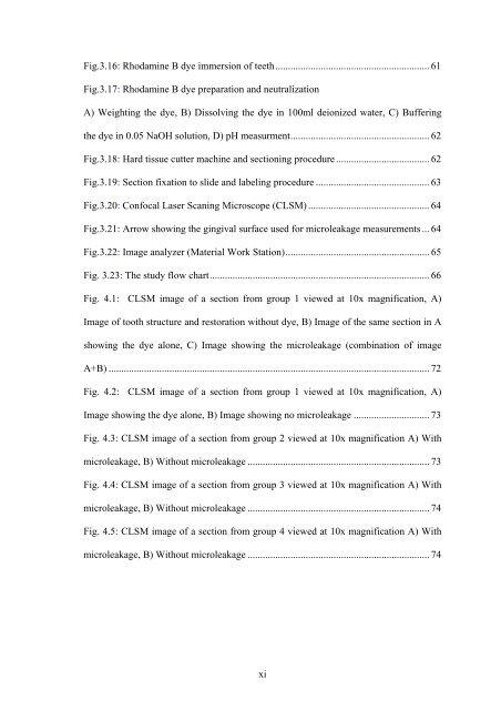
Fig.3.16: Rhodam<strong>in</strong>e B dye immersion of teeth ............................................................. 61Fig.3.17: Rhodam<strong>in</strong>e B dye preparation and neutralizationA) Weight<strong>in</strong>g the dye, B) Dissolv<strong>in</strong>g the dye <strong>in</strong> 100ml deionized water, C) Buffer<strong>in</strong>gthe dye <strong>in</strong> 0.05 NaOH solution, D) pH measurment ....................................................... 62Fig.3.18: Hard tissue cutter mach<strong>in</strong>e and section<strong>in</strong>g procedure ..................................... 62Fig.3.19: Section fixation to slide and label<strong>in</strong>g procedure ............................................. 63Fig.3.20: Confocal Laser Scan<strong>in</strong>g Microscope (CLSM) ................................................ 64Fig.3.21: Arrow show<strong>in</strong>g the g<strong>in</strong>gival surface used for <strong>microleakage</strong> measurements ... 64Fig.3.22: Image analyzer (Material Work Station) ......................................................... 65Fig. 3.23: The study flow chart ....................................................................................... 66Fig. 4.1: CLSM image of a section from group 1 viewed at 10x magnification, A)Image of tooth structure and restoration without dye, B) Image of the same section <strong>in</strong> Ashow<strong>in</strong>g the dye alone, C) Image show<strong>in</strong>g the <strong>microleakage</strong> (comb<strong>in</strong>ation of imageA+B) ............................................................................................................................... 72Fig. 4.2: CLSM image of a section from group 1 viewed at 10x magnification, A)Image show<strong>in</strong>g the dye alone, B) Image show<strong>in</strong>g no <strong>microleakage</strong> .............................. 73Fig. 4.3: CLSM image of a section from group 2 viewed at 10x magnification A) With<strong>microleakage</strong>, B) Without <strong>microleakage</strong> ........................................................................ 73Fig. 4.4: CLSM image of a section from group 3 viewed at 10x magnification A) With<strong>microleakage</strong>, B) Without <strong>microleakage</strong> ........................................................................ 74Fig. 4.5: CLSM image of a section from group 4 viewed at 10x magnification A) With<strong>microleakage</strong>, B) Without <strong>microleakage</strong> ........................................................................ 74xi


