Havva Dashtdar, Hussin A. Rothan, Terence Tay, Raja Elina Ahmad ...
Havva Dashtdar, Hussin A. Rothan, Terence Tay, Raja Elina Ahmad ...
Havva Dashtdar, Hussin A. Rothan, Terence Tay, Raja Elina Ahmad ...

You also want an ePaper? Increase the reach of your titles
YUMPU automatically turns print PDFs into web optimized ePapers that Google loves.
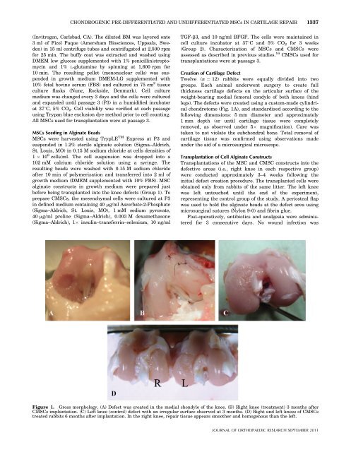
CHONDROGENIC PRE-DIFFERENTIATED AND UNDIFFERENTIATED MSCs IN CARTILAGE REPAIR 1337(Invitrogen, Carlsbad, CA). The diluted BM was layered onto3 ml of Ficol Paque (Amersham Biosciences, Uppsala, Sweden)in 15 ml centrifuge tubes and centrifugated at 2,500 rpmfor 25 min. The buffy coat was extracted and washed usingDMEM low glucose supplemented with 1% penicillin/streptomycinand 1% L-glutamine by spinning at 1,600 rpm for10 min. The resulting pellet (mononuclear cells) was suspendedin growth medium DMEM-LG supplemented with10% fetal bovine serum (FBS) and cultured in 75 cm 2 tissueculture flasks (Nunc, Rockside, Denmark). Cell culturemedium was changed every 3 days and the cells were culturedand expanded until passage 3 (P3) in a humidified incubatorat 378C, 5% CO 2 . Cell viability was verified at each passageusing Trypan blue exclusion dye method prior to cell counting.All MSCs used for transplantation were at passage 3.MSCs Seeding in Alginate BeadsMSCs were harvested using TrypLE TM Express at P3 andsuspended in 1.2% sterile alginate solution (Sigma–Aldrich,St. Louis, MO) in 0.15 M sodium chloride at cells densities of1 10 6 cells/ml. The cell suspension was dropped into a102 mM calcium chloride solution using a syringe. Theresulting beads were washed with 0.15 M sodium chlorideafter 10 min of polymerization and transferred into 2 ml ofgrowth medium (DMEM supplemented with 10% FBS). MSCalginate constructs in growth medium were prepared justbefore being transplanted into the knee defects (Group 1). Toprepare CMSCs, the mesenchymal cells were cultured at P3in defined medium containing 40 mg/ml Ascorbate-2-Phosphate(Sigma–Aldrich, St. Louis, MO), 1 mM sodium pyruvate,40 mg/ml proline (Sigma–Aldrich), 0.003 M dexamethasone(Sigma–Aldrich), 1 insulin–transferrin–selenium, 10 ng/mlTGF-b3, and 10 ng/ml BFGF. The cells were maintained incell culture incubator at 378C and 5% CO 2 for 3 weeks(Group 2). Characterization of MSCs and CMSCs wereassessed as described in previous studies. 16 CMSCs used fortransplantations were at passage 3.Creation of Cartilage DefectTwelve (n ¼ 12) rabbits were equally divided into twogroups. Each animal underwent surgery to create fullthickness cartilage defects on the articular surface of theweight-bearing medial femoral condyle of both knees (hindlegs). The defects were created using a custom-made cylindricalchondrotome (Fig. 1A), and standardized according to thefollowing dimensions: 5 mm diameter and approximately1 mm depth (or until cartilage tissue were completelyremoved, as observed under 5 magnification). Care wastaken to not violate the subchondral bone. Total removal ofcartilage tissue was confirmed using observations madeunder the aid of a microsurgical microscope.Transplantation of Cell Alginate ConstructsTransplantations of the MSC and CMSC constructs into thedefective areas (i.e., right knee in each respective group)were conducted approximately 3–4 weeks following theinitial defect creation procedure. The transplanted cells wereobtained only from rabbits of the same litter. The left kneewas left untouched until the end of the experiment,representing the control group of the study. A periosteal flapwas used to hold the alginate beads at the defect area usingmicrosurgical sutures (Nylon 9-0) and fibrin glue.Post-operatively, antibiotics and analgesia were administeredfor 3 consecutive days. No wound infection wasFigure 1. Gross morphology. (A) Defect was created in the medial chondyle of the knee. (B) Right knee (treatment) 3 months afterCMSCs implantation. (C) Left knee (control) defect with an irregular surface observed at 3 months. (D) Right and left knees of CMSCstreated rabbits 6 months after implantation. In the right knee, repair tissue appears smoother and homogenous than the left.JOURNAL OF ORTHOPAEDIC RESEARCH SEPTEMBER 2011