Havva Dashtdar, Hussin A. Rothan, Terence Tay, Raja Elina Ahmad ...
Havva Dashtdar, Hussin A. Rothan, Terence Tay, Raja Elina Ahmad ...
Havva Dashtdar, Hussin A. Rothan, Terence Tay, Raja Elina Ahmad ...

Create successful ePaper yourself
Turn your PDF publications into a flip-book with our unique Google optimized e-Paper software.
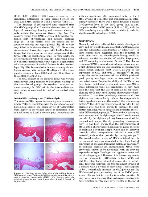
CHONDROGENIC PRE-DIFFERENTIATED AND UNDIFFERENTIATED MSCs IN CARTILAGE REPAIR 1339(11.8 1.47 vs. 8.67 1.86). However, there were nosignificant differences in these scores between theMSC and CMSC groups at 3 and 6 months (Table 1).The histology of the repaired sites obtained fromthe MSCs group after 3 months demonstrated a thinlayer of neocartilage forming and contained clusteredcells within the immature tissue (Fig. 2A). Therepaired tissue from CMSCs group at 6 months containedboth fibrocartilage and hyaline cartilage(Fig. 2C). In the control sites, the defects did notappear to undergo any repair process (Fig. 2B) or wereonly filled with fibrous tissue (Fig. 2B). Some sitesdemonstrated incomplete repair with hyaline like cartilage,but there were no vertical integration of thistissue with the subchondral bone. In some parts, thedefect was filled with bone (Fig. 3B). This repair tissueat 6 months demonstrated early signs of degenerationwith the presences of vertical fissures in the neocartilage(Fig. 3D). Immunohsitochemical staining showedhigher concentration of type II collagen in the transplantedtissues in both MSC and CMS than those inthe control sites (Fig. 3).The GAG content of the repaired tissue was verifiedqualitatively using Safranin O Fast green staining. Inthe transplanted groups, the repaired tissue stainedmore intensely for GAG within the intermediate anddeep zones as compared to that of the control sites(Fig. 4).Sulfated Glycoaminoglycans (GAG) AnalysisThe results of GAG quantitative analysis are summarizedin Table 1. Consistent with the morphological andhistological scores, the mean levels of GAG/proteinwere higher in the treated knees as compared to thecontrol knees at 3 and 6 months post-transplantation,Figure 2. Histology of the defect site of the rabbit’s knee inMSC-treated group (A,B) and CMSC-treated group (C,D) after 3and 6 months, respectively (H&E 10): Arrows point therepaired tissue in the right knees after 3 and 6 month ofimplantation.with no significant differences noted between the 2MSC groups at 3 months post-transplantation. Interestinglyhowever, there was a trend towards a higherGAG/protein level in the MSC group (1.67 0.14)compared to the CMSC groups (1.29 0.38), with thedifference being marginally close but did not reach thesignificance threshold (p ¼ 0.052).DISCUSSIONMesenchymal stem cells exhibit a stable phenotype invitro and have multilineage potential of differentiatinginto the adipocytes, chondrocytes, or osteocytes. 21 Invitro studies have suggested that the induction ofadult stem cells into chondrogenic lineage could beachieved via the introduction of soluble, biophysical,and 3D culturing environment factors. 22 The characteristicsof CMSCs were described in previous studies,which demonstrates an up-regulation of chondrogenicgenes, which included Sox9, COMP, as well as theexpression of GAG and type II collagen. 5,16 In thisstudy, the results demonstrated that CMSCs producedgood quality cartilage tissue as compared to theuntreated knees. Despite the ability of CMSCs to producebetter histological repair outcomes than MSCs,these differences were not significant. It may havebeen the case that the use of alginate gel for transplantingMSCs may have induced chondrogenic differentiation.It has been previously demonstrated thatalginate can independently induce chondrogenesis inBM stromal cells without the need of other stimulatingfactors. 23 The ideal microenvironment provided by thealginate gels has been shown to increase the cell–matrix signaling, which initiates and promotes the cellulardifferentiation process. 24 Because both cells typeswere encapsulated in alginate gel, the 3D environmentprovided by the alginate gel may have maintained therounded cell shape, thereby promoting chonrogenesis.22 It has been shown that the differentiation ofMSCs into a chondrogenic phenotype requires the cellsto maintain a rounded shape, which can be achievedthrough pellet encapsulation within a semi-solidmaterial, such as that provided by the alginate gel. 25The round nuclear shape of MSCs obtained in thisenvironment has been associated with a high level ofexpression of the chondrogenesis molecular markers. 15In addition, alginate gel encapsulating MSCs werepreviously shown to be associated with an increaseexpression of type II collagen and aggrecan genes andnot of collagen type I. However, it is interesting to notethat an increase in GAG/protein in both MSC- andCMSC-treated knees almost demonstrated a significantdifference between the two treatment groups at6 months with a p-value of only 0.052. There appearsto be a remarkable increase in GAG/protein for theMSC-treated group, exceeding that of the CMSC groupwhen cells are left in vivo for 6 months duration. Itmay have been the case that prolonged chondrogenicdifferentiation in vitro may have resulted in the lost ofJOURNAL OF ORTHOPAEDIC RESEARCH SEPTEMBER 2011