Havva Dashtdar, Hussin A. Rothan, Terence Tay, Raja Elina Ahmad ...
Havva Dashtdar, Hussin A. Rothan, Terence Tay, Raja Elina Ahmad ...
Havva Dashtdar, Hussin A. Rothan, Terence Tay, Raja Elina Ahmad ...

You also want an ePaper? Increase the reach of your titles
YUMPU automatically turns print PDFs into web optimized ePapers that Google loves.
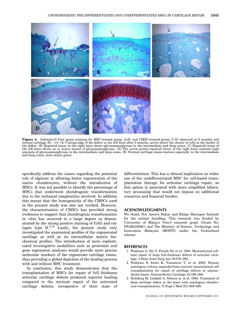
CHONDROGENIC PRE-DIFFERENTIATED AND UNDIFFERENTIATED MSCs IN CARTILAGE REPAIR 1341Figure 4. Safranin-O Fast green staining for MSC-treated group (A,B) and CMSC-treated group (C,D) observed at 6 months andnormal cartilage (E) 10. (A) Cutting edge of the defect in the left knee after 6 months, arrow shows the cluster of cells at the border ofthe defect. (B) Repaired tissue in the right knee shows glycosaminoglycans in the intermediate and deep zones. (C) Repaired tissue ofthe left knee shows no or scarce mount of glycosaminoglycans. (D) The arrow points repaired tissue of the right knee contains highamounts of glycosaminoglycans in the intermediate and deep zones. (E) Normal cartilage stains maroon especially in the intermediateand deep zones, bone stains green.specifically address the issues regarding the potentialrole of alginate in allowing better regeneration of thenative chondrocytes, without the introduction ofMSCs. It was not possible to identify the percentage ofMSCs that underwent chondrogenic transformationdue to the technical complexities involved. In additionthis meant that the homogenicity of the CMSCs usedin the present study was also not verified. However,the characterization of CMSCs has provided strongevidences to support that chondrogenic transformationin vitro has occurred to a large degree as demonstratedby the strong positive staining of GAG and collagentype II. 5,16 Lastly, the present study onlyinvestigated the anatomical profiles of the regeneratedcartilage as well as its extracellular matrix biochemicalprofiles. The introduction of more sophisticatedinvestigative modalities such as proteomic andgene expression analyses would provide more precisemolecular markers of the regenerate cartilage tissue,thus providing a global depiction of the healing processwith and without MSC treatment.In conclusion, this study demonstrates that thetransplantation of MSCs for repair of full thicknessarticular cartilage defects produced superior healingcompared to the intrinsic repair of the untreatedcartilage defects, irrespective of their state ofdifferentiation. This has a clinical implication on wideruse of the undifferentiated MSC for cell-based transplantationtherapy for articular cartilage repair, asthis option is associated with more simplified laboratoryprocessing that would not impose on additionalresources and financial burden.ACKNOWLEDGMENTSWe thank Nor Azeera Bakar and Balqis Mariapee Sarinahfor the animal handling. This research was funded byUniversity of Malaya Vote-f research grant (Grant No:PS169/2008C) and The Ministry of Science, Technology andInnovation Malaysia (MOSTI) under the Technofundscheme.REFERENCES1. Wakitani S, Gto T, Pineda SJ, et al. 1994. Mesenchymal cellbaserepair of large full-thickness defects of articular cartilage.J Bone Joint Surg Am 76:579–592.2. Wakitani S, Imoto K, Yamamoto T, et al. 2002. Humanautologous culture expanded bone marrow mesenchymal celltransplantation for repair of cartilage defects in osteoarthriticknees. Osteoarthritis Cartilage 10:199–206.3. Brittberg M, Lindahl A, Nilsson A, et al. 1994. Treatment ofdeep cartilage defect in the knee with autologous chondrocytetransplantation. N Engl J Med 331:889–895.JOURNAL OF ORTHOPAEDIC RESEARCH SEPTEMBER 2011