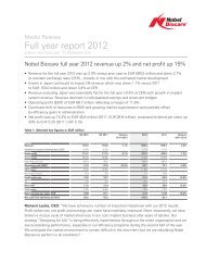
State of the art implant placement and restoration ... - Nobel Biocare
State of the art implant placement and restoration ... - Nobel Biocare
State of the art implant placement and restoration ... - Nobel Biocare

You also want an ePaper? Increase the reach of your titles
YUMPU automatically turns print PDFs into web optimized ePapers that Google loves.
Technology<strong>State</strong> <strong>of</strong> <strong>the</strong> <strong>art</strong> <strong>implant</strong> <strong>placement</strong> <strong>and</strong><strong>restoration</strong> through guided techniques:An ‘all-on-4’ clinical reportBy M<strong>art</strong>in Marcus DDS, Russell Baer DDS, Nelson Lo DDS - UniversityAssociates in Dentistry, Dental Implant Institute <strong>of</strong> Chicago, Chicago, IllinoisImplant dentistry has become a well provenaccepted st<strong>and</strong>ard <strong>of</strong> care for <strong>the</strong> singletooth, multiple missing teeth, as well as <strong>the</strong>fully edentulous arch <strong>of</strong> teeth. Constant changesare rapidly improving our ability to clinicallytreat patients in a more expeditious, efficient<strong>and</strong> less invasive manner. Technologies usingCBCT (cone beam computerized technology)3-D imaging, computer generated plannings<strong>of</strong>tware, guided surgery <strong>and</strong> immediate functionhave benefited our patients with reducedtreatment times, minimally invasive <strong>and</strong> simplifiedsurgical techniques, reduction <strong>of</strong> postsurgicaldiscomfort <strong>and</strong> <strong>the</strong> immediate <strong>placement</strong><strong>of</strong> a fixed <strong>restoration</strong>.The guided <strong>implant</strong> surgical <strong>and</strong> pros<strong>the</strong>tictechnique for <strong>the</strong> All on 4 <strong>restoration</strong> <strong>of</strong> anedentulous arch has gained greater acceptance<strong>and</strong> success 1 . Its benefits are many:• The ability to rehabilitate <strong>the</strong> completelyedentulous jaw with a minimum <strong>of</strong> bone volume<strong>and</strong> avoiding in most situations bonegrafting procedures 1 .• Flapless surgery which allows for <strong>the</strong> patientto experience less surgical trauma, painor swelling 2 .• Strategically placed <strong>implant</strong>s, 2 posteriorly<strong>and</strong> 2 anteriorly, with good anchorage haveshown very successful outcomes 3, 4 .• Angulations <strong>of</strong> <strong>the</strong> <strong>implant</strong>s allows for longer<strong>implant</strong>s to be placed with cortical boneanchorage. Greater anterior-posterior spread<strong>of</strong> <strong>implant</strong>s creates an ability to restore teethback to <strong>the</strong> first molar region with <strong>the</strong> reduction<strong>of</strong> cantilever effects 5, 6 .• The pros<strong>the</strong>sis allows for <strong>the</strong> re-establishment<strong>of</strong> vertical dimension most patients havelost to ei<strong>the</strong>r bone loss due to periodontal diseaseor existing old dentures.• Primary immediate <strong>implant</strong> stabilizationreducing <strong>the</strong> mechanical load by <strong>implant</strong>splinting 7, 8 .• An immediate function fixed <strong>restoration</strong> at<strong>the</strong> time <strong>of</strong> surgery due to <strong>implant</strong> stability 9 .• The immediately loaded <strong>restoration</strong> allowsan easier transition for patients from a removableto a fixed restorative state.• Simplifies <strong>the</strong> procedure for <strong>the</strong> clinician.Clinical reportPlanningA 70 year old woman presented with a maxillarydenture. The patient has been edentulousin <strong>the</strong> upper jaw for over 10 years (Fig.1). Shehas worn several different sets <strong>of</strong> dentures duringthat period <strong>of</strong> time. A preliminary evaluation<strong>of</strong> her dental <strong>and</strong> medical history wasobtained. Her medical history presents withno significant contraindications for <strong>implant</strong>surgery. A treatment plan was devised utilizingguided <strong>implant</strong> surgery <strong>and</strong> <strong>the</strong> All on 4(<strong>Nobel</strong> <strong>Biocare</strong>) protocols.The upper denture was relined in order to fitintimately with <strong>the</strong> tissue, at a corrected occlusalvertical dimension <strong>and</strong> with a pleasing, acceptable,es<strong>the</strong>tic alignment. This will duplicateas a radiographic template.Multiple 2-mm holes were placed into <strong>the</strong>dentures at different levels <strong>and</strong> in areas bothbuccally <strong>and</strong> palatally. These holes were filledwith gutta percha as radio-opaque markers.(Fig. 2) An occlusal record was <strong>the</strong>n taken usingvinyl polysiloxane at <strong>the</strong> appropriate centric<strong>and</strong> vertical dimensions. This allowed <strong>the</strong>patient to hold <strong>the</strong> radiographic template inposition during <strong>the</strong> CBCT scan <strong>and</strong> preventmovement.Our patient was now ready for a CBCT(Gendex CB-500; DENTSPLY) scan. CBCT allowsviews <strong>of</strong> different objects at different densitiesin a 3D view. We can visualize availablebone for <strong>implant</strong> <strong>placement</strong>, as well as pertinentanatomical structures such as <strong>the</strong> maxillarysinuses, <strong>the</strong> m<strong>and</strong>ibular inferior alveolarnerve canal, s<strong>of</strong>t tissue thickness <strong>and</strong> tooth positionrelative to <strong>the</strong> underlying bone 10 .A double scan technique was used 11 . Thefirst scan was <strong>of</strong> <strong>the</strong> patient with her denture(radiographic template) <strong>and</strong> <strong>the</strong> interocclusalrecord. (Fig.3) The second scan was <strong>of</strong> <strong>the</strong> dentureonly.The CBCT data was <strong>the</strong>n formatted <strong>and</strong>transferred into a 3-demensional <strong>implant</strong> plannings<strong>of</strong>tware program (Procera CAD Design;<strong>Nobel</strong> <strong>Biocare</strong>). Implant positions <strong>and</strong> sizes,as it relates to bone availability <strong>and</strong> associatedvital structures, can be digitally evaluated <strong>and</strong>placed. Super imposing <strong>the</strong> two sets <strong>of</strong> scansallowed us to correlate <strong>the</strong> correct planned <strong>implant</strong>position in relation to <strong>the</strong> position <strong>of</strong> <strong>the</strong>denture teeth on <strong>the</strong> pros<strong>the</strong>sis. (Fig.4)After <strong>the</strong> plan was completed, <strong>the</strong> data wastransferred to a milling center to fabricate astereolithography surgical guide <strong>and</strong> duplicatedenture.(Fig.5) This guide will: 1) at <strong>the</strong> time<strong>of</strong> surgery, direct <strong>the</strong> <strong>placement</strong> <strong>of</strong> <strong>implant</strong>s into<strong>the</strong> identical positions as planned for a flaplesssurgical approach 12 <strong>and</strong> 2) allow for creatinga cast from which <strong>the</strong> interim, immediatepros<strong>the</strong>sis will be created. (Fig.6) The duplicatedenture gives <strong>the</strong> technician <strong>the</strong> ability tomount <strong>the</strong> cast <strong>and</strong> shows tooth position to beduplicated into an all acrylic fixed hybrid interimpros<strong>the</strong>ses. (Fig. 7) A new silicone centricocclusal record was created with <strong>the</strong> surgicalguide <strong>and</strong> opposing arch. This allows properseating <strong>of</strong> <strong>the</strong> guide at <strong>the</strong> time <strong>of</strong> surgery.SurgeryAfter all our pre-surgical diagnostics <strong>and</strong> planningwere completed guided <strong>implant</strong> surgerywas performed. Initially appropriate pre-surgicalmedications were given which includedantibiotics, analgesics <strong>and</strong> an antimicrobialrinse.Local anes<strong>the</strong>tics were given ie. lidocaine<strong>and</strong> bupivacaine <strong>and</strong> <strong>the</strong> surgical guide inserted.Biting on <strong>the</strong> interocclusal record for1 – 2 minutes allowed for tissue dis<strong>placement</strong>insuring a more positive position <strong>of</strong> <strong>the</strong> guideto <strong>the</strong> planning data. Positioning <strong>and</strong> seating<strong>of</strong> <strong>the</strong> guide was confirmed. While <strong>the</strong> patientremained closed on <strong>the</strong> interocclusal recordour initial step was to place <strong>the</strong> 3 pre-plannedhorizontal stabilization pins. This secured <strong>the</strong>guide. The interocclusal record was removed,exposing <strong>the</strong> surgical sites <strong>of</strong> <strong>the</strong> guide.The first osteotomy was prepared, choosingone <strong>of</strong> <strong>the</strong> anterior positions. Appropriate sequencing<strong>of</strong> <strong>the</strong> tissues punch, drilling guides<strong>and</strong> twist drills were used according to <strong>the</strong>manufacturer’s directions (<strong>Nobel</strong>Guide; <strong>Nobel</strong><strong>Biocare</strong>). Once <strong>the</strong> osteotomy was completed,<strong>the</strong> pre-planned <strong>implant</strong> type, length <strong>and</strong> sizewere placed <strong>and</strong> a template abutment inserted.This now provided <strong>the</strong> guide with additionalstability from a vertical direction as well. The46 Implant practice August 2009 Volume 2 Number 3
TechnologyFigure 1 Figure 2 Figure 3Figure 4 Figure 5 Figure 6Figure 7 Figure 8 Figure 9remaining three <strong>implant</strong> osteotomy sites were<strong>the</strong>n prepared using <strong>the</strong> same sequential drillingprotocols <strong>and</strong> <strong>the</strong> appropriate <strong>implant</strong>swere placed. (Fig. 8)After <strong>implant</strong> <strong>placement</strong> <strong>the</strong> surgical guidewas removed <strong>and</strong> <strong>the</strong> 2 anterior straight multiunitabutments ( Multiunit abutment; <strong>Nobel</strong><strong>Biocare</strong>) were placed first, followed by <strong>the</strong>posterior 30 degree angled abutments (30 degreeMultiunit abutment; <strong>Nobel</strong> <strong>Biocare</strong>) usinga laboratory fabricated custom jig.(Fig. 8)The all acrylic hybrid pros<strong>the</strong>sis was tried infor fit <strong>and</strong> confirmed by radiographic evaluation.Occlusion <strong>and</strong> vertical dimension wereevaluated <strong>and</strong> minor adjustments made asneeded. The pros<strong>the</strong>sis was screwed in, accessholes covered <strong>and</strong> immediate functionobtained.(Fig.10)Post-operative instructions regarding hygienemaintenance, s<strong>of</strong>t diet <strong>and</strong> continuedantibiotics (daily for 1 week), anti-inflammatory(ibupr<strong>of</strong>en 600mg. 3 times a day for 3days) <strong>and</strong> antimicrobial (chlorhexidine rinse,daily for 1 week) were prescribed. Any o<strong>the</strong>rmedications thought to be necessary, such as anarcotic for pain, would be considered at thistime as well.RestorativeAfter 3 months <strong>of</strong> osseointegration <strong>the</strong> <strong>restoration</strong><strong>of</strong> a titanium reinforced fixed hybridpros<strong>the</strong>sis (Procera Implant Bridge; <strong>Nobel</strong><strong>Biocare</strong>) was created. The interim pros<strong>the</strong>siswas removed, tissue <strong>and</strong> <strong>implant</strong> site inspectionfor osseointegration were evaluated, <strong>and</strong><strong>the</strong> following sequence <strong>of</strong> appointments werenecessary for <strong>the</strong> completion <strong>of</strong> <strong>the</strong> final <strong>restoration</strong>.1. Custom tray impression using impressiontransfers for <strong>the</strong> positional relationship <strong>of</strong> <strong>the</strong><strong>implant</strong>s to <strong>the</strong> ridge <strong>and</strong> each o<strong>the</strong>r. (Fig 11)2. Verification jig to identify that <strong>the</strong> <strong>implant</strong>son <strong>the</strong> master cast are in <strong>the</strong> identical positionas <strong>the</strong>y are in <strong>the</strong> mouth. (Fig. 12)3. Bite block records to measure <strong>the</strong> vertical<strong>and</strong> centric relation positions.4. Wax try-in to ensure <strong>the</strong> es<strong>the</strong>tics, toothposition, phonetics <strong>and</strong> occlusion. Also thisidentified <strong>the</strong> spacial relationship between<strong>the</strong> correct tooth position, <strong>the</strong> ridge <strong>and</strong> <strong>the</strong><strong>implant</strong> abutments to create a custom milledAugust 2009 Volume 2 Number 3 Implant practice 47
TechnologyFigure 10 Figure 11 Figure 12Figure 14 Figure 15Figure 13titanium bar that was milled <strong>and</strong> fit within <strong>the</strong>parameters <strong>of</strong> <strong>the</strong> pros<strong>the</strong>sis.5. Try-in <strong>of</strong> <strong>the</strong> custom milled titanium retentionbar along with <strong>the</strong> transfer <strong>of</strong> <strong>the</strong> tooth setup for final evaluation <strong>of</strong> fit, es<strong>the</strong>tics <strong>and</strong> function(phonetics <strong>and</strong> occlusion).(Fig12)6. Delivery <strong>of</strong> <strong>the</strong> final pros<strong>the</strong>sis, minor occlusaladjustments, torquing <strong>the</strong> retention screwsto 15 Ncm. <strong>and</strong> restoring <strong>the</strong> screw accessholes.(Fig14)7. Radiographic confirmation <strong>of</strong> <strong>the</strong> fit <strong>of</strong> <strong>the</strong>pros<strong>the</strong>sis to <strong>the</strong> <strong>implant</strong> abutments, <strong>implant</strong><strong>placement</strong> <strong>and</strong> integration.(Fig.15)SummaryTechnological advancements continue to revolutionizeour abilities to treat our patients ina more efficient <strong>and</strong> less invasive manner.Through CBCT dual scan data visualization <strong>of</strong>available bone, anatomical issues that may beencountered during surgery, s<strong>of</strong>t tissue thickness<strong>and</strong> pros<strong>the</strong>tic tooth position may be revealed.3-D virtual planning allows us to diagnosticallyplan <strong>implant</strong> sites, develop surgicalguides to transfer that information intraorally<strong>and</strong> create a prefabricated, immediate functionpros<strong>the</strong>sis before <strong>the</strong> patient is even inour dental chair. By using surgical guides for<strong>implant</strong> <strong>placement</strong> <strong>the</strong> <strong>implant</strong>s may be placedthrough a minimally invasive flapless procedure,quickly, predictably <strong>and</strong> with a low level<strong>of</strong> complications. This minimizes <strong>the</strong> patient’spain, time <strong>and</strong> stress. Restoratively, we are ableto bring our patients from a removable dentureREFERENCES1. MaloP., NobreM.,Lope,A. The Use <strong>of</strong> Computer-Guided Flapless Implant Surgery <strong>and</strong> Four ImplantsPlaced in Immediate Function to Support a FixedDenture: Preliminary Results After a Mean FollowupPeriod <strong>of</strong> Thirteen Months. Journal <strong>of</strong> Pros<strong>the</strong>ticDentistry 2007 Volume 97 Issue 62. CampelLD, CamaraJR. Flapless Implant Surgery;A 10-year Clinical Retrospective Analysis. Int JournalOral Maxill<strong>of</strong>acial Implants 2002 17:271-63. BranemarkPI, SvenssonB, van SteenbergheD.TenyearSurvival Rates <strong>of</strong> Fixed Pros<strong>the</strong>ses on Four to SixImplants Ad Modum Branemark in Full Edentulism.Clin Oral Implants Res 1995; 6:227-314. Mal,P, RangertB, NombreM. ‘All on Four’Immediate Function Concept with BranemarkSystem Implants for Completely Edentulous Maxilla:A 1 Year Retrospective Clinical Study. Clin ImplantDent Relat Res 2003:7 Supplement 1:588-945. AparicioC, PeralesP, RangertB. Tilted Implants asan Alternative to Maxillary Sinus Grafting: A Clinical,Radiographic <strong>and</strong> Periotest Study. Clin Implant DentRelat Res 2001;3:39-496. KrekamanovL, KahanM, RangertB, Lindstrom,H.Tilting <strong>of</strong> Posterior M<strong>and</strong>ibular <strong>and</strong> MaxillaryImplants <strong>of</strong> Improved Pros<strong>the</strong>sis Support. Int Journal<strong>of</strong> Oral Maxill<strong>of</strong>acial Implants 2000;15:405-147. MortonD, JaffinR, WeberHP. ImmediateRestoration <strong>and</strong> Loading <strong>of</strong> Dental Implants:Clinical; Considerations <strong>and</strong> Protocols. Int Journal <strong>of</strong>to a functioning fixed pros<strong>the</strong>sis immediately.Considering <strong>the</strong> advantages <strong>and</strong> benefits <strong>of</strong><strong>implant</strong> <strong>placement</strong> <strong>and</strong> <strong>restoration</strong> throughguided techniques, we can recommend this asa viable alternative to treatment for rehabilitation<strong>of</strong> <strong>the</strong> edentulous jaws.IOral Maxill<strong>of</strong>acial Implants 2004;19 Supplemental1:103-88. AttardNJ, ZarbGA. Immediate <strong>and</strong> Early ImplantLoading Protocols: A Literature Review <strong>of</strong> ClinicalStudies. Journal <strong>of</strong> Pros<strong>the</strong>tic Dentistry 2005;94:242-589. SchnitmanDA, WohrlePS, RubensteinJE et al.Branemark Implants Immediately Loaded withFixed Pros<strong>the</strong>sis at Implant Placement: Ten YearResults. Int Journal Oral Maxill<strong>of</strong>acial Implants1997;12:495-50310. SonickM, AbrhamsJ, FaiellaR. A Comparison<strong>of</strong> <strong>the</strong> Accuracy Of Periapical, Panoramic, <strong>and</strong>Computerized Tomographic Radiographs in Locating<strong>the</strong> M<strong>and</strong>ibular Canal. Int Journal Oral Maxill<strong>of</strong>acialImplants. 1994;9(4);455-46011. Van SteenbergheD, NaertI, AnderrsonM,Brajnovic I, Van CleynenbreugelJ, SuetensP. ACustom Template <strong>and</strong> Definitive Pros<strong>the</strong>sis AllowingImmediate Loading in <strong>the</strong> Maxilla: A ClinicalReport Int Journal <strong>of</strong> Oral Maxill<strong>of</strong>acial Implants2002;17:663-7012. Van SteenbergheD, GlauserR, BlombackU, et al.A Computed Tomographic Scan-derived CustomizedSurgical Template <strong>and</strong> Fixed Pros<strong>the</strong>sis for FlaplessSurgery <strong>and</strong> Immediate Loading <strong>of</strong> Implants in FullyEdentulous Maxillae: A Prospective MulticenterStudy. Clin Implant Dent Relat Res. 2005;7 (Suppl1):S111-S12048 Implant practice August 2009 Volume 2 Number 3